CHAPTER 4 IN VIVO TOXICITY EVALUATION OF LAUHA...
26
114 CHAPTER 4 –IN VIVO TOXICITY EVALUATION OF LAUHA BHASMA 4.1 INTRODUCTION The usage of Ayurvedic herbo-metallic preparations has been in vogue over several centuries in clinical practice [1, 2]. There is a global perception that Ayurvedic medicines are toxic, based on several reports that indicate the presence of heavy metals like mercury and lead above the permissible limits in these preparations [2–4]. The toxic effects of metals in case of improper bhasma preparations have also been portrayed in the literature [5]. Iron is also known for its toxic nature, apart from its essential functions in the biological systems. Iron can produce free radicals, which ultimately result in alteration of metabolic pathways and alteration in the biological systems [6]. Due to these toxicity concerns, several questions have been raised about the safety of these preparations. Some of them are Do Ayurvedic purification and calcination procedures convert iron into medicine? Whether the prepared Lauha bhasma is safe or toxic? To address these questions, attempts have been made here to study the in vivo toxicity profile of Lauha bhasma in Wistar rats. Acute oral toxicity (single dose) and sub acute toxicity studies (repeated dose) were carried out by oral administration of Lauha bhasma. All the experiments were carried as per the WHO guidelines.
-
Upload
truongxuyen -
Category
Documents
-
view
221 -
download
4
Transcript of CHAPTER 4 IN VIVO TOXICITY EVALUATION OF LAUHA...
114
CHAPTER 4 –IN VIVO TOXICITY EVALUATION OF LAUHA BHASMA
4.1 INTRODUCTION
The usage of Ayurvedic herbo-metallic preparations has been in vogue over several
centuries in clinical practice [1, 2]. There is a global perception that Ayurvedic medicines
are toxic, based on several reports that indicate the presence of heavy metals like mercury
and lead above the permissible limits in these preparations [2–4]. The toxic effects of
metals in case of improper bhasma preparations have also been portrayed in the literature
[5]. Iron is also known for its toxic nature, apart from its essential functions in the biological
systems. Iron can produce free radicals, which ultimately result in alteration of metabolic
pathways and alteration in the biological systems [6]. Due to these toxicity concerns,
several questions have been raised about the safety of these preparations. Some of them are
Do Ayurvedic purification and calcination procedures convert iron into medicine?
Whether the prepared Lauha bhasma is safe or toxic?
To address these questions, attempts have been made here to study the in vivo toxicity
profile of Lauha bhasma in Wistar rats. Acute oral toxicity (single dose) and sub acute
toxicity studies (repeated dose) were carried out by oral administration of Lauha bhasma.
All the experiments were carried as per the WHO guidelines.

115
4.2 MATERIALS AND METHODS
4.2.1 Study design
Wistar rats of both sexes in the age group of 4-6 weeks (Acute oral toxicity) and 6- 8
weeks (Sub acute oral toxicity) were used for the study. The rats were housed in
polypropylene cages and kept in a 12 h light/dark cycle .The room temperature was
maintained at 21±2°C and they were fed with standard rat feed pellet and deionized water
ad libitum throughout the entire study. The animals were acclimatized for 5 days before
the initiation of the study. The procedures used were reviewed and approved by the
Institutional Animal ethics committee (120/SASTRA/IAEC/RPP, SASTRA University).
4.2.2 Acute oral toxicity study (14-days single dose toxicity study)
A total of 12 rats were divided into two groups (I & II) with equal number of male and
female. The body weight of male ranged from 105 - 125 g and female from 85 - 100 g.
Group I rats were treated as normal control where they received honey with distilled water
in the ratio of 2:3 through oral route. Group II animals were administered with single dose
of Lauha bhasma (2000 mg/ kg) as a suspension in honey with distilled water in the ratio
of 2:3 through oral route. Body weight was measured on Day 0, Day 7 and Day 14. The
fecal samples were collected for 14 days to determine the elimination profile of iron. On
the 14th day, animals were sacrificed and major organs including liver, spleen, intestine,
brain and kidney were collected for bio-distribution studies.

116
4.2.3 Bio-distribution studies and Fecal analysis
The organs and fecal samples were subjected to microwave digestion using ultra-pure nitric
acid (Merck) and 50 % Hydrogen peroxide(Merck) in the ratio of (4:1) using microwave
digestor (Anton-Paar, Graz, Austria). The digested samples were then quantitatively
estimated for the iron content using atomic absorption spectroscopy (AAnalyst 400, Perkin
Elmer, USA).
4.2.4 Sub acute oral toxicity study (28-days repeated dose toxicity study and 14 days
recovery study)
A total of 60 rats (n=10) were divided into six groups (Group I to Group VI) with equal
number of male and female. The therapeutic dose of Lauha bhasma for humans is 250mgs
[7]. This dose was converted to animal dose [8] as follows:
Human equivalent dose (mg kg⁄ ) =Rat dose (mg kg⁄ ) × Rat Km (6)
Human Km (37)
Human equivalent dose (mg/kg) = Rat dose (mg/kg) x Rat Km (6) / Human Km (37)
where, Km is the dose conversion factor based on body surface area.
The human equivalent dose was found to be 25 mg/kg. By fixing 25mg/kg as normal dose,
the medium and high doses were calculated as 100 mg/kg and 400 mg/kg respectively.
Group I was treated as normal control and received honey with distilled water in the ratio
of 2:3 for 28 days. Lauha bhasma was administered to Group II to Group IV animals at
different doses and was given as a suspension in honey and distilled water (2:3) for 28
days.

117
Group II received the therapeutic dose (25mg/kg body weight) of Lauha bhasma, Group
III received the medium dose (100 mg/kg) and Group IV received the high dose (400
mg/kg). Group V and Group VI were treated as satellite groups in which group V was
treated as satellite control and received honey with distilled water in the ratio of 2:3. Group
VI received the high dose (400 mg/kg body weight). Weekly body weight was measured.
On the 28th day animals from Group I to Group IV were sacrificed. Blood was collected by
retro orbitary puncture for biochemical and hematological analysis. Major organs were
collected for histopathological studies. Group V and Group VI were treated with standard
rat feed pellet and deionized water ad libitum for the next 14 days. On the 42nd day the
satellite group animals were sacrificed as mentioned above.
4.2.5 Biochemical and Hematological analysis
Biochemical analysis was performed using Auto Analyzer (A15 Biosystems, Spain) and
the kits were procured from Bio Systems. Hematological parameters were analyzed using
Mythic 22 Hematology Analyzer (Orphee SA, Switzerland)
4.2.6 Histopathology
Tissues were collected and preserved in 10 % buffered formalin. All tissues required for
histopathological evaluation were embedded in paraffin wax, sectioned at a thickness of
approximately 3-5 microns, stained with hematoxylin-eosin and examined by light
microscopy.
118
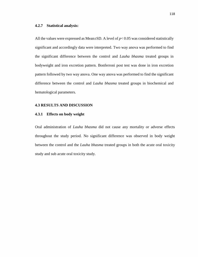
4.2.7 Statistical analysis:
All the values were expressed as Mean±SD. A level of p< 0.05 was considered statistically
significant and accordingly data were interpreted. Two way anova was performed to find
the significant difference between the control and Lauha bhasma treated groups in
bodyweight and iron excretion pattern. Bonferroni post test was done in iron excretion
pattern followed by two way anova. One way anova was performed to find the significant
difference between the control and Lauha bhasma treated groups in biochemical and
hematological parameters.
4.3 RESULTS AND DISCUSSION
4.3.1 Effects on body weight
Oral administration of Lauha bhasma did not cause any mortality or adverse effects
throughout the study period. No significant difference was observed in body weight
between the control and the Lauha bhasma treated groups in both the acute oral toxicity
study and sub acute oral toxicity study.

119
Figure 4.5 Weekly body weight in control and different doses of Lauha bhasma treated
male rats in sub acute oral toxicity study. The values are expressed as Mean± SD for 5
animals.
Figure 4.1 Body weight of control and Lauha bhasma treated animals in acute oral
toxicity study. The values are expressed as Mean ± SD for 3 animals.
Figure 4.2 Body weight of control and Lauha bhasma treated animals in acute oral
toxicity study. The values are expressed as Mean ± SD for 3 animals.
Figure 4.3 Body weight of control and Lauha bhasma treated animals in acute oral
toxicity study. The values are expressed as Mean ± SD for 3 animals.
Figure 4.4 Body weight of control and Lauha bhasma treated animals in acute oral
toxicity study. The values are expressed as Mean ± SD for 3 animals.

120
Figure 4.6 Weekly body weight in control and different doses of Lauha bhasma treated
female rats in sub acute oral toxicity study. The values are expressed as Mean± SD for 5
animals.
4.3.2 Bio-distribution studies
In acute oral toxicity studies there was no deposition of iron in major organs including
liver, kidney, brain intestine and spleen when compared to control group in both male and
female rats. From Figure 4.4 it was apparent that there was no significant difference
between the control and Lauha bhasma treated groups in the concentration of iron in the
organs. This also correlated reasonably very well with the iron excretion pattern (Figure
4.5) as high amount of iron excretion was seen on day 1 in Lauha bhasma treated group.
Machado et al. [9] reported that lower deposition of iron was observed in liver when
administered as iron peptide complex (30mg/kg and 60 mg/kg body weight). However
when administered as iron sulfate at same doses, excess deposition of iron was observed in
liver and spleen [9]. Papanastu et al. [10] reported that under normal circumstances, the

121
concentration of iron was found to be higher in spleen when compared to liver. After
administration with high dose of iron, the concentration was found to be higher in liver due
to excess deposition when compared to spleen [10].
Figure 4.7 Bio distribution studies of control and Lauha bhasma treated animals in acute
oral toxicity study. The values are expressed as Mean± SD for 3 animals in each group of
122
both male and female rats.
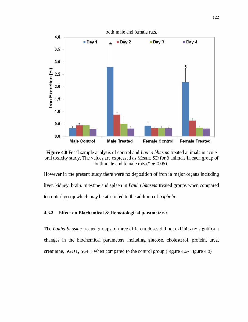
Figure 4.8 Fecal sample analysis of control and Lauha bhasma treated animals in acute
oral toxicity study. The values are expressed as Mean± SD for 3 animals in each group of
both male and female rats (* p<0.05).
However in the present study there were no deposition of iron in major organs including
liver, kidney, brain, intestine and spleen in Lauha bhasma treated groups when compared
to control group which may be attributed to the addition of triphala.
4.3.3 Effect on Biochemical & Hematological parameters:
The Lauha bhasma treated groups of three different doses did not exhibit any significant
changes in the biochemical parameters including glucose, cholesterol, protein, urea,
creatinine, SGOT, SGPT when compared to the control group (Figure 4.6- Figure 4.8)

123
Figure 4.9 Biochemical parameters of control and different doses of Lauha bhasma
treated groups, (A) Glucose, (B) Protein. The values are expressed as Mean± SD for 5
animals.
124
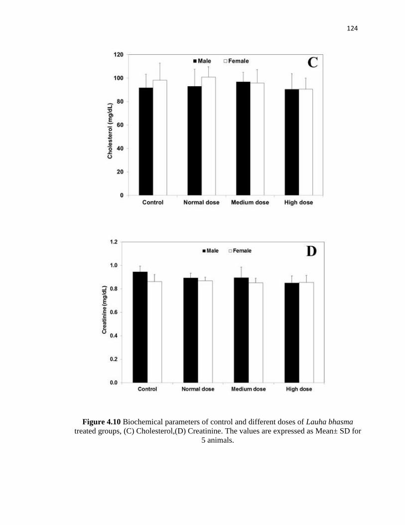
Figure 4.10 Biochemical parameters of control and different doses of Lauha bhasma
treated groups, (C) Cholesterol,(D) Creatinine. The values are expressed as Mean± SD for
5 animals.

125
Figure 4.11 Biochemical parameters of control and different doses of Lauha bhasma
treated groups, (E) ALT, (F) AST. The values are expressed as Mean± SD for 5 animals.

126
No significant changes were observed in major hematological parameters including RBC,
hemoglobin and hematocrit in Lauha bhasma treated groups when compared to control
group. Lymphocytes count was also found to be normal between the groups.
Figure 4.12 Hematological parameters of control and different doses of Lauha bhasma
treated groups (A) RBC, (B) Hemoglobin. The values are expressed as Mean± SD for 5
animals.

127
Figure 4.13 Hematological parameters of control and different doses of Lauha bhasma
treated groups (C) Hematocrit. The values are expressed as Mean± SD for 5 animals.
Sarkar et al. [11] performed investigations on toxicity of Lauha bhasma in experimental
animals, followed by recovery studies. Lauha bhasma was administered to experimental
animals (Albino rats) at a dosage of five times the therapeutic effective dose. The drug
was administered for 60 days and 45-day recovery period was observed. Hyperglycemia
was observed in Lauha bhasma treated group along with elevated levels of serum liver
enzymes and the levels were restored to normalcy in the recovery period [11].
In another study iron administered as iron dextran injection in mice resulted in iron
overload and caused liver injury that resulted in significant elevation of serum enzymes
including ALT, AST, ALP and bilirubin. Further an increase in liver iron content was also
observed. However when the iron overloaded mice was treated orally with Terminalia
chebula extract there was a decline in the level of serum enzymes and the liver iron content

128
also got lowered. Terminalia chebula scavenges free radicals and inhibits the free radical
formation by chelating excess iron with its active components [12]. Hence it is evident that
iron should not be administered alone.
It has been reported that iron supplementation when administered continuously to Sprague-
Dawley rats resulted in increased serum cholesterol and triglycerides level. This is due to
the decreased antioxidant levels which resulted in increased lipid peroxidation in liver and
leads to leakage of hepatic enzymes in the circulation [13].
However, in the present study, Lauha bhasma even at high doses did not cause any adverse
effects as evidenced from the biochemical and hematological parameters.
4.3.4 Histopathological Studies
Liver and spleen play a major role in iron metabolism[7,15]. Normal cyto architecture of
liver and spleen were observed in the high dose treated group. Liver exhibited normal
hepatocytes and sinusoids. No signs of iron deposition in the form of hemosiderin as brown
patches, were observed in liver and spleen in the high dose treated group (Figure 4.11)
Sarkar et al. [11] reported liver toxicity in Albino rats after administering Lauha bhasma
in high dose. Histopathological findings of liver showed congestion of central vein,
sinusoidal dilation with mild to moderate fatty changes [11].
Meltem et al. [15] studied the effect of chronic iron toxicity in Sprague-Dawley rat liver
by administering iron-sorbitol for two days per week for a period of eight weeks. Iron
overload in hepatocytes was observed by appearance of characteristic yellowish brown
depositions when stained with hematoxylin-eosin. The iron overload in liver was further

129
confirmed by Perls prussain blue stain, Massons trichrome stain and periodic acid Schiff
reagent (PAS) [15].
Figure 4.14 Representative histopathology images of liver: [A] - Control; [B] - High dose
of 400 mg/kg of Lauha bhasma. Hepatocytes (H) and sinusoids (S) are shown.
Representative histopathology images of spleen: [C] - Control; [D] - High dose of 400
mg/kg of Lauha bhasma. Peri-arteriolar lymphoid sheath (PALS) and central splenic
arteriole (A) are shown. Representative histopathology images of kidney: [E] - Control;
[F]- High dose of 400 mg/kg of Lauha bhasma. Glomerulus (G) and tubules (T) are shown.

130
In another study, attempts were made to study the role of iron supplementation in treatment
of anemia as well as when administered during normal conditions. Iron was administered
to rabbits in fumarate form for a period of three weeks. Signs of anemia characterized by
decrease in animal size and activeness, decolorized organs (pale) were observed in starved
group. After iron supplementation, the starved animals showed moderate recovery from
the anemia during the first week. During the third week, the cyto architecture of liver and
kidney were restored along with increased iron deposition with time. In case of well-fed
groups, excess iron deposition with disruption of liver and kidney cyto architecture was
observed. Liver sections of such animals showed dark stained iron accumulation with
ruptured hepatic cell membrane. Iron deposition in the form of granules scattered in the
cytoplasm of kidney was noticed, which increased in volume and number by time. The
renal tubules appeared irregular, dilated with flattened epithelial lining and shrinkage of
the glomerolus leading to dilated urinary space [16].
It has been reported in a 28-day study that the iron deposition in liver and spleen was
reduced when supplemented as iron-peptide complex rather than iron sulphate [9].
Aggressive behavior was observed in ferrous sulfate treated group [9].
Feeding rats with iron rich diets led to the accumulation of iron in periportal region of the
liver, mostly in the cytoplasmic region of the hepatocytes. In spleen, splenic atrophy was
observed by loss of cells in white pulp region and deposition of iron as hemosiderin in
sinusoidal macrophages[17]. Iron administration either as diet or intravenous or
intraperitoneal administration led to deposition of iron as hemosiderin in hepatocytes and
kuppfer cells surrounding the entire hepatic lobule of liver of Wistar male rats.
Hemosiderin laden macrophages were observed in the spleen in iron overloaded conditions
131
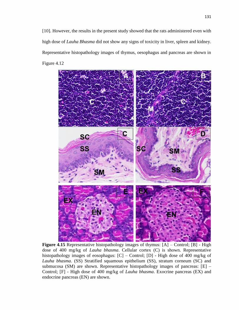
[10]. However, the results in the present study showed that the rats administered even with
high dose of Lauha Bhasma did not show any signs of toxicity in liver, spleen and kidney.
Representative histopathology images of thymus, oesophagus and pancreas are shown in
Figure 4.12
Figure 4.15 Representative histopathology images of thymus: [A] – Control; [B] - High
dose of 400 mg/kg of Lauha bhasma. Cellular cortex (C) is shown. Representative
histopathology images of eosophagus: [C] – Control; [D] - High dose of 400 mg/kg of
Lauha bhasma. (SS) Stratified squamous epithelium (SS), stratum corneum (SC) and
submucosa (SM) are shown. Representative histopathology images of pancreas: [E] –
Control; [F] - High dose of 400 mg/kg of Lauha bhasma. Exocrine pancreas (EX) and
endocrine pancreas (EN) are shown.

132
In an earlier study, porta caval shunt was surgically created in male Sprague-Dawley rats
to study iron accumulation in pancreas [18].The rats were fed with carbonyl iron diet for
17 weeks. Histopathological section revealed chronic pancreatitis characterized by acinar
atrophy, proliferation of intercalated ducts, mononuclear cell infiltration and fibrosis.
Oxidative stress was also confirmed with the elevated levels of glutathione disulfide [18] .
However, the results in the present study showed that the rats administered even with high
dose of Lauha Bhasma did not show any signs of toxicity in pancreas.
Representative histopathology images of intestine, brain and ovary are shown in Figure
4.13.
Mohammed et al. [19] studied the effect of changes in brain neuro transmitters of rats due
to administration of iron fortified diet. The levels of serotonin and dopamine were found
to be reduced significantly. Increase in lipid peroxidation in brain as a result of excess iron
deposition led to oxidative stress that ultimately resulted in neuro degenerative disorders.
Histopathology studies revealed the conditions of meningeal haemorrhage, congestion and
edema due to excessive iron overload. Further degenerated neurons, satellitosis and
neuronophagia were also observed in cerebal cortex [19].
Sobotka et al. [20] investigated toxicity and neuro behavioral changes in rodents fed with
different concentrations of dietary iron. Dose dependent toxicity and behavioral changes
were observed [20]. Another study revealed behavioral impairments combined with brain
iron accumulation in rats injected with ferrous sulphate for five days [21].

133
Figure 4.16 Representative histopathology images of intestine: [A] – Control; [B] - High
dose of 400 mg/kg of Lauha bhasma. Intestinal crypts (C) are shown. Representative
histopathology images of brain: [C] – Control; [D] - High dose of 400 mg/kg of Lauha
bhasma. Neurons (N), eosinophilic cytoplasmic processes (C) and glial cells (G) are
shown. Representative histopathology images of ovary: [E] – Control; [F] - High dose of
400 mg/kg of Lauha bhasma. Ovarian stroma (OS) and ovarian follicle (OF) are shown.
134
In vitro studies were carried out using PC 12 cells incubated with ferrocene and dopamine.
The addition of Fe2+ (in the form of ferrocene) increased the oxidation of monoamines.
The oxidation products were bound covalently to sulfhydryl groups of serotonin binding
proteins leading to apoptosis in PC-12 cells [22].
The effect of ferrous fumarate at low dose on dextran sulfate sodium (DSS) induced colitis
in rats has been reported [23]. The signs of intestinal inflammation and plasma redox status
were observed in DSS-administered groups with respect to control groups. A significant
increase in colitis score was observed in DSS-administered groups. Colitis score in DSS-
administered groups was further increased due to administration of ferrous fumarate. Rats
that received ferrous fumarate only showed normal cyto –architecture with no excess iron
deposition. No changes were observed in plasma redox status in DSS induced colitis rats
when compared to control groups [23].
In another study, the effect of oral iron supplementation at two different doses was
investigated in DSS induced colitis rats. Iron with doses of 0.3 % and 3 % showed higher
histological scores of colitis accompanied by heavier rectal bleeding and shortening of the
colon. The levels of antioxidant vitamins were also decreased with increased lipid
peroxidation [24]. The free radical generating capacity and lipid peroxidation of the
intestine in rats were assessed through administration of iron enriched diets for six months
[25]. Though the chronic administration of iron did not increase crypt cell proliferation,
increased free radical generation in colon and lipid peroxidation in caecum were observed
[25]. However, the results in the present study showed that the rats administered even with
high dose of Lauha Bhasma did not show any signs of toxicity in intestine and brain.

135
Histopathology of other organs were also carried out and normal cyto articheture was
observed in high dose Lauha bhasma treated group, when compared to control group.
Representative histopathology images of thyroid, lungs and heart are shown in Figure 4.14
Figure 4.17 Representative histopathology images of thyroid: [A] – Control; [B] - High
dose of 400 mg/kg of Lauha bhasma. Cuboidal epithelium (CE) and homogenous colloid
(C) are shown. Representative histopathology images of lungs: [C] – Control; [D] - High
dose of 400 mg/kg of Lauha bhasma. Alveolar wall (Arrow mark) and alveolar lumen (AL)
are shown. Representative histopathology images of heart: [E] – Control; [F] - High dose
of 400 mg/kg of Lauha bhasma. Cardiac myocytes (CM) and cytoplasm (C) are shown.

136
Representative histopathology images of adrenal gland, pituitary gland and bone
marrow are shown in Figure 4.15
Figure 4.18 Representative histopathology images of adrenal gland: [A] – Control; [B] -
High dose of 400 mg/kg of Lauha bhasma. Cortex (C) and medulla (M) are shown.
Representative histopathology images of pituitary gland: [C] – Control; [D] - High dose of
400 mg/kg of Lauha bhasma. Pars distalis, (D) pars intermedia (I) andpars nervosa (N) are
shown. Representative histopathology images of bone marrow: [E] – Control; [F] - High
dose of 400 mg/kg of Lauha bhasma. Erythroid and myeloid precursors (P) and
megakaryocytes (M) are shown.

137
4.4 CONCLUSION
Iron, despite its essential role in biological system can cause adverse toxic effects ranging
from male sterility to tumorogenesis. Lauha bhasma even when administered at a high dose
was not toxic to the vital organs of the rats. Histopathological studies showed that the cyto
architecture of rats treated with high dose of Lauha bhasma was comparable to normal
control. This was further supported by the biochemical and hematological parameters
which showed that Lauha bhasma did not cause any significant changes in the levels of
those parameters.
4.5 REFERENCES
1. Nagarajan S, Pemiah B, Krishnan UM, Rajan KS, Krishnaswamy S, Sethuraman S.
Physico-Chemical Characterization of Lead based Indian Traditional Medicine-
Naga bhasma. Int J Pharm Pharm Sci 2012;4:69–74.
2. Kales SN, Christophi CA, Saper RB. Hematopoietic toxicity from lead-containing
Ayurvedic medications Stefanos. Med Sci Monit 2008;13:1–8.
3. Saper RB, Phillips RS, Khouri N, Davis RB, Paquin J. Ayurvedic Medicines Sold
via the Internet. JAMA 2008;300:915–24.
4. Saper RB, Kales SN, Paquin J, Burns MJ, Eisenberg DM, Davis RB, et al. Heavy
metal content of ayurvedic herbal medicine products. JAMA 2004;292:2868–73.
5. Sarkar PK, Das S, Prajapati PK. Ancient concept of metal pharmacology based on
Ayurvedic literature. Anc Sci Life 2010;29:1–6.
6. Papanikolaou G, Pantopoulos K. Iron metabolism and toxicity. Toxicol Appl
Pharmacol 2005;202:199–211.
7. Anonymous. The Ayurvedic Formulary of India. Second edi. Ministry of Health and
Family Welfare, Government of India, New Delhi; 2003.
8. Reagan-Shaw S, Nihal M, Ahmad N. Dose translation from animal to human studies
revisited. FASEB J 2008;22:659–61.

138
9. Machado AA, Izumi C, Zucoloto S, Freitas O de. Iron depositions in rat liver and
spleen were lower when the mineral was supplied as a derivative of casein
hydrolysate in place of iron sulfate. Alim Nutr, 2005;16:111–6.
10. Papanastasiou D a, Vayenas D V, Vassilopoulos a, Repanti M. Concentration of iron
and distribution of iron and transferrin after experimental iron overload in rat tissues
in vivo: study of the liver, the spleen, the central nervous system and other organs.
Pathol Res Pract 2000;196:47–54.
11. Sarkar PK, Prajapati PK, Shukla VJ, Ravishankar B, Choudhary a K. Toxicity and
recovery studies of two ayurvedic preparations of iron. Indian J Exp Biol
2009;47:987–92.
12. Sarkar R, Hazra B, Mandal N. Reducing power and iron chelating property of
Terminalia chebula (Retz.) alleviates iron induced liver toxicity in mice. BMC
Complement Altern Med 2012;12:144.
13. Whittaker P, Chanderbhan RF. Effect of increasing iron supplementation on blood
lipids in rats. Br J Nutr 2007;86:587.
14. Andrews NC, Schmidt PJ. Iron homeostasis. Annu Rev Physiol 2007;69:69–85.
15. Ozguner M, Sayin N. Histological changes in rat liver after chronic iron- sorbitol
overload. J Ankara Med Sch 2002;24:49–54.
16. Al-shaikh TM. Accumulation and Toxicity of Extra Iron Fed as Fumarate in Rabbits.
Glob Adv Res J Environ Sci Toxicol 2012;1:190–8.
17. Whittaker P, Dunkel VC, Bucci TJ, Kusewitt DF, Thurman JD, Warbritton A, et al.
Genome-Linked Toxic Responses to Dietary Iron Overload. Toxicol Pathol
1997;25:556–64.
18. Horne WI, Tandler B, Dubick M a, Niemelä O, Brittenham GM, Tsukamoto H. Iron
overload in the rat pancreas following portacaval shunting and dietary iron
supplementation. Exp Mol Pathol 1997;64:90–102.
19. Elseweidy MM, El-Baky AEA. Effect of dietary iron overload in rat brain:
Oxidative stress, neurotransmitter level and serum metal ion in relation to
neurodegenerative disorders. Indian J Exp Biol 2008;46:855–8.

139
20. Sobotka TJ, Whittaker P, Sobotka JM, Brodie RE, Quander DY, Robl M, et al.
Neurobehavioral dysfunctions associated with dietary iron overload. Physiol Behav
1996;59:213–9.
21. Maaroufi K, Ammari M, Jeljeli M, Roy V, Sakly M, Abdelmelek H. Impairment of
emotional behavior and spatial learning in adult Wistar rats by ferrous sulfate.
Physiol Behav 2009;96:343–9.
22. Velez-Pardo C, Jimenez Del Rio M, Verschueren H, Ebinger G, Vauquelin G.
Dopamine and iron induce apoptosis in PC12 cells. Pharmacol Toxicol 1997;80:76–
84.
23. Erichsen K, Milde AM, Arslan G, Helgeland L, Gudbrandsen OA, Ulvik RJ, et al.
Low-dose oral ferrous fumarate aggravated intestinal inflammation in rats with
DSS-induced colitis. Inflamm Bowel Dis 2005;11:744–8.
24. Carrier J, Aghdassi E, Platt I, Cullen J, Allard JP. Effect of oral iron supplementation
on oxidative stress and colonic inflammation in rats with induced colitis. Aliment
Pharmacol Ther 2001;15:1989–99.
25. Lund EK, Fairweather-Tait SJ, Wharf SG, Johnson IT. Chronic exposure to high
levels of dietary iron fortification increases lipid peroxidation in the mucosa of the
rat large intestine. J Nutr 2001;131:2928–31.