Lecture 6 Fluid as skeleton: hydrostatic, hydraulic skeletons and muscular hydrostats

Circulation & Cardiovascular
Systems
Chapter 32: pp. 593 - 612
1(Normal): © Yorgos Nikas/Getty Images; (Leukemia): © SPL/Photo Researchers, Inc.
Copyright © The McGraw-Hill Companies, Inc. Permission required for reproduction or display.
SEM 5,270 ×
Leukemia
Outline• Transport in Invertebrates
• Open versus Closed Circulatory Systems• Transport in Vertebrates• Transport in Humans
• Heartbeat• Vascular Pathways• Blood Pressure
• Cardiovascular Disorders• Blood
• Components• Clotting
2

TransportinInvertebrates• Small aquatic animals with no circulatory system
• May rely on external water in gastrovascular cavity to service cells
• Roundworms and other pseudocoelomates• Use a fluid‐filled body cavity as a means of transporting substances
• Fluid‐filled cavity can also act as a hydrostatic skeleton • Animals that have a rigid skeleton
• May still rely on body fluids for the purpose of locomotion
• Bivalves pump hemolymph into the foot for digging into mud 3

AquaticOrganismsWithoutaCirculatorySystem
4
Copyright © The McGraw-Hill Companies, Inc. Permission required for reproduction or display.
c. Red sea star, Mediastar
coelomic fluid
b. Flatworm
a. Hydra
mouthenzymes
food
food
7×
undigestedwaste products
nutrientuptake byendocytosis
gastrovascularcavity
a: © CABISCO/Visuals Unlimited; b: © B. Runk/S. Schoenberger/Grant Heilman Photography; c: © Randy Morse, GoldenStateImages.com;

Openvs.ClosedInvertebrateCirculation• Two types of circulatory fluids:
• Blood ‐ contained within blood vessels
• Hemolymph ‐ flows into hemocoel
• Open Circulatory System• Heart pumps hemolymph via vessels
• Vessels empty into tissue spaces
• Closed Circulatory System• Heart pumps blood to capillaries
• Gases and materials diffuse to and from nearby cells
• Vessels return blood to heart without it contacting tissues 5

Openvs.ClosedCirculatorySystems
6
Copyright © The McGraw-Hill Companies, Inc. Permission required for reproduction or display.
valve
heart
a. Open circulatory system b. Closed circulatory system
ostia
hemocoel
hemolymph
pump pumpostia
dorsalaorta
tubularheart
ventralbloodvessel
dorsalbloodvessel
lateralvessel

TransportintheVertebrates• All vertebrates have a closed cardiovascular system• Vertebrate heart:
• Atrial chamber(s) of heart receive blood from general circulation• Ventricle chamber(s) of heart pump blood out through blood vessels
• Vertebrate vessels:• Arteries ‐ Carry blood away from heart• Arterioles – Lead to capillaries• Capillaries ‐ Exchange materials with tissue fluid• Venules ‐ Lead to veins• Veins ‐ Return blood to heart
7

TransportinVertebrates
8
Copyright © The McGraw-Hill Companies, Inc. Permission required for reproduction or display.
endothelium
endotheliumfibrous connective tissue
b. Capillary
Inner layerMiddle layerOuter layer
endothelium
closed valve
fibrous connective tissue
Inner layerMiddle layerOuter layer
smoothmuscle
elastictissue
a. Artery
elastictissue
smoothmuscle
c. Vein

AnatomyofaCapillaryBed
9
artery
arteriole
vein
O2-richblood flow
O2-poorblood flow
precapillarysphincter
arteriovenousshunt
Copyright © The McGraw-Hill Companies, Inc. Permission required for reproduction or display.
venule

ComparisonofCirculatoryCircuitsinVertebrates
10
Copyright © The McGraw-Hill Companies, Inc. Permission required for reproduction or display.
gill capillaries
ventricle
atrium
aorta
heart
a.
ventricle
aorta
heart
aorta
b. c.
pulmonarycapillaries
pulmonarycircuit
rightatrium
leftatrium
leftventricle
rightventricle
systemiccapillaries
systemiccircuit
rightatrium
leftatrium
systemiccapillaries
systemiccircuit
pulmonarycircuit
pulmonarycapillaries
systemiccapillaries

ComparisonofCirculatoryPathways• Fish ‐ Blood flows in single loop
• Single atrium and single ventricle
• Amphibians ‐ Blood flows in double loop
• Two atria with single ventricle
• Other vertebrates ‐ Blood flows in a double loop
• Heart divided by septum into separate sides11

TransportinHumans• Human Heart
• Fist‐sized
• Cone‐shaped
• Very muscular organ (special cardiac fibers)
• Lies within a fluid‐filled sac (the pericardium)
12

HumanHeart:GrossAnatomy• Septum separates heart into left & right halves
• Each half has two chambers
• Upper two chambers are the atria
• Thin‐walled
• Receive blood from circulation
• Lower two chambers are the ventricles
• Thick‐walled
• Pump blood away from heart 13

ExternalHeartAnatomy
14
Copyright © The McGraw-Hill Companies, Inc. Permission required for reproduction or display.
a.
left subclavian arteryleft common carotid arterybrachiocephalic artery
superior vena cavaaortic archaortaleft pulmonary arterypulmonary trunkleft pulmonary veinsright pulmonary artery
right pulmonary veins
left atriumleft cardiac vein
right atriumright coronary artery
left ventricle
right ventricle
Inferior vena cava
apex
b.
lung
pericardium heart
sternum
b: © SIU/Visuals Unlimited

InternalViewoftheHeart
15
Copyright © The McGraw-Hill Companies, Inc. Permission required for reproduction or display.
b.
right pulmonary artery
right atrium
chordae tendineae
right ventricle
inferior vena cava
right pulmonary veins
brachiocephalic artery
superior vena cava
left common carotid artery
semilunar valve
left pulmonary veins
aorta
left pulmonary artery
pulmonary trunk
mitochondrion
gap junction
left atrium
left subclavian artery
left ventricle
septum
a.
papillary muscles
cardiacmuscle cell
intercalateddisk
atrioventricular(bicuspid) valveatrioventricular(tricuspid) valve
b: © Dr. Don W. Fawcett/Visuals Unlimited;

HumanHeart:Valves• Valves open and close to control blood flow through heart
• Atrioventricular valves
• Tricuspid
• Bicuspid
• Semilunar valves
• Pulmonary
• Aortic
16

TransportinHumans• Blood returning to heart from systemic circuit
• Enters right atrium• Right atrium pumps through tricuspid valve to right ventricle
• Right ventricle pumps blood through pulmonary valve to the pulmonary circuit
• Blood returning to heart from pulmonary circuit• Enters left atrium• Left atrium pumps through mitral valve to left ventricle• Left ventricle pumps blood through aortic valve to the systemic circuit
• Oxygen‐poor blood never mixes with oxygen‐rich blood (in humans)
17

Heartbeat• Systole ‐ Contraction of heart chambers• Diastole ‐ Relaxation of heart chambers• Pulse ‐ Two‐part pumping action that takes about a second• Blood collects in atria, the atria contract
• Pushes blood through tricuspid and mitral valves into the resting lower ventricles
• This phase (the longer of the two) is called the diastole• Second part begins when ventricles fill
• Ventricles contract• This is called systole
• After blood moves into the pulmonary artery and aorta, the ventricles relax
18

StagesintheCardiacCycle
19
Copyright © The McGraw-Hill Companies, Inc. Permission required for reproduction or display.
a.
b.
c.
d.
aortic semilunar valve bicuspid valve
aorta
aorta
pulmonaryvein
superiorvena cava
semilunarvalves
leftatrium right
atrium
leftventricle
inferiorvena cava
semilunarvalves close(“dub”)
atrioventricular (AV)valves close(“lub”)
representscontraction
rightventricle
rightatrium
pulmonaryvein
d: © Biophoto Associates/Photo Researchers, Inc.

Animation

Animation
Please note that due to differing operating systems, some animations will not appear until the presentation is viewed in Presentation Mode (Slide Show view). You may see blank slides in the “Normal” or “Slide Sorter” views. All animations will appear after viewing in Presentation Mode and playing each animation. Most animations will require the latest version of the Flash Player, which is available at http://get.adobe.com/flashplayer.

ConductionSystemoftheHeart
22
Copyright © The McGraw-Hill Companies, Inc. Permission required for reproduction or display.
Purkinje fibers
a.
b. Normal ECG
d. Recording of an ECG
R
P
Q
T
S
c. Ventricular fibrillation
SA node
AV node
branches ofatrioventricularbundle
d: © David Joel/MacNeal Hospital/Getty Images

Animation
Please note that due to differing operating systems, some animations will not appear until the presentation is viewed in Presentation Mode (Slide Show view). You may see blank slides in the “Normal” or “Slide Sorter” views. All animations will appear after viewing in Presentation Mode and playing each animation. Most animations will require the latest version of the Flash Player, which is available at http://get.adobe.com/flashplayer.
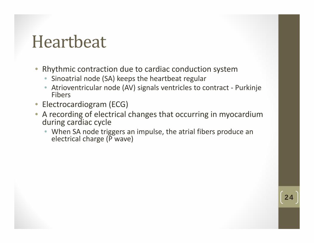
Heartbeat• Rhythmic contraction due to cardiac conduction system
• Sinoatrial node (SA) keeps the heartbeat regular• Atrioventricular node (AV) signals ventricles to contract ‐ Purkinje Fibers
• Electrocardiogram (ECG)• A recording of electrical changes that occurring in myocardium during cardiac cycle• When SA node triggers an impulse, the atrial fibers produce an electrical charge (P wave)
24

Animation
Please note that due to differing operating systems, some animations will not appear until the presentation is viewed in Presentation Mode (Slide Show view). You may see blank slides in the “Normal” or “Slide Sorter” views. All animations will appear after viewing in Presentation Mode and playing each animation. Most animations will require the latest version of the Flash Player, which is available at http://get.adobe.com/flashplayer.

VascularPathways• Human cardiovascular system includes two major circular pathways:• Pulmonary Circuit
• Takes oxygen‐poor blood to the lungs and returns oxygen‐rich blood to the heart
• Systemic Circuit• Takes blood throughout the body from the aorta to the vena cava
26

PathofBlood
27
Copyright © The McGraw-Hill Companies, Inc. Permission required for reproduction or display.
jugular vein(also subclavianvein from arms)
head and arms carotid artery(also subclavianartery to arms)
pulmonaryartery
pulmonaryvein
superiorvena cava aorta
inferiorvena cava
hepaticvein mesenteric
arteries
hepaticportalvein
liver
renalartery
digestivetract
renalvein kidneys
trunk and legs
iliacartery
Iliac vein
heart
lungs
CO2 O2
CO2 O2
CO2O2
CO2 O2

VelocityandBloodPressure
28
Copyright © The McGraw-Hill Companies, Inc. Permission required for reproduction or display.
Mag
nitu
de
totalcross-sectionalarea ofvessels
ariteries
Blood Flow
ariterioles capillaries veinsvenules
bloodpressure
velocity

CrossSectionofaValveinaVein
29
Copyright © The McGraw-Hill Companies, Inc. Permission required for reproduction or display.
to heart to heart
a. Contracted skeletal musclepushes blood past open valve.
b. Closed valve preventsbackward flow of blood.

BloodPressure• The beat of the heart supplies pressure that keeps blood moving in the arteries• Systolic Pressure results from blood forced into the arteries during ventricular systole
• Diastolic Pressure is the pressure in the arteries during during ventricular diastole
• Skeletal muscle contraction pushes blood in the veins toward the heart
• Blood pressure• Normally measured with a sphygmomanometer on the brachial artery
• Expressed in the form: Systolic “over” Diastolic30

CardiovascularDisorders• Hypertension ‐ High blood pressure• Atherosclerosis ‐ Accumulation of fatty materials in inner linings of arteries
• Stroke ‐ Cranial arteriole bursts or is blocked by an embolus• Heart attack – (Myocardial infarction) Coronary artery becomes partially blocked
• Angina pectoris – Painful squeezing sensation from myocardial oxygen insufficiency
31

CoronaryArteriesandPlaque
32
Copyright © The McGraw-Hill Companies, Inc. Permission required for reproduction or display.
coronary artery ulceration
fat
lumen of vessel
cholesterolcrystals
atheroscleroticplaque
© Biophoto Associates/Photo Researchers, Inc.

Blood:HomeostasisFunctions• Transports substances to and from capillaries for exchange with tissue fluid
• Guards against pathogen invasion• Regulates body temperature• Buffers body pH• Maintain osmotic pressure• Clots prevent blood/fluid loss
33

RedBloodCells• Small, biconcave disks
• Lack a nucleus and contain hemoglobin
• Hemoglobin contains
• Four globin protein chains
• Each associated with an iron‐containing heme
• Manufactured continuously in bone marrow of skull, ribs, vertebrae, and ends of long bones
34

WhiteBloodCells• Most types larger than red blood cells• Contain a nucleus and lack hemoglobin• Important in inflammatory response
• Neutrophils enter tissue fluid and phagocytize foreign material• Lymphocytes (T Cells) attack infected cells• Antigens cause body to produce antibodies
35

CompositionofBloodBlood
Plasma 46-63% Formed Elements 37-54%
Plasma Protein 7% Water 92% Other Solutes 1% Platelets RBC 99.9% WBC
Albumin
Fibrinogen
Globulin
Regulatory Proteins
Eg. Electrolytes
Monocytes
Basophils
Eosinophils
Neatrophils
Lymphocytes
36

Platelets• Platelets
• Result from fragmentation of megakaryocytes• Involved in coagulation
• Blood clot consists of:• Platelets• Red blood cells• All entangled within fibrin threads
37

BloodClotting
38
Copyright © The McGraw-Hill Companies, Inc. Permission required for reproduction or display.
1. Blood vessel is punctured.
platelet plug fibrin threads
2. Platelets congregate andform a plug.
3. Fibrin threads form andtrap red blood cells.
fibrinthreads
redblood cell
© Eye of Science/Photo Researchers, Inc.

CapillaryExchange• Capillaries very narrow – Tiny RBCs must go through single file• Wall of capillaries very thin to facilitate diffusion of nutrients, gases, and wastes• Oxygen and nutrients exit a capillary near the arterial end• Carbon dioxide and waste molecules enter a capillary near the venous end
39

BloodType• Determined by the presence or absence of surface antigens (agglutinogens) • Antigens A, B and Rh (D)
• Antibodies in the plasma (agglutinins)• Cross‐reactions occur when antigens meet antibodies
40

BloodType
41

Agglutination
42
Copyright © The McGraw-Hill Companies, Inc. Permission required for reproduction or display.
Agglutination
binding
500×
clumping
antigen
Type A bloodof donor
anti- A antibody oftype B recipient

BloodType• During pregnancy, if the mother is Rh negative and the fatheris Rh positive, the child may be Rh positive.• Rh‐positive red blood cells may leak across the placenta• The mother will produce anti‐Rh antibodies.• Antibodies may attack the embryo in a subsequent pregnancy
43

Review• Transport in Invertebrates
• Open versus Closed Circulatory Systems• Transport in Vertebrates• Transport in Humans
• Heartbeat• Vascular Pathways• Blood Pressure
• Cardiovascular Disorders• Blood
• Components• Clotting
44

Circulation & Cardiovascular
Systems
Chapter 32: pp. 593 - 612
45(Normal): © Yorgos Nikas/Getty Images; (Leukemia): © SPL/Photo Researchers, Inc.
Copyright © The McGraw-Hill Companies, Inc. Permission required for reproduction or display.
SEM 5,270×
Leukemia