Chapter 27 Fibular Resections - Sarcoma.orgsarcoma.org/publications/OTOS_Book/13282_ON-27.pdf1...
10
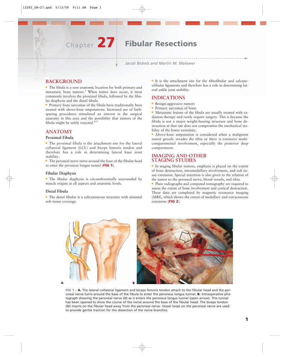
1 Chapter 27 Jacob Bickels and Martin M. Malawer Fibular Resections BACKGROUND ■ The fibula is a rare anatomic location for both primary and metastatic bone tumors. 1 When tumor does occur, it most commonly involves the proximal fibula, followed by the fibu- lar diaphysis and the distal fibula. ■ Primary bone sarcomas of the fibula have traditionally been treated with above-knee amputations. Increased use of limb- sparing procedures stimulated an interest in the surgical anatomy in this area and the possibility that tumors of the fibula might be safely resected. 2–7 ANATOMY Proximal Fibula ■ The proximal fibula is the attachment site for the lateral collateral ligament (LCL) and biceps femoris tendon and therefore has a role in determining lateral knee joint stability. ■ The peroneal nerve turns around the base of the fibular head to enter the peroneus longus tunnel (FIG 1). Fibular Diaphysis ■ The fibular diaphysis is circumferentially surrounded by muscle origins at all aspects and anatomic levels. Distal Fibula ■ The distal fibular is a subcutaneous structure with minimal soft tissue coverage. ■ It is the attachment site for the tibiofibular and calcane- ofibular ligaments and therefore has a role in determining lat- eral ankle joint stability. INDICATIONS ■ Benign-aggressive tumors ■ Primary sarcomas of bone ■ Metastatic lesions of the fibula are usually treated with ra- diation therapy and rarely require surgery. This is because the fibula is not a major weight-bearing structure and bone de- struction at that site does not compromise the mechanical sta- bility of the lower extremity. ■ Above-knee amputation is considered when a malignant tumor grossly invades the tibia or there is extensive multi- compartmental involvement, especially the posterior deep compartment. IMAGING AND OTHER STAGING STUDIES ■ In staging fibular tumors, emphasis is placed on the extent of bone destruction, intramedullary involvement, and soft tis- sue extension. Special attention is also given to the relation of the tumor to the peroneal nerve, blood vessels, and tibia. ■ Plain radiographs and computed tomography are required to assess the extent of bone involvement and cortical destruction. These data are completed by magnetic resonance imaging (MRI), which shows the extent of medullary and extraosseous extension (FIG 2). A B FIG 1 • A. The lateral collateral ligament and biceps femoris tendon attach to the fibular head and the per- oneal nerve turns around the base of the fibula to enter the peroneus longus tunnel. B. Intraoperative pho- tograph showing the peroneal nerve (N) as it enters the peroneus longus tunnel (open arrow). This tunnel has been opened to show the course of the nerve around the base of the fibular head. The biceps tendon (Bi) inserts on the fibular head away from the peroneal nerve. Vessel loops on the peroneal nerve are used to provide gentle traction for the dissection of the nerve branches. 13282_ON-27.qxd 5/13/09 9:11 AM Page 1
Transcript of Chapter 27 Fibular Resections - Sarcoma.orgsarcoma.org/publications/OTOS_Book/13282_ON-27.pdf1...
1
Chapter 27Jacob Bickels and Martin M. Malawer
Fibular Resections
BACKGROUND■ The fibula is a rare anatomic location for both primary andmetastatic bone tumors.1 When tumor does occur, it mostcommonly involves the proximal fibula, followed by the fibu-lar diaphysis and the distal fibula.■ Primary bone sarcomas of the fibula have traditionally beentreated with above-knee amputations. Increased use of limb-sparing procedures stimulated an interest in the surgicalanatomy in this area and the possibility that tumors of thefibula might be safely resected.2–7
ANATOMYProximal Fibula■ The proximal fibula is the attachment site for the lateralcollateral ligament (LCL) and biceps femoris tendon andtherefore has a role in determining lateral knee jointstability.■ The peroneal nerve turns around the base of the fibular headto enter the peroneus longus tunnel (FIG 1).
Fibular Diaphysis■ The fibular diaphysis is circumferentially surrounded bymuscle origins at all aspects and anatomic levels.
Distal Fibula■ The distal fibular is a subcutaneous structure with minimalsoft tissue coverage.
■ It is the attachment site for the tibiofibular and calcane-ofibular ligaments and therefore has a role in determining lat-eral ankle joint stability.
INDICATIONS■ Benign-aggressive tumors■ Primary sarcomas of bone■ Metastatic lesions of the fibula are usually treated with ra-diation therapy and rarely require surgery. This is because thefibula is not a major weight-bearing structure and bone de-struction at that site does not compromise the mechanical sta-bility of the lower extremity.■ Above-knee amputation is considered when a malignanttumor grossly invades the tibia or there is extensive multi-compartmental involvement, especially the posterior deepcompartment.
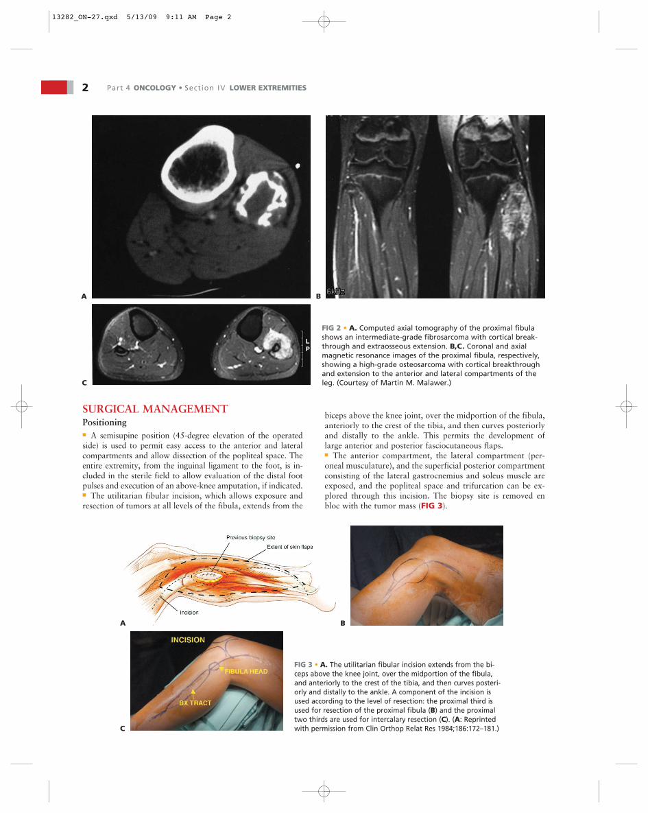
IMAGING AND OTHER STAGING STUDIES■ In staging fibular tumors, emphasis is placed on the extentof bone destruction, intramedullary involvement, and soft tis-sue extension. Special attention is also given to the relation ofthe tumor to the peroneal nerve, blood vessels, and tibia.■ Plain radiographs and computed tomography are required toassess the extent of bone involvement and cortical destruction.These data are completed by magnetic resonance imaging(MRI), which shows the extent of medullary and extraosseousextension (FIG 2).
A B
FIG 1 • A. The lateral collateral ligament and biceps femoris tendon attach to the fibular head and the per-oneal nerve turns around the base of the fibula to enter the peroneus longus tunnel. B. Intraoperative pho-tograph showing the peroneal nerve (N) as it enters the peroneus longus tunnel (open arrow). This tunnelhas been opened to show the course of the nerve around the base of the fibular head. The biceps tendon(Bi) inserts on the fibular head away from the peroneal nerve. Vessel loops on the peroneal nerve are usedto provide gentle traction for the dissection of the nerve branches.
13282_ON-27.qxd 5/13/09 9:11 AM Page 1
2 Part 4 ONCOLOGY • Section IV LOWER EXTREMITIES
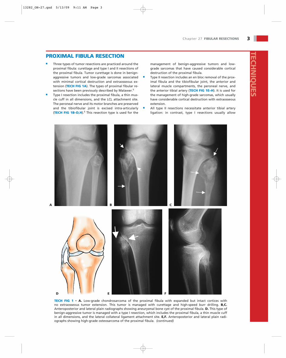
SURGICAL MANAGEMENTPositioning■ A semisupine position (45-degree elevation of the operatedside) is used to permit easy access to the anterior and lateralcompartments and allow dissection of the popliteal space. Theentire extremity, from the inguinal ligament to the foot, is in-cluded in the sterile field to allow evaluation of the distal footpulses and execution of an above-knee amputation, if indicated.■ The utilitarian fibular incision, which allows exposure andresection of tumors at all levels of the fibula, extends from the
biceps above the knee joint, over the midportion of the fibula,anteriorly to the crest of the tibia, and then curves posteriorlyand distally to the ankle. This permits the development oflarge anterior and posterior fasciocutaneous flaps.■ The anterior compartment, the lateral compartment (per-oneal musculature), and the superficial posterior compartmentconsisting of the lateral gastrocnemius and soleus muscle areexposed, and the popliteal space and trifurcation can be ex-plored through this incision. The biopsy site is removed enbloc with the tumor mass (FIG 3).
FIG 2 • A. Computed axial tomography of the proximal fibulashows an intermediate-grade fibrosarcoma with cortical break-through and extraosseous extension. B,C. Coronal and axialmagnetic resonance images of the proximal fibula, respectively,showing a high-grade osteosarcoma with cortical breakthroughand extension to the anterior and lateral compartments of theleg. (Courtesy of Martin M. Malawer.)
A B
C
FIG 3 • A. The utilitarian fibular incision extends from the bi-ceps above the knee joint, over the midportion of the fibula,and anteriorly to the crest of the tibia, and then curves posteri-orly and distally to the ankle. A component of the incision isused according to the level of resection: the proximal third isused for resection of the proximal fibula (B) and the proximaltwo thirds are used for intercalary resection (C). (A: Reprintedwith permission from Clin Orthop Relat Res 1984;186:172–181.)
A B
C
13282_ON-27.qxd 5/13/09 9:11 AM Page 2
TECH
NIQ
UES
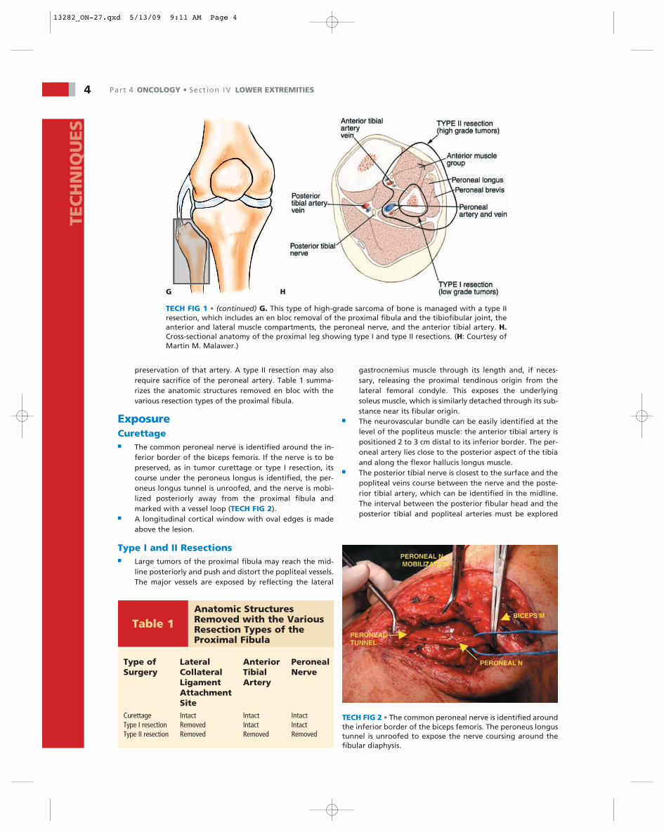
TECH FIG 1 • A. Low-grade chondrosarcoma of the proximal fibula with expanded but intact cortices with no extraosseous tumor extension. This tumor is managed with curettage and high-speed burr drilling. B,C.Anteroposterior and lateral plain radiographs showing aneurysmal bone cyst of the proximal fibula. D. This type ofbenign-aggressive tumor is managed with a type I resection, which includes the proximal fibula, a thin muscle cuffin all dimensions, and the lateral collateral ligament attachment site. E,F. Anteroposterior and lateral plain radi-ographs showing high-grade osteosarcoma of the proximal fibula. (continued)
A B C
D E F
PROXIMAL FIBULA RESECTION■ Three types of tumor resections are practiced around the
proximal fibula: curettage and type I and II resections ofthe proximal fibula. Tumor curettage is done in benign-aggressive tumors and low-grade sarcomas associatedwith minimal cortical destruction and extraosseous ex-tension (TECH FIG 1A). The types of proximal fibular re-sections have been previously described by Malawer.5
■ Type I resection includes the proximal fibula, a thin mus-cle cuff in all dimensions, and the LCL attachment site.The peroneal nerve and its motor branches are preservedand the tibiofibular joint is excised intra-articularly(TECH FIG 1B–D,H).5 This resection type is used for the
management of benign-aggressive tumors and low-grade sarcomas that have caused considerable corticaldestruction of the proximal fibula.
■ Type II resection includes an en bloc removal of the prox-imal fibula and the tibiofibular joint, the anterior andlateral muscle compartments, the peroneal nerve, andthe anterior tibial artery (TECH FIG 1E–H). It is used forthe management of high-grade sarcomas, which usuallyhave considerable cortical destruction with extraosseousextension.
■ All type II resections necessitate anterior tibial arteryligation: in contrast, type I resections usually allow
Chapter 27 FIBULAR RESECTIONS 3
13282_ON-27.qxd 5/13/09 9:11 AM Page 3
4 Part 4 ONCOLOGY • Section IV LOWER EXTREMITIESTE
CH
NIQ
UES
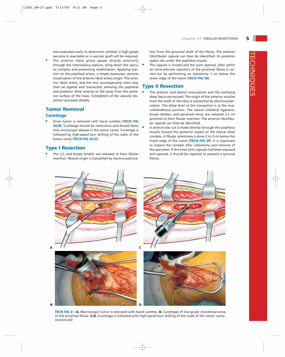
TECH FIG 1 • (continued) G. This type of high-grade sarcoma of bone is managed with a type IIresection, which includes an en bloc removal of the proximal fibula and the tibiofibular joint, theanterior and lateral muscle compartments, the peroneal nerve, and the anterior tibial artery. H.Cross-sectional anatomy of the proximal leg showing type I and type II resections. (H: Courtesy ofMartin M. Malawer.)
G H
Type of Lateral Anterior PeronealSurgery Collateral Tibial Nerve
Ligament ArteryAttachmentSite
Curettage Intact Intact IntactType I resection Removed Intact IntactType II resection Removed Removed Removed
Table 1Anatomic StructuresRemoved with the VariousResection Types of theProximal Fibula
TECH FIG 2 • The common peroneal nerve is identified aroundthe inferior border of the biceps femoris. The peroneus longustunnel is unroofed to expose the nerve coursing around thefibular diaphysis.
preservation of that artery. A type II resection may alsorequire sacrifice of the peroneal artery. Table 1 summa-rizes the anatomic structures removed en bloc with thevarious resection types of the proximal fibula.
ExposureCurettage■ The common peroneal nerve is identified around the in-
ferior border of the biceps femoris. If the nerve is to bepreserved, as in tumor curettage or type I resection, itscourse under the peroneus longus is identified, the per-oneus longus tunnel is unroofed, and the nerve is mobi-lized posteriorly away from the proximal fibula andmarked with a vessel loop (TECH FIG 2).
■ A longitudinal cortical window with oval edges is madeabove the lesion.
Type I and II Resections■ Large tumors of the proximal fibula may reach the mid-
line posteriorly and push and distort the popliteal vessels.The major vessels are exposed by reflecting the lateral
gastrocnemius muscle through its length and, if neces-sary, releasing the proximal tendinous origin from thelateral femoral condyle. This exposes the underlyingsoleus muscle, which is similarly detached through its sub-stance near its fibular origin.
■ The neurovascular bundle can be easily identified at thelevel of the popliteus muscle: the anterior tibial artery ispositioned 2 to 3 cm distal to its inferior border. The per-oneal artery lies close to the posterior aspect of the tibiaand along the flexor hallucis longus muscle.
■ The posterior tibial nerve is closest to the surface and thepopliteal veins course between the nerve and the poste-rior tibial artery, which can be identified in the midline.The interval between the posterior fibular head and theposterior tibial and popliteal arteries must be explored
13282_ON-27.qxd 5/13/09 9:11 AM Page 4
Chapter 27 FIBULAR RESECTIONS 5TEC
HN
IQU
ES
and evaluated early to determine whether a high-gradesarcoma is resectable or a vascular graft will be required.
■ The anterior tibial artery passes directly anteriorlythrough the interosseous septum, tying down the vascu-lar complex and preventing mobilization. Applying trac-tion on the popliteal artery, a simple maneuver, permitsvisualization of the anterior tibial artery origin. The ante-rior tibial artery and the two accompanying veins maythen be ligated and transected, allowing the poplitealand posterior tibial arteries to fall away from the poste-rior surface of the mass. Completion of the vascular dis-section proceeds distally.
Tumor RemovalCurettage■ Gross tumor is removed with hand curettes (TECH FIG
3A,B). Curettage should be meticulous and should leaveonly microscopic disease in the tumor cavity. Curettage isfollowed by high-speed burr drilling of the walls of thetumor cavity (TECH FIG 3C,D).
Type I Resection■ The LCL and biceps tendon are released at their fibular
insertion. Muscle origin is transected by electrocauteriza-
A
B
TECH FIG 3 • A. Macroscopic tumor is removed with hand curettes. B. Curettage of low-grade chondrosarcomaof the proximal fibula. C,D. Curettage is followed with high-speed burr drilling of the walls of the tumor cavity.(continued)
C
D
tion from the proximal shaft of the fibula. The anteriortibiofibular capsule can then be identified: its posterioraspect lies under the popliteus muscle.
■ The capsule is incised and the joint opened, after whichan intra-articular resection of the proximal fibula is car-ried out by performing an osteotomy 1 cm below thelower edge of the lesion (TECH FIG 3E).
Type II Resection■ The anterior and lateral musculature and the overlying
deep fascia are excised. The origin of the anterior musclesfrom the shaft of the tibia is transected by electrocauter-ization. The distal level of the transaction is at the mus-culotendinous junction. The lateral collateral ligament,biceps tendon, and peroneal nerve are released 2.5 cmproximal to their fibular insertion. The anterior tibiofibu-lar capsule can then be identified.
■ A semicircular cut is made directly through the popliteusmuscle toward the posterior aspect of the lateral tibialcondyle. A fibular osteotomy is done 2 to 3 cm below thelower edge of the tumor (TECH FIG 3F). It is importantto inspect the condyle after osteotomy and removal ofthe specimen. If the knee joint capsule had been exposedand opened, it should be repaired to prevent a synovialfistula.
13282_ON-27.qxd 5/13/09 9:11 AM Page 5
6 Part 4 ONCOLOGY • Section IV LOWER EXTREMITIES
E F
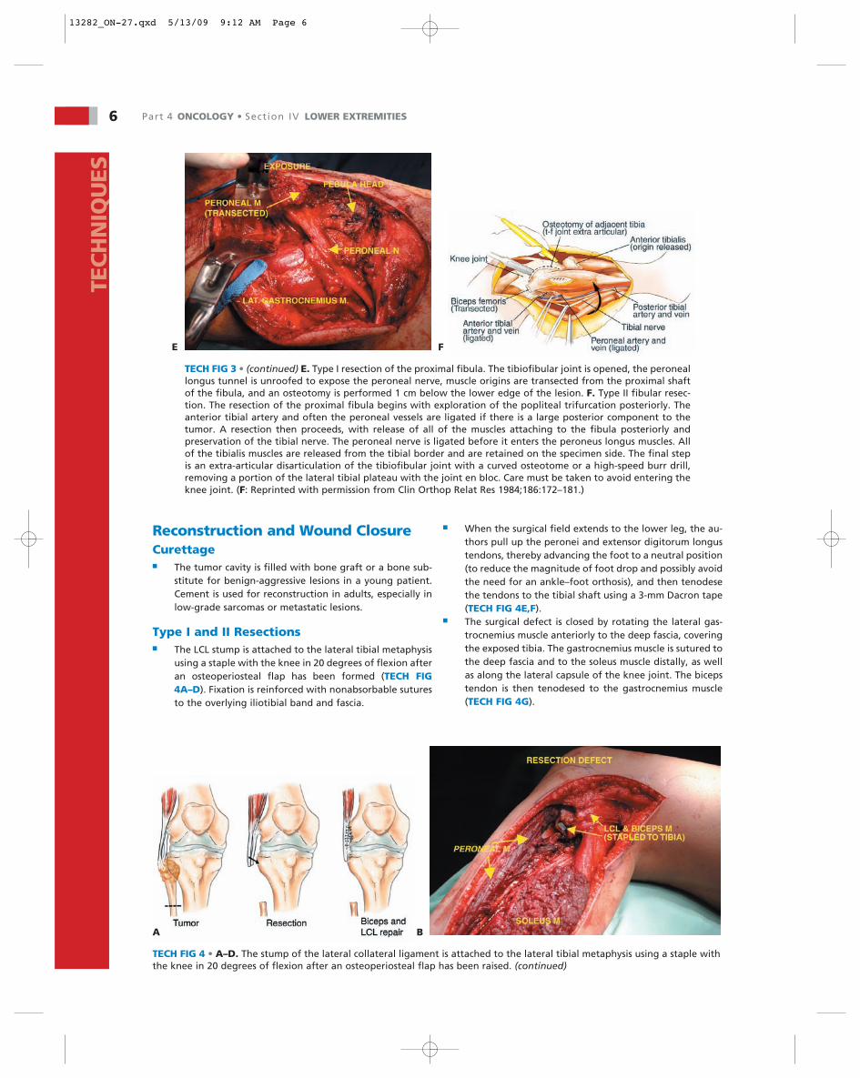
TECH FIG 3 • (continued) E. Type I resection of the proximal fibula. The tibiofibular joint is opened, the peroneallongus tunnel is unroofed to expose the peroneal nerve, muscle origins are transected from the proximal shaftof the fibula, and an osteotomy is performed 1 cm below the lower edge of the lesion. F. Type II fibular resec-tion. The resection of the proximal fibula begins with exploration of the popliteal trifurcation posteriorly. Theanterior tibial artery and often the peroneal vessels are ligated if there is a large posterior component to thetumor. A resection then proceeds, with release of all of the muscles attaching to the fibula posteriorly andpreservation of the tibial nerve. The peroneal nerve is ligated before it enters the peroneus longus muscles. Allof the tibialis muscles are released from the tibial border and are retained on the specimen side. The final stepis an extra-articular disarticulation of the tibiofibular joint with a curved osteotome or a high-speed burr drill,removing a portion of the lateral tibial plateau with the joint en bloc. Care must be taken to avoid entering theknee joint. (F: Reprinted with permission from Clin Orthop Relat Res 1984;186:172–181.)
A B
■ When the surgical field extends to the lower leg, the au-thors pull up the peronei and extensor digitorum longustendons, thereby advancing the foot to a neutral position(to reduce the magnitude of foot drop and possibly avoidthe need for an ankle–foot orthosis), and then tenodesethe tendons to the tibial shaft using a 3-mm Dacron tape(TECH FIG 4E,F).
■ The surgical defect is closed by rotating the lateral gas-trocnemius muscle anteriorly to the deep fascia, coveringthe exposed tibia. The gastrocnemius muscle is sutured tothe deep fascia and to the soleus muscle distally, as wellas along the lateral capsule of the knee joint. The bicepstendon is then tenodesed to the gastrocnemius muscle(TECH FIG 4G).
Reconstruction and Wound ClosureCurettage■ The tumor cavity is filled with bone graft or a bone sub-
stitute for benign-aggressive lesions in a young patient.Cement is used for reconstruction in adults, especially inlow-grade sarcomas or metastatic lesions.
Type I and II Resections■ The LCL stump is attached to the lateral tibial metaphysis
using a staple with the knee in 20 degrees of flexion afteran osteoperiosteal flap has been formed (TECH FIG4A–D). Fixation is reinforced with nonabsorbable suturesto the overlying iliotibial band and fascia.
TECH FIG 4 • A–D. The stump of the lateral collateral ligament is attached to the lateral tibial metaphysis using a staple withthe knee in 20 degrees of flexion after an osteoperiosteal flap has been raised. (continued)
TEC
HN
IQU
ES
13282_ON-27.qxd 5/13/09 9:12 AM Page 6
Chapter 27 FIBULAR RESECTIONS 7TEC
HN
IQU
ES
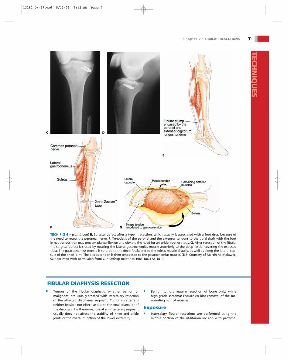
TECH FIG 4 • (continued) E. Surgical defect after a type II resection, which usually is associated with a foot drop because ofthe need to resect the peroneal nerve. F. Tenodesis of the peronei and the extensor tendons to the tibial shaft with the footin neutral position may prevent plantarflexion and obviate the need for an ankle–foot orthosis. G. After resection of the fibula,the surgical defect is closed by rotating the lateral gastrocnemius muscle anteriorly to the deep fascia, covering the exposedtibia. The gastrocnemius muscle is sutured to the deep fascia and to the soleus muscle distally, as well as along the lateral cap-sule of the knee joint. The biceps tendon is then tenodesed to the gastrocnemius muscle. (E,F: Courtesy of Martin M. Malawer;G: Reprinted with permission from Clin Orthop Relat Res 1984;186:172–181.)
E
GF
■ Tumors of the fibular diaphysis, whether benign ormalignant, are usually treated with intercalary resectionof the affected diaphyseal segment. Tumor curettage isneither feasible nor effective due to the small diameter ofthe diaphysis. Furthermore, loss of an intercalary segmentusually does not affect the stability of knee and anklejoints or the overall function of the lower extremity.
■ Benign tumors require resection of bone only, whilehigh-grade sarcomas require en bloc removal of the sur-rounding cuff of muscles.
Exposure■ Intercalary fibular resections are performed using the
middle portion of the utilitarian incision with proximal
FIBULAR DIAPHYSIS RESECTION
DC
13282_ON-27.qxd 5/13/09 9:12 AM Page 7
8 Part 4 ONCOLOGY • Section IV LOWER EXTREMITIES
A B
C
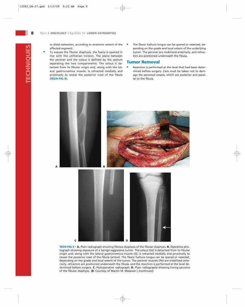
TECH FIG 5 • A. Plain radiograph showing fibrous dysplasia of the fibular diaphysis. B. Operative pho-tograph showing exposure of a benign-aggressive tumor. The soleus (So) is detached from its fibularorigin and, along with the lateral gastrocnemius muscle (G), is retracted medially and proximally toreveal the posterior crest of the fibula (arrow). The flexor hallucis longus can be spared or resected,depending on the grade and local extent of the tumor. The peronei muscles (Pe) are mobilized ante-riorly, retractors are positioned underneath the fibula, and the resection is performed at the level de-termined before surgery. C. Postoperative radiograph. D. Plain radiographs showing Ewing sarcomaof the fibular diaphysis. (B: Courtesy of Martin M. Malawer.) (continued)
D
or distal extension, according to anatomic extent of theaffected segment.
■ To expose the fibular diaphysis, the fascia is opened inline with the utilitarian incision. The plane betweenthe peronei and the soleus is defined by the septumseparating the two compartments. The soleus is de-tached from its fibular origin and, along with the lat-eral gastrocnemius muscle, is retracted medially andproximally to reveal the posterior crest of the fibula(TECH FIG 5).
■ The flexor hallucis longus can be spared or resected, de-pending on the grade and local extent of the underlyingtumor. The peronei are mobilized anteriorly, and retrac-tors are positioned underneath the fibula.
Tumor Removal■ Resection is performed at the level that had been deter-
mined before surgery. Care must be taken not to dam-age the peroneal vessels, which are posterior and paral-lel to the fibula.TE
CH
NIQ
UES
13282_ON-27.qxd 5/13/09 9:12 AM Page 8
Chapter 27 FIBULAR RESECTIONS 9TEC
HN
IQU
ES
E
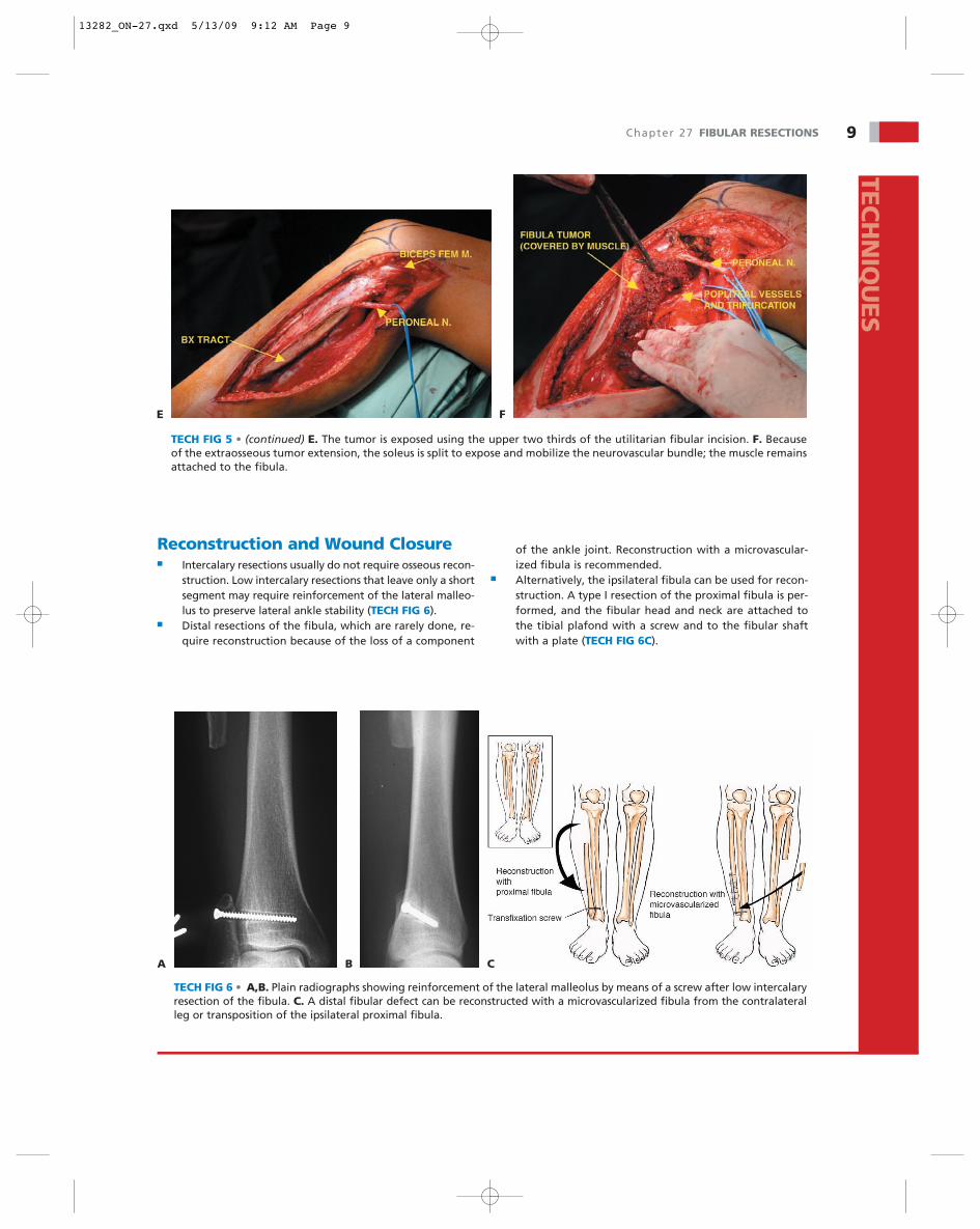
TECH FIG 5 • (continued) E. The tumor is exposed using the upper two thirds of the utilitarian fibular incision. F. Becauseof the extraosseous tumor extension, the soleus is split to expose and mobilize the neurovascular bundle; the muscle remainsattached to the fibula.
F
TECH FIG 6 • A,B. Plain radiographs showing reinforcement of the lateral malleolus by means of a screw after low intercalaryresection of the fibula. C. A distal fibular defect can be reconstructed with a microvascularized fibula from the contralateralleg or transposition of the ipsilateral proximal fibula.
CA B
Reconstruction and Wound Closure■ Intercalary resections usually do not require osseous recon-
struction. Low intercalary resections that leave only a shortsegment may require reinforcement of the lateral malleo-lus to preserve lateral ankle stability (TECH FIG 6).
■ Distal resections of the fibula, which are rarely done, re-quire reconstruction because of the loss of a component
of the ankle joint. Reconstruction with a microvascular-ized fibula is recommended.
■ Alternatively, the ipsilateral fibula can be used for recon-struction. A type I resection of the proximal fibula is per-formed, and the fibular head and neck are attached tothe tibial plafond with a screw and to the fibular shaftwith a plate (TECH FIG 6C).
13282_ON-27.qxd 5/13/09 9:12 AM Page 9
10 Part 4 ONCOLOGY • Section IV LOWER EXTREMITIES
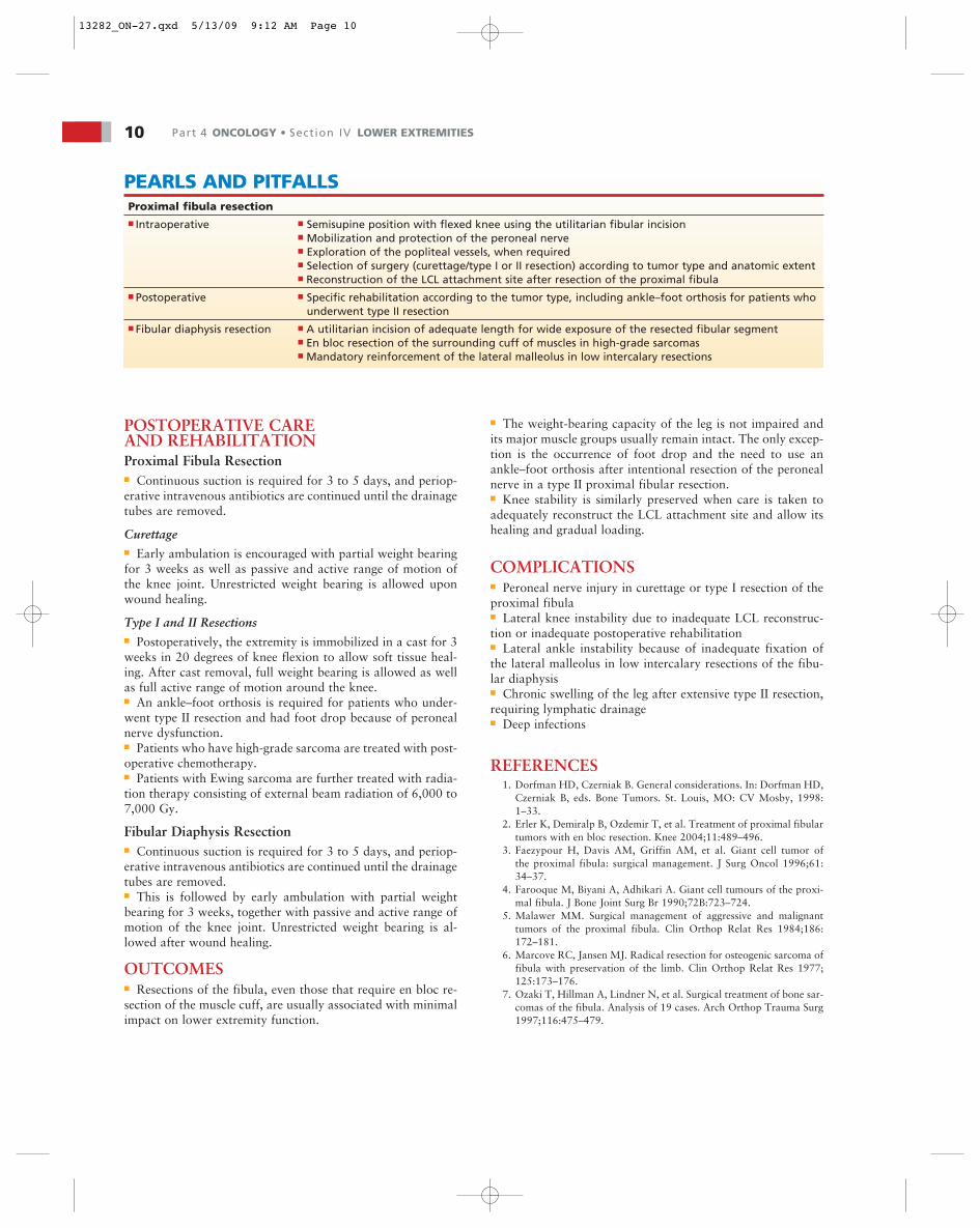
PEARLS AND PITFALLSProximal fibula resection■ Intraoperative ■ Semisupine position with flexed knee using the utilitarian fibular incision
■ Mobilization and protection of the peroneal nerve■ Exploration of the popliteal vessels, when required■ Selection of surgery (curettage/type I or II resection) according to tumor type and anatomic extent■ Reconstruction of the LCL attachment site after resection of the proximal fibula
■ Postoperative ■ Specific rehabilitation according to the tumor type, including ankle–foot orthosis for patients whounderwent type II resection
■ Fibular diaphysis resection ■ A utilitarian incision of adequate length for wide exposure of the resected fibular segment■ En bloc resection of the surrounding cuff of muscles in high-grade sarcomas■ Mandatory reinforcement of the lateral malleolus in low intercalary resections
POSTOPERATIVE CAREAND REHABILITATIONProximal Fibula Resection■ Continuous suction is required for 3 to 5 days, and periop-erative intravenous antibiotics are continued until the drainagetubes are removed.
Curettage■ Early ambulation is encouraged with partial weight bearingfor 3 weeks as well as passive and active range of motion ofthe knee joint. Unrestricted weight bearing is allowed uponwound healing.
Type I and II Resections■ Postoperatively, the extremity is immobilized in a cast for 3weeks in 20 degrees of knee flexion to allow soft tissue heal-ing. After cast removal, full weight bearing is allowed as wellas full active range of motion around the knee.■ An ankle–foot orthosis is required for patients who under-went type II resection and had foot drop because of peronealnerve dysfunction.■ Patients who have high-grade sarcoma are treated with post-operative chemotherapy.■ Patients with Ewing sarcoma are further treated with radia-tion therapy consisting of external beam radiation of 6,000 to7,000 Gy.
Fibular Diaphysis Resection■ Continuous suction is required for 3 to 5 days, and periop-erative intravenous antibiotics are continued until the drainagetubes are removed.■ This is followed by early ambulation with partial weightbearing for 3 weeks, together with passive and active range ofmotion of the knee joint. Unrestricted weight bearing is al-lowed after wound healing.
OUTCOMES■ Resections of the fibula, even those that require en bloc re-section of the muscle cuff, are usually associated with minimalimpact on lower extremity function.
■ The weight-bearing capacity of the leg is not impaired andits major muscle groups usually remain intact. The only excep-tion is the occurrence of foot drop and the need to use anankle–foot orthosis after intentional resection of the peronealnerve in a type II proximal fibular resection.■ Knee stability is similarly preserved when care is taken toadequately reconstruct the LCL attachment site and allow itshealing and gradual loading.
COMPLICATIONS■ Peroneal nerve injury in curettage or type I resection of theproximal fibula■ Lateral knee instability due to inadequate LCL reconstruc-tion or inadequate postoperative rehabilitation■ Lateral ankle instability because of inadequate fixation ofthe lateral malleolus in low intercalary resections of the fibu-lar diaphysis■ Chronic swelling of the leg after extensive type II resection,requiring lymphatic drainage■ Deep infections
REFERENCES1. Dorfman HD, Czerniak B. General considerations. In: Dorfman HD,
Czerniak B, eds. Bone Tumors. St. Louis, MO: CV Mosby, 1998:1–33.
2. Erler K, Demiralp B, Ozdemir T, et al. Treatment of proximal fibulartumors with en bloc resection. Knee 2004;11:489–496.
3. Faezypour H, Davis AM, Griffin AM, et al. Giant cell tumor ofthe proximal fibula: surgical management. J Surg Oncol 1996;61:34–37.
4. Farooque M, Biyani A, Adhikari A. Giant cell tumours of the proxi-mal fibula. J Bone Joint Surg Br 1990;72B:723–724.
5. Malawer MM. Surgical management of aggressive and malignanttumors of the proximal fibula. Clin Orthop Relat Res 1984;186:172–181.
6. Marcove RC, Jansen MJ. Radical resection for osteogenic sarcoma offibula with preservation of the limb. Clin Orthop Relat Res 1977;125:173–176.
7. Ozaki T, Hillman A, Lindner N, et al. Surgical treatment of bone sar-comas of the fibula. Analysis of 19 cases. Arch Orthop Trauma Surg1997;116:475–479.
13282_ON-27.qxd 5/13/09 9:12 AM Page 10