CHAPTER 23 Circulation It transports O 2 and nutrients to cells It takes away CO 2 and other wastes...
32
CHAPTER 23 Circulation It transports O 2 and nutrients to cells It takes away CO 2 and other wastes Artery and vein, cross-section •Blood smear
-
Upload
tracey-francis -
Category
Documents
-
view
215 -
download
0
Transcript of CHAPTER 23 Circulation It transports O 2 and nutrients to cells It takes away CO 2 and other wastes...

CHAPTER 23Circulation
It transports O2 and nutrients to cells
It takes away CO2 and other wastes
Artery and vein, cross-section •Blood smear

Circulatory system
made up of 3 parts organ
heart tissues & cells
blood vessels arteries veins capillaries
blood red blood cells plasma

The circulatory system associates intimately with all body tissues
Capillaries are microscopic blood vessels They form an intricate network among the tissue
cells
Capillary
Redbloodcell

The circulatory system associates intimately with all body tissues II
No substance has to diffuse far to enter or leave a cell
Diffusion ofmolecules
INTERSTITIALFLUID
Capillary
Tissuecell
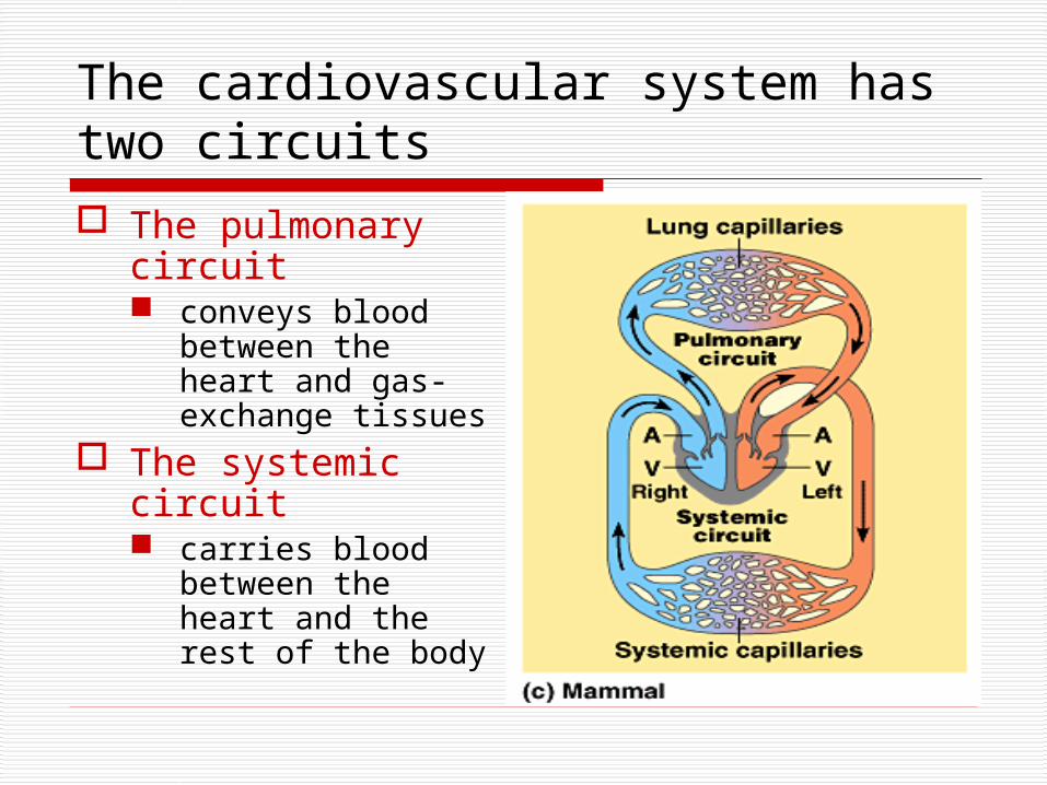
The cardiovascular system has two circuits
The pulmonary circuit conveys blood
between the heart and gas-exchange tissues
The systemic circuit carries blood
between the heart and the rest of the body
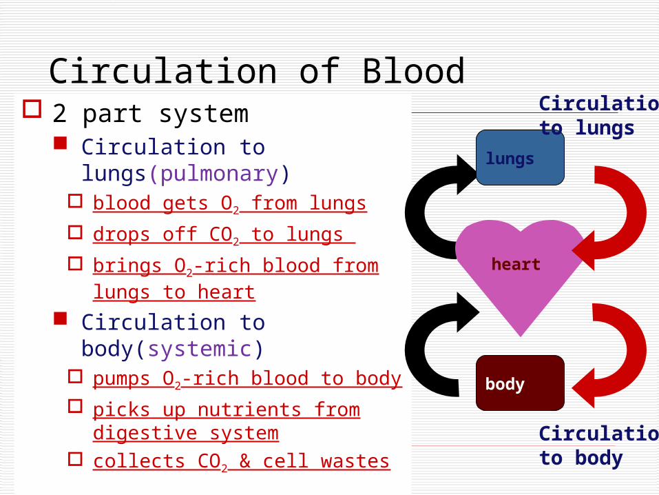
2 part system Circulation to
lungs(pulmonary) blood gets O2 from lungs
drops off CO2 to lungs
brings O2-rich blood from lungs to heart
Circulation to body(systemic)
pumps O2-rich blood to body
picks up nutrients from digestive system
collects CO2 & cell wastes
Circulation of Blood
heart
lungs
body
Circulationto lungs
Circulationto body

The human heart 4-Chambered heart
atria (atrium) thin wall collection chamber receive blood
ventricles thick wall pump pump blood out right
atrium
leftatrium
rightventricle
leftventricle

AV
SL
AV
Lub-dub, lub-dub 4 valves in the heart
flaps of connective tissue prevent backflow
Heart sounds closing of valves “Lub”
force blood against closed AV valves
“Dub” force of blood against
semilunar valves
Heart murmur leaking valve causes hissing sound blood squirts backward through valve

Blood’s path through the heart
1. vena cavae 2. right atrium 3. valve 4. right ventricle 5. valve 6. pulmonary artery (to
lungs) 7. pulmonary veins 8. left atrium 9. valve 10. left ventricle 11. valve 12. aorta largest blood
vessel in the body.

Blood vesselsarteries
arterioles
capillaries
venules
veins
artery
arteriolesvenules
veins

Arteries: Built for their job Arteries
blood flows away from heart thicker walls
provide strength for high pressure pumping of blood
elastic & stretchable maintains blood
pressure even when heart relaxes

Major arteries
pulmonaryartery
pulmonary
artery =to lungs
aortacarotid = to headto brain & left arm to right arm
coronary arteries
to body
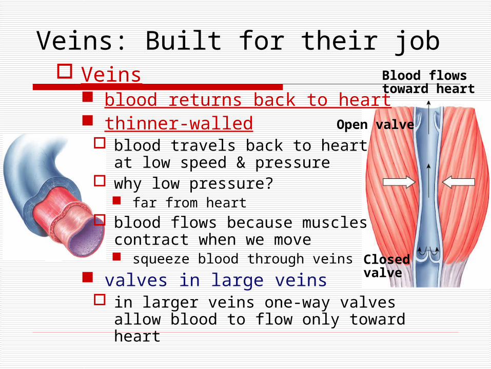
Veins: Built for their job Veins
blood returns back to heart thinner-walled
blood travels back to heart at low speed & pressure
why low pressure? far from heart
blood flows because muscles contract when we move squeeze blood through veins
valves in large veins in larger veins one-way valves
allow blood to flow only toward heart
Open valve
Blood flowstoward heart
Closed valve

Major Veins
pulmonaryvein =
from lung
superiorvena cava = from upper body
pulmonaryvein = from lung
inferiorvena cava = from lower body
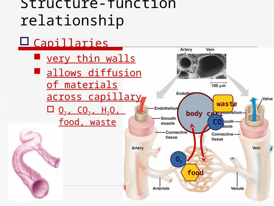
Structure-function relationship Capillaries
very thin walls allows diffusion of
materials across capillary O2, CO2, H2O,
food, wastebody cell
O2
food
waste
CO2

The heart contracts and relaxes rhythmically
Diastole Blood flows from
the veins into the heart chambers
Systole The atria briefly
contract and fill the ventricles with blood
Then the ventricles contract and propel blood out
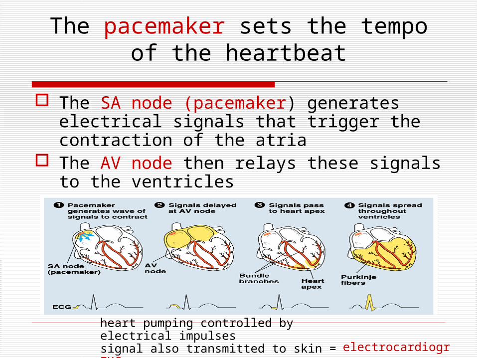
The pacemaker sets the tempo of the heartbeat
The SA node (pacemaker) generates electrical signals that trigger the contraction of the atria
The AV node then relays these signals to the ventricles
heart pumping controlled by electrical impulses signal also transmitted to skin = EKG electrocardiogram

Connection: What is a heart attack?
A heart attack is damage that occurs when a coronary feeding the heart is blocked
Aorta
Leftcoronaryartery
Blockage
Dead muscle tissue
Rightcoronaryartery

Cardiovascular diseases are the leading cause of death in the United States
Blood vessel blockage is usually due to blood clots Atherosclerosis: Growths called plaques develop in the inner
wall of the arteries, narrowing their bore In some cases, plaques also become hardened by calcium
deposits, leading to arteriosclerosis, commonly known as hardening of the arteries
PlaqueEpithelium
Smoothmuscle
Connectivetissue

Women & Heart Disease
Heart disease is 3rd leading cause of death among women aged 25–44 years & 2nd leading cause of death among women aged 45–64 years.
Risk factorsSmokingLack of exercise
High fat dietOverweight
Death rates for heart disease per 100,000 women, 2002

Blood exerts pressure on vessel walls
Blood pressure depends on cardiac output resistance of
vessels
Pressure is highest in the arteries It drops to zero by
the time the blood reaches the veins
Systolicpressure
Diastolicpressure
Relative sizes andnumbersof blood vessels
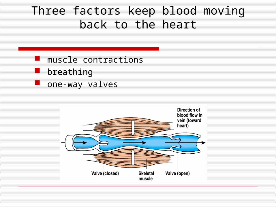
Three factors keep blood moving back to the heart
muscle contractions breathing one-way valves
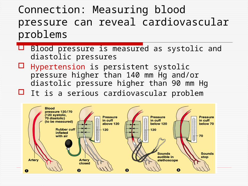
Connection: Measuring blood pressure can reveal cardiovascular problems Blood pressure is measured as systolic and diastolic
pressures Hypertension is persistent systolic pressure higher
than 140 mm Hg and/or diastolic pressure higher than 90 mm Hg
It is a serious cardiovascular problem

STRUCTURE AND FUNCTION OF BLOOD
Blood consists of cells suspended in plasma Plasma is an aqueous solution of various
substances

Blood Cell production
ribs, vertebrae, breastbone & pelvis
Stem cells “parent”
cells in bone marrow
differentiate into many different types of cells
white bloodcells
red bloodcells
white blood cells

Blood & blood cells Blood is a tissue of fluid & cells
plasma liquid part of blood dissolved salts, sugars, proteins, and more
cells red blood cells (RBC)
transport O2 in hemoglobin
white blood cells (WBC) defense & immunity
platelets blood clotting
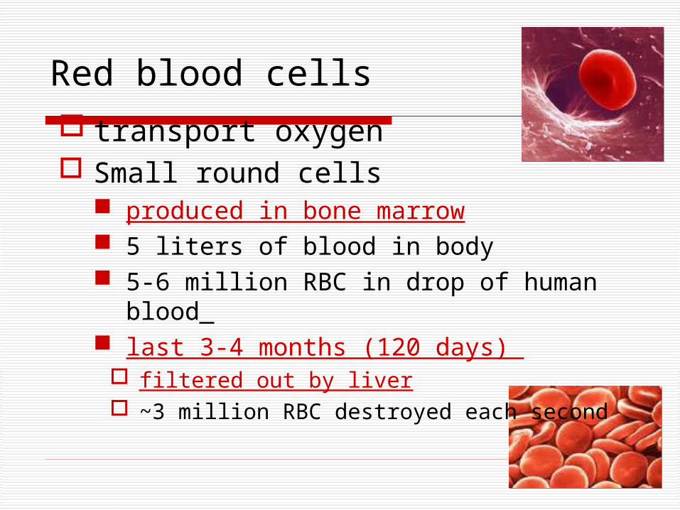
Red blood cells transport oxygen Small round cells
produced in bone marrow 5 liters of blood in body 5-6 million RBC in drop of human blood last 3-4 months (120 days)
filtered out by liver ~3 million RBC destroyed each second
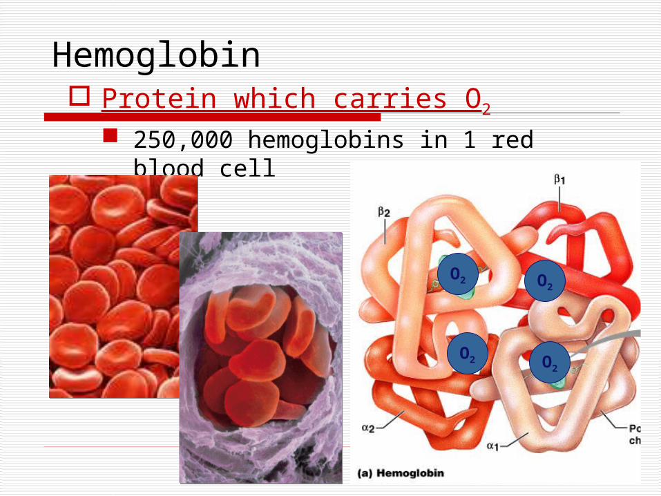
Hemoglobin Protein which carries O2
250,000 hemoglobins in 1 red blood cell
O2
O2O2
O2
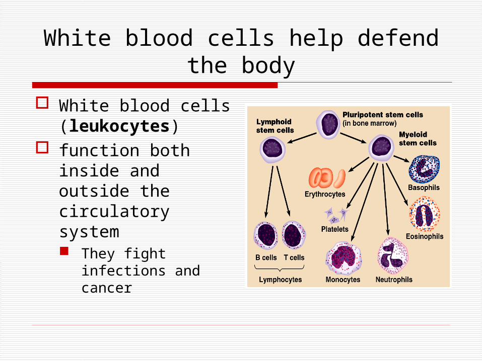
White blood cells help defend the body
White blood cells (leukocytes)
function both inside and outside the circulatory system They fight
infections and cancer
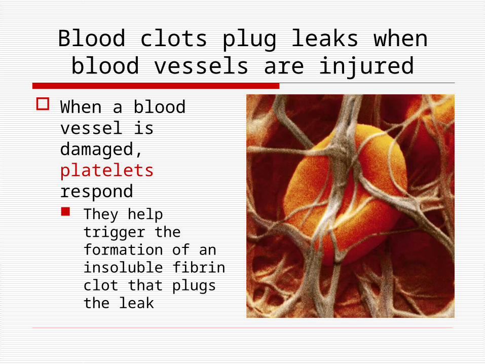
Blood clots plug leaks when blood vessels are injured
When a blood vessel is damaged, platelets respond They help trigger
the formation of an insoluble fibrin clot that plugs the leak

Connection: Stem cells offer a potential cure for leukemia and other blood cell
diseases
All blood cells develop from stem cells in bone marrow Such cells may
prove valuable for treating certain blood disorders

2008-2009
Have a heart?Ask Questions!!