CHAPTER 2 : CELL STRUCTURE AND CELL ORGANISATIONsasbadisb.com/download/page-34.pdf · ©Sasbadi...
12
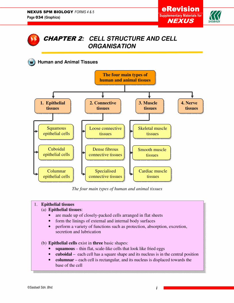
©Sasbadi Sdn. Bhd. i CHAPTER 2: CELL STRUCTURE AND CELL ORGANISATION Human and Animal Tissues The four main types of human and animal tissues NEXUS SPM BIOLOGY FORMS 4 & 5 Page 034 (Graphics) Supplementary Materials for 1. Epithelial tissues 2. Connective tissues 3. Muscle tissues 4. Nerve tissues Skeletal muscle tissues Smooth muscle tissues Cardiac muscle tissues Loose connective tissues Dense fibrous connective tissues Specialised connective tissues Squamous epithelial cells Cuboidal epithelial cells Columnar epithelial cells The four main types of human and animal tissues 1. Epithelial tissues (a) Epithelial tissues: • are made up of closely-packed cells arranged in flat sheets • form the linings of external and internal body surfaces • perform a variety of functions such as protection, absorption, excretion, secretion and lubrication (b) Epithelial cells exist in three basic shapes: • squamous – thin flat, scale-like cells that look like fried eggs • cuboidal – each cell has a square shape and its nucleus is in the central position • columnar – each cell is rectangular, and its nucleus is displaced towards the base of the cell
Transcript of CHAPTER 2 : CELL STRUCTURE AND CELL ORGANISATIONsasbadisb.com/download/page-34.pdf · ©Sasbadi...
©Sasbadi Sdn. Bhd. i
CHAPTER 2: CELL STRUCTURE AND CELL
ORGANISATION
Human and Animal Tissues
The four main types of human and animal tissues
NEXUS SPM BIOLOGY FORMS 4 & 5
Page 034 (Graphics)
Supplementary Materials for
1. Epithelial
tissues
2. Connective
tissues
3. Muscle
tissues
4. Nerve
tissues
Skeletal muscle
tissues
Smooth muscle
tissues
Cardiac muscle
tissues
Loose connective
tissues
Dense fibrous
connective tissues
Specialised
connective tissues
Squamous
epithelial cells
Cuboidal
epithelial cells
Columnar
epithelial cells
The four main types of
human and animal tissues
1. Epithelial tissues
(a) Epithelial tissues:
• are made up of closely-packed cells arranged in flat sheets
• form the linings of external and internal body surfaces
• perform a variety of functions such as protection, absorption, excretion,
secretion and lubrication
(b) Epithelial cells exist in three basic shapes:
• squamous – thin flat, scale-like cells that look like fried eggs
• cuboidal – each cell has a square shape and its nucleus is in the central position
• columnar – each cell is rectangular, and its nucleus is displaced towards the
base of the cell

©Sasbadi Sdn. Bhd. ii
Human epithelial tissues
The three basic shapes of epithelial cells
(a) Epithelial tissues lining the outer skin, mouth, pharynx, oesophagus, vagina and
anus
Stratified squamous epithelium
of the human skin
Squamous
epithelium
Cuboidal
epithelium
Columnar
epithelium
squamous cells
cuboidal cells
Structure:
• also known as stratified
(formed into layers)
squamous epithelium
• newly produced cuboidal
cells, which are pushed
towards the surface of the
tissue, are gradually
transformed into flat,
squamous cells
Functions:
• protects against abrasion
• forms the first line of defence
against microorganisms (see
Figure 10.3, page 348,
Nexus SPM Biology)
NEXUS SPM BIOLOGY FORMS 4 & 5
Page 034 (Graphics)
Supplementary Materials for

©Sasbadi Sdn. Bhd. iii
(b) Epithelial tissues lining the alveoli of the lungs and the renal glomeruli
Simple squamous epithelium
(c) Epithelial tissues lining the small intestine and the stomach
Simple columnar epithelium of
the small intestine (LS)
squamous cells
Structure:
• also known as simple squamous
epithelium (simple refers to only one
layer of squamous epithelial cells)
Functions:
• diffusion (see Figure 7.24, page 228,
Nexus SPM Biology)
• filtration (see Figure 12.31, page 440,
Nexus SPM Biology)
• forms the first line of defence against
microorganisms
Functions:
• secretes digestive enzymes into
the intestine
• goblet cells secrete mucus
(see Figure 6.32, page 173,
Nexus SPM Biology
• absorbs digested food
Structure:
• also known as simple columnar
epithelial tissue
NEXUS SPM BIOLOGY FORMS 4 & 5
Page 034 (Graphics)
Supplementary Materials for
columnar cells
goblet cells
©Sasbadi Sdn. Bhd. iv
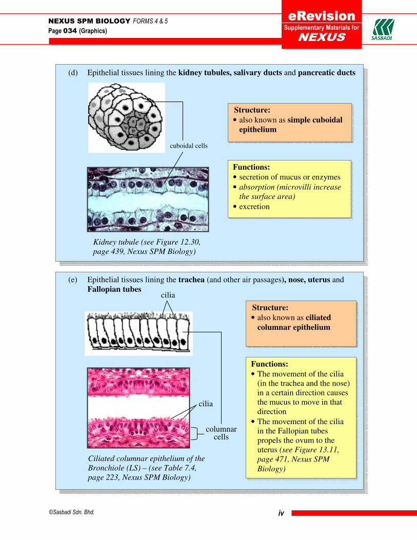
(d) Epithelial tissues lining the kidney tubules, salivary ducts and pancreatic ducts
Kidney tubule (see Figure 12.30,
page 439, Nexus SPM Biology)
(e) Epithelial tissues lining the trachea (and other air passages), nose, uterus and
Fallopian tubes
Ciliated columnar epithelium of the
Bronchiole (LS) – (see Table 7.4,
page 223, Nexus SPM Biology)
Structure:
• also known as simple cuboidal
epithelium
Functions:
• secretion of mucus or enzymes
• absorption (microvilli increase
the surface area)
• excretion
cuboidal cells
cilia
cilia
columnar cells
Structure:
• also known as ciliated
columnar epithelium
Functions:
• The movement of the cilia
(in the trachea and the nose)
in a certain direction causes
the mucus to move in that
direction
• The movement of the cilia
in the Fallopian tubes
propels the ovum to the
uterus (see Figure 13.11,
page 471, Nexus SPM
Biology)
NEXUS SPM BIOLOGY FORMS 4 & 5
Page 034 (Graphics)
Supplementary Materials for

©Sasbadi Sdn. Bhd. v
2. Connective tissues
• serve to ‘connect’ (support and bind) other tissues
• consist of various types of cells scattered throughout an extracellular matrix,
which is made of a complex mixture of carbohydrates and proteins (plus
minerals in the case of a bone)
• are widely distributed in the body
The three main types of connective tissues (see diagram above) are as follows:
(a) Loose fibrous connective tissue:
• It is the most widespread connective tissue in the body.
• It holds the organs in place and attaches the epithelial tissue to other underlying
tissues.
(b) Dense fibrous connective tissue:
• It is found in tendons and ligaments.
• It consists of large amounts of closely packed collagenous fibres.
(c) Specialised connective tissues which consist of:
• adipose tissue – is a type of loose connective tissue that stores fat
• blood tissue – consists of blood cells (erythrocytes, leucocytes and platelets)
suspended in the plasma (the extracellular matrix)
• bone – is a type of mineralized connective tissue that contains collagen and
calcium phosphate (which makes the bone hard)
• cartilage – is a strong and flexible connective tissue which supports the nose, ears
and covers the ends of the bones at the joints
The three main types of connective
tissues in humans and animals
NEXUS SPM BIOLOGY FORMS 4 & 5
Page 034 (Graphics)
Supplementary Materials for
Adipose tissue Cartilage
Blood tissue Bone tissue
The three main connective tissues
Dense fibrous
connective tissue
Loose fibrous
connective tissue
Specialised connective tissues

©Sasbadi Sdn. Bhd. vi
3. Muscle tissues
The three types of muscle tissues are as follows:
(a) Skeletal muscle tissue
• It is found attached to the skeleton.
• It contracts and relaxes to move the bones at the joints.
• It is found in the arms, legs and body parts where there is movement.
(b) Smooth muscle tissue
• It contracts and relaxes to enable all involuntary body movements, e.g.
peristalsis.
• It is found in the walls of the digestive, urinary and reproductive tracts, and the
blood vessels.
(c) Cardiac muscle tissue
• It is only found in the wall of the heart.
• It contracts to pump blood to all parts of the body.
(see Figures 11.20, 11.21 and 11.22, page 389, Nexus SPM Biology)
The three main types of muscle tissues
NEXUS SPM BIOLOGY FORMS 4 & 5
Page 034 (Graphics)
Supplementary Materials for
Skeletal muscles
Cardiac muscles
Smooth muscles

©Sasbadi Sdn. Bhd. vii
4. Nerve tissue
• It consists of neurones or nerve cells.
• A neurone is a very long cell that transmits impulses (electrical signals).
• It controls and coordinates body activities (see unit 12.2, page 419, Nexus SPM
Biology).
Nerve tissue
Plant Tissues
The three main types of plant tissues
(see Figure 11.48, page 403, Nexus SPM Biology)
NEXUS SPM BIOLOGY FORMS 4 & 5
Page 034 (Graphics)
Supplementary Materials for
Ordinary epidermal
cells
Epidermal tissue
Root hair cells
Guard cells
Parenchyma tissue
Sclerenchyma tissue
Collenchyma tissue
1. Ground tissue
2. Epidermal tissue
3. Vascular tissue
The three main types
of plant tissues
Xylem tissue
Phloem tissue

©Sasbadi Sdn. Bhd. viii
1. Ground tissue
General functions: Various functions including photosynthesis, support and storage
NEXUS SPM BIOLOGY FORMS 4 & 5
Page 034 (Graphics)
Supplementary Materials for
Parenchyma tissue
Parenchyma tissue (XS)
Structure:
• cells have thin flexible primary cell
walls and large central vacuoles
• consists of unspecialised cells found
in all plant organs
Functions:
• e.g. palisade mesophyll cells and spongy mesophyll cells contain
chloroplasts to carry out photosynthesis (see Table 6.28, pages 187 - 188,
Nexus SPM Biology)
• e.g. forms packing tissue and provides support and shape to herbaceous
plants; also to store food (see no. 7, unit 11.3.3, page 404, Nexus SPM
Biology)
Collenchyma tissue
Collenchyma tissue (XS)
Structure:
• cells have unevenly thickened walls,
especially at the corners
• found just under the epidermis of the stem
and along leaf veins
Function:
• supports herbaceous plants, young stems
and petioles (see no. 7(b), page 404 Nexus
SPM Biology)

©Sasbadi Sdn. Bhd. ix
Epidermal tissue
2. Epidermal tissue
General functions: Covers and protects the young plant parts
NEXUS SPM BIOLOGY FORMS 4 & 5
Page 034 (Graphics)
Supplementary Materials for
Sclerenchyma tissue
Sclerenchyma tissue (XS)
Structure:
• cells have uniformly thickened secondary walls
and large vacuoles
• cells may be dead at maturity
Function:
• provides support to the plant
(see no. 7(c), page 404 Nexus SPM Biology)
Structure:
• forms the outermost
layer covering stems,
leaves, and roots
• most epidermal cells
are flat and have
large vacuoles
• cells are closely
packed; some are
covered with a waxy
cuticle layer
Functions:
• e.g. ordinary epidermal cells covering stems,
leaf petioles and leaves help to reduce water loss
(by the cuticle) and protect against mechanical
injury and invasion of microorganisms
• e.g. root hairs for the absorption of water and
minerals (see Figure 10.46, page 360, Nexus
SPM Biology)
• e.g. guard cells control the opening and closing
of the stomata (see Figure 10.62 and 10.63, page
367, Nexus SPM Biology)
cuticle
upper epidermis
lower epidermis stoma
guard cell
The cuticle, epidermis and guard cells of the leaf Root hairs (XS, tip
of the root)
©Sasbadi Sdn. Bhd. x
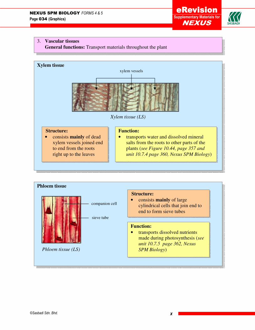
3. Vascular tissues
General functions: Transport materials throughout the plant
Xylem tissue
Xylem tissue (LS)
NEXUS SPM BIOLOGY FORMS 4 & 5
Page 034 (Graphics)
Supplementary Materials for
Structure:
• consists mainly of dead
xylem vessels joined end
to end from the roots
right up to the leaves
Function:
• transports water and dissolved mineral
salts from the roots to other parts of the
plants (see Figure 10.44, page 357 and
unit 10.7.4 page 360, Nexus SPM Biology)
xylem vessels
Phloem tissue
Phloem tissue (LS)
Structure:
• consists mainly of large
cylindrical cells that join end to
end to form sieve tubes
Function:
• transports dissolved nutrients
made during photosynthesis (see
unit 10.7.5 page 362, Nexus
SPM Biology)
companion cell
sieve tube

©Sasbadi Sdn. Bhd. xi
Human and Animal Organs
Some major human and animal organs (in yellow boxes)
NEXUS SPM BIOLOGY FORMS 4 & 5
Page 034 (Graphics)
Supplementary Materials for
Major organs
in humans and
animals
Male reproductive
system
• Seminal vesicle
• Penis
• Testes
• Thyroid
• Pituitary
• Pancreas
• Hypothalamus
• Adrenal glands
• Bones
• Cartilage
• Ligaments
• Tendons
• Nose
• Trachea
• Lungs
• Lymph
• Lymph
nodes
• Lymph
vessels
• Kidneys
• Ureters
• Urethra
• Bladder
Female reproductive
system
• Ovaries
• Oviducts
• Uterus
• Mammary glands
• Heart
• Blood
vessels
• Blood
• Mouth
• Oesophagus
• Stomach
• Intestines
• Skeletal
muscles
• Smooth
muscles
• Peripheral
nerves
• Spinal cord
• Brain
Nervous
system
Circulatory
system
Digestive
system
Reproductive
system
Excretory
system
Muscular
system
Respiratory
system
Lymphatic
system
Endocrine
system
Skeletal
system

©Sasbadi Sdn. Bhd. xii
Plant Organs
The organs of a plant (Impatiens balsamina)
NEXUS SPM BIOLOGY FORMS 4 & 5
Page 034 (Graphics)
Supplementary Materials for
1 Plant organs are divided into vegetative and reproductive organs.
2. Vegetative organs produce growth in plants and reproductive organs are involved in
sexual reproduction.
3. The vegetative organs are…
(a) the roots (b) the stems (c) the leaves
4. The reproductive organs are…
(a) the flowers (b) the fruits (c) the seeds
Leaf:
• To photosynthesise
• For gaseous exchange
and excretion
• To regulate transpiration
Stem:
• To transport water,
nutrients and organic
materials
• To lift the leaves
above the ground to
trap sunlight
Roots:
• To anchor the plant
• To absorb water and
nutrients
• To store excess sugar as
starch
Flower:
• Contains the organs of
plant sexual
reproduction
• To attract insects for
pollination
Fruit:
• To protect and
disperse seeds for
reproduction
Seeds:
• To protect and nourish the
embryo or baby plant
• Aids in dispersal
a burst
fruit fruit
wall