Chapter 19
41
Principles of Human Anatomy and Physiology, 11e 1 Chapter 19 The Cardiovascular System: The Blood
-
Upload
rhona-mooney -
Category
Documents
-
view
32 -
download
2
description
Chapter 19. The Cardiovascular System: The Blood. What is blood?. Blood is a connective tissue composed of a liquid extracellular matrix called plasma. It has cells and cell fragments dissolved in it. The cardiovascular system is made up of blood, the heart, and blood vessels. - PowerPoint PPT Presentation
Transcript of Chapter 19

Principles of Human Anatomy and Physiology, 11e 1
Chapter 19
The Cardiovascular System: The Blood

What is blood?
• Blood is a connective tissue composed of a liquid extracellular matrix called plasma. It has cells and cell fragments dissolved in it.
• The cardiovascular system is made up of blood, the heart, and blood vessels.
Principles of Human Anatomy and Physiology, 11e 2

Principles of Human Anatomy and Physiology, 11e 3
Functions of Blood
• Transportation– O2, CO2, metabolic wastes, nutrients, heat & hormones
• Regulation– helps regulate pH through buffers– helps regulate body temperature– helps regulate water content of cells
• Protection from disease & loss of blood
• Hematology is study of blood and blood disorders

Principles of Human Anatomy and Physiology, 11e 4
Physical Characteristics of Blood
• Thicker than water and therefore flows more slowly than water
• Feels sticky• Temperature of 100.4 degrees F• pH 7.4 (7.35-7.45)• 8 % of total body weight• Blood volume
– 5 to 6 liters in average male– 4 to 5 liters in average female

Principles of Human Anatomy and Physiology, 11e 5
Techniques of Blood Sampling
• Venipuncture– Most common method– sample taken from vein with hypodermic needle &
syringe
• Finger or heel stick– Used by diabetics to monitor blood sugar– Used to test blood of infants
• Arterial stick: used to determine oxygen levels

Principles of Human Anatomy and Physiology, 11e 6
COMPONENTS OF BLOOD
• Blood consists of 55% plasma and 45% of cells.• Blood plasma consists of 91.5% water and 8.5% solutes.• Principal solutes include nutrients, enzymes, hormones,
respiratory gases, electrolytes, and waste products.

Principles of Human Anatomy and Physiology, 11e 7
Blood Cells• Red blood cells, 40% ( erythrocytes )• White blood cells, 20% ( leukocytes )
• neutrophils, eosinophils, basophils• lymphocytes = T cells, B cells, and natural killer cells• monocytes
• Platelets, 30% (special cell fragments)

8
Hematocrit• Hematocrit is the percentage of total blood volume occupied by
RBCs.
• Anemia
– not enough RBCs or not enough hemoglobin
– Can be due to leukemia, iron deficiency, or B12 deficiency
– Vitamins or injections of iron can treat anemia
• Polycythemia
– too many RBCs (over 65%)
– dehydration, blood loss (including menstruation), blood doping in athletes (common in professional cycling to increase O2 supplies to the blood, muscles, and lungs)

Principles of Human Anatomy and Physiology, 11e 9
Formation of Blood Cells
• Most blood cells types need to be continually replaced– die within hours, days or weeks– process of blood cells formation is hematopoiesis or
hemopoiesis• In the embryo
– occurs in yolk sac, liver, spleen, thymus, lymph nodes & red bone marrow
• In adults– occurs only in red marrow of flat bones like sternum,
ribs, skull & pelvis and ends of long bones

Principles of Human Anatomy and Physiology, 11e 10
• Contain oxygen-carrying protein hemoglobin that gives blood its red color– 1/3 of cell’s weight is hemoglobin
• Biconcave disk 8 microns in diameter– increased surface area/volume ratio – flexible shape for narrow passages– no nucleus or other organelles
Red Blood Cells or Erythrocytes

Principles of Human Anatomy and Physiology, 11e 11
RBC Life Cycle
• RBCs live only 120 days– wear out from bending to fit through capillaries– no repair possible due to lack of organelles
• Worn out cells removed by macrophages in spleen & liver

Principles of Human Anatomy and Physiology, 11e 12
Erythropoiesis: Production of RBCs
• Erythrocyte formation, called erythropoiesis, occurs in adult red bone marrow of certain bones.
• It takes 1-2 days for a mature red blood cell to be formed and released into the blood stream.

Principles of Human Anatomy and Physiology, 11e 13
Hemoglobin
• Each RBC has 280 million hemoglobin molecules• Each one can bind 4 molecules of oxygen (to the iron at the center of the
molecule).• Hemoglobin removes CO2 from the blood• Hemoglobin also regulates blood pressure by regulating nitric oxide in the
blood

Principles of Human Anatomy and Physiology, 11e 14
WHITE BLOOD CELLS
• Leukocytes (WBCs) have a nucleus but do not have hemoglobin.
– Granular leukocytes include eosinophils, basophils, and neutrophils based on the straining of the granules.
– Agranular leukocytes do not have cytoplasmic granules and include the lymphocytes and monocytes.

Principles of Human Anatomy and Physiology, 11e 15
WBC Physiology• Less numerous than RBCs
– 5000 to 10,000 cells per drop of blood– 1 WBC for every 700 RBC
• Leukocytosis is a high white blood cell count– microbes, disease, strenuous exercise, anesthesia, cancer
or surgery• Leukopenia is low white blood cell count
– radiation, shock or chemotherapy• Only 2% of total WBC population is in circulating blood at any
given time– rest is in lymphatic fluid, skin, lungs, lymph nodes &
spleen

Principles of Human Anatomy and Physiology, 11e 16
Neutrophils (Granulocyte)
• The most abundant WBC• 60 to 70% of circulating WBCs• First cells to migrate to the site of bacterial infection or cancer
formation.• Also involved in healing injuries
– release lysozymes which destroy/digest bacteria– release defensin proteins that act like antibiotics & poke
holes in bacterial cell walls destroying them– release strong oxidants (bleach-like, strong chemicals)
that destroy bacteria

Principles of Human Anatomy and Physiology, 11e 17
Eosinophils (Granulocyte)
• 2 to 4% of circulating WBCs• Combat parasites and infections• Fight allergies and asthma

Principles of Human Anatomy and Physiology, 11e 18
Basophils (Granulocyte)
• Less than 1% of circulating WBCs• Store histamine and can contribute to allergic reactions• Also contain heparin, which prevents blood from clotting
too quickly

Principles of Human Anatomy and Physiology, 11e 19
Lymphocyte (Agranulocyte)
• 20 to 25% of circulating WBCs• The main soldiers in the immune system battles• B cells
– destroy bacteria and their toxins– turn into plasma cells that produces antibodies
• T cells– attack viruses, fungi, transplanted organs, cancer cells
& some bacteria• Natural killer cells (NKC)
– attack many different microbes & some tumor cells– destroy foreign invaders by direct attack

Principles of Human Anatomy and Physiology, 11e 20
Monocyte (Agranulocyte)
• 3 to 8% of circulating WBCs• Stored in the spleen• Produce macrophages which attack bacteria• Destroy microbes and clean up dead tissue following an
infection

Principles of Human Anatomy and Physiology, 11e 21
PLATELETS
• Platelets help stop blood loss from damaged vessels by forming a platelet plug.
• • Their granules also
contain chemicals that promote blood clotting.

Principles of Human Anatomy and Physiology, 11e 22
Platelet (Thrombocyte) Anatomy
• Disc-shaped cell fragment with no nucleus
• They are produced in the bone marrow and live 5 to 9 days in the blood stream before being recycled by the spleen and liver.

Principles of Human Anatomy and Physiology, 11e 23
Bone Marrow Transplant
• Bone marrow transplant replaces diseased marrow with healthy marrow.
• Patient’s diseased marrow is destroyed by cancer or disease.
• Healthy marrow is supplied by a donor or the patient. It is removed (painfully) by sticking a needle into the hipbone.
• Risks to recipient include:– Infection due to decreased WBC– T cells might attack new bone marrow– Must take immunosuppressant drugs for life
Principles of Human Anatomy and Physiology, 11e 24
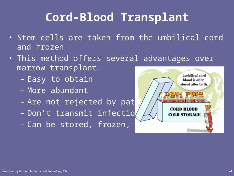
Cord-Blood Transplant
• Stem cells are taken from the umbilical cord and frozen• This method offers several advantages over marrow
transplant.– Easy to obtain– More abundant– Are not rejected by patient– Don’t transmit infections– Can be stored, frozen, forever

Principles of Human Anatomy and Physiology, 11e 25
Hemostasis
• Stoppage of bleeding in a quick & localized fashion when blood vessels are damaged
• Prevents hemorrhage (loss of a large amount of blood)• Methods utilized
– vascular spasm– platelet plug formation– blood clotting

26
Vascular Spasm
• Damage to blood vessel produces stimulates pain receptors
• Small blood vessels are contracted which reduces blood flow to the injury site

Principles of Human Anatomy and Physiology, 11e 27
Platelet Plug Formation
• Steps in the process– (1) platelet adhesion (2) platelet release reaction
(3) platelet aggregation

Principles of Human Anatomy and Physiology, 11e 28
Platelet Adhesion
• Platelets stick to exposed collagen underlying damaged cells in vessel wall
Principles of Human Anatomy and Physiology, 11e 29
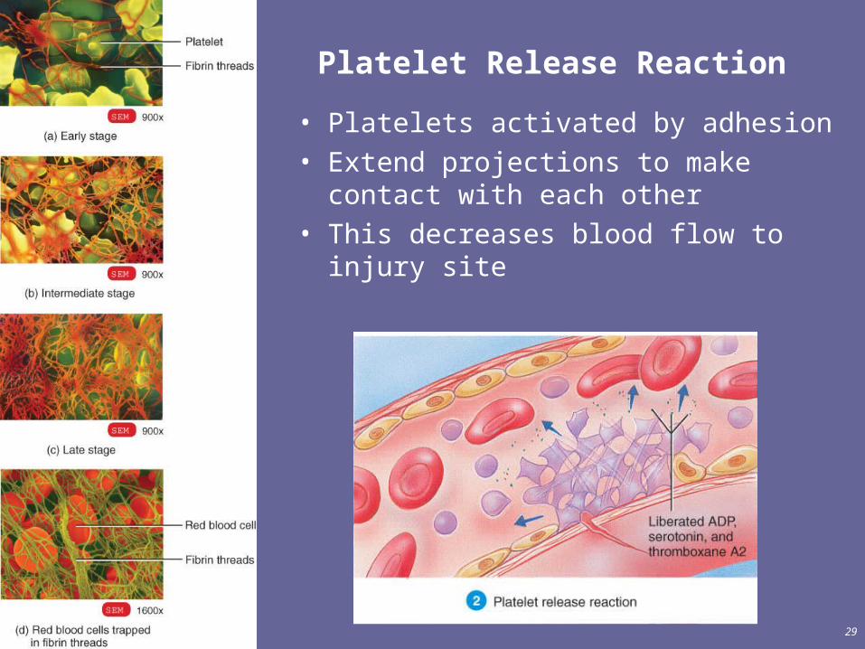
Platelet Release Reaction
• Platelets activated by adhesion• Extend projections to make contact
with each other • This decreases blood flow to injury site

Principles of Human Anatomy and Physiology, 11e 30
Platelet Aggregation
• Activated platelets stick together and activate new platelets to form a mass called a platelet plug
• Plug reinforced by fibrin threads formed during clotting process

Principles of Human Anatomy and Physiology, 11e 31
Clot Retraction & Blood Vessel Repair
• The plug stops the bleeding, then…
• Edges of damaged vessel are pulled together
• Fibroblasts & endothelial cells repair the blood vessel

Principles of Human Anatomy and Physiology, 11e 32
Blood Clotting
• Blood drawn from the body thickens into a gel– gel separates into liquid (serum) and a clot of insoluble
fibers (fibrin) in which the cells are trapped• If clotting occurs inside the body, it is called a thrombosis• Having vitamin K in your diet can help prevent blood
clotting

Principles of Human Anatomy and Physiology, 11e 33
Blood Groups and Blood Types
• RBC surfaces are marked by antigens, which determine blood type

Principles of Human Anatomy and Physiology, 11e 34
ABO Blood Groups
• Blood types in humans are:– display only antigen A -- blood type A– display only antigen B -- blood type B– display both antigens A & B -- blood type AB– display neither antigen -- blood type O
– Type O is the universal donor– Type AB is the universal receiver

Principles of Human Anatomy and Physiology, 11e 35
RH blood groups
• Antigen was discovered in blood of Rhesus monkey• People with Rh agglutinogens on RBC surface are Rh+. • Most people are Rh +• If an Rh - woman is pregnant and the baby is Rh+, the mother’s
body may reject the pregnancy; she must take anti-rejection drugs and be closely monitored.

Principles of Human Anatomy and Physiology, 11e 36
DISORDERS: HOMEOSTATIC IMBALANCES
• Anemia • Sickle-cell • Hemophilia • Disseminated intravascular clotting • Acute leukemia• chronic leukemia

Principles of Human Anatomy and Physiology, 11e 37
Anemia = Not Enough RBCs• Symptoms
– oxygen-carrying capacity of blood is reduced– fatigue, cold intolerance & paleness
• Types of anemia– iron-deficiency =lack of absorption or loss of iron– pernicious = lack of intrinsic factor for B12 absorption– hemorrhagic = loss of RBCs due to bleeding (ulcer)– hemolytic = defects in cell membranes cause rupture– thalassemia = hereditary deficiency of hemoglobin– aplastic = destruction of bone marrow (radiation/toxins)

Principles of Human Anatomy and Physiology, 11e 38
Sickle-cell Anemia
• Genetic defect in hemoglobin molecule– RBC is deformed
• sickle-shaped cells rupture easily = causing anemia & clots
• Found among populations in malaria belt– Mediterranean Europe, sub-Saharan Africa & Asia

Principles of Human Anatomy and Physiology, 11e 39
Hemophilia
• Inherited deficiency of clotting factors – bleeding spontaneously or after minor trauma– subcutaneous & intramuscular hemorrhaging– nosebleeds, blood in urine, articular bleeding & pain
• Treatment is transfusions of fresh plasma or concentrates of the missing clotting factor

40
Disseminated Intravascular Clotting
• Life threatening paradoxical presence of blood clotting and bleeding at the same time throughout the whole body
• Associated with infections, hypoxia, low blood flow rates, trauma, hypotension & hemolysis
• Clots cause necrosis leading to multisystem organ failure
Principles of Human Anatomy and Physiology, 11e 41
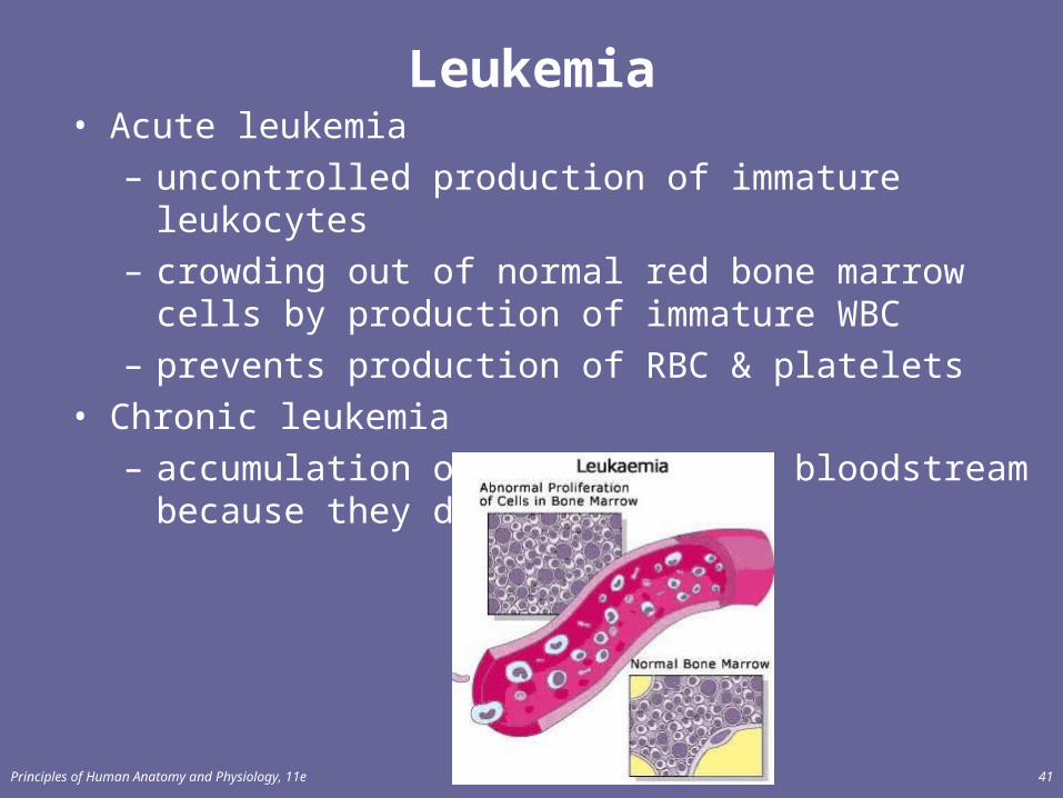
Leukemia• Acute leukemia
– uncontrolled production of immature leukocytes– crowding out of normal red bone marrow cells by
production of immature WBC– prevents production of RBC & platelets
• Chronic leukemia– accumulation of mature WBC in bloodstream because
they do not die