Chapter 17 CPT: Integumentary System
58
CHAPTER OUTLINE Surgical Terminology Integumentary Anatomy Integumentary Organization Modifiers Common Diseases and Disorders of the Integument Common Procedures Performed on the Integument Pulling It Together: Excision and Repair Code Assignment Skin Grafts and Flaps Burns Wound Care and Debridement Mohs Micrographic Surgery Breast Procedures Skin Resurfacing Cosmetic Procedures LEARNING OBJECTIVES After studying this chapter, you should be able to: 1. Define the surgical procedures common to the integumentary system: incision and drainage, biopsy, destruction, debridement, excision, repair, and Mohs micrographic surgery. 2. Differentiate among simple, intermediate, and complex wound repairs. 3. Explain guidelines for coding excision of lesions. 4. Accurately calculate measurements for lesion excisions. 5. Differentiate among the various types of skin grafts. 6. Describe the difference between a flap and a graft. CHAPTER 17 CPT: Integumentary System riskms/iStock/Thinkstock 577 03_Jurek_Part_3_Ch17.indd 577 15/11/17 5:43 pm
Transcript of Chapter 17 CPT: Integumentary System

Chapter OutlineSurgical TerminologyIntegumentary AnatomyIntegumentary OrganizationModifiersCommon Diseases and Disorders of the IntegumentCommon Procedures Performed on the IntegumentPulling It Together: Excision and Repair Code AssignmentSkin Grafts and FlapsBurnsWound Care and DebridementMohs Micrographic SurgeryBreast ProceduresSkin ResurfacingCosmetic Procedures
learning ObjeCtivesAfter studying this chapter, you should be able to:1. Define the surgical procedures common to the integumentary system: incision
and drainage, biopsy, destruction, debridement, excision, repair, and Mohs micrographic surgery.
2. Differentiate among simple, intermediate, and complex wound repairs.3. Explain guidelines for coding excision of lesions.4. Accurately calculate measurements for lesion excisions.5. Differentiate among the various types of skin grafts.6. Describe the difference between a flap and a graft.
Chapter 17
CPT: Integumentary System
riskm
s/iS
tock
/Thi
nkst
ock
577
03_Jurek_Part_3_Ch17.indd 577 15/11/17 5:43 pm

578 Part III: CPT and HCPCS Coding CPT © 2017 American Medical Association, All Rights Reserved.
7. Determine when it is appropriate to assign lesion excisions with flap and graft closure.8. Use the Rule of Nines to assign codes for burn treatment.9. Describe the four types of debridement codes.
10. Describe the difference between selective and nonselective wound debridement.11. Understand both the therapeutic and cosmetic breast procedures and the methods for treat-
ment of the breasts.
This chapter focuses on the procedural coding and terminology of surgical cases involving prob-lems of the integumentary system. It covers anatomical structures, common conditions that
are surgically treated, and surgical procedures performed on the skin and subcutaneous tissue as well as the breasts and nails. Chapter 16 introduced key CPT surgical concepts such as the surgi-cal package, separate procedures, modifiers, global period, and recognizing when a procedure is a diagnostic or therapeutic service. This chapter introduces additional terminology that is used throughout CPT and encountered when reading surgical documentation.
Two main specialties treat diseases, disorders, and injuries of the skin and underlying tissue: der-matology, and plastic and reconstructive surgery. Dermatology is a branch of medicine that specializes in diagnosing and treating diseases and disorders of the skin, its structure, and its function; it includes nails, hair, and sweat glands. The plastic and reconstructive surgery specialty concentrates on the repair and reconstruction of defects of the integument and its underlying musculoskeletal system to restore function and appearance. Plastic surgeons perform procedures on the following body parts:
• Skin, subcutaneous tissue, and musculature (e.g., scar revisions, lesion removals, complex repairs, flaps, grafts, wound care, and tattoo removal)
• Craniofacial structures (from trauma or birth defects)• Breast (e.g., reconstruction, reduction, and augmentation)• Hand (e.g., injury repairs and replantation)• Lips, palate, and oropharynx• Microvasculature
Plastic surgeons are most known for their aesthetic or cosmetic surgery to reconstruct, smooth, augment, tighten, or lift undesirable features of one’s body by performing procedures such as body contouring, facelifts, abdominoplasty (tummy tuck), rhinoplasty (nose job), and blepharoplasty (eyelid lift).
Integumentary codes are not reserved exclusively for use by dermatologists or plastic surgeons. Many of these codes are also routinely assigned by emergency department (ED) physicians and primary care physicians.
surgiCal terminOlOgy
Coders are expected to have a broad knowledge of medical terminology in order to read and inter-pret medical documentation. Differentiating between surgical terms and techniques is paramount to proper code assignment. When assigning codes from the Surgery section, it is advantageous to have a medical dictionary and an anatomy atlas available for reference. A foundation in anatomy and medical terminology is essential when coding in general but particularly for surgical services. A coder who masters the terminology can visualize and mentally follow along with what is taking place during the surgery as documented in the medical record. This enables the coder to identify all procedures that should be coded and to recognize when a procedure is a component of a more comprehensive procedure. Knowledge of these terms also promotes efficiency in locating codes in CPT, because many of these terms are main headings or subheadings.
The terminology can seem overwhelming. When reading medical documents, coders are bound to come across words they do not recognize. Coders must at a minimum memorize basic prefixes, suffixes, and root words to be able to break complicated words into smaller parts to decipher their meaning. Some of these common terms are located near the front of the CPT manual and should be reviewed. Also review the suffixes noted in Table 17.1.
Do not assign codes for documentation that contains technical
words and procedures you do not understand. Take the time to look them up in references.
03_Jurek_Part_3_Ch17.indd 578 15/11/17 5:43 pm

CPT © 2017 American Medical Association, All Rights Reserved. Chapter 17: CPT: Integumentary System 579
Directional termsWhen coders read medical documentation, they must master medical terms describing location and direction, as shown in Table 17.2 and Figure 17.1. Directional terms are widely used in operative reports and x-rays to indicate the precise location of a body part. CPT codes also contain these terms.
eXamples• 11755 Biopsy of nail unit (e.g., plate, bed, matrix, hyponychium, proximal and lateral nail
folds) (separate procedure).• 11750 Excision of nail and nail matrix, partial or complete (e.g., ingrown or deformed nail),
for permanent removal; with amputation of tuft of distal phalanx.• 12020 Treatment of superficial wound dehiscence; simple closure.• 19020 Mastotomy with exploration or drainage of abscess, deep.• 49560 Repair initial incisional or ventral hernia; reducible.• 54160 Circumcision, surgical excision other than clamp, device, or dorsal slit; neonate.• 21282 Lateral canthopexy.
table 17.1 Common Surgical SuffixesSUFFIX DEFINITION EXAMPLE
-centesis Puncturing a cavity to remove or inject fluid arthrocentesis-desis Fixation or fusing permanently arthrodesis-ectomy Partial or total removal of a body part, bone, or tissue colectomy-graphy Use of x-ray or fluoroscopy for viewing anatomy during a procedure radiography-lysis Destruction, breaking down, or freeing enterolysis-ostomy Creating a hole or opening from the inside to the outside of the body or
creating an opening within the bodycolostomy
-otomy Making an incision or opening into laparotomy-pexy Surgical fixation of an organ mastopexy-plasty Repair or reconstruct mammaplasty-rrhaphy Use of sutures to repair or reduce herniorrhaphy-scopy Surgical intervention using an endoscope to visualize internal structures laparoscopy-tripsy Crushing, pulverizing, destroying lithotripsy
table 17.2 Directional TermsTErM DEScrIPTION
Anterior Toward the front or in front ofDistal Farthest from where that point attaches to the trunk of the bodyDorsal (dorsum) Toward the backside of the body Inferior BelowLateral Toward the outer edges of the body or away from the middleMedial Toward the middle of the bodyPosterior Toward the back of or the back surfaceProximal Closest to where that point attaches to the trunk of the bodySuperior AboveVentral Toward the front side of the body
03_Jurek_Part_3_Ch17.indd 579 15/11/17 5:43 pm

580 Part III: CPT and HCPCS Coding CPT © 2017 American Medical Association, All Rights Reserved.
integumentary anatOmy
When reading an operative note, the coder must be able to recognize what type of repair was performed or how deep a debridement was based on the anatomical landmarks and descriptions. Recognition begins with the basic anatomy of the skin, as shown in Figure 17.2.
The integument is made up of two parts: the cutaneous membrane, or skin, and the accessory structures. The cutane-ous membrane is made up of two layers: epidermis or super-ficial epithelium (epithelial tissues) and the dermis, which is composed of connective tissues. The epidermis is the outside layer of skin. The dermis, or dermal layer, is the second layer of skin containing blood vessels (capillaries), hair follicles, nerves, and glands. The dermal layer is dense with connective tissue and is woven to this subcutaneous layer to secure the skin in place, stabilizing the position of the skin but allowing separate movement. It is strong from the collagen fibers and elastic, with water aiding in flexibility. Bruises visible in the skin are the result of damage to the tiny capillaries. Below the dermis is a subcutaneous layer of loose connective tissue, also known as the superficial fascia or hypodermis. The subcu-taneous layer contains mostly fat cells and superficial fascia. The hypodermis is not technically part of the integumentary system, despite its name.
Sebaceous gland
Epidermis
Dermis
Subcutaneouslayer
Apocrine gland
Figure 17.2 Layers of the integument.
Figure 17.1 Directional terms in anatomy.
Superior: Above
Anterior (ventral):
Toward the front of
the body
Posterior (dorsal):
Toward the back of
the body
Superficial: At or near
the body’s surface
Deep: Away from the
body’s surface
Inferior: Below
LeftRight
Midline
Medial: Toward the
body’s midline
Lateral: Away from
the body’s midline
Proximal:
Closest to the
point of origin
Distal: Farthest
from the point
of origin
03_Jurek_Part_3_Ch17.indd 580 15/11/17 5:43 pm

CPT © 2017 American Medical Association, All Rights Reserved. Chapter 17: CPT: Integumentary System 581
The accessory structures include hair, nails, and exocrine glands. These accessory structures are considered part of the integumentary system, because they originate in the dermis and extend through the epidermis to the skin’s surface.
The skin is the largest organ of the body and performs these functions:
• Protects the body as a barrier against the environment, fluid loss, abrasion, and shock• Maintains body temperature by keeping in heat and allowing sweat out to cool the body
• Produces vitamin D3
• Stores lipids
The skin repairs minor abrasions and injuries itself. Cells from the epidermis break away and form a sheet across the wound. For deeper wounds, a clot forms in the wound bed, and blood flow increases, bring-ing cells into the wound to form a scab. Granulation tissue fills the wound and there is intense growth of epithelial cells beneath the scab.
integumentary OrganizatiOn
Integumentary codes are arranged by anatomical site and category of pro-cedure. The main headings are Skin, Subcutaneous and Accessory Struc-tures; Nails; Pilonidal Cyst; Introduction; Repair (Closure); Destruction; and Breast. These sections are further broken down into subheadings, as shown in Box 17.1.
mODiFiers
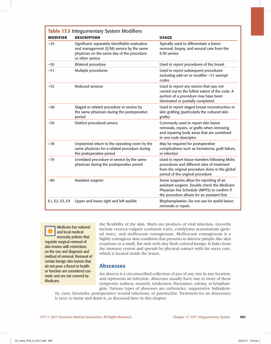
Table 17.3 outlines the CPT modifiers most commonly used in the Integumentary System section and why. Documentation must support the use of the modifiers.
Note that modifier –50 is commonly misused in the Integumentary System section when re-porting lesion removals or skin repairs when performed on both arms and/or both legs. The code descriptors for lesion removals and skin repairs include several body parts that are not paired body organs, so it is inappropriate to use LT or RT or –50 in these situations. You may recall that these modifiers are used when performing procedures on paired body parts or organ systems. The skin is one organ system. The code descriptions do not accommodate these modifiers because many body parts are grouped under one code and would not describe in any further detail to which one it applies.
eXampleExcision of 2.5-cm benign lesion of the right shoulder. The correct code assignment is 11403 Excision, benign lesion including margins, except skin tag (unless listed elsewhere), trunk, arms or legs, excised diameter 2.1 to 3.0 cm. Here, the RT modifier would not differentiate arms from legs. Without the operative report, the insurance carrier or other third party would not glean any clarification by appending this modifier.
Modifier –59 is used with skin lesion excisions to identify lesions removed from separate inci-sions in the same body category, as in code 11403 in the previous paragraph that describes several anatomical sites. By appending modifier –59, the provider is alerting the payer that additional work was involved because each lesion was removed individually through separate incisions from a dif-ferent area on the body but incorporated within the one CPT code.
Modifier –51 is also used to identify multiple procedures. For physician coders, the –51 modifier goes on any code not listed as the primary procedure, with the exception of those not permitting –51 modifier assignments such as add-on codes and those that are –51 modifier exempt.
eXampleThe physician excises a 1.5-cm lesion from the left arm, a 2.0-cm lesion from the upper back, and a 1.0-cm lesion from the right leg. According to the code description in base code 11400, the back, arms, and legs are grouped into one category. A code should be submitted for each of these excisions because they were separate lesions on parts of the
Codes for skin repair and certain types of wound debridement
are determined by the depth of the skin involved.
Coding guidelines and instructional notes are located throughout
the code listings and should be reviewed before assigning integumentary system codes.
03_Jurek_Part_3_Ch17.indd 581 15/11/17 5:43 pm

582 Part III: CPT and HCPCS Coding CPT © 2017 American Medical Association, All Rights Reserved.
body requiring removal through separate incisions; they just so happen to fall into the same body category according to CPT and must be distinguished by appending the –59 modifier to the second and third codes. Codes: 11402, 11402–59, –51, 11401–59, –51.
Codes in this section would not typically be reported with global surgery modifiers –54, –55, and –56 because of the nature of the specialty and procedures performed.
COmmOn Diseases anD DisOrDers OF the integument
A wide variety of conditions affect the skin, including skin injuries. The most common of these conditions are presented here. Treat-ments for these conditions are discussed imme-diately following this section. Burns, wound care, breast procedures, and cosmetic surgery are discussed separately in this chapter.
skin neoplasmsBenign lesions, or neoplasms, are abnormal noncancerous growths on the skin that tend not to spread to surrounding tissue. These le-sions are removed because of their cosmetic appearance, suspicion of malignancy, or in-convenience of location on the skin. These include seborrheic keratosis, pigmented nevus (mole; plural nevi), dermatofibroma, and keratoacanthoma.
Lipomas (tumor) also fall into this cate-gory. These are benign tumors composed of fatty tissue. Code assignment is based on the location and depth of the tumor.
Malignant lesions or neoplasms are can-cerous lesions and are dangerous because of their unregulated growth and invasion of nearby tissues (e.g., basal cell carcinoma, melanoma, squamous cell carcinoma) and must be removed.
Other nonneoplastic lesions and growthsLesion is a “catchall” word for many things removed from the body. This is a universal term to look up in CPT in lieu of the following words: macule, papule, nodule, plaque, wheal, vesicle, bulla, scales, fissure, crust, erosion, ulcer, atrophy, scars, and warts. The term lesion is used to locate pro-cedure codes in the CPT Index because entries for individual skin ailments are not usually indexed. Scars are revised surgically to reduce disfigurement or to improve function when a scar limits
skin, subcutaneous, and accessory structures
Introduction and Removal
Incision and Drainage
Debridement
Paring or Cutting
Biopsy
Removal of Skin Tags
Shaving of Epidermal or Dermal Lesions
Excision—Benign Lesions
Excision—Malignant Lesions
nails
pilonidal Cyst
introduction
repair (closure)
Repair—Simple
Repair—Intermediate
Repair—Complex
Adjacent Tissue Transfer or Rearrangement
Skin Replacement Surgery
Surgical Preparation
Autografts/Tissue Cultured Autograft
Skin Substitute Grafts
Flaps (Skin and/or Deep Tissues)
Other Flaps and Grafts
Other Procedures
Pressure Ulcers (Decubitus Ulcers)
Burns, Local Treatment
Destruction
Destruction, Benign or Premalignant Lesions
Destruction, Malignant Lesions, Any Method
Mohs Micrographic Surgery
Other Procedures
breast
Incision
Excision
Introduction
Mastectomy Procedures
Repair and/or Reconstruction
Other Procedures
BOX 17.1 integumentary system sections and subsections
03_Jurek_Part_3_Ch17.indd 582 15/11/17 5:43 pm
CPT © 2017 American Medical Association, All Rights Reserved. Chapter 17: CPT: Integumentary System 583
table 17.3 Integumentary System ModifiersMODIFIEr DEScrIPTION USAgE
–25 Significant, separately identifiable evaluation Typically used to differentiate a lesion and management (E/M) service by the same removal, biopsy, and wound care from the physician on the same day of the procedure E/M service or other service
–50 Bilateral procedure Used to report procedures of the breast
–51 Multiple procedures Used to report subsequent procedures excluding add-on or modifier –51 exempt codes
–52 Reduced services Used to report any service that was not carried out to the fullest extent of the code. A portion of a procedure may have been eliminated or partially completed.
–58 Staged or related procedure or service by Used to report staged breast reconstruction or the same physician during the postoperative skin grafting (particularly the cultured skin period grafts)
–59 Distinct procedural service Commonly used to report skin lesion removals, repairs, or grafts when removing and repairing body areas that are combined in one code descriptor
–78 Unplanned return to the operating room by the May be required for postoperative same physician for a related procedure during complications such as hematoma, graft failure, the postoperative period or infection
–79 Unrelated procedure or service by the same Used to report tissue transfers following Mohs physician during the postoperative period procedures and different sites of treatment from the original procedure done in the global period of the original procedure
–80 Assistant surgeon Some surgeries allow for reporting of an assistant surgeon. Double check the Medicare Physician Fee Schedule (MPFS) to confirm if the procedure allows for an assistant first.
E1, E2, E3, E4 Upper and lower right and left eyelids Blepharoplasties. Do not use for eyelid lesion removals or repair.
the flexibility of the skin. Warts are products of viral infection. Growths include verruca vulgaris (common wart), condyloma acuminatum (geni-tal wart), and molluscum contagiosum. Molluscum contagiosum is a highly contagious skin condition that presents as discrete pimple-like skin eruptions or a small, flat rash with tiny flesh-colored bumps. It hides from the immune system and spreads by physical contact with the waxy core, which is located inside the lesion.
abscessesAn abscess is a circumscribed collection of pus of any size in any location, and represents an infection. Abscesses usually have one or more of these symptoms: redness, warmth, tenderness, fluctuance, edema, or lymphan-gitis. Various types of abscesses are carbuncles, suppurative hidradeni-
tis, cysts, furuncles, postoperative wound infections, or paronychia. Treatment for an abscess(es) is (are) to incise and drain it, as discussed later in this chapter.
Medicare has national and local medical necessity policies that
regulate surgical removal of skin lesions with restrictions on the size and diagnosis and method of removal. Removal of certain benign skin lesions that do not pose a threat to health or function are considered cos-metic and are not covered by Medicare.
03_Jurek_Part_3_Ch17.indd 583 15/11/17 5:43 pm

584 Part III: CPT and HCPCS Coding CPT © 2017 American Medical Association, All Rights Reserved.
CystsCysts are soft, raised cavities usually filled with a liquid or semisolid material. These are usually caused by infection, the presence of a foreign body, or blockage of a gland. Cysts can either be excised or incised and drained. Common types of cysts are sebaceous and epidermoid cysts.
hidradenitisHidradenitis is a chronic abscessing and subsequent infection of the sweat gland in the axilla, groin, or abdomen. Codes for this are provided at the end of the skin, subcutaneous, and accessory structures section.
pilonidal Cyst or sinusA pilonidal cyst is an abscess, a pocket of pus below the skin. It consists of entrapped epidermal tissue. A sinus cavity is usually present and may have fluid or tracts. These types of cysts are usu-ally located at the base of the tail bone in the area where the buttocks begin. In some cases, these cysts have accompanying sinus tracts. These sinuses are not at all associated with the sinuses in your head! A sinus is a cavity or a passageway that links the abscess with the outer skin, which allows it to drain. Not everyone who has a pilonidal abscess has a pilonidal sinus. In differenti-ating which codes to assign, there are three from which to choose. For a small or simple sinus, the physician uses a scalpel to completely excise the cyst and sinus and any inflamed tissue. The wound is sutured in a single layer, and the procedure is reported with code 11770. Code 11771 would be assigned if a sinus is superficial to the underlying fascia but has subcutaneous extensions. The physician uses a scalpel to completely excise the tract and affected tissue. The wound may be sutured in several layers or packed and left open to heal from the inside out. Code 11772 is used when sinuses are present with subcutaneous extensions and/or if tissue re-arrangement was necessary to close the wound. This procedure would result in a larger defect that would require a flap or complex closure. The surgeon may elect to pack the wound and allow the deeper tissue to heal from the inside out with a staged closure once all drainage has stopped and granulation tissue has formed. If the cyst is only incised and none of it is removed, use codes 10080–10081.
ulcersSkin ulcers can be caused by a variety of factors, such as trauma, exposure to heat or cold, prob-lems with blood circulation, constant pressure, and neuropathy. Three common types are pressure ulcers, venous stasis ulcers, and nonhealing wounds.
skin tagsSkin tags are small, usually benign, painless flesh-colored growths of tissue on the skin’s surface that are usually located on the neck, face, and axilla.
internet resOurCes: Descriptions of skin DiseasesElectronic Textbook of Dermatologywww.telemedicine.org/stamford.htmAtlas of Dermatologyhttp://atlases.muni.cz/atl_en/sect_main.html
skin WoundsThere are six types or classifications of skin injuries. Each differs according to depth, entry into the skin, and whether there are clean or jagged edges of skin. The type and extent of injury are the basis for accurately assigning codes for level of treatment provided.
1. Laceration—an open wound caused by blunt trauma to the body or by cutting or tearing of the skin and subcutaneous layers with a saw or other sharp object, leaving jagged edges
2. Puncture—an injury sustained from a needle, pipe, nails, or objects that penetrate the skin and create a hole but do not tear the skin
Most insurance carriers do not cover treatment related to skin tags.
Check carrier medical policies for specific coverage.
03_Jurek_Part_3_Ch17.indd 584 15/11/17 5:43 pm

CPT © 2017 American Medical Association, All Rights Reserved. Chapter 17: CPT: Integumentary System 585
3. Abrasion—superficial damage to the epidermis that may bleed slightly but does not scar, such as carpet burns
4. Avulsion—a sudden, traumatic tearing away of tissue5. Incision—an open wound caused by cutting into the skin by a sharp object such as a knife,
glass, or razor blade, leaving clean skin edges6. Burn—an injury or destruction to layers of skin from intense heat as a result of exposure
to a fire or too much sun or contact with hot objects or corrosive chemicals. Burns are classified based on the depth of damage to integument and total body surface area injured.
nailsThe nail consists of structures such as the nailbed, matrix, and nail plate. Nail care is a very important aspect of care for people with diabetes. Trim-ming of nondystrophic nails or debridement of nails using an electrical sander or grinder is performed every 3 months. The nail codes are as-signed based on how the nails are trimmed and the number of nails. Common procedures carried out on nails include evacuation of subungual hematoma (11740), matrixectomy (11750), and avulsion of the nail plate for chronic ingrown nails or for severe onychomycosis (11730–11732). Evacuation of a hematoma is carried out by using a needle, drill, or other instrument such as a cautery probe to puncture a hole into the nail to remove the trapped blood under the nail. Avulsion here involves using a nail elevator to tear the nail from the nail plate. Code descriptions state specific numbers of nails or “each.”
The CPT Professional Edition provides two illustrations of the nail for reference when reading medical documentation pertaining to the nail.
■ Case sCenariOA patient presents complaining of chronic ingrown toenail of the right great toe. After digi-tal anesthesia is administered, a nail splitter is used to cut proximally and create a smooth, straight edge. The free lateral nail now is grasped with a hemostat and removed. The lateral nailbed and matrix are now exposed for ablation. Code 11730–T5 is assigned.
COmmOn prOCeDures perFOrmeD On the integument
incision and Drainage and Foreign body removalThe surgical technique of drainage involves removal of liquid, pus, or blood from an area by inserting a needle or catheter and sucking or aspirating the material. Key words to look for are stabbing, suction, needle puncture, evacuation, aspiration, arthrocentesis, or creation of a window. When the surgeon performs an incision, a cut is made into the body with a sharp instrument such as a scalpel or scissors for purposes of gaining access to a site or to drain a site. Words ending in -otomy depict incision.
Incision and drainage (I&D) is a minor procedure commonly performed under local anesthe-sia in an office setting, although in some cases general anesthesia and an operating room (OR) are required. Conditions treated by I&D include abscess, cyst, boil, hematoma, acne, carbuncle, and wounds. I&D codes require that an incision has been done, as compared to puncture aspira-tion, another form of drainage carried out by inserting a needle into the site and drawing out fluid.
The code descriptions for I&D and foreign body (FB) removal refer to a superficial procedure. If the procedure involves a deep incision, the coder should check the Musculoskeletal System section for a more
• HCPCS Level II anatomical modifiers (TA–T9
and FA–F9) should be used to indicate which fingers and toes are biopsied or repaired to avoid claim denials.
• Medicare generally excludes routine nail care, but certain conditions are covered.
• Medicare requires G0127 to be reported for trimming of dystrophic nails.
It is easy to confuse I&D of a site with aspi-ration. These are not
the same technique. Aspiration does not involve incising the skin. Read the documentation carefully.
03_Jurek_Part_3_Ch17.indd 585 15/11/17 5:43 pm

586 Part III: CPT and HCPCS Coding CPT © 2017 American Medical Association, All Rights Reserved.
specific code depending on the depth of the procedure and anatomical site. A foreign body re-moval usually includes I&D, which is then not reported unless the documentation states that more than a single incision was made for the procedure. CPT considers removing joint prostheses and implants as retained FB removals.
eXamples• 10120 Incision and removal of foreign body, subcutaneous tissues; simple.• 11450 Excision of skin and subcutaneous tissue for hidradenitis, axillary; with simple or
intermediate repair.
■ Case sCenariOA patient is experiencing pain and complaining of a growth that weeps fluid at the base of his tail bone. Upon examination, it is determined that he has a pilonidal cyst. The physician shaves this area and incises the skin to allow drainage of the cystic fluid. Curettage is also performed and the skin is closed primarily. Code: 10081.
biopsyA biopsy—the process of obtaining living tissue samples for analysis—is a very common procedure. A biopsy may be done independently to make a diagnosis or as a related part of another surgical procedure, often to confirm a suspected diagnosis. A biopsy does not treat or repair a prob-lem. The sample of tissue removed is sent to a pathologist for examina-tion under a microscope and interpretation. Many surgical procedures routinely involve removing tissue that may be submitted for pathological examination. This is a component of the procedure and routinely carried out, so a biopsy code would not be appropriate to assign in addition to the procedure code. Biopsy coding requires the coder to read and interpret the documentation to determine what type of biopsy occurred. There are five types of biopsy techniques:
1. Incisional biopsy—In an incisional biopsy, performed by an incision, a portion or a sample of the lesion or tumor is removed and sent for pathological examination. Key words to look for include sample, portion, piece, and partial.
eXample11100 Biopsy of skin, subcutaneous tissue, and/or mucous membrane (including simple closure), unless otherwise listed; single lesion.
2. Excisional biopsy—In contrast, in an excisional biopsy, the entire lesion, tumor, or other tissue is removed as one specimen and sent to pathology. Key words to look for include removed in toto (meaning the whole thing), entirely dissected free, excised en-block, margins are clear, and words that end in -ectomy.
eXample11600 Excision, malignant lesion including margins, trunk, arms, or legs; excised diameter 0.5 cm or less.
3. Aspiration—Fluid or tissue is aspirated with a needle and cells are sent to pathology to be examined under a microscope (Fig. 17.3). This may be done with or without x-ray imaging guidance. Key words to look for include needle, syringe, fluid withdrawal, and cytopathology.
eXample10021 Fine needle aspiration; without imaging guidance.
Report code 10030 if a catheter is used for percutaneous image
guidance to drain an abscess, hematoma, seroma, cyst, or lymphocele of the soft tissue. This code is reported for each separate catheter collection. This is not reported for needle drainage or fine needle aspiration.
03_Jurek_Part_3_Ch17.indd 586 15/11/17 5:43 pm

CPT © 2017 American Medical Association, All Rights Reserved. Chapter 17: CPT: Integumentary System 587
4. Percutaneous needle core biopsy—A large-bore needle is inserted into a suspicious site and a core of tissue is removed (see Fig. 17.3). A small nick or incision is made to place the needle through to retrieve tissue, not fluid. A radiologist typically performs this procedure. Key words to look for include stereotactic x-ray, ultrasound, and probe.
eXample19100 Biopsy of breast; percutaneous, needle core, not using image guidance.
5. Punch biopsy—This is used to biopsy small areas where a 3- to 4-mm cylindrical core of tissue is removed. It can be used to re-move an entire lesion if it is small enough. This technique differs from needle core biopsies because it does not go deeper than the subcutaneous fat. Key words to look for include trephine, biopsy punch, and stretching of skin perpendicular to skin tension lines.
eXampleThere is no code assigned just for punch biopsies. These fall under
11100 Biopsy of skin, subcutaneous tissue and/or mucous membrane (including simple closure), unless otherwise listed; single lesion.
In many cases, biopsies are initially performed in an office setting, per-haps with the punch and aspiration techniques. Once pathology results are received, the physician will then determine if further surgery is re-quired such as an excision of the entire lesion. Physicians routinely will remove an entire lesion or mass and refer to this as an “excisional biopsy.” This is somewhat of a misnomer, because, by definition, a biopsy is re-moving a sample from the suspicious site, not the entire thing. However, it is a biopsy in the sense that the physician is removing something that requires pathological examination before a diagnosis can be confirmed.
Pay close attention to the details in the operative report. A biopsy code is not assigned separately if an excision code description includes “with or without biopsy.” In using the wording “unless otherwise listed” in the code descriptions, CPT is suggesting that the coder should reference a specific anatomical site for a more specific biopsy code before assigning codes 11100–11101.
Figure 17.3 Biopsies. A. Punch biopsy. B. Needle aspiration.
If a code exists that specifically describes a biopsy of a particu-
lar site, it should be assigned rather than a code from the biopsy range.
Simple repair or clo-sure is always included in the biopsy service
and is not coded separately.
A biopsy and excision performed on the same site are not both
coded; only the excision code is assigned. Excision procedures include biopsies.
a b
03_Jurek_Part_3_Ch17.indd 587 15/11/17 5:43 pm
588 Part III: CPT and HCPCS Coding CPT © 2017 American Medical Association, All Rights Reserved.
eXampleBiopsy of skin, outer ear, would be assigned 69100 instead of 11100 because it is exclusive to that anatomical site and better describes “where” the biopsy was taken.
Some biopsy sites, particularly incisional and excisional, require clo-sure or repair. If more than simple repair or closure is needed, then the repair would be coded separately. (The levels of repair are discussed later.) A biopsy code can be assigned separately from an excision if the excision is performed on a different site from the biopsy. A –59 modifier is required on the second code to identify the separate and distinct site.
eXampleBiopsy of the neck performed followed by excision of a 0.4-cm lesion of the scalp. Codes: 11420, 11100–59.
DestructionDestruction involves eradicating or obliterating all or a portion of a lesion, growth, or structure. De-struction of lesions uses various methods, such as ablation, obliteration, vaporization, cryosurgery, laser or chemical procedures, and electrosurgical annihilation. Tissue is destroyed by force, chemi-cals, heat, or freezing. Key words are destruction, ablation, desiccation, fulguration, and cauterization (to cauterize is to use heat or chemicals to burn or cut). Ablation uses energy such as radio frequency or heat to remove tissue on the surface of the body or organ while not harming the surrounding tissue. Cryosurgery uses liquid nitrogen to freeze a lesion or growth by applying it to the skin with a cotton swab, sprayer, or cryoprobe.
Destruction procedures can be performed in an office setting with local anesthesia, but, depend-ing on the area treated and the number of lesions, general anesthesia may be required. In most cases, closure or repair is not required after destruction.
To correctly code these procedures, the coder must know the following:
• Whether the lesion(s) is premalignant, benign, or malignant• Lesion site• The number of lesion(s)• The depth of the lesion(s)• The size of the lesion(s)
CPT instructions refer the coder to a specific anatomical site for a more appropriate code before assigning a code from this general lesion range. For example, genital condylomata are coded from the genital surgery codes.
Codes 17000–17004 are assigned for premalignant lesions based on the number of lesions. Codes 17110–17111 are used for benign lesions (which include scars, which are cicatricial lesions) and are assigned based on the number of lesions that were treated. Codes from both ranges can be reported for the same patient on the same day of service when separate diagnoses are appropriate. Codes 17260–17286 are specific to malignant lesions only and are assigned for each lesion destroyed based on the size of the lesion diameter.
eXamples• Ten actinic keratosis lesions on the hands, ears, and face were de-
stroyed by laser. Codes: 17000, 17003 × 9. The code 17003 would be assigned nine times. The code description for 17003 indicates that this code is listed separately in addition to the first lesion for each lesion removed.
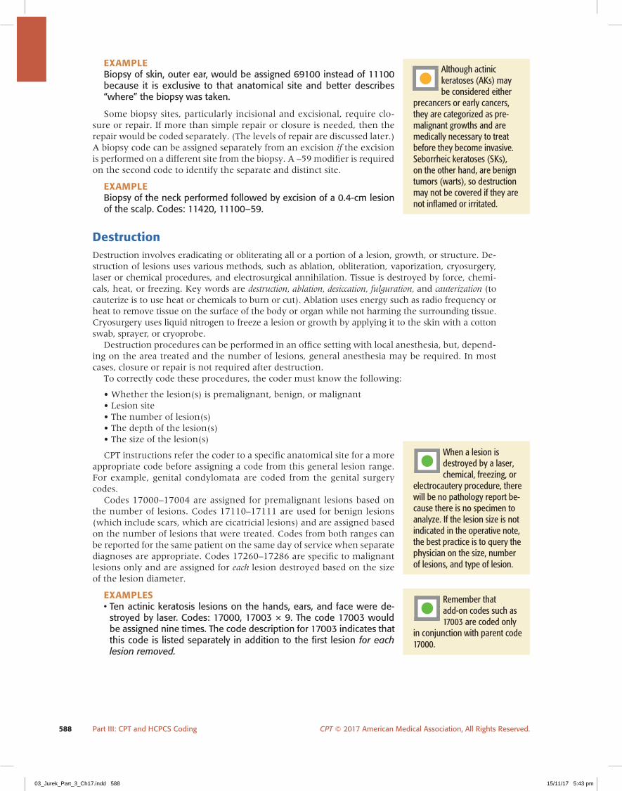
Although actinic keratoses (AKs) may be considered either
precancers or early cancers, they are categorized as pre-malignant growths and are medically necessary to treat before they become invasive. Seborrheic keratoses (SKs), on the other hand, are benign tumors (warts), so destruction may not be covered if they are not inflamed or irritated.
When a lesion is destroyed by a laser, chemical, freezing, or
electrocautery procedure, there will be no pathology report be-cause there is no specimen to analyze. If the lesion size is not indicated in the operative note, the best practice is to query the physician on the size, number of lesions, and type of lesion.
Remember that add-on codes such as 17003 are coded only
in conjunction with parent code 17000.
03_Jurek_Part_3_Ch17.indd 588 15/11/17 5:43 pm
CPT © 2017 American Medical Association, All Rights Reserved. Chapter 17: CPT: Integumentary System 589
introductionIn surgical terminology, introduction means insertion of an instrument, substance, or appliance into the body that performs a function, assists in treatment, monitors body functions, or marks a site. Key words: insertion, injection, percutaneous placement, intubation, and catheterization.
eXample11900 Injection, intralesional; up to and including 7 lesions.
paringParing means trimming or gradual reduction in size by slicing. Picture a potato or carrot peeler and how a thin layer is trimmed off with each stroke.
eXample11055 Paring or cutting of benign hyperkeratotic lesion (e.g., corn or callus); single lesion.
shavingShaving involves slicing a raised lesion or mole entirely off at skin level (i.e., flush with the skin) by using a razor or surgical blade; this is in contrast to paring, where layers are sliced off gradually. Shaving is also carried out in orthopedic procedures of the joints where cartilage is removed with a shaver.
eXample11300 Shaving of epidermal or dermal lesion, single lesion, trunk, arms, or legs; lesion diameter 0.5 cm or less.
Be careful! CPT directs the coder out of the Integumentary System
section to other surgery sections for destruction of lesions of specific anatomical sites for a more appropriate code.
chEckPOINT 17.1
Assign codes to the following procedures. Include any necessary modifiers.
1. A patient has been complaining of a nonhealing surgical wound. The patient underwent an
arthroplasty of the left index finger proximal interphalangeal (PIP) joint. The skin over the joint
has been chronically infected. The physician debrides the skin surface. _______________________
2. A patient presents to the office with a tender 2.0-cm cyst located in the right axilla. The patient
says she has had this for 2 weeks and thinks it is from an ingrown hair. The physician uses a
scalpel to puncture the cyst and proceeds to drain the pustule. _______________________
3. A patient was hit in the left upper arm with an 85-mph fastball while playing baseball. Over the
course of the day, the arm became bruised and began to swell. Two days later, he went to the
physician who diagnosed him with a hematoma and recommended draining the pooled blood.
The patient tolerated the procedure well. _______________________
4. A women is seen in the dermatologist’s office for a chronic rash with raised lesions on her
stomach. The physician examines the rash under magnification and decides to biopsy two
lesions. He stretches the skin perpendicular to the tension lines and inserts the trephine to
sample the skin to rule out lupus. _______________________
5. A patient presents to the office with two dermatofibroma on her right leg and one on her left.
The nurse practitioner uses liquid nitrogen to destroy them. _______________________
6. Laser destruction of four premalignant lesions. _______________________
03_Jurek_Part_3_Ch17.indd 589 15/11/17 5:43 pm

590 Part III: CPT and HCPCS Coding CPT © 2017 American Medical Association, All Rights Reserved.
excisionAn excision involves surgically removing or cutting out part or all of a tumor, lesion, organ, or tis-sue with a scalpel, sharp instrument, laser, loop electrode, or hot knife. Key words to look for are debulking, trimming, wedging, removing, excising, dissecting, and biopsy.
Three common surgical techniques are used for excising lesions of the integument. Two meth-ods are located in the Integumentary System section, and both methods are represented by the same group of codes:
• Simple• Wide
Simple excision removes a full-thickness lesion (through the dermis) and includes direct closure of the skin. Wide excision removes a lesion or tumor, its capsule, and a margin of normal tissue.
eXample11401 Excision, benign lesion including margins, except skin tag (unless listed elsewhere)trunk, arms or legs: excised diameter 0.6cm or to 1.0cm.
Radical excision is the third method. As the name indicates, radical excision involves the removal of the growth, its capsule, and the entire area of tissue or bone encompassing the growth. A radical excision is located within the Musculoskeletal System section of CPT.
To properly code excisions, the coder must have the following data:
• Morphology (type) of lesion: malignant or benign• Anatomical site of lesion• Number of lesions• Depth of the lesion• Size of the lesion• Surgical technique (shaving, excision, destruction, etc.)• Type of closure or repair to site
The operative report or procedure note along with the pathology report will provide this necessary information. Note that all excision codes include simple closure. Repair by intermediate or complex closure is separately reportable.
Two distinctly separate code ranges are available for excisions: benign lesions and malignant le-sions. After this division, codes are grouped by body part and then by size of lesion. The codes are assigned based on the diameter of the lesion being excised, including any margins, not according to the size of the incision necessary to access the lesion. Asymmetrical or irregular-shaped lesions are assigned codes based on the longest dimension. The margin of skin is the additional skin excised beyond or bordering the actual lesion. This is done to ensure that the entire lesion is removed. Surgeons typi-cally make the excision slightly larger than the actual lesion, creating a larger defect. The resulting defect is called an ellipse. The ellipse is drawn so that the resulting scar runs parallel with existing skin creases. This ensures that the scar is as narrow and short as possible. A visual example of this is shown in Figure 17.4. This is also illustrated in the CPT Professional Edition, Example B, located adjacent to the guidelines for excising benign lesions. The ellipse also allows for the surgeon to sew a straight line when closing the wound. If this technique is ap-plied, the size of the repair for other than a simple repair would be the length of one end of the ellipse to the other.
Other forms of lesion removal that do not involve a full-thickness excision of the skin, such as shaving, paring, and removal of skin tags, are located under those respective headings prior to the Excision heading. Codes for excision should be assigned to each lesion excised, regardless of the number of lesions removed.
CPT codes 11400–11446 should be used when the excision is a full-thickness (through the dermis) removal of a lesion, including margins, and includes simple (nonlayered) closure and is specific to benign lesions only.
Figure 17.4 Ellipse.
Pay particular attention to how the size of a lesion is stated.
Physicians may report the size as inches, centimeters, or millimeters.
03_Jurek_Part_3_Ch17.indd 590 15/11/17 5:43 pm

CPT © 2017 American Medical Association, All Rights Reserved. Chapter 17: CPT: Integumentary System 591
If a mix of benign and malignant excisions is performed, each lesion excision is reported individually.
Following an excision, any closure other than a simple closure is billed separately.
measurement of lesionsMany of the codes in the Integumentary System sec-tion involve measurements. All integumentary codes are based on centimeters excised, repaired, or otherwise treated. If the measurements are in millimeters, they must be converted to centimeters to accurately code the service (Box 17.2). For the most part, it is a matter of moving the decimal point to the left.
eXamplesA physician excises a 1.5-cm (width) × 2.0-cm (length) benign lesion from the top of the hand. The largest dimension is 2.0 cm; therefore, the code is 11422.
2-in × 3-in lesion = 6 sq in
6 sq in × 6.452 = 38.71 sq cm
If the diameter of a lesion is 5 mm, then it would be 0.5 cm
Converting a 5-in laceration would look like this: 5 in × 2.54 cm = 12.7 cm
The following points should be observed:
• The size of the lesion should be taken from the physician’s op note or office notes. If there is no documentation, query the physician for this information.
• Never guess or assume the size of the specimen or lesion; instead, query the surgeon.
• The pathology report is also a source for measurements.• If both sources have a size listed and they are not the same, use the
physician’s measurement. The formalin preservative may shrink the size of the tissue sample, so always report the pre-excision size if it is available.
Study the illustration shown in Figure 17.5 and review the following ex-ample. The CPT Professional Edition also provides an illustration of this within the Excision-Benign Lesions section, Example A.
eXampleRemoval of a benign lesion of the back with an actual diameter of 2.0 cm × 1.0 cm. The physician determines that the appropriate mar-gin required for this excision is 0.2 cm. The greatest clinical diameter of the lesion is 2.0 cm. An additional 0.2-cm margin is excised around the entire lesion. The excised diameter is then equal to the greatest clinical diameter (2.0 cm) and an additional margin of 0.4 cm (0.2 cm added on either side of the lesion). The total excised diameter is 2.4 cm, which codes to CPT code 11403.
1 mm = 0.1 cm10 mm = 1 cm1 in. = 2.54 cm1 sq. in. = 6.452 sq cm
BOX 17.2 Converting to metric units
There is one exception to how measurements are described in In-
tegumentary System codes. Codes for flaps and grafts are reported in square centimeters (cm2), not by the diameter of the lesion or length of repair.
It is important for facility and surgeon claims to match. It is not uncom-
mon for the surgeon not to wait for the pathology report to re-port a final diagnosis for a biopsy or lesion excision; therefore, the diagnosis that the facility bills might not match the surgeon’s bill, which would cause claim denials to either entity. Ideally, the diagnoses and CPT codes that both parties submit should match for the same ser-vice performed on the same day.
1 cm 0.2 cm 2 cm 0.2 cm
0.2 + 2 + 0.2 = 2.4 cm total diameter
Figure 17.5 Diameter of a lesion.
03_Jurek_Part_3_Ch17.indd 591 15/11/17 5:43 pm

592 Part III: CPT and HCPCS Coding CPT © 2017 American Medical Association, All Rights Reserved.
When multiple lesions are treated, report the most complex (largest lesion) procedure first and the others with a –51 or –59 modifier depending on the code description and the location of the lesions.
eXampleIf two lesions of the hand (0.5 cm each) and two lesions of the neck are removed (0.7 cm each), assign a –59 modifier for each separate lesion and site because the code description includes several body sites. Codes: 11420, 11420–59, 11421–59, 11421–59.
If two lesions are removed through one incision, only one code is as-signed. The size of the two lesions and the space between the lesions are added together.
eXampleTwo 0.3-cm hand lesions located 0.5 cm apart are removed through one incision. 0.3 cm + 0.3 cm + 0.5 cm = 1.10 cm. Code: 11422.
In most cases, modifiers –RT and –LT do not apply to lesion removals or repairs in the integumentary section. Because many different anatomical sites are “grouped” or classified into codes for removal and repair, assign-ing one of these modifiers would not be describing a paired body part.
eXampleExcision of a 0.5-cm nevus from the right ear is coded as 11440. The code description for 11440 includes lesion removal from the face, ears, eyelids, nose, lips, and mucous membrane. Applying the RT modifier does not differentiate the right ear from the right eyelid.
Lipomas (tumors) are tricky to code. Pay very close attention to the code description. If the description says, “unless listed elsewhere,” refer to the most specific code in the most specific body category. Code to the most appropriate anatomical site regardless of the depth of the lipoma (tumor) even if the term skin is mentioned. Use the Integumentary System code only when there is no specific code in the Musculoskeletal System section.
eXample11406 Excision, benign lesion including margins, except skin tag (unless listed elsewhere), trunk, arms, or legs; excised diameter over 4.0 cm.
a note on malignant lesionsIf a physician excises a malignant lesion and sends the specimen to pathol-ogy intraoperatively (during the procedure) to determine if the margins are “clear,” and further excision is done until the pathologist confirms for the surgeon that the margins are “clear,” only one excision code is as-signed. This code is for the final greatest width of the lesion.
EXAMPLESThe surgeon excises a 2.0-cm × 1.0-cm lesion of the shoulder with a 0.1-cm margin on both sides and sends it to pathology. The pathologist says the margins are not clear. Surgeon excises an additional 0.1-cm margin all around. Pathologist finally confirms that the margins are free of disease. What is the widest final diameter? 2.0 + 0.1 + 0.1 + 0.1 + 0.1 = 2.4 cm. Code: 11603.
A 1.0-cm malignant lesion of the neck with a margin of 2.0 cm is excised. The excised diameter is the sum of the greatest diameter (1.0 cm) plus the margin of 2.0 cm from each side of the lesion (2.0 + 2.0). Total diameter is 5.0 cm. Code: 11626.
What happens if a physician removes a lesion and the pathology report states that the lesion is of unknown neoplastic behavior? This
Note that when a le-sion is removed from a previous mastectomy
site, the site is considered the “trunk” and not the “breast” because the breast is no longer there, unless a partial mas-tectomy or lumpectomy was performed and the woman still has breast tissue remaining.
If a malignant lesion is excised on one visit and a re-excision is
performed on a separate visit (could be weeks or months later) to verify that all the cancer is gone, code to the excision of a malignant lesion, even if the second re-excision pathology report states that margins are benign or clear.
If lesion size is not in-dicated in the op note, the best thing to do
is query the physician on the size and type of lesion. If this is not done, you are forced to use a code for the smallest size lesion (0.5 cm), which in most cases is not reimbursable in the outpatient setting.
Remember to add the margins (M) for both ends of the lesion to
the diameter (D) of the lesion itself. Think times two! Total diameter × D + (M × 2).
03_Jurek_Part_3_Ch17.indd 592 15/11/17 5:43 pm

CPT © 2017 American Medical Association, All Rights Reserved. Chapter 17: CPT: Integumentary System 593
documentation means it is uncertain as to whether it is benign or malignant, which has a di-rect impact on CPT code assignment. In this case, the surgeon’s documentation should reflect the surgical approach. The CPT code that best describes the extent of the procedure performed should be reported. A lesion that is judged probably benign might be excised with minimal surrounding tissue removal, but one judged more suspicious might be excised with greater tissue margins.
Wound repair (Closure)A repair surgically closes a defect, restores shape or functionality, or reconnects to an anatomical body part, which usually involves suturing and possibly replacement of tissue. Key words to look for include stitching, closure, correction, anastomosis, restoration, suturing, reduction, and words ending in –rrhaphy.
Codes for repair are not based on whether the lesion removed is malignant or benign. Instead, code assignment is based on depth of skin, technique, and materials used. As mentioned earlier, CPT defines three levels of wound repair complexity:
• Simple• Intermediate• Complex
Codes are grouped within each level by anatomical sites and size of wound. The size of the wound is determined by adding the length of all wounds in the same classification or anatomical group. With wound re-pairs, the lengths of multiple wounds are added together by repair group or classification and by complexity.
eXample13160 Secondary closure of surgical wound or dehiscence, extensive or complicated.
simple repairA simple repair is made for a superficial wound repair involving the epi-dermis, dermis, and a minimal amount of subcutaneous tissue requiring one-layer suturing. This type of repair is referred to as “direct” or “primary” closure. If the closure was done with tape, staples, Dermabond, glue, or adhesive strips, this service is included in the E/M code and not billed sepa-rately. Likewise, when lesions were removed and only simple repair was necessary to close the wound, the simple repair is not coded separately. A simple repair is included in the lesion removal “package” and payment. Running subcuticular suture involves the epidermis and part of the dermis.
eXampleExcision of a 3.0-cm nevus from the left shoulder. Only the epidermal and part of the dermal layers were repaired. Code: 11403. The simple repair is included in the code for the lesion removal.
intermediate repairAn intermediate repair involves closure of one or more of the subcutaneous layers and superficial fascia, in addition to the skin. Often, two different types of suture are used to close the wound. There are several names or types that serve varying purposes. Some are used for closing deep tissue and dissolve internally, while others are used for the superficial layers of skin and require removal. Common suture brands include Chromic, Vicryl, Monocryl, Prolene, and Ethilon. An intermediate repair indicates that a layered closure took place. One layer is closed with one type of suture and the second layer is closed with a different suture type. Reference to buried suture indicates a lay-ered closure took place. The buried sutures provide support to the wound and prevent tension on the skin wound edges. An intermediate repair code can also be reported when the wound has to be extensively cleaned due to contamination or removal of small pieces of foreign material before su-turing, even if the suturing is simple. This is an important exception that is often overlooked in the coding process. Running subcutaneous suture is used to close the deep portion of incisions created during surgery. Sizes 3-0 and 4-0 suture materials are typically used for subcutaneous suturing.
“Bring together, sew together, add together.”
Medicare requires code G0168 to be submitted when a
repair is performed with tissue adhesive only. However, G0168 is bundled to all wound repair codes and cannot be reported separately if also assigning a code in the 12001–13160 range. For non-Medicare patients, if the repair is performed solely with tissue adhesive, only the E/M code for the visit is assigned.
03_Jurek_Part_3_Ch17.indd 593 15/11/17 5:43 pm

594 Part III: CPT and HCPCS Coding CPT © 2017 American Medical Association, All Rights Reserved.
eXampleLook at code 12031 in CPT. It includes intermediate repair of wounds of the scalp, axillae, trunk, and/or extremities (excluding hands and feet). As you can see, many significantly different sites are “grouped” or “classified” into this code. For example, a 2.5-cm wound of the right leg and a 3.1-cm wound of the back were both repaired with intermediate repair. The coder should add the lengths of both of these wounds together and use the sum to base code assignment. In this case 2.5 + 3.1 = 5.6 cm. Code: 12032.
Complex repairA complex repair or closure includes scar revision, debridement, extensive undermining, stents, or retention sutures, and more than layered closure. Repair of traumatic avulsions or lacerations may be in this category. When the wound is complex, repair of extensive nerve, blood vessel, or tendon damage may also be reported. Irregular or jagged wounds often require complex repairs.
eXampleThe physician repairs a 7.6-cm knife laceration of the right forearm. The physician had to re-pair all layers of the skin and debride foreign material from the wound. Because the length of the repair is 7.6 cm, two codes are required: 13121 and 13122. Note that add-on code 13122 specifies “each additional 5cm or less.”
Wound repairs include simple ligation of vessels; simple exploration of surrounding tissue, nerves, vessels, and tendons; and simple debridement (cleaning or removing of tissue until normal-appearing, healthy tissue is exposed). If gross contamination is encountered and requires exten-sive debridement, the wound requires extensive cleansing, or significant amounts of tissue are removed, this is coded separately.
Lengths of wounds or defects caused by lesion removal are only added together if the repair is the same complexity and from the same body category or group depicted in the code description.
eXampleA 2.5-cm simple repair of the arm and 3.1-cm intermediate repair of the leg. These procedures cannot be added together because they are two different complexities of repair even though they are in the same anatomical group.
If multiple types of repair are performed, the most complex type is coded as the primary procedure. Report modifier –59 for any additional repairs. Although modifier –51 is the multiple procedure modifier, CPT specifies the use of modifier –59 for multiple repair codes.
eXampleSimple repair of the lower leg and intermediate repair of the foot are performed. The intermediate repair of the foot is coded and listed first followed by the simple repair of the leg.
assigning Codes for Wound repairTo properly assign codes for wound repair, the following factors (Pinpoint the Code 17.1) must be considered:
1. What is the length of wound in centimeters (cm)? If the size is reported in millimeters (mm), it must be converted to cm by moving the decimal point. For example, if the lesion is 11 mm, it is 1.1 cm. (Do not confuse the size of the lesion with the size of the specimen or size of the defect left.)
2. What type of wound (wound dehiscence, burn, laceration) is it? 3. Where is the site of the wound repair? 4. How many wounds were repaired?
Medicare will not pay for intermediate or complex wound repair
if associated with a lesion removal of less than 0.5 cm.
Repair sites and types must match to be added. Lengths cannot
be added together if the type of repair is not the same or if the repairs were done on dif-ferent sites.
Debridement is coded separately from wound repairs if gross
contamination is encoun-tered and requires extensive debridement, the wound requires extensive cleansing, significant amounts of tissue are removed, or the wound is packed and closed at a later encounter.
03_Jurek_Part_3_Ch17.indd 594 15/11/17 5:43 pm
CPT © 2017 American Medical Association, All Rights Reserved. Chapter 17: CPT: Integumentary System 595
PINPOINT ThE cODE 17.1: Wound Repair
Group wounds with same anatomical grouping, with the same complexity of repair, and add lengths of all wounds together for one total repair length.
How many wounds?
Determine the complexity of repair(s).
For multiple wounds, a code from each level of complexity is assigned, listing the most complex first.
Was gross contamination encountered requiring extensive debridement? Did the wound require extensive cleansing or significant amounts of
tissue to be removed? Was the wound packed and closed at a later encounter?
Is debridement also performed?
Code separately in addition to repair by depth of debridement (full thickness, partial thickness, etc.): 11042–11047.
Where is the wound(s)?
What kind of wound is it?
If it is an open fracture, no repair is coded. This is included in the service
for fracture care. Debridement for fractures is assigned to 11010–11012.
Don’t code debridement separately.
What is the length of the wound?
Add –58 modifier.
Assign code for debridement and repair. It is a good idea to append –59 modifier to prevent bundling.
Assign code for debridement only without a repair code unless the
debridement is done at a different site from repair. If so, append –59 modifier.
WithoutWith
NO
NO
NO
YES
YES
Staged debridement?
With/without closure?
03_Jurek_Part_3_Ch17.indd 595 15/11/17 5:43 pm

596 Part III: CPT and HCPCS Coding CPT © 2017 American Medical Association, All Rights Reserved.
5. What was the complexity of repair for all wounds? Group the same anatomical groups, with the same complexity of repair, and add lengths of all wounds together for one total repair length.
6. The most complex repair is listed first as the primary procedure and then the lesser repair codes with a modifier –59 per CPT guidelines.
7. Was debridement provided? If this is documented and extensive, it can be coded separately. 8. Simple ligation or cautery of blood vessels and simple exploration of nerves or tendons
through the wound site are routine and are not coded separately. 9. If nerves, blood vessels, and tendons are repaired, then these are coded separately from
complex wound repairs.10. If the wound requires enlargement or extension of the dissection to determine penetra-
tion or depth of the wound, then the wound exploration codes 20100–20103 should be reported instead of the wound repair codes.
11. If the wound requires debridement, removal of FBs, or ligation or coagulation of minor subcutaneous and/or muscular blood vessels(s) of the subcutaneous tissue, muscle fascia, and/or muscle, use the wound exploration codes 20100–20103.
12. If the wound requires a thoracotomy or laparotomy, report those services and not the wound repair codes.
pulling it tOgether: eXCisiOn anD repair CODe assignment
Pinpoint the Code 17.2 demonstrates the thought process a coder should follow when coding excisions and repairs. This decision tree is only used for surgical excisions and is not meant for shaving, destruction, or paring. Before assigning any codes for excisions or repairs, make sure measurements are in centimeters; if they are not, they must be con-verted. New coders can use this decision tree when assigning codes from this section because it uses a systematic approach that can be applied time and again. Key words to use in locating these codes in the Index are Excision then Lesion followed by Skin. The coder will then have to choose from Benign or Malignant.
Shaving of lesions requires no closure because no incision has been made.
If multiple types of repairs are done at the same session, sequence the most
extensive repair first.
chEckPOINT 17.2
Assign codes to the following procedures. Include any necessary modifiers.
1. A 7-year-old boy fell from his tree house, lacerating his forehead, right hand, and left forearm. He
was taken to the ED. The ED physician used Dermabond and butterflies to close the superficial
2.0-cm laceration of the forehead. The 2.5-cm laceration of the skin and subcutaneous tissue
of the left forearm required a layered closure using a Vicryl suture. The 3.0-cm laceration of the
right hand had bark debris that had to be washed out before suturing the skin and subcutaneous
tissue. __________________________
2. A 19-year-old patient goes to the physician complaining of two warts on the left index and
middle fingers. He wants these removed. The physician applies Verruca-Freeze for 1 minute to
each wart. __________________________Continued
03_Jurek_Part_3_Ch17.indd 596 15/11/17 5:43 pm

CPT © 2017 American Medical Association, All Rights Reserved. Chapter 17: CPT: Integumentary System 597
SkIN grAFTS AND FLAPS
The skin is the body’s first defense against injury and infection by microorganisms. When skin is damaged, the body can regenerate only the epidermal layer. At times, skin injuries and defects from lesion removal destroy deeper structures such as the dermal and subcutaneous tissues. This damage requires more than simple, intermediate, or complex closure. Alternative methods of repair are adjacent tissue transfer or tissue rearrangement and skin grafting with either the patient’s own skin or skin substitutes. The success of the skin graft will depend upon the location and size of the injury, along with the patient’s healing abilities and blood supply.
Codes for tissue transfers/arrangements (flaps) are assigned based on location of the defect and surface area to cover (size of defect). To assign codes for skin grafts, the type of graft is located first (i.e., split-thickness, full-thickness, xenograft) followed by the location of the defect, and surface area (size of defect). It is critical to read the operative report in its entirety to determine the type of graft, flap, or tissue rearrangement carried out, where the tissue is coming from (donor site), the recipient site, the size of the defect where the graft is placed, and if any additional repair to the donor site is required. It is also important to ascertain if a skin substitute is used in the repair. The area is measured in square centimeters (cm2 or sq cm). The coder is often required to calculate the square centimeters to derive the correct flap or graft size before assign-ing the code. If the operative report describes deeper structures such as muscle, fat or fascia, or microvascular anastomosis of other than skin and vessels, other CPT codes may be more appropriate from the Muscu-loskeletal section of CPT.
Skin flap and graft codes are not located centrally in one subsection of the Integumentary chapter. Instead, they are located in different areas under codes 14000–15777, 15840–15845, 15922, 15934, 15944, 15952, 19364, 19637, and 19369 so the coder needs to have a good understanding of this and not skip the Index and go right to the graft section of the chapter.
Skin grafts are obtained by using an instrument called a dermatome to slice the skin away from the donor site at very precise thicknesses. It is similar to a hand-held cheese slicer or vegetable peeler that is dragged across the skin. The surgeon may mesh the graft by running it through a special processor that places holes in the tissue to allow it to expand to cover more area.
3. A 63-year-old female noticed a purplish irregular lesion on her lower leg. She has already had
a basal cell carcinoma removed from her left shoulder. The 1.7-cm lesion was removed with
a 0.2-cm margin. Simple repair was performed. Pathology confirmed basal cell carcinoma.
__________________________
4. A patient has been complaining of a nonhealing surgical wound. The patient underwent an
arthroplasty of the left index finger PIP joint. The skin over the joint has been chronically infected.
The doctor debrides 10 sq cm of skin and subcutaneous tissue and performs a complex layered
closure. __________________________
5. A 3.0-cm, full-thickness benign lesion is removed from the patient’s upper back. A second benign
lesion is also removed from the midback measuring 2.2 cm. The physician uses Vicryl to repair
the subcutaneous layer and Monocryl to close the skin in both sites. __________________________
chEckPOINT 17.2 Continued
Be sure that the measurements used are square centimeters
and not just centimeters. Finding square centimeters is like measuring a room for carpet in your house. Multiply length (L) times width (W) to get the square area. L x W = square centimeters. For example, if a 2 x 3 cm defect was closed, the code assigned is based on 6 sq cm (2 x 3 = 6).
03_Jurek_Part_3_Ch17.indd 597 15/11/17 5:43 pm

598 Part III: CPT and HCPCS Coding CPT © 2017 American Medical Association, All Rights Reserved.
PINPOINT ThE cODE 17.2: Excision and Repair
If lesions from same bodycategory but excised from
different incisions, use modifier–59 for each additional lesion.
If excised through same incision,add widths and space betweenlesions for one total width and
assign only one excision code.
Two ormore
If excised through different incisions, assign code for
each lesion. If excised through same incision, add widths and space between lesions for one total width.
Where?
Decide size by greatest dimension including margins.
Choose excision code only.
Benign
Skin/subcutaneous stay inIntegumentary System section.
If more than one lesion is excisedand repaired from the same body
category, add sum of lengthstogether to get the longest
measurement. Assign the repaircode in addition to the lesion
removal. If different body category,assign excision and repair codeaccording to the measurementsfrom each body area applicable.
Don’t add anything!
Deeper or elsewhere listed? SeeMusculoskeletal System section.
Skin/subcutaneous, deeperor elsewhere listed?
Depth/location of lesion?
One
One
Where?
Initial?
Initial orre-excision?
Decide size by greatest dimension including margins.
Even if margins come back
clear, still code malignant.
Re-excision?Malignant
Intermediate or complex.
Benign/malignant
Simple repair?
How many?
How many?
YES NO
Two ormore
03_Jurek_Part_3_Ch17.indd 598 15/11/17 5:43 pm

CPT © 2017 American Medical Association, All Rights Reserved. Chapter 17: CPT: Integumentary System 599
Free skin graftsSkin grafting procedures are assigned from code range 15040–15278. There are many types of grafting procedures and types discussed in this section. Free skin grafts get their name because they are completely freed from the donor site and placed at a recipient site. No vascular connection from the donor site is left intact with these grafts. Free skin grafts are sections of skin that fall into one of these four categories:
1. Autograft/Tissue Cultured Autograft2. Acellular Dermal Replacement3. Allograft/Tissue Cultured Allogeneic Skin Substitute4. Xenograft
An autograft is a piece of skin taken from the patient’s own body to cover a defect so the donor and recipient site is the same individual. The patient’s own skin is used whenever possible to reduce the risk of rejection. The term allograft refers to a graft obtained from a source other than the patient’s body. It is either a donation from a cadaver or is a manmade synthetic material.
To understand free skin grafting, the coder must be able to distinguish between the recipient site (site receiving the graft) and the donor site (site where graft was harvested from). If the donor site requires more than simple repair after harvesting the tissue, an additional repair code is required. Sometimes, a graft is necessary to close the defect at the donor site. If so, this is coded separately in addition to the original skin graft. If a lesion is excised and immediately closed with a free skin graft, code the excision of the lesion in addition to the graft.
surgical preparationCodes 15002–15005 are used to report the initial preparation of the graft recipient site, which needs healthy bleeding tissues to receive the graft. The initial preparation includes cleansing, re-moving any nonviable tissue, or excision of tissue or incisional or excisional release of scar con-tracture resulting in an open wound. Surgical preparation maximizes the chances of survival of
the graft to be placed. Usually, the recipient site contains uneven layers or multiple layers that could pose a problem with connecting the surfaces of the donor graft to the recipient site. The layers may also cause the graft site to be very visible with a less than adequate cosmetic effect.
As stated in the CPT Surgical Preparation section, 15002, “Surgical preparation or creation of recipient site by excision of open wounds, burn eschar, or scar (including subcutaneous tissues); first 100 sq cm or 1% of body area of infants and children” and 15004, “Surgical preparation or creation of recipient site by excision of open wounds, burn eschar, or scar (including subcutaneous tissues, or incisional release of scar contracture, face, scalp, eyelids, mouth, neck, ears, orbits, genitalia, hands, feet and/or multiple digits); first 100 sq cm or 1% of body area of infants and children.” The operative words here are excision of tissue or incisional or excisional release. This type of preparation requires the use of a scalpel.
CPT provides parenthetical instruction to report 15002–15005 in con-junction with the codes for skin graft or replacement when preparation of the recipient site is performed.
Debridement of skin that is nonintact to prepare the site to receive a skin graft is generally included in the skin graft service and not coded separately. However, if the recipient site requires debridement of intact skin to prepare for a free skin graft, use code 15002–15005 in addition to the code for the graft when the site is prepared prior to receiving the graft (i.e., removing scar tissue or eschar).
Codes 15002–15005 are not used to report debridement of nonintact necrotic skin lesion remov-als, or for chronic wounds that are intentionally allowed to heal secondarily from the inside out. These services are reported with codes from the Medicine section.
Table 17.4 provides a short list of graft categories and descriptions to assist coders in determining the uses and respective CPT code ranges.
Report codes 15002–15005 in addition to the code
for the respective skin grafts or replacement codes 15050–15261, 15271–15278 if the skin is intact.
Do not assign codes 15002–15005 for non-incisional or excisional
debridement (VERSAJET™ or manual removal) of tissue reported with a code from the Medicine section because these are not considered to meet the definition of surgical preparation.
03_Jurek_Part_3_Ch17.indd 599 15/11/17 5:43 pm

600 Part III: CPT and HCPCS Coding CPT © 2017 American Medical Association, All Rights Reserved.
autograftsThe autograft can be of various thicknesses, shapes, and sizes. The purpose of the autograft is to actually replace the skin that was damaged or removed. There are four categories of autograft in-dicative of the source of tissue: split thickness, full thickness, epidermal, and dermal. Each category of autograft has its respective CPT code range.
Split-Thickness Grafts (STGs or STSGs). Split-thickness skin grafts get their name because they contain only part of the dermal layer in some areas of the graft. It is used for areas that do not require a complete dermal covering but do require a complete epidermal layer. Split-thickness grafts are most typically used for non-weight-bearing parts of the body or for infected or large wounds resulting from burns. The code range is 15100–15101, 15120–15121. This code range is also assigned for thin grafts (0.010–0.015 inches), moderately thick grafts (up to 0.015 inch, which includes more of the dermis), and thick grafts (greater than 0.015 inches but does not include all of the dermis) because these all contain varying percentages of epidermis and dermis.
Full-Thickness Grafts. Full-thickness grafts (FTGs or FTSGs) are used to replace areas that are missing epidermal and dermal layers of skin. The code range is 15200–15261. They include an equal and continuous section of both layers of the skin and all of the skin elements including blood ves-sels but none of the underlying fat. The blood vessels will begin to grow from the recipient area into the transplanted skin within 36 hours of grafting. Full-thickness grafts are used for weight-bearing portions of the body and friction-prone areas, such as feet and joints. Donor sites are typically the thigh, abdomen, postauricular skin, and inguinal folds.
Full-thickness grafts are also located in the Eye and Ocular Adnexa section of CPT and reported with codes 67961 or 67966.
table 17.4 Free Skin Grafts and Skin SubstitutesgrAFT cATEgOrY DEScrIPTION cPT cODE(S)
Pinch Small (typically 2 cm or smaller) pieces of tissue that are used to repair tips of digits or other small areas
15050
Epidermal Grafts composed of the epidermis, the outermost layer of skin 15110–15116
Dermal Grafts composed of the dermis the second layer of skin immediately below the epidermis
15130–15136
Split thickness Grafts composed of the full layer of epidermis and part of the dermis 15100–15101, 15120–15121
Full thickness Grafts composed of the full layer of both the epidermis and dermis 15200–15261
Tissue-cultured epidermal autograft
Cultured skin with only an epidermal layer (e.g., CEA, Epicel®, EpiDex®). For treatment of deep dermal or full-thickness wounds where adequate donor sites not unavailable.
15150–15157
Acellular dermal replacement
A tissue-derived or manufactured device that provides immediate temporary wound closure. It incorporates into the wound and promotes generation of new skin that can support epidermal tissue (e.g., Integra®).
15271–15278
Allograft Cadaveric human skin (from skin banks) 15271–15278
Skin substitute grafts Non-autologous human skin, non-human skin substitute grafts (xenograft), and biological products that form a sheet scaffolding for skin growth.
15271–15278
03_Jurek_Part_3_Ch17.indd 600 15/11/17 5:43 pm

CPT © 2017 American Medical Association, All Rights Reserved. Chapter 17: CPT: Integumentary System 601
let’s Code it
DIAgNOSIS: Full-thickness skin injury, right elbow.OPErATION: Skin graft.
This 33-year-old male was pushed through a glass storm door window and sliced skin off his right medial elbow area. He was seen in the ED and sent home using saline moistened dressings and referred to a plastic surgeon. The patient is seen today in the outpatient surgery center. The wound was full thickness, partially granulating, and covered with fibrinous debris with uneven skin margins and required a 60 sq cm. full thickness skin graft. The wound was painted with ethylene blue as a marker and then the VERSAJET was used to remove the outer layer of granulation tissue. There are no gouges or exposed fat in the wound bed. Using a dermatome, a full-thickness skin graft was harvested from the right left shoulder and passed through the mesher. It was then placed over the wound. Fibrin glue was used to adhere the graft to the wound bed. A foam bolster was applied.
Answer: 15220, 15221 x 2 for the skin graft. Documentation notes a 60 sq cm. full-thickness auto-graft from the patient’s own shoulder. Do not assign 15002 because surgical preparation was per-formed by using the VERSAJET and it is not considered to be surgical preparation. With skin grafts, codes are assigned based on the recipient site and the depth or thickness of the graft.
Epidermal Graft. Epidermal grafts (15110–15116) are composed of the epidermis, the outermost layer of skin. This type of closure is used when direct suture closure or adjacent tissue transfer is not feasible. The graft is used in many cases to perform the final stage of wound closure. Fre-quently, it is used in conjunction with dermal allografts in treating ulcers of the lower extremities.
Dermal Graft. Dermal grafts (15130–15136) are composed of the dermis, the second layer of skin immediately below the epidermis. The epidermis and subcutaneous fat are removed. This type of graft is used instead of fascia in various plastic procedures. It serves as a filler and provides a local-ized correction to an area, such as the border of the lip, forehead lines, depressed scars, or the deep hollow beneath the eyes.
Pinch Graft. Pinch grafts are very small (typically 2 cm or smaller) pieces of tissue that are used to repair tips of digits or other small areas. They heal quickly and resist infection. Refer to CPT code 15050 to report pinch graft services. As indicated in the code descriptor, this code would be used only once, regardless of the number of pinch grafts performed to cover a defect 2 cm or less.
Other Grafts. There are other autografts located in this chapter but these grafts have different compositions of tissue and are not just skin or subcutaneous tissue. These particular grafts, similar to the previously mentioned autografts, serve a specific purpose of replacing tissue and are chosen based on their size, location of defect, and physical composition.
Composite grafts (15760–15770) include more than one tissue type, such as the cartilaginous skin found in the ear or nose. This graft type is used to fill in a defect and to provide skin and struc-tural support (cartilage) in the recipient site; therefore, it minimizes scar contraction and distortion because it imitates the tissue and function of the tissue in the recipient area.
Derma-fascia-fat grafts (code 15770) are composed of the dermal layer, fascia, and subcutane-ous fat. Indications for this graft are primarily the same as the composite graft in that the purpose is to blend-in or fill-in defects left behind by surgical excisions, atrophy, and so on. The tissue may be grafted in three ways: complete portion (containing all three of the layered components), indi-vidual parts (grafted layer by layer), or inserted in combination (such as a fascia-fat layer, later cov-ered by a dermal layer). The technique used to attach the graft is not relevant to code assignment.
Tissue-cultured AutograftAn autograft as described earlier is harvested from the patient’s own body and applied to another area requiring coverage. A tissue-cultured autograft is one that has been first cultured in the laboratory
03_Jurek_Part_3_Ch17.indd 601 15/11/17 5:43 pm
602 Part III: CPT and HCPCS Coding CPT © 2017 American Medical Association, All Rights Reserved.
from skin cells harvested from the patient and under controlled conditions grown into sheets of graft material to produce “new skin.” The autograft is then applied to the patient’s wound.
When the surgeon harvests skin—or more specifically, keratinocytes—and dermal tissue for the cultured autograft, CPT code 15040 Harvest of skin for tissue cultured skin autograft, 100 sq cm or less is reported. The skin is removed by using a dermatome to obtain a split-thickness graft. The tissue is sent to a lab and the layers of skin are separated and placed in a special solution that stimulates the skin cells to divide and generate tissue.
Surface area and location matter when assigning codes for the placement of the cultured au-tograft. The code range is 15150–15157. Codes are reported according to the number of square centimeters used. Either code 15150 or 15155 is reported for the first 25 sq cm. If more than 25 sq cm is used, add-on codes are required for reporting additional square centimeters from 1 up to 75 (15151 or 15156). The measurements of the two codes—base code (25 sq cm) plus the add-on code (additional 1 to 75 sq cm)—can add up to 100 sq cm. If the graft tissue required is larger than 100 sq cm (total sq cm of base code plus add-on), then either 15152 or 15157 is also reported to capture each 100 sq cm beyond the first 100 sq cm and/or each additional 1% of body surface or fraction thereof of each. Laboratories can only culture a limited amount of autograft; therefore, for large surface areas this procedure is often performed in stages. Modifier –58 is required to indicate that the procedure is being conducted in stages.
■ Case sCenariOA patient suffered extensive road rash to the back when she crashed her motorcycle. The surgeon provided temporary wound closure until the patient’s cultured skin was ready to be permanently placed. He placed 150 sq cm of cultured epidermal autograft to the upper and mid-back. This requires reporting three codes: 15150, 15151, 15152. Note that add-on code 15152 states “or part thereof” indicating that the additional sq. cm. autograft does not have to be 100 sq. cm. to use code 15152
skin substitute graftsSkin substitutes (15271–15278) are successful in improving wound healing, controlling pain, and improving function and cosmetic appearance. Skin and dermal substitutes refer to nonpermanent grafts. A bilaminate skin substi-tute is a manufactured product composed of human cells in a bovine (cow) collagen matrix. Skin substitutes are favored for some wound coverage be-cause they are flexible, pliable, and biodegradable. They also resist the stress of stretching and pulling, and allow fibroblast in-growth. Skin substitutes have less risk of disease transmission than traditional cadaver allografts. The substitute has two layers: an outer epidermal layer of silicon rubber and an inner layer of flexible nylon fabric. The inner fabric contains porcine colla-gen, which promotes fibroblast in-growth and patient acceptance. The artifi-cial skin product TransCyte is made by culturing human neonatal fibroblasts on a synthetic nylon mesh dressing. Once optimal growth is achieved, it is then transplanted or grafted to the patient’s wound. When applying a tissue-cultured allogenic skin substitute, report 15002–10005 for preparation of the graft site prior to applying a tissue-cultured allogenic skin substitute—do not report an excisional debridement code.
Acellular dermal replacements are bioengineered artificial skin sub-stitutes. They may go by the names neodermis, skin substitute, AlloDerm, Integra, Oasis, Surgisis, or Dermagraft. These are temporary skin substitutes used to cover wounds until skin autografts can be harvested and placed. CPT defines acellular dermal replacements as “A tissue-derived or manu-factured device that provides immediate, temporary wound closure and that incorporates into the wound and promotes the generation of a neo-dermis that can support epidermal tissue.” These products usually consist
The FDA as well as CMS state that skin substitutes are a one-
time use item because they have a finite shelf life. Even if the surgeon does not use all of the material to cover the ulcer, wound, or burn, the entire piece of material is billed regardless of the size of the piece applied.
HCPCS codes Q4100–Q4114 should be reported per sq cm
for the graft material in addi-tion to the CPT code for the grafting procedure if the physician is performing the procedure in his office or is bringing the material with him to the facility. Facility codes will be assigned based on charges entered via the charge master for OR supplies and services. Reporting the HCPCS code for the supply is challenging; it requires the coder to know the brand name of the material and the amount of material applied or the entire size of the material. Because these codes are reported by sq cm, the quantity in the unit’s column of the claim form must reflect the correct amount applied.
03_Jurek_Part_3_Ch17.indd 602 15/11/17 5:43 pm
CPT © 2017 American Medical Association, All Rights Reserved. Chapter 17: CPT: Integumentary System 603
of a silicone outer epidermis and an inner layer of porous lattice fibers made from a collagen/glycosamine or bovine cartilage mix. They are used to provide full-thickness skin coverage in burn victims when autografting is not an option. The products are also FDA approved for treatment of noninfected partial and full-thickness skin ulcers due to venous insufficiency and neuropathic diabetic foot ulcers. The dermal replacement is applied and in time the patient’s dermal cells travel into the graft area. Once new dermal growth has appeared, an epidermal graft may be applied. Wound care centers see many patients receiving this type of graft. Codes are assigned based on anatomic site and upon documentation of the size of the total defect treated. These codes are re-ported for the total body surface area treated, not per wound site.
Allografts are used to temporarily cover wounds from burns, traumatic skin injuries, and areas of soft tissue infection or necrosis. An allograft (homograft or allogeneic graft) is a graft transplanted between genetically nonidentical individuals of the same species (i.e., cadaver). The graft pro-tects the exposed recipient site from infection and maintains the overall viability of the underlying tissue until a more suitable autograft can be obtained or cultured later.
A xenograft is a graft transferred from an animal of one species to one of another species, typically porcine (pig) or bovine (cow). Acellular xeno-grafts actually incorporate into the skin during the healing process are not removed but rather become a permanent implant of the skin. This is not the case with regular xenografts. Allograft and xenograft tissue, once placed, only lasts a few days, at which time the material is either replaced with more xeno- or allograft material or the autograft procedure can be performed.
Flaps and adjacent tissue transfer or rearrangementFlaps and tissue transfers or arrangements differ from skin grafts because they carry their own blood supply. The procedures are carried out when the donor site and the recipient site are in close proximity. The flaps or tissue transfers are not completely severed from the donor site, so the vas-cular bed is still attached to feed the tissue with adequate blood supply. The CPT Professional Edition provides illustrations and descriptions of adjacent tissue repairs.
Skin flaps are used for wound coverage when inadequate vascularity of the wound bed prevents free skin graft survival. Usually, surgeons use tissue transfer to minimize scarring when repairing wounds that are too large or deep for a complex repair. This procedure is most likely to be used for the hand, face, or neck areas, but can be placed anywhere on the body. Each flap is different in the way the skin is incised, undermined, and moved to cover the defect area, leaving the base or connected portion intact. Tissue rearrangement involves freeing skin and subcutaneous tissue and reconfiguring a flap. Flaps are categorized into two major categories: local and distant. Local is what the name implies because the wound or defect is adjacent to, in close proximity to, or in the local area of the donor tissue. Local flaps remain attached to their blood supply. Distant flaps are used to cover wounds that are not adjacent to the defect. These may be transferred directly, tubed, or transferred by the microvascular technique.
adjacent tissue transfer or rearrangementAn adjacent tissue transfer (ATT) or rearrangement includes excising the lesion; therefore, an excision code is not reported in addition to codes 14000–14032. The defect size includes both the original defect being repaired and the secondary defect created by the ATT or rearrangement.
eXampleA lesion was excised from the trunk leaving a 7 sq cm defect requiring a rotation flap. After placing the rotation flap, a second defect of 12 sq cm required closure. The ATT code to be reported will be 14001 to include the size of both defects (19 sq cm).
• Codes are reported based both on the anatomical site and the size of the defect repaired, not the size of the tissue flap used.
• The defect size is measured in square centimeters. The code descriptors allow for adjacent tissue transfer for rearrangement of a defect measuring 10 square cm or less, and also for defects measuring 10.1 square cm to 30.0 square cm.
Unlike the adjacent tissue transfer cod-ing guidelines, when
lesions are excised and then repaired with a free skin graft (split thickness or full thick-ness), the excision of the lesion(s) may be reported separately with codes from the 11400–11471 and 11600–11646 code ranges.
03_Jurek_Part_3_Ch17.indd 603 15/11/17 5:43 pm

604 Part III: CPT and HCPCS Coding CPT © 2017 American Medical Association, All Rights Reserved.
• Adjacent tissue transfer or rearrangement, more than 30 sq cm, unusual or complicated, any area, as stated in the code descriptor, should be reported when the size of the defect is greater than 30 sq cm.
• Additionally, this code may be reported when the physician performs an unusual or complex tissue transfer or rearrangement. CPT does not define unusual or complicated; instead, this determina-tion is made by the physician. • Code 14300 may be reported for any anatomical
area.
Adjacent Tissue Transfer/Flaps. To perform an adjacent tissue transfer (e.g., rotation flap, ad-vancement flap and double pedicle flap), a tissue flap is created by surgically freeing the skin and underlying subcutaneous tissue and/or fascia.
• The base of the tissue flap remains connected to one or more borders of the donor site, thus maintaining the blood supply to the surgically created flap.
• Once the flap has been freed from the donor site, it is moved to cover the site of the defect, where it is sutured into place.
• The recipient site may be a laceration or a defect created by removal of a lesion. • Transfer of the tissue flap may create an exposed area, or secondary defect, at the donor site. • This may be closed by primary suturing of the wound edges. In some cases, however, a sepa-
rate graft or flap may be required to close the tissue flap donor site. • When another graft or flap is required for closure of the donor site, this is considered an ad-
ditional procedure, and should be reported with a separate CPT code.
Surgeons may use one of four major types of local flaps:
1. Advancement flap2. Rotation flap3. Transposition flap4. Interpolation flap
The rotation, transposition, and interpolation flaps are very similar. Each has a pivot point where the flap is still attached to the vascular bed and rotated (Fig. 17.6). The difference between these flaps is the degree of rotation or arch.
Adjacent tissue transfer involves freeing tissue from around the wound and literally rearranging/transferring it to an adjacent area to cover the defect. Look for all elements of the procedure: lesion removal, development of the flap, and transfer or rearrangement of the surrounding tissue to ac-complish final closure. Look for key words such as the following:
• A-T flap• Z-plasty• W-plasty• V-Y plasty• O-Z plasty• Rotation flaps• Advancement flaps (single or double)• Rhomboid flaps• Bilobed flaps• Double pedicle flap
If two lesions from the same site category are removed and each defect requires an adjacent tissue transfer, each transfer is reported with a –59 modifier on the second code as long as each defect has distinct margins and is not contiguous.
Advancement Flap. An advancement flap moves directly forward without lateral movement. It requires undermining and advancing or
Any excision of le-sion that is repaired by an adjacent tissue
transfer or flap is included in the transfer code and is not coded separately. The NCCI includes a modifier indicator of “1” for the edits bundling 11400–11646 to 14000–14350. This means that you may use modifier –59 (Distinct proce-dural service) to override the edits “when the lesion excision and adjacent tissue transfer occur at different anatomical locations or during separate operative sessions.”
Donorsite
Pivot Recipientsite
Figure 17.6 Flaps and pivot point.
03_Jurek_Part_3_Ch17.indd 604 15/11/17 5:43 pm

CPT © 2017 American Medical Association, All Rights Reserved. Chapter 17: CPT: Integumentary System 605
moving a flap of tissue forward in the direction of the wound. These flaps are used to repair rela-tively square wounds. The defect is located at the tip of the flap. Two skin incisions are made in the donor skin parallel to the wound edges. One edge of the wound is thus used as the leading edge of the flap. Pedicle flaps cannot be performed in areas with movement or variable tension such as hands and neck or over joints. An example is the Kutler flap, which is a form of the V-Y advance-ment flap. An incision is made in the shape of “V” and sutured in the shape of “Y” to lengthen an area. Kutler flaps are used for repairs on the fingers where flaps are created on each side of the finger and then moved toward the tip of the finger to create a normally shaped fingertip.
Rotation Flap. A rotation flap is a semicircular flap of skin that is freed by incising the skin on three sides to create flap and rotated into position over the wound site. In Figure 17.6, the flap with the largest arch would represent a rotation flap.
EXAMPLEA surgeon removes a lesion measuring 2.5 cm x 2 cm from a patient’s left forearm. To repair this primary defect, the surgeon creates a rotation flap measuring 4 cm x 2.5 cm. To determine the total square area of the primary defect, multiply 2.5 x 2 for a total of 5 sq cm. To determine the size of the secondary defect multiply 2.5 x 4 for a total of 10 sq cm. Code 14021 is assigned representing 15 sq cm.
Transposition Flap. A transposition flap is a rectangular skin flap that is rotated into the defect around its pivot point, most commonly at a 90-degree angle. A transposition flap moves sideways about a pivot point into an adjacent defect. Usually, it is designed as a rectangle. Examples of this type of flap are the bilobed flap (Fig. 17.7), the Z-plasty, melolabial flap (nasolabial flap), and the rhomboid flap (actually shaped like the geometric shape with 60-degree and 120-degree angles). The Z-plasty is commonly used to treat scars by lengthening, straightening, or realigning the scar to help reduce tension on the wound. A “Z” type of incision is made and two triangular flaps are interposed to exchange width and length. The bilobed flap has two lobes or pedicles. The primary lobe is transposed to cover the defect and the secondary lobe is used to close the defect left from the donor site.
Interpolation Flap. The interpolation flap rotates about a pivot point into a nearby but not ad-jacent defect, with the pedicle passing above or below a skin bridge. In this type of flap, the flap does not border the defect and has to cover intact skin between the donor and recipient site. An example is an island pedicle flap where the pedicle consists solely of the supplying artery and veins and possibly a nerve.
Distant and Other FlapsCPT codes 15570–15776 are used for flap grafts. The pedicle is created anywhere on the body, stretching the skin to a nearby site, and suturing it in place. The flap is actually staged whereby the pedicle is severed once the recipient site begins to create its own blood supply to the flap. Codes 15733–15738 and 15756 refer specifically to muscle, myocutaneous, or fasciocutaneous flaps. In order to assign these codes, these tissues must be specified in the documentation of the donor tis-sue flap development being used to graft.
Secondary lobe
Primary lobe
Figure 17.7 The bilobed flap.
03_Jurek_Part_3_Ch17.indd 605 15/11/17 5:43 pm
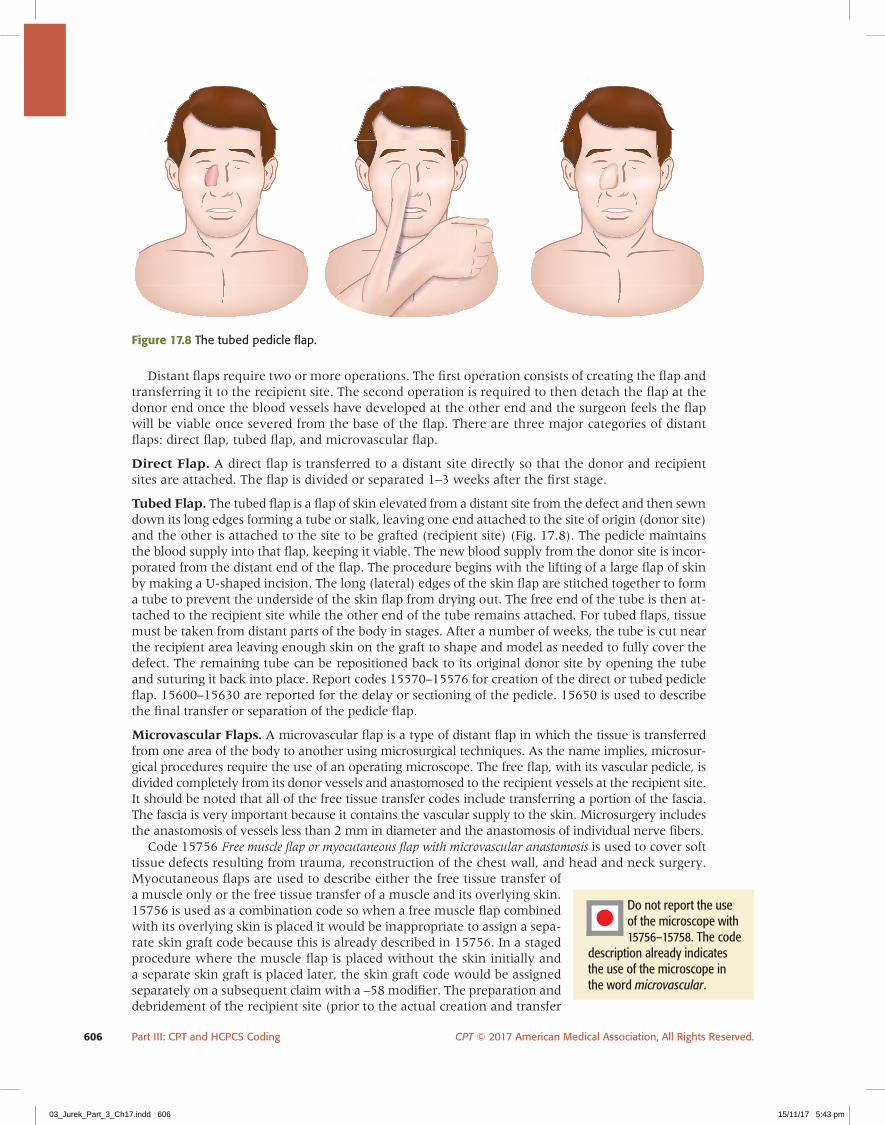
606 Part III: CPT and HCPCS Coding CPT © 2017 American Medical Association, All Rights Reserved.
Distant flaps require two or more operations. The first operation consists of creating the flap and transferring it to the recipient site. The second operation is required to then detach the flap at the donor end once the blood vessels have developed at the other end and the surgeon feels the flap will be viable once severed from the base of the flap. There are three major categories of distant flaps: direct flap, tubed flap, and microvascular flap.
Direct Flap. A direct flap is transferred to a distant site directly so that the donor and recipient sites are attached. The flap is divided or separated 1–3 weeks after the first stage.
Tubed Flap. The tubed flap is a flap of skin elevated from a distant site from the defect and then sewn down its long edges forming a tube or stalk, leaving one end attached to the site of origin (donor site) and the other is attached to the site to be grafted (recipient site) (Fig. 17.8). The pedicle maintains the blood supply into that flap, keeping it viable. The new blood supply from the donor site is incor-porated from the distant end of the flap. The procedure begins with the lifting of a large flap of skin by making a U-shaped incision. The long (lateral) edges of the skin flap are stitched together to form a tube to prevent the underside of the skin flap from drying out. The free end of the tube is then at-tached to the recipient site while the other end of the tube remains attached. For tubed flaps, tissue must be taken from distant parts of the body in stages. After a number of weeks, the tube is cut near the recipient area leaving enough skin on the graft to shape and model as needed to fully cover the defect. The remaining tube can be repositioned back to its original donor site by opening the tube and suturing it back into place. Report codes 15570–15576 for creation of the direct or tubed pedicle flap. 15600–15630 are reported for the delay or sectioning of the pedicle. 15650 is used to describe the final transfer or separation of the pedicle flap.
Microvascular Flaps. A microvascular flap is a type of distant flap in which the tissue is transferred from one area of the body to another using microsurgical techniques. As the name implies, microsur-gical procedures require the use of an operating microscope. The free flap, with its vascular pedicle, is divided completely from its donor vessels and anastomosed to the recipient vessels at the recipient site. It should be noted that all of the free tissue transfer codes include transferring a portion of the fascia. The fascia is very important because it contains the vascular supply to the skin. Microsurgery includes the anastomosis of vessels less than 2 mm in diameter and the anastomosis of individual nerve fibers.
Code 15756 Free muscle flap or myocutaneous flap with microvascular anastomosis is used to cover soft tissue defects resulting from trauma, reconstruction of the chest wall, and head and neck surgery. Myocutaneous flaps are used to describe either the free tissue transfer of a muscle only or the free tissue transfer of a muscle and its overlying skin. 15756 is used as a combination code so when a free muscle flap combined with its overlying skin is placed it would be inappropriate to assign a sepa-rate skin graft code because this is already described in 15756. In a staged procedure where the muscle flap is placed without the skin initially and a separate skin graft is placed later, the skin graft code would be assigned separately on a subsequent claim with a –58 modifier. The preparation and debridement of the recipient site (prior to the actual creation and transfer
Figure 17.8 The tubed pedicle flap.
Do not report the use of the microscope with 15756–15758. The code
description already indicates the use of the microscope in the word microvascular.
03_Jurek_Part_3_Ch17.indd 606 15/11/17 5:43 pm
CPT © 2017 American Medical Association, All Rights Reserved. Chapter 17: CPT: Integumentary System 607
of the flap) including the microdissection of arteries, veins, and nerves of the recipient site is not included in the free flap graft code, and should be reported separately. Examples of free muscle flaps are latissimus dorsi flap, serratus anterior flap, gracilis flap, rectus abdominis flap, tensor fascia lata flap, and extensor digitorum brevis. As with other flap and graft procedures, if a skin graft is needed to close the donor muscle flap site, a separate graft code is assigned to close the secondary defect. The donor site is always closed at the time of the initial free flap, either directly or with a skin graft.
Code15757 is used to report free skin flaps with microvascular anastomosis. Examples of free skin flaps are: free scapula, parascapula, groin flap, lateral arm, gluteal flap, and medial thigh flap.
Free fascial flaps (15758) involve only the elevation and transfer of the fascia. They are used when soft tissue coverage is needed without the bulk of muscle. Free fascial flaps are limited in use compared to other types of free flap grafts. The fascial flaps are usually covered with a skin graft, which would be coded separately. These could be used to cover joints and the dorsum of the hand. Examples of fascial flaps are: lateral and radial forearm flap and temporalis flap.
skin graft and tissue transfer Coding stepsSuccessful graft and tissue transfer code assignment requires that you follow the steps below. See Pinpoint the Code 17.3.
1. Determine if there was a lesion removal also performed. If lesion removal is per-formed simultaneously with a flap or tissue transfer, do not assign a separate code for the lesion removal. It is included in the flap procedure. It is, however, separately reportable with the free skin graft. There are three exceptions to this rule. The first exception is if the flap or transfer is staged, which means the surgeon removed the lesion at a previous opera-tive session. In this case, both the lesion removal and the repair can be separately reported; however, the flap code must have a –58 modifier if the tissue transfer occurs during the excision’s 10-day global period. The second exception pertains to lesion removals and adjacent tissue transfers performed on the same day during the same operative session but on separate sites. For example, if a lesion is removed from the shoulder as well as from the forearm and the forearm defect requires an adjacent tissue transfer, both can be reported but the lesion removal code must be appended by the –59 modifier to indicate that these are separate lesions and sites. The third exception applies to lesion removal with complex repair. Both codes may be reported if it is performed at the same session.
2. Determine what type of graft or tissue transfer/flap is performed. Differentiate transfers from repairs/grafts and flaps.
3. Determine the anatomic area and the size of the defect in square centimeters. Consider each repair separately if more than one repair type is performed at different sites. These should each be calculated separately by type and not added together. If the surgeon uses more than one type of flap to close a defect, a code for each flap or graft is separately assigned based on the defect’s size.
4. Report surgical preparation for skin grafts separately if the surgeon removed intact tissue.
5. Assign the HCPCS code for the allograft if supplied by the physician. Facility coders should check the charges in the facility computer system to see if this was captured in the chargemaster. If not, assign the HCPCS code for the supply.
6. Append modifiers if necessary for staged procedures (–58) or separate sites (–59).
chEckPOINT 17.3
Assign codes to the following procedures:
1. A patient is treated for a chronic venous stasis ulcer of the lower calf. The patient has been
managed in the wound care clinic for 8 weeks with debridement, whirlpool therapy, and
compression dressings and is now ready today for graft closure. An acellular graft is applied to the
5 cm x 12 cm ulcer site. ______________________
2. Split-thickness autograft, 80 sq cm., lower leg, staged procedure. ______________________
03_Jurek_Part_3_Ch17.indd 607 15/11/17 5:43 pm

608 Part III: CPT and HCPCS Coding CPT © 2017 American Medical Association, All Rights Reserved.
PINPOINT ThE cODE 17.3: Flap and Graft Repair Following a Lesion Removal
Simplerepair?
Intermediateor complex?
Tissue transferor rearrangement?
Choose excisioncode only.
Multiple lesions,same bodycategory
Don't addmeasurements
across bodycategories orrepair types!
NO
Freeskin graft?
Surgical preparationof recipient site?
NO
YES
Assign code from15002–15005.
Determine whichtype of graft.
YES
YES
NO
NOYES
YES YES
YES
YES
NO
NO
Same repair type:add lengths
together for totalrepair length.Report repair
code inaddition to
excision code.
Determine totalsq cm of defect
areas. If primary and secondary defects require repair, add the sq cm for each area together for
one total.Assign code from 14000–14300,15570–15776by type of flap.
Depending on visit, assign 15040 for
1st stage and15150–15157for 2nd stage
with –58 modifier.
Autograft
STG15100–101,15120–121
Epidermal15110–116
Skin substitute15271–15278
Assign HCPCScode for graft
supply.
Dermal15130–136
Lesionremovalseparate
site?
Append –59to lesion
removal code.
Pinch15050
FTG15200–261
Donor site requireskin graft?
Composite15760–770
Tissue-culturedautograft
Assign code from 15100–15278 based on sq cm of donor defect
in addition to flap.
Assign code for lesion removal 11400–11646 in addition to
composite 15760–770.
03_Jurek_Part_3_Ch17.indd 608 15/11/17 5:43 pm

CPT © 2017 American Medical Association, All Rights Reserved. Chapter 17: CPT: Integumentary System 609
burns
Burns are a type of injury caused by heat, cold, electricity, chemicals, light, radiation, or friction. Two million people in the United States each year are treated for burn injuries. According to WebMD, burn injuries account for an estimated 700,000 annual ED visits per year, with 45,000 re-quiring hospitalization. It is important to learn about burn types and treatments because it is likely
coders will encounter these when coding ED visits and inpatient records. Table 17.5 and Figure 17.9 illustrate the type of burns.
Burn acuity is judged by the amount of skin surface affected, the sever-ity, the depth of the burn, and resultant complications. Burns that extend deep may cause permanent injury and scarring and not allow the skin in that area to return to normal function. First-degree burns are minor in nature and only involve the epidermis and can be treated at home with basic first aid and over-the-counter pain relievers. Second-degree burns involve the epidermis and part of the dermis. These can also be treated at home unless the skin becomes infected. Third-degree burns involve the epidermis, dermis, and subcutaneous layers of skin. These layers are
destroyed and precipitate complications such as dehydration, infection, and loss of limb. Muscle, bone, blood vessel, and epidermal tissue can all be damaged, with subsequent pain due to profound injury to nerves. The more body surface area (BSA) involved in a burn, the greater the morbidity and mortality rates and the greater the management challenge.
CPT contains a section that includes local treatment of burns, but this section does not include skin grafting and medical management services. CPT codes 16000–16030 describe burn dressing
table 17.5 Types of BurnsBUrN TYPE SkIN LAYEr(S) FEATUrE
First-degree Epidermis Superficial reddened area of skin. Redness, swelling, pain to touch. Similar to a sunburn.
Second-degree Epidermis, superficial dermis, Redness, blistering, pallor. Skin blanches with pressure and is and/or partial deep reticular still moist. Extremely painful. dermis. Partial-thickness superficial or partial-thickness deep
Third-degree Complete dermis, full thickness Grayish white ash or black char with necrosis. Leather appearance. Loss of feeling. The epidermis is lost with damage to the subcutaneous tissue, which requires surgical intervention for wound healing such as debridement and grafting. Dermis and underlying tissue and possibly fascia, bone, or muscles are damaged.
Fourth-degree Complete dermis, fat, and Dry, crispy, black charred tissue. Loss of feeling because muscle; may include bone nerves are destroyed. Function of area is lost or severely and joints limited. Amputation or extensive surgical intervention is required. Death may result.
Scald Epidermis, superficial dermis Caused by hot liquids or gases such as hot water, grease, or radiator fluid.
Chemical Epidermis, dermis Direct skin exposure to strong acid or base solutions causing redness, pain, blistering, and in some cases necrosis of skin. Some agents cause deep-tissue burns.
Electrical Can damage all layers of skin Occur to the skin when directly exposed to an electrical current from, for example, a live wire or electrical outlet.
Contact Epidermis Deep burns that are contained where the skin comes in direct contact with hot objects such as curling irons, muffler tail pipes, hot irons, cigarettes, and stoves.
It is important for the ICD-10 diagno-sis codes specifying the percentage of
body area burned to be linked to the respective dressing or debridement code for demon-stration of medical necessity.
03_Jurek_Part_3_Ch17.indd 609 15/11/17 5:43 pm

610 Part III: CPT and HCPCS Coding CPT © 2017 American Medical Association, All Rights Reserved.
Figure 17.9 Burn. A. First-degree burn. B. Second-degree burn. C. Third-degree burn.
Degree of damage
Epidermis
Dermis
Subcutaneous
Degree of damage
Epidermis
Dermis
Subcutaneous
Degree of damage
Epidermis
Dermis
Subcutaneous
a
b
C
03_Jurek_Part_3_Ch17.indd 610 15/11/17 5:43 pm

CPT © 2017 American Medical Association, All Rights Reserved. Chapter 17: CPT: Integumentary System 611
and/or debridement of burned tissue and pertain to local treatment of burned surfaces only. These codes refer to treatment of the skin surface or dressings and/or debridement of partial-thickness burns. Full-thickness debridement of skin is not located in this subsection of CPT. It is reported
separately, however, if the surgeon must debride deeper layers of tissue; use CPT codes 11042–11047.
The codes are differentiated by size: small, medium, or large. Code 16000 is the only code in this series that refers to the “specific type of burn; first degree.” Dressing and debridement codes 16020–16030 are used for either initial or subsequent treatments and are assigned based on what percentage of body surface was involved. Body surfaces are divided into small (less than 5%), medium (5%–10%), and large (over 10%). The coder should be familiar with the Rule of Nines, which is used to calculate the percentage of a body burned by dividing the total BSA into 9% or multiples of 9% segments. Refer to your codebook for the Rule of Nines used to estimate body surface. In an infant or child, the “rule” deviates because of the large surface area of a child’s head. CPT Professional Edition includes a table in this section titled the “Lund-Browder Diagram and Classification Method Table for Burn Estimations.” The coder can locate the body part affected and the age of the patient to assist in selecting the appropriate code based on percentage of body surface burned.
Small burns or minor burns are treated with a silver sulfadiazine (Silvadene) dressing in the ED or the physician office. In this case, CPT code 16020, Dressings and/or debridement of partial-thickness burns, initial or subsequent; small, would be reported.
■ Case sCenariOA patient was camping and poured lighter fluid on to a charcoal grill, causing the flame to flare up and burn his hands, arms, neck, and face. He was taken to the local ED and deter-mined to have 25% total body surface area (TBSA) first-degree burns and 4% second-degree burns on the hands. First-degree burn treatment was provided to his face, arms, and neck and second-degree treatment with Silvadene and gauze was provided to the hands. Codes: 16000, 16020.
Burn patients commonly undergo an escharotomy to relieve pain and pressure at the burn site when eschar is prominent. Escharotomy (16035) is an incision made through the eschar (dead tissue or scab) into the fat layer below. The aim of the escharotomy is to release the pressure over the involved deeper tissues and to restore circulation to this tissue. Escharotomy is indicated for circumferential full-thickness burns. Escharotomy may be reported for each anatomical area in which it is performed. Use modifier –59 to identify each area treated.
As described earlier, codes 15002–15005 describe burn and wound preparation or incisional or excisional release of scar contracture, resulting in an open wound requiring a skin graft. Such wound preparation is required when the site has uneven layers or multiple layers. Uneven or mul-tiple layers could cause the graft not to adhere or connect to the wound surface and thus fail or “die.” Report this procedure in addition to the code for the actual graft. This excisional preparation is coded once per recipient area by the size of the excision.
chEckPOINT 17.4
Assign codes to the following procedure. Include any necessary modifiers.
1. The patient was burned on his upper arm and now suffers pain from the burn scar (eschar) in
that area. The surgeon makes three incisions around the scar to release the constriction and pain
caused by the scar. _____________________
E/M codes reported with burn codes 16000–16036 will be denied unless
the E/M code is reported with a –25 modifier. As always, the E/M service must be documented and be separately identifiable from the burn care.
When 16035 is billed, the ICD-10 code reported should indicate
the burn was full thickness or deeper; otherwise, carriers will deny 16035 and 16036.
03_Jurek_Part_3_Ch17.indd 611 15/11/17 5:43 pm

612 Part III: CPT and HCPCS Coding CPT © 2017 American Medical Association, All Rights Reserved.
WOunD Care anD DebriDement
The primary goals of active wound care are to debride the wound of devitalized tissue, cleanse the wound, promote coverage of the skin defect, and restore function to the tissue and surrounding area. Wound care services include assessment of the wound, debridement (selective or nonselec-tive), cleansing of the wound, and dressing of the wound (including application of topical oint-ments, wound bed protection, and bulk dressing).
Debridement is the removal of dead, necrotic, devitalized, or damaged tissue (including carti-lage) and/or foreign material from a wound. This can be accomplished surgically by using a sharp instrument such as a scalpel or scissors, or nonsurgically by using wet-to-dry dressings, whirlpool baths, or application of medications that contain enzymes to dissolve dead tissue. A debridement is done to leave behind viable tissue and promote healing. Phrases to indicate this procedure in-clude “tissue was removed until healthy-appearing tissue was encountered” or “tissue was sharply debrided until bleeding tissue was visualized.”
The objective is to preserve granulation tissue (pink) in the wound and remove fibrin tissue (white, yellow, or green tissue) or necrotic (black) tissue. Debridement is indicated when a wound is erythematous, shows fluctuance, or has discolored discharge. Debridement is performed at the bedside, in the OR, or in a wound care clinic. Anesthesia is necessary when debriding deep wounds or when bone biopsies are obtained.
eXample11000 Debridement of extensive eczematous or infected skin; up to 10% of body surface.
Several codes in the Integumentary System section of CPT and in the Medicine section encompass some level of surgical debridement. The coder must make an educated decision about which code to assign based on the following:
• Why the debridement is being performed• Where it is being performed• Who performed the procedure• The depth of the debridement• The type of wound dressing applied
Debridement is commonly performed on soft tissues of the body. This tissue connects, supports, or surrounds other structures and organs of the body. Partial-thickness debridement includes the epider-mis and part but not all of the dermal layer of skin. Full-thickness debridement involves epidermis and dermis layers. Debridement of a wound may be any type of debridement, such as mechanical, surgical, autolytic, or chemical. Refer to Table 17.6 for a list of common surgical and excisional debridement methods and their respective CPT codes (see Chapter 29 for a discussion of nonexcisional methods). Selective debridement refers to controlled debridement of a targeted area of skin where tissue is re-moved leaving behind healthy, undamaged tissue. Instruments such as scissors and forceps are used to cut the tissue along the margin of viable and nonviable tissue, which requires a greater set of skills by a qualified health-care provider. In contrast, nonselective debridement methods are not as controlled; therefore, they do not have the ability to target certain tissues and run the risk of damaging healthy tissue. Debridement is also carried out on joints and nerves to remove scar tissue or other material.
Debridement CodesSurgical debridement codes are divided into four sections:
1. Codes 11000 and +11001• Based on body surface percentage
2. Codes 11004 through +11008• Specific to necrotizing soft tissue infections• Specific to anatomical locations
3. Codes 11010–11012• Specific to debridement associated with open fractures and/or dislocations• Based on depth
03_Jurek_Part_3_Ch17.indd 612 15/11/17 5:43 pm

CPT © 2017 American Medical Association, All Rights Reserved. Chapter 17: CPT: Integumentary System 613
4. Codes 11042–11047• For all other types of debridement (ulcers, injuries, infections, etc.)• Based on depth, not on location.
• If reporting a single wound/ulcer, and so on, base the code on the depth of the wound. Examples: Subcutaneous, muscle, bone
• If reporting multiple wounds/ulcers, and so on, combine (sum) the surface area of the wounds that are at the same depth.
■ Case sCenariOA patient has two ulcers, one on the heel and one on the hip; both ulcers go down to the bone. The heel ulcer is 4 sq cm. The hip ulcer is 10 sq cm. The coder will report code 11044, Debridement, bone (includes epidermis, dermis, subcutaneous tissue, muscle, and/or fascia, if performed); first 20 sq cm or less. This code was selected based on the total square centi-meters of the two debridement (14 cm). Note that code 11044 is an out-of-sequence code.
■ Case sCenariOA patient has two wounds, one on the abdomen and one on the thigh. The abdominal wound is 16 sq cm. The thigh wound is 10 sq cm. The coder will report code 11042 for the first 20 sq cm and add-on code +11045 to represent the additional 6 sq cm. If the physician treated all four wounds during the same session, the coder would report 11042, 11042-59, +11045-59.
Wounds are classified into stages developed by the Agency for Health Care Research and Quality (AHRQ) Clinical Practice Guidelines (Table 17.7).
Clinical studies show that as deep ulcers heal, the lost muscle, fat, and dermis are not replaced. Per the National Pressure Ulcer Advisory Panel (NPUAP) Report, Vol. 4, No. 2, September 1995, granulation tissue fills the defect before re-epithelialization. Knowing this, it is not appropriate to reverse-stage a healing ulcer when documenting wound status. In other words, a stage III pressure ulcer does not become a stage II or stage I pressure ulcer during healing. The provider must methodically record the wound’s healing progress by noting an improvement in the characteristics (size, depth, amount of necrotic tissue, amount of exudate, etc.). Invariably, stage III and IV wounds require skin grafting, skin flaps, musculocutaneous flaps, or free flaps for final closure.
Codes 11000–11001 refer to debridement of extensive eczematous or infected skin and are as-signed according to the percentage of body surface affected. Codes 11004–11006 are assigned for debridement of necrotizing soft tissue of the genitalia or abdominal wall only. Codes 11010–11012
table 17.6 Surgical/Excisional MethodsCode(s) Description Code(s)
97602 Debridement of extensive 11000–11001 eczematous or infected skin
97602 Debridement of skin, subcutaneous 11004–11006 muscle, and fascia for necrotizing tissue
97602 Debridement of skin by depth 11010–11012 including removal of foreign material for open fracture and/or dislocation
97602 Debridement of skin by depth 11042–11047
97602 Excision of pressure ulcers 15920–15999
03_Jurek_Part_3_Ch17.indd 613 15/11/17 5:43 pm

614 Part III: CPT and HCPCS Coding CPT © 2017 American Medical Association, All Rights Reserved.
involve debridement associated only with open fractures or dislocations. This type of debridement can be performed at any stage of the frac-ture care—before, during, or after the surgical treatment of the fracture. Codes 11042–11047 are based on thickness of the debridement. These codes are assigned for debridement of gangrene, ulcers, and nonhealing wounds. According to many carriers, this type of surgical debridement is only medically necessary if infected, necrotic, devitalized, or nonvi-able tissue is present. Do not report these codes for washing bacterial or fungal debris from lesions, paring or cutting of corns or calluses, inci-sion and drainage of abscess including paronychia, trimming or debride-ment of nails, avulsion of nail plates, acne surgery, destruction of warts, or burn debridement. Providers instead should report these procedures when reasonable and necessary using the appropriate CPT or HCPCS codes that accurately describe the service such as those located in the Medicine section.
■ Case sCenariOA physician sees a patient in the office and debrides 15 sq cm of skin and subcutaneous tissue from a nonhealing surgical wound. Code: 11042.
Code range 15920–15999 is used specifically to code debridement or excision of pressure ulcers. Note, however, that these codes are only for pressure ulcers of the coccyx, sacrum, ischium, or trochanter. What about the other sites of the body previously mentioned that are prone to pressure ulcers? Debridement codes from the 11042–11047 range would apply as well as additional codes from the flap or graft subsection if repair was also done. Codes are specific to the site of the ulcer. Pay close attention to the wording of these code descriptors. These codes are combination codes in that the code description includes excision and repair (e.g., flap closure, ostectomy) all in one code. It is incorrect to assign codes for tissue flaps, grafts, or repairs in addition to these codes. CPT does direct the coder to assign codes for muscle or myocutaneous flap closure in addition to these services. Many of these codes also depict excision with ostectomy. In the case of ischial ulcer excision without os-tectomy, report code 15946 with a –52 modifier to reflect that the ostec-tomy was not performed because this code does not have a companion code for without ostectomy.
table 17.7 Wound Stagesstage Description
I Nonblanchable erythema of intact skin, the heralding lesion of skin ulceration. In individuals with darker skin, discoloration of the skin, warmth, edema, induration, or hardness may also be indicators.
II Partial-thickness skin loss involving epidermis, dermis, or both. The ulcer is superficial and presents clinically as an abrasion, blister, or shallow crater.
III Full-thickness skin loss involving damage to or necrosis of subcutaneous tissue that may extend down to, but not through, underlying fascia. The ulcer presents clinically as a deep crater with or without undermining of adjacent tissue.
IV Full-thickness skin loss with extensive destruction, tissue necrosis, or damage to muscle, bone, or supporting structures (e.g., tendon, joint capsule). Undermining and sinus tracts also may be associated with Stage IV pressure ulcers.
Debridement performed as part of a primary procedure
is included in that procedure unless the debridement is extensive.
It is very common to provide multiple de-bridement in a span of
several days or weeks, as in the case of open fracture and non-healing wounds. In these cases, the –58 modifier would be ap-plied during the global period. Debridement of superficial tissue is not coded separately in this case.
Debridement codes are reported for each site treated. The ad-
ditional sites would be coded and appended with the –59 modifier.
Codes 97597–97606 cannot be reported in conjunction with
11040–11044.
97602 has a status indicator “B” on the MPFS database, mean-
ing that it is not separately pay-able under Medicare.
03_Jurek_Part_3_Ch17.indd 614 15/11/17 5:43 pm

CPT © 2017 American Medical Association, All Rights Reserved. Chapter 17: CPT: Integumentary System 615
■ Case sCenariOA 45-year-old quadriplegic patient presents with a stage II pressure ulcer of the tailbone. The ulcer requires debriding. Surgical debride-ment is carried out in the wound care center under local anesthesia. The skin was cleansed and prepped. The incision was carried down to the subcutaneous tissue where devitalized tissue was sharply de-brided until normal-appearing bleeding tissue was encountered. The wound was irrigated and closed with direct suture. Code: 15931.
Wound DressingSurgical dressings must be ordered by a physician or an NP, clinical nurse specialist, certified nurse-midwife, or PA who is acting within the scope of his or her legal authority as defined by state law or regu-lation. Alloplastic dressings are man-made coverings used to aid heal-ing of ulcers or wounds. As discussed earlier, these are not skin grafts but rather temporary wound coverings. Composite dressings such as Tegaderm, Telfa, and Viasorb are used for wounds with minimal to heavy drainage, healthy granulation tissue, or necrotic tissue. Most composite dressings have three layers: (1) a semiadherent or nonad-herent layer that touches the wound and protects the wound from adhering to other material—drainage passes through it into the next layer, which is absorptive; (2) an absorptive layer that wicks drain-age and debris away from the wound’s surface and helps liquefy es-char and necrotic debris, facilitating autolytic debridement; and (3) an outer bacterial barrier layer, which may have an adhesive border, that allows moisture vapor to pass from the wound to the air and keeps bacteria and particles out of the wound.
A signed order for the dressing must be present along with a treat-ment plan for the wound care. The order should include type of dress-ing, size of dressing, and frequency of changes. Both primary and secondary dressings are covered when either of the following criteria is met: (1) They are medically necessary for the treatment of a wound caused by, or treated by, a surgical procedure; or (2) they are medi-cally necessary when debridement of a wound is medically necessary.
A new order is required for wound dressing in the following circumstances:
1. When a new dressing is added2. When the quantity of an existing dressing increases3. Every 3 months for each dressing being used, even if the quan-
tity remains unchanged
Dressings may be used as long as necessary until the wound is healed or a permanent skin graft is performed.
Many wound care items are not covered. Review the list below and make sure that the physician is aware. An advance beneficiary notice (ABN) may be required.
1. Dressings for cutaneous fistula drainage that has not been caused by or treated by a surgical procedure
2. Dressings for stage I pressure ulcers3. Dressings for first-degree burns4. Dressings for traumatic wounds that do not require surgical clo-
sure or debridement5. Skin sealants or barriers, wound cleansers or irrigating solutions,
solutions used to moisten gauze (e.g., saline), topical antiseptics, and topical antibiotics
Medicare does not consider the following services to be debridement
services:
• Removal of necrotic tissue by cleansing, scraping (other than with a scalpel or a curette), chemical application, and/or wet-to-dry dressing technique
• Trimming of callous or fibrinous material from ulcer margins
• Cleaning and dressing of small or superficial lesions
• Removing coagulation serum from normal skin surrounding an ulcer
Local infiltration, metacarpal/digital block, and topical anesthesia are included in the reimbursement for debridement services and are not separately payable.
Medical necessity must be documented in the patient’s medical record
based on the following criteria:
1. Treatment will significantly improve the wound in a reasonable period of time.
2. The expectation is that the dressing will significantly improve healing and tissue viability, reduce or control tissue infection, and allow tissue preparation for surgical grafting.
3. Additional precautions, such as appropriate bedding, padding, and patient turning, have been taken to prevent the formation of new or perpetuation of existing ulcers or wounds.
4. A treatment plan is established at the beginning of any debride-ment therapy outlining specific goals, duration, frequency of dressing, number/amount of dressings to be used at one time (if more than one), size of dressing, modalities, expected duration of need, and other pertinent factors as they may apply. An explanation should be provided for any deviation from this plan.
03_Jurek_Part_3_Ch17.indd 615 15/11/17 5:43 pm

616 Part III: CPT and HCPCS Coding CPT © 2017 American Medical Association, All Rights Reserved.
6. Enzymatic debriding agents7. Surgical tray items such as gauze or other dressings used to debride a wound, but not left
on the wound
According to CMS policy, the use of skin substitutes (15271–15278), or skin substitute material (Q4100–Q4111) is considered not reasonable and necessary when used for or with the presence of cellulitis, osteomyelitis, necrotic ulcer, draining wound, or bone-exposed wound bed. Q4100–Q4111 are payable by contractor discretion. Dressings used for mechanical debridement are typically covered. Check your local CMS contractor for specific Local Coverage Determinations (LCDs).
hyperbaric Oxygen therapy (hbO2)Hyperbaric oxygen therapy utilizes a pressure chamber to administer oxygen under pressure. It boosts wound healing by hyperoxygenation of tissue, which promotes vasoconstriction and has antibacterial effects. These chambers provide tissue oxygen levels of greater than 11 times normal, quickly feeding tissue that is hypoxic, or oxygen deprived, with much needed oxygen to stimulate wound healing. Hypoxic or ischemic wounds such as diabetic wounds, venous stasis ulcers, failing grafts and flaps, nec-rotizing soft tissue infections, and refractory osteomyelitis can all benefit from this therapy. It is primarily used to aid in healing of diabetic ulcers, venous stasis ulcers, and arterial insufficiency ulcers.
To report this service, 99183 is submitted for each session during which the physician is in personal attendance. Per CPT, any E/M service provided in a hyperbaric oxygen facility in conjunction with this service should be reported with the appropriate E/M code.
States have specific rules for if and when a physician must be present during this service. These rules vary from requiring the physician to check in with the patient at least every 30 minutes dur-ing each patient treatment and at the end with an expected response time of 1 minute for an emer-gency to requiring the physician to be in continuous attendance within the immediate hyperbaric oxygen area. Coverage is also limited to place of service and diagnosis.
mOhs miCrOgraphiC surgery
Mohs micrographic surgery is a surgical technique used to remove the three most common types of skin cancer: basal cell carcinoma (BCC), squamous cell carcinoma (SCC), and melanoma. It of-fers a strong cure rate of approximately 98% and spares healthy tissue. Mohs is the standard of care when a tumor is either in a critical location (either cosmetic or functional), recurrent, has ill-defined margins, or is large (over 2 cm) or aggressive in its growth. The surgery is unique in that the surgeon acts as both the surgeon and the pathologist.
procedureThe surgery is typically performed in stages in the outpatient office under local anesthetic by a der-matologist. A thin layer of the tumor is removed, marked, and frozen and stained so that it can be examined under the microscope. A two-dimensional diagram is drawn of the specimen. The saucer-shaped specimen is further divided, numbered, and colored into units called tissue blocks. All of these steps are mapped on the Mohs operative map. This map precisely outlines the residual tumor and allows for examination of 100% of the surgical margin, thus sparing normal surrounding tissue. If malignant cells are still found at the margins, the surgeon will repeat the process. Each layer that is removed and examined is called a “stage,” and it may take any number of stages to remove an ag-gressive tumor.
Code assignmentEach stage of the Mohs procedure is coded separately. Code descriptions for the number of speci-mens and the stage should be read carefully. When additional layers are removed, add-on codes 17312 or 17314 are used. Should any stages require more than five tissue blocks, use code 17315 and indicate the number of units over five taken in each layer.
CMS has very strict coverage policies for HBO2 therapy, requir-
ing specific diagnoses. Check for LCDs in your state.
03_Jurek_Part_3_Ch17.indd 616 15/11/17 5:43 pm
CPT © 2017 American Medical Association, All Rights Reserved. Chapter 17: CPT: Integumentary System 617
eXampleCancers of the neck and right upper back are treated by Mohs micro-graphic surgery in one stage each. Codes: 17311, 17313.
eXampleA cancer of the nose requires a first and second stage of Mohs micro-graphic surgery. Codes: 17311, 17312.
Repairs and grafts/flaps may be done to close the defect created by the removal of the tumor. In this case, these are coded separately. Any such reconstructions are not routine or expected work in conjunction with Mohs micrographic surgery.
If a definitive diagnosis of a tumor is not available, the Mohs surgeon may obtain a biopsy to confirm the diagnosis of skin cancer before initiat-ing the surgery. The biopsy and the frozen section surgical pathology code 88331 may be additionally reported with a modifier –59. Code 11100–59 is reported for the biopsy procedure, and 88331–59 for the pathology
interpretation of the skin biopsy specimen. Any other pathology service associated with MOHS surgery is included in the MOHS codes.
internet resOurCe: visit the mohs College website for a patient education video explaining mohs surgery.www.mohscollege.org/about/video_patient_education.php
chEckPOINT 17.5
Assign codes to the following procedures. Include any necessary modifiers.
1. A patient is being treated for injuries sustained in a motor vehicle accident earlier in the day.
Debridement is necessary on his right foot down to the muscle and on his right hand down to
the subcutaneous tissue. _______________________________
2. The surgeon performed Mohs surgery on an upper extremity malignancy. He performed a first-
stage excision with seven tissue blocks. The frozen section examined by the surgeon revealed that
the tumor was still present. A second-stage excision was performed with four tissue blocks and the
tumor was still present. A third-stage excision with four tissue blocks revealed no tumor present in
the margins. _______________________________
breast prOCeDures
Procedures performed on the breast may be for therapeutic or cosmetic purposes. Mammaplasty refers to various surgical techniques used to change the size or shape of a patient’s breast(s). Cosmetic procedures on the breast are most often performed by plastic surgeons but may also be performed by dermatologists. Breast mastectomies are primarily performed by general surgeons but may also be performed by plastic surgeons. Breast reconstruction procedures are performed by plastic surgeons.
The diagnostic and therapeutic procedures used to diagnose, stage, and treat breast disease are rapidly becoming less drastic and less invasive. Breast imaging procedures such as mammography, ultrasonography, magnetic resonance imaging (MRI), fine needle aspira-tion (FNA), core biopsy, and surgical excisional biopsy are routinely performed in diagnosis and management of breast disease. Many surgical physician practices can perform breast ultrasonog-raphy and ultrasound-guided biopsy in the office setting. Trends now show that excisional breast
Breast procedures are inherently unilateral. If the same procedures
are done on both breasts, a –50 modifier is needed.
Do not assign surgical pathology codes in addition to the Mohs
surgical codes. The definition of Mohs micrographic surgery includes the pathology services.
There is no global period with Mohs micrographic surgery,
so follow-up visits can be billed and the –58 modifier is not applicable.
03_Jurek_Part_3_Ch17.indd 617 15/11/17 5:43 pm

618 Part III: CPT and HCPCS Coding CPT © 2017 American Medical Association, All Rights Reserved.
biopsy is being superseded by FNA biopsy for palpable breast lesions and by percutaneous biopsy for nonpalpable breast lesions. Stereotactic and ultrasound-guided needle core biopsies are less-invasive alternatives to open surgical biopsies for most patients with nonpalpable breast lesions whereby tissue must be sampled to achieve a diagnosis.
Breast biopsies are performed using the following methods:
• FNA• Needle core biopsy• Open biopsy (excisional or incisional)
Fine-needle aspirationFine-needle aspiration (FNA) biopsy samples cells from lesions for cytologi-cal analysis rather than removing the lesion itself. FNA is not a biopsy where tissue is taken and examined. CPT codes 10021–10022 include preparation of smears, if prepared. These codes do not specify an anatomical part and are used if FNA is performed anywhere on the body. FNA is typically the first step in the evaluation of breast masses. A local anesthetic is usually not needed. The tip of the needle is advanced into the lesion before any suction is performed to avoid collection of tissue outside the lesion. Once the tip is in place, strong suction is applied, and the needle is moved back and forth within the lesion repeatedly along a 5- to 10-mm long track to loosen and collect cells. The needle is then withdrawn from the breast and the sample expelled onto prepared glass slides, spread into a thin smear, and fixed accord-ing to the preferences of the cytology laboratory. Assign codes 10021–10022 depending on whether imaging guidance was utilized.
internet resOurCe: visit MedlinePlus to view illustrations of Fna.http://nlm.nih.gov/medlineplus/ency/imagepages/17016.htm
needle Core biopsyNeedle core biopsy removes a narrow cylinder of tissue that is submit-ted for standard pathological rather than cytological analysis. Needle core biopsy is ideal for sampling large lesions and not ideal for small, nonpalpable lesions. If the mass is palpable, the surgeon can perform the biopsy in the office. Injection of a local anesthetic is required. A large needle is placed either by hand or with a biopsy gun device into the mass. A small “nick” is made in the skin with a scalpel to create an entry point for the biopsy needle into breast tissue and then into the lesion.
If the lesion is nonpalpable but detected on mammography, stereotac-tic image-guided biopsy is the preferred approach. When possible, nee-dle core biopsy is performed with ultrasound guidance, which confirms needle position within the lesion as the procedure is done. Stereotactic mammography-guided needle core biopsy is performed if the lesion is not visualized on ultrasound.
Biopsy code 19100 is assigned for each lesion or site biopsied. The code description is specific to a biopsy not using imaging guidance and is a des-ignated separate procedure.
Open biopsyOpen biopsy can be either incisional or excisional. The difference is based on the amount of breast tissue or lesion removed.
incisional breast biopsyIncisional biopsy (19101) is cutting of breast tissue where a small portion or slice of a lesion is removed. Incisional biopsies are typically performed on large lesions.
Cytological analysis is conducted on specimens obtained
through FNA. This involves spreading a fluid specimen on a glass slide for examination at the cellular level under a microscope. Pathological analysis is conducted on tissue obtained from core biopsies, excisional biopsies, and surgical resections. It is performed by a combination of macroscopic and histological examination of the tissue and at times may involve other chemistry and other laboratory tests.
When coding breast biopsies requiring imaging assistance, be
sure to note that the codes now include the imaging portion of the biopsy, if performed. Coders will no longer report the radiology imaging codes 76942, 77002, 77012, or 77021. CPT codes 19081–19086 now include the imaging services when performed.
Remember that add-on codes such as 19291 and 19295 are
coded only in conjunction with a parent code.
Do not assign 19100 to report FNA. 19100 is for reporting needle
core biopsies only.
03_Jurek_Part_3_Ch17.indd 618 15/11/17 5:43 pm

CPT © 2017 American Medical Association, All Rights Reserved. Chapter 17: CPT: Integumentary System 619
excisional breast biopsyExcisional biopsy (19120) involves removing an entire lesion (male or female). This type of biopsy is carried out when the mass is larger than 4 cm or so in diameter, is deeper and harder to reach, or contains microcalcifications.
excisional biopsy With needle localizationNeedle localization is the placement of a guide wire. Insertion of a localization device (e.g., guide wire) is most often done preoperatively in the radiology department, and if that is the case, would not be coded separately by the surgeon. This procedure is coded separately if done in a different location. Codes for insertion of the guide wire (19281–19288) vary based on the method of guid-ance used: mammogram, stereotactic, ultrasound, or MRI. All specimens excised with a previously placed marker are immediately radiographed (76098) to confirm that the lesion has been excised in its entirety. In rare circumstances, the surgeon may place the localization device and surgically excise the lesion, in which case he would report both services with 19125/19126 for the excision and a code from 19281–19288 for the localization. Device placement and surgical excision is also discussed in Chapter 27.
When coding for this procedure, it is important to differentiate whose work is being coded (i.e., radiologist, surgeon, or facility). Each entity codes for its portion of the service and uses modifiers to indicate such.
■ Case sCenariOA female patient undergoes an excisional breast biopsy of the right breast with placement of a preoperative needle guide wire in the radiology department via stereotactic guidance. The radiologist places the guide wire before the patient is taken to the OR. The surgeon removes the suspicious lesion that is indicated by the guide wire and confirms via x-ray that the lesion has indeed been removed. The radiologist would bill for the guide wire insertion: code 19283–26–RT. The surgeon would bill for the excisional biopsy: code 19125–RT. The facility would report codes 19125–RT–TC, 19283–RT–TC, and 76098.
image-guided placement of metallic markerAfter needle core biopsy, areas where lesions were biopsied can be difficult to re-identify, particu-larly after needle core biopsy, because the lesions may be altered and be made even less conspicu-ous. Placement of a marker (clip, pellet, wire) to aid in relocating the lesion is reported separately from the biopsy code for each site marked. Code selection depends on the type of imaging used to place the marker: mammography (19281–19282), stereotactic (19283–19284), ultrasound (19285–19286), or magnetic resonance (19287–19288). In some cases, once the mass is excised from the breast, the surgeon will place a clip in the area where the mass was removed for future reference or marking
when additional surgery or follow-up is likely.
internet resOurCe: read the American Journal of Roentgenology article at the following website for information and illustrations of metallic clip insertions. www.ajronline.org/cgi/content/full/175/5/1353
breast reductionReduction mammaplasty, more commonly referred to as breast reduction, is the reduction of large breasts by excising excessive tissue and fat, thereby
reducing overall cup size. Most women who have the surgery have very large, sagging breasts re-ferred to as pendulous breasts that restrict their activities and cause back, neck, and shoulder ailments. Basically four different techniques are used in reduction mammaplasty:
• Pedicle method (and all its variations)• Free nipple graft• Stevens Laser Bra• Liposuction only
Be sure to read the pathology report before finalizing an
incisional biopsy code. If the report indicates the lesion was removed entirely or in toto, an excisional biopsy code must be assigned instead.
03_Jurek_Part_3_Ch17.indd 619 15/11/17 5:43 pm

620 Part III: CPT and HCPCS Coding CPT © 2017 American Medical Association, All Rights Reserved.
There are surgical and scar pattern differences with these methods and not everyone is a candidate for all of the different types of reductions. Size of the breast and the patient’s medical history are considered when determin-ing the best approach. Regardless of technique (with the exception of the free nipple graft), CPT code 19318 is as-signed for this procedure. The breasts are a paired body part, so if the reduction is performed on both breasts, append the –50 modifier.
pedicle methodThe pedicle method of breast reduction reduces breast appearance, volume, and contour, while maintaining breast function and nipple sensation. The pedicle method refers to the location of the blood and nerve supply—not where the incisions are placed and subsequent scars will result. This is the method that the major-ity of surgeons choose.
Pedicle methods vary by the way the blood and nerves are maintained from different areas of the breast and where the incisions are made in relation to this (Box 17.3).
Free nipple graftIn the case of very large breasts, however, women are sometimes faced with the free nipple graft (FNG) procedure. The pedicle, along with its blood and nerve supply, must be severed, and the nipple-areola complex detached and grafted back on as a skin graft after the reduction is com-pleted. Function and sensation are lost with this method.
stevens laser braThe Stevens Laser Bra is a new technique that uses a laser and the skin that would normally be discarded during breast reduction surgery. This tech-nique creates an “internal bra” to provide a permanent support system and a more natural effect in the long term for patients. The incision patterns are the same as with most of the anchor-style pedicle methods, as is the preser-vation of the pedicle. The Laser Bra is attached internally to the chest wall.
liposuction-Only breast reductionLiposuction can be used to remove excess breast tissue. It is sometimes referred to as the “scar-less” breast reduction procedure. Two small inci-sions large enough for the cannulas to be inserted are all that is visible compared to the large incisions under the breast and around the nipple in other methods. Cannulas are inserted and tumescent fluid is injected, which separates the tissue from the fat. Liposuction is then performed on each breast. This procedure takes less time than traditional breast reduc-tion methods; however, it does not include the benefit of a breast lift.
nipple reconstructionNipple reconstruction is performed for two purposes: as the last phase of breast reconstruction postmastectomy or to correct inverted nipples. Reconstruction is accomplished by harvesting a skin graft from various donor sites on the body. A thin circular layer of skin is removed from the recipient breast and the graft is placed onto the reconstructed breast to resemble the areola or it is molded into a represen-tative nipple. Code 19350 includes repair of the donor site with suture. Code 19355 is reported for correction of inverted nipples. These codes are unilateral so modifiers LT, RT, or –50, if performed bilaterally, are applicable. The surgeon may elect to insert a biological implant to support the nipple; this is reported with +15777. Surgical tattooing may also be used during reconstruction to tint the areolar grafted skin to the pigment that most resembles that of the areola. Report codes 11920–11922 for the tattooing based on the area (in square centimeters) of skin treated. Notice that these codes are not specific to breasts or a site other than skin so anatomical modifiers do
If breast reduction is done with areolar re-construction, code both
19318 and 19350.
Many insurance car-riers have specific requirements for
the amount of breast tissue removed during breast reduc-tion. Some require as little as 200 g for each, while others are requiring up to 500 g of tis-sue to be removed from each breast. Preauthorization is typi-cally required for mastopexy and mammaplasty procedures. Many of these procedures are covered by carriers, although many carriers still have a no-payment benefit for these procedures because they are classified as cosmetic.
Anchor method LollipopBipedicle McKissockCentral pedicle RegnoughtCircumareolar RobertsonDonut Short scarInferior pedicle Superior pedicleInverted T pattern T-scarKeyhole VerticalLateral pedicle Wise methodLejour
BOX 17.3 pedicle methods
03_Jurek_Part_3_Ch17.indd 620 15/11/17 5:43 pm

CPT © 2017 American Medical Association, All Rights Reserved. Chapter 17: CPT: Integumentary System 621
not apply. Tattooing is also used in reconstruction following trauma or removal of cancer from an eyelid, eyebrow, or lip.
mastectomyBreast cancer continues to be a top national health concern. It ranks number one in occurrence and number two in death among women. Mastectomies are performed for carcinoma of the breast, severe fibrocystic disease, or as preventative therapy in women who have a very strong family his-tory of breast cancer. Each code differs in the amount of tissue removed and if lymph nodes and underlying muscle were taken. Mastectomy procedures are categorized by CPT into four areas:
1. Gynecomastia2. Partial (lumpectomy)3. Simple, Complete4. Subcutaneous5. Radical
mastectomy for gynecomastia Gynecomastia is a condition that affects enlarged breasts in males. Sur-gical treatment involves performing a mastectomy (19300), otherwise known as breast reduction, whereby the excess breast tissue is removed. This can be done through a periaereolar incision. The surgeon cuts away the excess glandular tissue, fat and skin from around the areola and from the sides and bottom of the breast. Major reductions that involve the removal of a significant amount of tissue and skin usually require larger incisions placed under the breast.
partial mastectomyPartial mastectomy (19301–19302) is also referred to as wide local exci-sion lumpectomy, segmental mastectomy, or quadrantectomy. Lumpec-tomy involves excision of all cancerous tissue and a margin of normal tissue around the tumor to ensure microscopically clean margins. It in-volves removal of the breast tumor and a small amount of surrounding tissue that may include the underarm lymph nodes. An incision is made in the skin over the site of malignancy. This procedure may be performed with the patient under local anesthesia, but sedation or general anesthesia is usually advisable if a significant amount of tissue is to be excised.
19302 is assigned if the surgeon also performs axillary lymphadenec-tomy. The documentation may state partial mastectomy with sentinel node biopsy. Lymph nodes are connected as a chain with the sentinel node being the first of the axillary lymph node chain. Even after the lymph nodes are located, it is difficult to determine where the lymph node chain starts. Dye is injected to assist the surgeon in identifying the sentinel node. The surgeon can observe the flow of dye through the lymph nodes to locate the first node. The surgeon can then remove the
first few nodes sequentially to test for metastasis. If the first few lymph nodes are benign then that means the cancer has not spread. The injection is coded separately with 38792. A separate inci-sion is made and lymph nodes between the pectoralis major and minor muscles and nodes in the axilla are removed. Not all axillary lymph nodes have to be removed in order to assign this code.
simple, Complete mastectomyA simple, complete mastectomy (19303) procedure entails removing the entire breast tissue, but axillary tissue is not removed. At times, the sur-geon may remove the sentinel lymph node. This node is the first axillary node into which the breast drains.
subcutaneous mastectomySubcutaneous mastectomy (19304) (also called a skin-sparing mastec-tomy) is a procedure in which the surgeon dissects the breast tissue from the pectoral fascia and from the skin preserving the nipple and areolar
Do not assign code 19300 to female patients.
Physicians need to specifically state that they paid attention
to the surgical margins to help the coder differentiate between an excisional biopsy and a lumpectomy. Excision is a removal of the lesion without specific attention to adequate surgical margins. During excision, the visible mass is removed but unless frozen section was performed, there is no guarantee that the margins are clear before the case is ended. Additionally, the intraoperative placement of a clip (or clips) is not separately reported with partial mastectomy procedures.
If the surgeon per-formed and docu-mented extra work to
spare the nipple—which can be difficult because of the need for an adequate blood supply—re-port the -22 modifier and send the operative report.
03_Jurek_Part_3_Ch17.indd 621 15/11/17 5:43 pm
622 Part III: CPT and HCPCS Coding CPT © 2017 American Medical Association, All Rights Reserved.
tissue but removing the rest of the breast. Breast tissue is removed but the skin and pectoral fascia are not removed. In a male subQ mastectomy, only about 200 grams of tissue are removed versus moderate to extensive amounts of tissue or anterior chest wall in a mastectomy in this range. This procedure is routinely performed for women who have a high risk for breast cancer or those with benign disease when the physician is con-cerned about increased risk cancer development in the breast tissue.
radical mastectomyRadical mastectomy (19305–19306) involves removing the entire breast, the axillary lymph nodes, and the pectoral tissue behind the breast. It is a procedure of last resort for recurrent breast cancer that has spread to the chest wall or for those with tumors of the pectoralis muscle. This proce-dure is the most disfiguring of all mastectomies. In 19305, the breast tissue including pectoral muscles, axillary pectoral major and minor and axillary lymph node is removed. In 19306, breast tissue is dissected from the pecto-ral fascia and pectoral muscles and axillary pectoral muscles are removed. All tissue within the vicinity of the internal mammary lymph nodes, ster-num, rectus fascia, latissimus dorsi muscle and the clavicle are removed.
Modified radical mastectomy requires that the entire breast tissue is removed along with the axillary contents (fatty tissue and lymph nodes) and may involve the pectoralis minor muscle but does not resect the pec-toralis major muscle. This saves the patient from having a more extensive procedure but is more difficult for the surgeon to accomplish.
Additionally, intraoperative placement of clips is not separately re-ported with total mastectomy procedures.
internet resOurCes: visit the breastcancer.org website to view illustrations of the radical and modified radical mastecomy procedure.
http://www.breastcancer.org/radical_mastectomy.htmlhttp://www.breastcancer.org/modified_radical_mastectomy.html
breast reconstructionsMastectomy patients are offered the opportunity to reconstruct the ab-sent breast either immediately after the mastectomy or at some time after the procedure once the patient has recovered or completed adjuvant therapy. According to the New York Times, “66,000 women in the U.S. had mastectomies in 2006 and 57,000 had reconstructive breast surgery.” These procedures are quite complex and vary in technique. Implant surgery is the most popular recon-struction and often performed immediately after the mastectomy by first placing a tissue expander to stretch the tissue ad prepare it for subsequent prosthetic implant insertion. Other techniques require transferring abdominal tissue to the breast or latissimus tissue with flap reconstruction. Patients may elect to have a mold of their breast made prior to mastectomy to be used to create a custom breast implant. Report 19396 for the preparation of the moulage. The actual insertion of the custom implant will be reported at a separate session using 19340 or 19342.
When assigning codes for breast reconstruction, the coder needs to read the documentation and determine which breast was treated, what type of reconstruction technique was applied, and whether or not an implant was inserted.
tissue expandersA tissue expander is a plastic balloon that is placed under the skin and filled with saline. Through a small injection port under the skin, saline is injected incrementally into the expander over a pe-riod of weeks or months, gradually filling it to the desired size while stretching the overlying skin. Tissue expanders may be inserted under the skin anywhere on the body. The purpose is to stretch or expand the skin in preparation of a staged reconstruction or to receive an implant. 11960 is used to report insertion of tissue expanders in other parts of the body aside from the breast. 19357 is used to report insertion of a tissue expander in preparation for later breast reconstruction with permanent mammary implant. It also includes subsequent expansion of the balloon. If for some reason a tissue expander is removed without immediate insertion of a permanent implant as in the case of an infection in the breast capsule, assign code 11971.
If a breast biopsy is performed at the same session as a
mastectomy, it is not coded separately unless the biopsy is performed to decide whether the mastectomy is to be done and if a previous biopsy had not been obtained. If a mastectomy is performed as a result of a positive breast biopsy performed at the same session, append a –58 modifier to the mastectomy code to show a staged procedure.Breast biopsy 19101 (Biopsy of breast; incisional) will always be denied when billed with 19316 (Mastopexy), 19318 (Reduction mammaplasty), 19324 (Mammaplasty; without implant), 19325 (Mammaplasty; with implants) or 19355–19369 (Breast reconstruction codes).
Excisions of breast le-sions are not coded separately from a mas-
tectomy.
03_Jurek_Part_3_Ch17.indd 622 15/11/17 5:43 pm

CPT © 2017 American Medical Association, All Rights Reserved. Chapter 17: CPT: Integumentary System 623
insertion of prosthetic implantImplant insertion post-mastectomy is performed the same way as a cosmetic breast augmentation. When the physician has determined that the tissue has been stretched adequately enough to allow for insertion of a permanent implant, report 11970 for removal of the temporary tissue expander and exchanging it with a permanent implant. For cases when a tissue expander is removed at a prior visit, the surgeon will place the permanent implant at a separate visit. In this case, when the patient does come back later for insertion of a breast implant, 19342 Delayed insertion of breast pros-thesis following mastopexy, mastectomy, or in reconstruction is reported.
Implants come with the risk of complications and like-lihood of future operations. According to studies, within five years of implant placement, more than one third of patients have undergone a second operation for ruptured implants or infections.
tranverse rectus abdominis myocutaneous (tram) FlapThe TRAM flap is a form of a pedicle flap where the rec-tus abdominis muscle along with soft tissue and blood
vessels is rotated and used to reconstruct a new breast (Fig. 17.10). An oval section of skin, fat, and muscle is taken from the lower half of the abdomen and slid up through a tunnel created
under the skin of the abdomen to the breast area. Blood vessels remain attached to the base of the flap an anywhere else along the pedicle when possible. The tissue is pulled out through the mastectomy site and shaped into a natural-looking breast and sewn into place. The procedure takes 3-4 hours to complete. 19367 is assigned for this procedure. All TRAM procedures include the following components:
• Creation of the breast pocket (capsule)• Elevation of the abdominal flap • Muscle dissection • Flap transfer • Fascial closure (donor site) with or without mesh • Abdominal closure including umbilical repair or reconstruction • Breast contouring
Blood vessels that are cut during the elevation of the flap and trans-fer are reanastomosed in the chest area using a microscope. The term supercharging appears in the code descriptor for 19368. This means that an additional artery is included with the flap when it is harvested from the abdomen to provide more blood supply to the flap. To report the micro-vascular anastomosis use code 19368.
Code 19369, Breast reconstruction with transverse rectus abdominis myocutaneous flap (TRAM), double pedicle, including closure of donor site, is reported for a unilateral breast reconstruction when both rectus muscles are used instead of one or the other as in the above TRAM procedure.
Deep inferior epigastric perforator (Diep) Free FlapThe DIEP flap (19364) utilizes the epigastric perforator, which is the main blood vessel that runs through the deep abdominal tissue used to reconstruct the breast. In DIEP flap reconstruction, only skin, fat, and blood vessels are removed from the lower abdomen below the belly button to the mons pubis. Muscle is spared in this technique. Because no abdominal muscle is removed, most women recover more quickly from DIEP compared to TRAM reconstruction.
Lines ofreconstructed
breast incisions
Left TRAMmuscle is swungover to re-create
new breast
Left transverserectus abdominis
muscle
Incision circleof repositioned“belly button”
Line ofabdominal
surgery
Figure 17.10 TRAM flap. From Gylys, B. Medical Terminology Systems, 8E. F.A. Davis, 2017.
Most local CMS car-riers cover the TRAM flap but they don't
cover a revision to the abdo-men when the TRAM flap creates dog ears. Dog ears are triangular shaped ends of poorly healed incisions that stick up on each end of the incision resembling a dog’s ears. CMS as well as many commercial carriers view this as cosmetic and not medically necessary.
Do not report the use of the microscope (69990) separately
because it is included in breast reconstruction surgeries 19367–19369.
03_Jurek_Part_3_Ch17.indd 623 15/11/17 5:43 pm

624 Part III: CPT and HCPCS Coding CPT © 2017 American Medical Association, All Rights Reserved.
The TRAM and DIEP have the same goal in mind and utilize tissue obtained from the same area but are very different in technique and execution. The main difference between the DIEP and TRAM procedures is the TRAM is a flap and the DIEP is a free graft. During the DIEP, tissue is completely detached from the abdomen and reanastomosed to the chest area. This procedure requires a highly skilled plastic surgeon specially trained to perform this intricate procedure. The surgeon must use the microscope to microsurgically repair the blood vessels. CPT instructs coders to not assign 69990 in addition to 19364 because harvesting of the flap, microvascular transfer, closure of the donor site, and shaping of the breast are all inclusive in this code.
■ Case sCenariOUnder high-power magnification, the pectoralis major muscle was split along the fourth rib. The deep perichondrium was then incised and the internal mammary artery and veins were identified. An oval incision was made in the lower abdomen approximately 2 cm below the umbilicus. Dissection was carried down through the subcutaneous tissue and fat down to the rectus abdominis fascia. The fascia was incised and the perforators were dissected through the rectus abdominis muscle. The flap was harvested with the deep inferior epigastric ves-sels visualized as they entered the muscle. The flap was brought with its vessels through the rectus abdominus muscle leaving all muscle behind and intact. The flap was transferred to the chest with microvascular anastomosis and secured.
latissimus Dorsi FlapThe latissimus dorsi muscle located on the side of trunk just adjacent to the breast tissue can be used to reconstruct the breast (19361). A section of skin, fat, and latissimus dorsi muscle is de-tached and slid around through a tunnel under the skin to the breast area. The muscle-skin flap remains attached to the blood vessels and is then rotated to the front of the chest through a tunnel under the patient's armpit and is then pulled out through the mastectomy incision. As with all flaps, blood vessels remain attached and preserved as much as possible. The tissue is shaped into a natural-looking breast and sewn into place. If blood vessels were severed during the elevation of the flap and transfer, they can be reanastomosed to the vessels in the chest area; however, 69990 cannot be reported in addition. In most cases, when using this type of flap reconstruction, patients also require insertion of an implant because there isn’t an abundance of fatty tissue on this part of the back to create a sizable breast. If an implant is also placed, assign 19340 in addition.
revision of reconstructed breastIt is not uncommon in patients that have had breast reconstruction for them to have subsequent surgery or staged surgery where the breast is revised. Tissue may be rearranged to correct asym-metry. Tissue may be revised or scars may be excised because of contracture or wound breakdown. Implants may be removed and replaced with a different kind or size to improve the appearance or fit. Common diagnoses submitted with this procedure are scar or fibrosis of skin, mechanical complication of breast implant, and late effect of surgical care.
mastopexyMastopexy (19316) is a breast lift usually done for cosmetic purposes. Various factors play a role in the sagging of a woman’s breast(s): preg-nancy, nursing, and the forces of gravity and aging. As the skin loses its elasticity, the breasts often lose their shape and firmness and begin to sag. Mastopexy raises and reshapes sagging breasts. At times, this procedure may be done in an effort to make breasts symmetrical after a woman has had a mastectomy—surgical removal of the breast—with reconstruction on the opposite side.
augmentationBreast augmentation is a cosmetic procedure to enhance the existing breast. A pocket is created between the breast and the pectoral muscles,
Some states require every health policy to provide coverage
for reconstructive surgery on each breast on which a mas-tectomy has been performed and reconstructive surgery on a nondiseased breast to produce a symmetrical appearance. Surgery on the nondiseased breast should be preautho-rized, because carrier and state rules differ.
03_Jurek_Part_3_Ch17.indd 624 15/11/17 5:43 pm

CPT © 2017 American Medical Association, All Rights Reserved. Chapter 17: CPT: Integumentary System 625
and an implant is inserted with the end result being a larger breast. Augmentation can also be performed by rearranging the fat and mammary tissue, which does not require the insertion of an implant. This procedure is reported using codes 19324 and 19325. This procedure is most com-monly performed in women; however, with the explosion of the cosmetic industry, men are also undergoing this procedure to obtain more defined “pecs” (pectoralis major muscles). Medicare covers implants inserted in a postmastectomy reconstruction for breast cancer patients.
breast CapsulotomyCapsule formation and contracture are common complications of breast augmentation. Formation of a capsule can cause distortion of the breast. A capsulotomy is usually performed in this case: An incision is made in the capsule to relieve tightness around the implant, but no capsule tissue is removed. The implant may be temporarily removed and replaced or not replaced at all. Manual capsulotomy can also be performed by applying pressure to the breast without using anesthesia. To report this service, use code 19370 with the respective LT or RT modifier.
breast CapsulectomyPeriprosthetic capsulation is the most common complication resulting from breast implantation. Fibrous sacs of scar tissue that encapsulate the implant become contracted, causing pain and distortion of the breast, and, depending on the degree of contracture, may require removal. Cap-sulectomy (19371) is the surgical removal of the capsular contracture or adhesive scar tissue. The implant is temporarily removed and the surgeon removes this tissue and revises the pocket and then places the implant back into the breast. This code does not include the reinsertion of the breast implant. The code assigned for reinsertion of the breast implant depends on whether the patient had the initial breast proce-dure done for reconstruction postmastectomy or post–cosmetic breast augmentation.
■ Case sCenariOs• A patient has a unilateral breast capsulectomy postaugmentation with removal of the
implant and reinsertion of the breast implant. Codes: 19371, 19325–51.
• A patient has a unilateral breast capsulectomy with removal of the implant post reconstruction and a new implant is replaced. Codes: 19371, 19340–51.
implant removal or exchangeUnfortunately, breast augmentation surgeries run the risk of complications with the mammary implant rippling, rupturing, or becoming dislodged from the breast capsule. If the implant was silicone and the material leaks beyond the capsule, infiltrating surrounding tissue into the breast capsule, assign 19330, Removal of mammary implant material, to account for the work entailed in re-trieving all of the silicone material. Do not assign this code when a saline implant ruptures. Saline implants may leak or develop ripples and can be removed and exchanged/replaced with a new implant. To report this service, use code 19325 for cosmetic augmentation patients and 19340 for postmastectomy patients.
skin resurFaCing
Dermatologists and plastic surgeons are performing these procedures more often now for a couple of reasons. Some individuals who want to appear younger are looking for noninvasive ways to turn back the clock. Once thought of as cosmetic procedures, insurance carriers are now paying for many of these procedures to
HCPCS codes for the implant supply should also be reported.
Code L8600 is used to describe breast silicone or saline implants.
It is considered incor-rect to bill a peripros-thetic capsulectomy
(19371) with delayed insertion of breast prosthesis (19342) when the procedures are ac-complished during the same operative session. The reason the breast implant was placed originally will determine the correct code. Choose from either 19325 or 19340.
03_Jurek_Part_3_Ch17.indd 625 15/11/17 5:43 pm

626 Part III: CPT and HCPCS Coding CPT © 2017 American Medical Association, All Rights Reserved.
treat surface skin conditions such as actinic keratosis before they progress to more serious conditions requiring invasive surgery.
DermabrasionDermabrasion is a surgical procedure that scrapes away the outermost layer of skin with a motorized instrument that uses a wire brush or a burr. The instrument sands down the skin and penetrates the dermis layer of skin to reach smoother, wrinkle-free skin. This technique is very similar to sanding and stripping wood to reveal the smooth untainted wood beneath the rough or painted outer surface. Dermabrasion is most often used to improve the look of scars on the face or to smooth out fine facial wrinkles, especially around the mouth. This procedure lowers raised lesions or thins tissue to regenerate skin with a smoother appearance.
It can also be utilized to remove the precancerous growths called keratoses that reside on the superficial layers of the skin. CO2 laser derm-abrasion to treat a basal cell carcinoma is also assigned with codes 15780–15783. Segmental dermabrasion may be covered as treatment for conditions such as rhinophyma.
Coding for dermabrasion is based on the location and the size of the area. Use code 15780, Dermabrasion; total face (e.g., for acne scarring, fine wrin-kling, rhytids, general keratosis), for the total face, 15781 for a segment of the face, and 15782 for regional dermabrasion of an area other than the face. Use 15783, Dermabrasion; superficial, any site (e.g., tattoo removal), for a superficial site. This code indicates that it should be used to remove a tattoo; however, codes 15780–15782 are more suitable for tattoo re-moval because these procedures penetrate into the dermal layer of skin where the tattoo resides. Code 15783 only affects the superficial layer of skin.
DermaplaningDermaplaning is used to treat deep acne scars. Dermaplaning utilizes a handheld instrument called a dermatome to slice off layers of skin with an oscillating blade. The area of skin is “skimmed” off evenly, making the skin flush with the deepest point of the scar or skin defect. This provides a smoother appearance of the entire area. It is not uncommon for the procedure to be performed more than once, or in stages, especially when scarring is deep or a large area of skin is involved.
Both dermabrasion and dermaplaning can be performed on small areas of skin or on the entire face. They can be used alone or in conjunction with other procedures, such as a facelift, scar re-moval or revision, or chemical peel.
COsmetiC prOCeDures
abdominoplastyAbdominoplasty is considered to be, in most cases, a cosmetic procedure performed on the abdomen to remove excess skin and fat, in addition to plicating, or tightening, the stomach muscles to obtain a flatter, tighter abdomen. Many women have historically undergone this procedure after having multiple children such that the abdominal wall is stretched so much it no longer retains its elasticity or if they developed rectus diastasis dur-ing pregnancy. Rectus diastasis is separation between the left and right sides of the rectus abdominis muscle, leaving a ridge-like appearance down the front of the abdomen from the breastbone to the belly but-ton. Abdominoplasty repairs weakened or separated muscles, creating an abdominal profile that is smoother and firmer. Report code 17999
Most carriers consider dermabrasion proce-dures to be cosmetic
and will only reimburse for them under certain medical circumstances. For example, Aetna states that it consid-ers dermabrasion—whether by dermaplaning or CO2 laser—medically necessary “for removal of superficial basal cell carcinomas and precancerous actinic keratoses” only when the patient meets two criteria: (1) Conventional methods of removal such as cryotherapy, curettage, and excision are impractical due to the number and distribution of the lesions, and (2) the patient has failed a trial of 5-fluorouracil (5-FU; Efudex). Check with individual carriers for specific policies.
CPT codes 15780, 15782, and 15783 have an assistant surgeon
indicator of 0, which means an assistant surgeon fee is payable for 15780 but requires documentation to substantiate medical necessity. These codes include 90 days of follow-up care.
Check carrier websites and LCDs for payer policies specific to this
procedure.
03_Jurek_Part_3_Ch17.indd 626 15/11/17 5:43 pm

CPT © 2017 American Medical Association, All Rights Reserved. Chapter 17: CPT: Integumentary System 627
for abdominoplasty. Abdominoplasty is never covered by insurance.
panniculectomyA panniculectomy is a lipectomy or removal of excess skin that can be performed on the abdomen, thighs, legs, upper arms, buttocks, and other areas. Panniculectomy, on the other hand, is not primarily a cosmetic procedure. This pro-cedure differs from abdominoplasty in that it is performed to reduce the health risk of the patient. More and more pa-tients are undergoing bariatric surgeries, resulting in drastic weight reduction and leaving behind an apron of redundant skin around the abdomen called abdominal panniculus. These skinfolds become problematic because they retain moisture
and become irritated or inflamed when they rub against the underlying skin. The skinfolds can harbor bacteria, resulting in chronic infections. Abdominoplasty tightens the muscle and removes excess skin and fat, but a panniculectomy only removes excess skin and fat. Aprons are graded on a scale of 1 to 5—the higher the grade, the higher the health risk associated with the condition. The larger the apron or panniculus, the more difficult it is for the patient to walk, dress, exercise, bathe, or perform other activities. See Table 17.8 for further details. This procedure is usually considered to be medically necessary for the patient who has had significant weight loss following the treat-ment of morbid obesity and when there are medical complications such as candidiasis, intertrigo, or tissue necrosis that is unresponsive to oral or topical medication. CPT instructs the coder to report codes 15830, +15847 for panniculectomy with abdominoplasty.
liposuctionLiposuction, or suction-assisted lipectomy, refers to the removal of unwanted fatty tissue, using suc-tion by inserting a cannula under the skin. Tumescent fluid is injected to break up the fatty tissue and liquefy it, making it easy to suction out. It also has some anesthetic properties. Liposuction is com-monly performed on the abdomen, the breasts during reduction mammaplasty, the thighs, the but-tocks, and under the chin. Liposuction is primarily a cosmetic procedure, but it is also used to enhance or contour during therapeutic procedures. Liposuction differs from lipectomy in that it uses suction to remove the tissue instead of using a scalpel or electrosurgery (Bovie) to excise the tissue surgically. Liposuction is reported by using codes 15876–15879, depending on which area of the body is treated.
rhytidectomyA facelift, or rhytidectomy, is a cosmetic surgical procedure performed on the face to improve the ap-pearance of the skin, giving it a more youthful look. This is accomplished by removing excess facial skin, with or without the tightening of underlying tissues of the face and neck, resulting in tighter-looking skin. During the procedure, the surgeon makes incisions at the temples above the hairline continuing down in front of the ear, around the earlobe, and to the lower scalp. If the patient has sagging of the neck tissue and the neck is also lifted, another incision is made under the chin. The surgeon then separates the skin from the underlying fat and muscle and any excess fat is trimmed or suctioned away around the neck and chin to contour the area. Tissue is repositioned and the muscles are tightened. Excess skin is removed. Codes used to report rhytidectomies are located in the 15824–15829 series. Codes are assigned based on where on the face and/or neck the procedure
was performed. If the surgeon frees the superficial musculoaponeurotic system (SMAS) from the skin and creates a flap, assign 15829. What is interesting is that CPT considers facelift procedure codes to be unilateral, so if the lift is done on both sides of the face, the –50 modifier is appended.
For a very few conditions, rhytidectomy is considered medically nec-essary to correct a functional impairment as a result of a disease such as facial paralysis. Often, this procedure is performed in conjunction with other procedures to correct the impairment.
table 17.8 Abdomen Panniculus/ Apron Grading ScalegrADE DEScrIPTION
1 Covers only the hairline to the mons pubis but does not cover the genitalia
2 Covers genitalia in line with upper thigh crease
3 Covers upper thigh
4 Covers midthigh
5 Covers knees or beyond
When liposuction is performed as part of a facelift repair, it is not
reported separately. It is con-sidered an integral component of the facelift procedure.
03_Jurek_Part_3_Ch17.indd 627 15/11/17 5:43 pm

628 Part III: CPT and HCPCS Coding CPT © 2017 American Medical Association, All Rights Reserved.
internet resOurCes: visit the american academy of plastic surgeons website to view pictures of common conditions treated by plastic surgeons. this site also has before-and-after photos and 3D imaging of many of the procedures discussed in this chapter. www.plasticsurgery.orgvisit Dr. steven Denenberg’s website to view tutorials on various cosmetic and therapeutic procedures of the face and nose. this website has surgical photos of facelift, rhinoplasty, and blepharoplasty procedures: www.facialsurgery.com/intraOp1.htmvisit aapC’s website to learn more about the specialty certification exams for coders who want to specialize in dermatology or plastic and reconstructive surgery: Certified plastic and reconstructive surgery Coder (CprC) and Certified professional Coder in Dermatology (CpCD).www.aapc.com/certification/specialty/plastics-and-reconstructive -surgery-coding-certification.aspx
Chapter summary
1. I&D procedures require an incision to open the af-fected site and allow for drainage. Biopsy is a di-agnostic procedure that allows a sample of tissue to be removed and sent for pathology testing and interpretation. Destruction uses various methods to eradicate a lesion but is not considered to be a full-thickness excision. Excisions are the removal of full-thickness (i.e., through the dermis) lesions. This method is used for both benign and malignant lesions, and understanding the guidelines for le-sion measurement is essential to correct coding. Debridement is the removal of devitalized tissue to promote repair and healing as well as func-tion to the affected area. Mohs micrographic surgery is a technique where the surgeon acts as surgeon and pathologist. If either the surgery or the pathology review is performed by different physicians, Mohs codes cannot be reported. Mohs is specific to basal cell carcinomas, squamous cell carcinomas, and melanomas.
Assign codes to the following procedures. Include any necessary modifiers. In some cases, more than one code is required.
1. A 47-year-old female patient underwent a routine mammogram. The patient has had chronic
fibrocystic disease, but this mammogram showed a small calcification not palpable to the touch.
The radiologist performed an ultrasound-guided percutaneous needle core biopsy of this lesion of
the right breast. ___________________________________
2. A patient undergoes a lumpectomy of the right breast for biopsy suspicious for malignancy. The
4.0-cm lesion with 0.2-cm margins was sent for frozen section. The pathologist confirmed the
specimen to be malignant and that the margins were clear. ___________________________________
3. Surgical removal of excess skin and tissue, upper arm and hand. ______________________________
4. Rhytidectomy of glabellar frown lines. ___________________________________
chEckPOINT 17.6
03_Jurek_Part_3_Ch17.indd 628 15/11/17 5:43 pm

CPT © 2017 American Medical Association, All Rights Reserved. Chapter 17: CPT: Integumentary System 629
2. There are basically three types of wound repairs: simple, intermediate, and complex. Simple repairs are for superficial wound repair involving the epidermis, dermis, and a minimal amount of subcutaneous tissue requiring one-layer suturing. The simple repair is included in the lesion removal “package” and payment. Intermediate repair is closure of one or more of the subcutaneous layers and superficial fascia, in addition to the skin. This type of repair can also be assigned when the wound has to be extensively cleaned due to contamination or removal of small pieces of foreign material before suturing, even if the suturing is simple. A complex repair is associated with complicated closure including scar revision, debridement, extensive undermining, stents, or retention sutures, and is more than layered closure.
3. Code to the most appropriate anatomical site regardless of the depth of lesion, which may require making a decision between using codes in the Integumentary System or Musculo-skeletal System sections. With excisions, a specimen is removed from the body using varying techniques, whether it is a partial or complete removal, and sent to pathology for examina-tion. Lesion excision codes are based on the following seven basic factors:
a. Where the lesion is located b. Type of lesion c. Morphology of the lesion (benign or malignant) d. Number of lesions e. Diameter
4. Measuring a lesion requires that you determine the greatest clinical diameter of the lesion plus that margin required for complete excision. In CPT, this is defined as the lesion diameter plus the narrowest margins required. The measurement is made prior to excision.
5. Skin graft coding differentiates between the autografts from the patient’s own skin, such as split thickness skin grafts, full thickness skin grafts, epidermal only grafts, dermal only grafts, and adjacent tissue transfers. The non autografts are skin substitute grafts.
6. Flaps and tissue transfers or arrangements differ from skin grafts because they carry their own blood supply. They are most commonly used when the donor site and the recipient site are in close proximity. The flaps or tissue transfers are not completely severed from the donor site, so the vascular bed is still attached to feed the tissue with adequate blood supply. The CPT Professional Edition provides illustrations and descriptions of adjacent tissue repairs. Skin grafts are completely freed from the donor site and placed onto the recipient site, no connection remains.
7. When an excision is performed and the method of closure is an adjacent tissue transfer, the excision procedure is included in the adjacent tissue transfer procedure. When the method of closure is a graft or other type of flap, the excision is reported in addition to the closure procedure.
8. The Rule of Nines is used to calculate the percentage of a body burned by dividing the total body surface area into multiples of 9% segments. For example, if both an arm and a leg are burned, this represents 27% of the body surface area. The arm is 9% and each leg is 18%.
9. The debridement codes are divided into four sections. The first is based on the body surface percentage debrided, the second is based on debridement of necrotizing soft tissue infections, the third is specific to open fractures and dislocations, and the fourth is for all other types of debridement such as ulcers, injuries, and infections.
10. Selective debridement refers to controlled debridement of a targeted area of skin where tis-sue is removed leaving behind healthy, undamaged tissue. Instruments such as scissors and forceps are used to cut the tissue along the margin of viable and nonviable tissue, which requires a greater set of skills by a qualified health-care provider. In contrast, nonselective debridement methods are not as controlled; therefore, they do not have the ability to target certain tissues and run the risk of damaging healthy tissue. Whirlpool therapy and maggot therapy are some examples of this type of debridement.
11. The diagnostic and therapeutic procedures used to diagnose, stage, and treat breast disease are rapidly becoming less drastic and less invasive. Breast procedures are inherently unilat-eral; therefore, if the same procedures are done on both breasts, a –50 modifier is needed. Some of the breast procedures are fine needle aspiration, needle core biopsy, open biopsy, placement of metallic marker(s), breast reduction and mastectomy. Understanding the dif-ference between these procedures and their coding requirements is essential to the coder.
03_Jurek_Part_3_Ch17.indd 629 15/11/17 5:43 pm
630 Part III: CPT and HCPCS Coding CPT © 2017 American Medical Association, All Rights Reserved.
1. ______ Slicing a raised lesion or mole entirely off at skin level
2. ______ Trimming or gradual reduction in size by slicing
3. ______ Surgically removing or cutting out part or all from the body
4. ______ Removal of dead, necrotic tissue
5. ______ Closest to where that point attaches to the trunk of the body
6. ______ Toward the backside of the body
7. ______ Entire lesion, tumor, and so on, is removed
8. ______ Layered closure/repair of a wound
9. ______ Portion or a sample of the lesion or tumor is removed through an incision
10. ______ Scraping away the outermost layer of skin with a motorized instrument
true or FalseDecide whether each statement is true or false.
1. ______ I&D of abscess of the left thigh would always be coded as 27301; the code is specific to the thigh region.
2. ______ Excision of a 3.0-cm hyperkeratosis of the scalp is coded as 11423.
3. ______ Needle cauterization of a subungual hematoma is coded as 11740.
4. ______ Suture repair of a 12.0-cm laceration of the right lower leg in which fascia, subcutaneous layer, and skin are closed is coded 13121 and 13122.
5. ______ The dermis is the thick, outer layer of the skin.
6. ______ Layered closure is an example of an intermediate wound repair.
7. ______ Debridement is never coded as a separate procedure.
8. ______ A shallow wound in the epidermis that could be repaired with a simple repair required extensive cleansing due to contamination. An intermediate wound repair code should be assigned.
9. ______ Before you can assign a code for shaving a lesion, you must know if the lesion was benign or malignant.
10. ______ One 0.5-cm benign lesion of the hand was removed and another 0.5-cm benign lesion of the foot was removed. Multiple lesions were excised and both anatomical areas are grouped into the same code descriptor, so the diameters of the lesions are added together.
11. ______ When coding for skin grafts, simple closure of the donor site is included in the service for the graft and is not coded separately.
12. ______ Simple closure is not included in the excision of a lesion.
Review Questions
matchingMatch the key terms with their definitions.
A. paringB. dermabrasionC. dorsalD. shavingE. incisional biopsy
F. excisionG. proximalH. excisional biopsyI. debridementJ. intermediate repair
03_Jurek_Part_3_Ch17.indd 630 15/11/17 5:43 pm

CPT © 2017 American Medical Association, All Rights Reserved. Chapter 17: CPT: Integumentary System 631
multiple ChoiceSelect the letter that best completes the statement or answers the question.
1. In which of these cases would the coder add the lengths of the defects?A. Removal of benign lesions from neckB. Simple closure of lacerations on the trunkC. Intermediate closures of lacerations on arm and faceD. Excision of lesion on left leg with simple repair and excision of lesion on back with intermediate
repair
2. Wound exploration does not include which of the following?A. LaparotomyB. DebridementC. Foreign body removalD. Wound enlargement
3. The physician excised a benign lesion on the face measuring 2.0 cm × 0.6 cm × 3.0 cm. The correct code would beA. 17000B. 11444C. 11643D. 11443
4. When coding wound repairs with varying complexity, which of the following is true?A. The codes are listed with the largest repair size listed first.B. Only the most complex repair code is listed.C. The most complex repair code is listed first followed by the other repairs.D. The order in which the codes are listed does not matter.
5. Per CPT guidelines, if a lesion is biopsied and the remainder of the lesion is removed, what code(s) is (are) assigned?A. Code for the biopsy and the lesion excision with a –59 modifier on the excision code.B. Code for the lesion excision only.C. Code for the biopsy only.D. Code for the biopsy and the lesion excision with a –59 modifier on the biopsy code.
6. Which of the following procedures describes destruction of a lesion?A. ParingB. ShavingC. RemovalD. Laser removal
7. A patient is seen in the ED after cutting his hand with a band saw. The ED physician indicates that the wound required extensive cleansing and multiple-layer closure. What kind of wound repair was done?A. IntermediateB. SimpleC. Tissue rearrangementD. Complex
8. For a superficial incision and drainage of the shoulder, the code would be assigned from which range?A. 21501–21510B. 23030–23031C. 10040–10180 D. None of the above
9. In an excision of a 3.0- x 2.5-cm lipoma of the back, the incision was carried down to and through the subfacial fascia. A layered closure was performed with 2-0 Vicryl and 1-0 Monocryl. Which codes apply to this procedure?A. 11403, 12032B. 21932, 12032C. 21925, 12031D. 21930, 12031
03_Jurek_Part_3_Ch17.indd 631 15/11/17 5:43 pm

632 Part III: CPT and HCPCS Coding CPT © 2017 American Medical Association, All Rights Reserved.
10. In a suture repair of a 9.0-cm laceration of the left lower leg, fascia, subcutaneous layer, and skin were closed. Which code(s) apply(ies) to this procedure?A. 12034B. 13121, 13122C. 12004D. 13120, 13121
11. A patient has two benign lesions on the left leg: one is 2 cm, the other is 2.5 cm. The surgeon will use chemosurgery to perform destruction of the lesions. Which code(s) apply to this procedure?A. 17263, 17262–51B. 17000 x 2C. 17000, +17003D. 17110
12. A patient was treated at the ambulatory surgery center for a 3.3-cm lesion on his cheek. A previous biopsy indicated the lesion was a malignant melanoma and the patient is now seen for Mohs surgery. The lesion required three stages of excision with four blocks in the first stage, three blocks in the second stage, and two blocks in the third stage. Which codes apply to this procedure?A. 17311, +17312 x 2, +17315 x 9B. 17311, +17312 x 2C. 17313, +17314 x 2, +17315 x 9D. 17313, +17314 x 2
13. A layered closure is an example of which type of repair?A. Skin graftB. Intermediate closureC. Rotation flapD. Adjacent tissue transfer
14. What is the code or codes for suction lipectomy of the right and left thighs?A. 15877, 15878B. 15878, 15877C. 15879–50 D. 15879
15. What is the code for mastectomy with removal of the right axillary lymph nodes including the pectoralis minor muscle but not the pectoralis major?A. 19305B. 19307C. 19303D. 19306
16. A surgeon performs three percutaneous needle core biopsies on the right breast and two percutaneous needle core biopsies on the left breast. One incision was made on each breast. The surgeon supervised and interpreted the ultrasound imaging. The biopsies were performed in the outpatient surgery setting. Which codes apply to this procedure?A. 19100, 19100–59 B. 19081, +19082C. 19083, +19084D. 19085, +19086
17. Mohs surgery was provided to a patient with a malignancy on his head. The surgeon provided both the surgery and the pathology services. There was a first-stage excision with 10 tissue blocks and a second-stage excision with 6 tissue blocks. Which codes apply to this procedure?A. 17311, 17312, 17315 x 6B. 17311 x 2, 17315 x 6C. 17313, 17314, 17315D. 17313, 17314, 17315 x 6
03_Jurek_Part_3_Ch17.indd 632 15/11/17 5:43 pm

CPT © 2017 American Medical Association, All Rights Reserved. Chapter 17: CPT: Integumentary System 633
18. A patient has a squamous cell carcinoma on his left thigh. The 2.1-cm lesion is excised and a simple repair is performed to repair the resulting defect. The total defect size for both defects is 10.1 cm2. Which code(s) apply(ies) to this procedure?A. 14021B. 11603C. 14001D. 11603, 14001
19. A patient with insulin-dependent diabetes has a 4- x 4-cm (16-cm2) ulcer involving the skin and subcutaneous tissue of the left foot. The surgeon debrides the area. Which code(s) apply(ies) to this procedure?A. 11042B. 11043C. 11044D. 11042, 11045
20. A patient suffered burns on the arm, torso, and leg. The surface size of the area to be excised involves 20 sq cm of the arm, 80 sq cm on the torso, and 30 sq cm on the leg. The total area to be excised is 130 sq cm. Treatment included excision of burn eschar and then placement of a split-thickness skin graft to each of the three areas. Which codes apply to this procedure?A. 15002, 15003, 15100, 15101B. 15002, 15003, 15100, 15100, 15100C. 15001, 15101D. 15002, 15002, 15002, 15100, 15100, 15100
21. A male patient was treated at the ambulatory surgery center for a 3.3-cm lesion on his cheek. It was excised, found to be basal cell carcinoma, and a full-thickness skin graft is excised from his shoulder and placed on the 10-sq cm defect. Which codes apply to this procedure?A. 15240, 11646B. 15240, 15000C. 15350, 15000, 11644D. 15240, 11644
03_Jurek_Part_3_Ch17.indd 633 15/11/17 5:43 pm

03_Jurek_Part_3_Ch17.indd 634 15/11/17 5:43 pm