

Chapter 17 Blood. Overview of Blood Circulation Blood leaves the heart via arteries that branch...
68
Chapter 17 Blood
-
Upload
shanna-mckinney -
Category
Documents
-
view
220 -
download
3
Transcript of Chapter 17 Blood. Overview of Blood Circulation Blood leaves the heart via arteries that branch...
Chapter 17
Blood
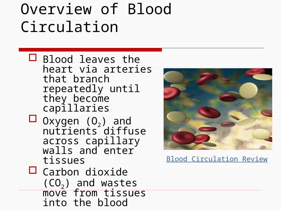
Overview of Blood Circulation
Blood leaves the heart via arteries that branch repeatedly until they become capillaries
Oxygen (O2) and nutrients diffuse across capillary walls and enter tissues
Carbon dioxide (CO2) and wastes move from tissues into the blood
Blood Circulation Review

Overview of Blood Circulation
Oxygen-deficient blood leaves the capillaries and flows in veins to the heart
This blood flows to the lungs where it releases CO2 and picks up O2
The oxygen-rich blood returns to the heart

Composition of Blood
Blood is the body’s only fluid tissue It is considered a connective tissue
Contains cells: formed elements Extracellular matrix: blood plasma
Formed elements include: Erythrocytes, or red blood cells (RBCs) Leukocytes, or white blood cells (WBCs) Platelets

Components of Whole Blood
Figure 17.1
• Hematocrit – the percentage of RBCs out of the total blood volume

Physical Characteristics and Volume
Blood is a sticky, opaque fluid with a metallic taste
Color varies from scarlet to dark red The pH of blood is 7.35–7.45 Temperature is 38C Blood accounts for approximately 8%
of body weight Average volume: 5–6 L for males, and
4–5 L for females
Functions of Blood
Blood performs a number of functions dealing with: Substance distribution Regulation of blood levels of particular
substances Body protection

Distribution
Blood transports: Oxygen from the lungs and nutrients
from the digestive tract Metabolic wastes from cells to the lungs
and kidneys for elimination Hormones from endocrine glands to
target organs

Regulation
Blood maintains: Appropriate body temperature by
absorbing and distributing heat Normal pH in body tissues using buffer
systems Adequate fluid volume in the circulatory
system
Protection Blood prevents blood loss by:
Activating plasma proteins and platelets Initiating clot formation when a vessel is
broken Blood prevents infection by:
Synthesizing and utilizing antibodies Activating complement proteins Activating WBCs to defend the body
against foreign invaders

Blood Plasma Blood plasma contains over 100 solutes,
including: Proteins – albumin, globulins, clotting
proteins, and others Metabolic wastes - Lactic acid, urea,
creatinine Organic nutrients – glucose, carbohydrates,
amino acids Electrolytes – sodium, potassium, calcium,
chloride, bicarbonate Respiratory gases – oxygen and carbon
dioxide
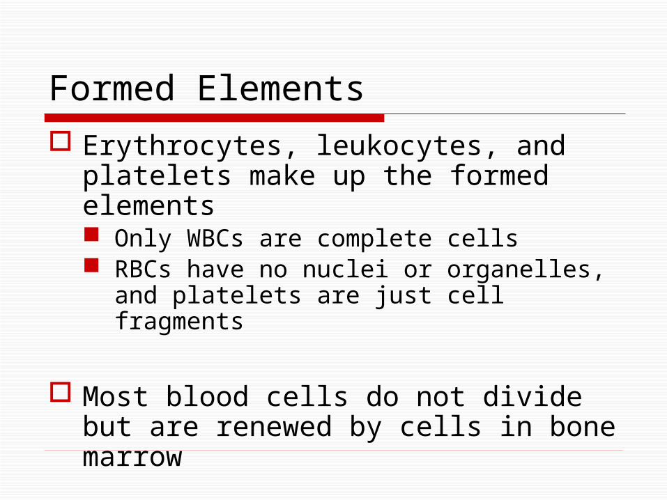
Formed Elements Erythrocytes, leukocytes, and
platelets make up the formed elements Only WBCs are complete cells RBCs have no nuclei or organelles, and
platelets are just cell fragments
Most blood cells do not divide but are renewed by cells in bone marrow

Erythrocytes (RBCs)
Figure 17.3

Erythrocytes (RBCs) Biconcave discs, anucleate,
essentially no organelles Filled with hemoglobin (Hb), a
protein that functions in gas transport
Contain the plasma membrane protein SPECTRIN and other proteins that: Give erythrocytes their flexibility Allow them to change shape as
necessary

Components of Whole Blood
Figure 17.2

Erythrocytes (RBCs)
Structural characteristics contribute to its gas transport function Biconcave shape has a huge surface area
relative to volume Erythrocytes are more than 97%
hemoglobin ATP is generated ANAEROBICALLY, so the
erythrocytes do not consume the oxygen
they transport

Erythrocyte Function
RBCs are dedicated to respiratory gas transport
Hb reversibly binds with oxygen and most oxygen in the blood is bound to Hb
Hb is composed of the protein globin, made up of two alpha and two beta chains, each bound to a heme group
Each heme group bears an atom of iron, which can bind to one oxygen molecule
Each Hb molecule can transport four molecules of oxygen
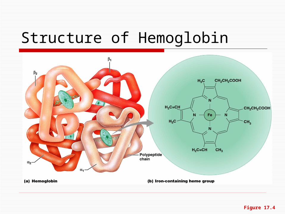
Structure of Hemoglobin
Figure 17.4
Hemoglobin (Hb) Oxyhemoglobin – Hb bound to oxygen
Oxygen loading takes place in the lungs Deoxyhemoglobin – Hb after oxygen
diffuses into tissues (reduced Hb) Carbaminohemoglobin – Hb bound to
carbon dioxide Carbon dioxide loading takes place in the
tissuesYouTube - Respiratory System

Production of Erythrocytes
Hematopoiesis – blood cell formation
Hematopoiesis occurs in the red bone marrow of the: Axial skeleton and girdles Epiphyses of the humerus and femur
Hemocytoblasts give rise to all formed elements

Production of Erythrocytes: Erythropoiesis
Figure 17.5

Homeostasis: Normal blood oxygen levels
IncreasesO2-carryingability of blood
Erythropoietinstimulates redbone marrow
Reduces O2 levelsin blood
Kidney (and liver to a smallerextent) releases erythropoietin
Enhancederythropoiesisincreases RBC count
Stimulus: Hypoxia due todecreased RBC count,decreased amount of hemoglobin, or decreased availability of O2
Start
Imbalance
Imbalance
Erythropoietin Mechanism
Figure 17.6
Fate and Destruction of Erythrocytes
The life span of an erythrocyte is 100–120 days
Old RBCs become rigid and fragile, and their Hb begins to degenerate
Dying RBCs are engulfed by macrophages
Heme and globin are separated and the iron is salvaged for reuse

Fate and Destruction of Erythrocytes
Globin is metabolized into amino acids and is released into the circulation
Hb released into the blood is phagocytized

Fate and Destruction of Erythrocytes
Heme is degraded to a yellow pigment called bilirubin
The liver secretes bilirubin into the intestines as bile
The intestines metabolize it into urobilinogen
This degraded pigment leaves the body in feces, in a pigment called stercobilin (makes feces brown)

Hemoglobin
Aminoacids
Globin
Raw materials aremade available inblood for erythrocytesynthesis.
Iron is bound to transferrin and released to blood from liver as needed for erythropoiesis
Food nutrients,including aminoacids, Fe, B12,and folic acidare absorbedfrom intestineand enter blood
Heme
Circulation
Iron storedas ferritin,hemosiderin
Bilirubin
Bilirubin is picked up fromblood by liver, secreted intointestine in bile, metabolizedto stercobilin by bacteriaand excreted in feces
Erythropoietin levelsrise in blood.
Erythropoietin and necessaryraw materials in blood promoteerythropoiesis in red bone marrow.
New erythrocytesenter bloodstream;function about120 days.
Low O2 levels in blood stimulatekidneys to produce erythropoietin.
Aged and damaged redblood cells are engulfed bymacrophages of liver, spleen,and bone marrow; the hemoglobinis broken down.
1
2
3
4
5
6
Figure 17.7

Leukocytes (WBCs) Leukocytes, the only blood
components that are complete cells: Are less numerous than RBCs Make up 1% of the total blood volume Can leave capillaries via diapedesis Move through tissue spaces
Leukocytosis – WBC count over 11,000 / mm3
Normal response to bacterial or viral invasion

Percentages of Leukocytes
Figure 17.9

Granulocytes
Granulocytes1. neutrophils, 2. eosinophils, and 3. basophils
Are larger and usually shorter-lived than RBCs
Have lobed nuclei Are all phagocytic cells
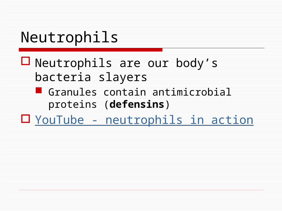
Neutrophils
Neutrophils are our body’s bacteria slayers Granules contain antimicrobial proteins
(defensins) YouTube - neutrophils in action

Eosinophils account for 1–4% of WBCs Lead the body’s
counterattack against parasitic worms
Lessen the severity of allergies
Eosinophils

Account for 0.5% of WBCs and: Have U- or S-shaped nuclei
with two or three conspicuous constrictions
Contain histamine Histamine – inflammatory
chemical that acts as a vasodilator and attracts other WBCs (antihistamines counter this effect)
Basophils
Explanation of Allergies

Account for 25% or more of WBCs and: Have large, dark-purple, circular
nuclei Are found mostly in lymphoid
tissue There are two types of lymphocytes: T cells and B cells T cells function in the immune response B cells give rise to plasma cells, which
produce antibodies (like gamma globulin)
Lymphocytes-agranulocyte

Monocytes account for 4–8% of leukocytes
Largest leukocytes They have purple-staining, U- or kidney-
shaped nuclei They leave the circulation, enter tissue,
and differentiate into macrophages Activate lymphocytes to mount an
immune response
Monocytes
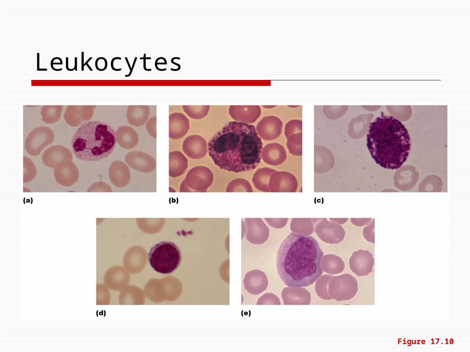
Leukocytes
Figure 17.10
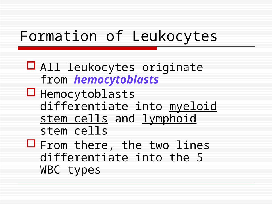
Formation of Leukocytes
All leukocytes originate from hemocytoblasts
Hemocytoblasts differentiate into myeloid stem cells and lymphoid stem cells
From there, the two lines differentiate into the 5 WBC types

(a) (b) (c) (d) (e)
Hemocytoblast
Myeloid stem cell Lymphoid stem cell
Myeloblast MyeloblastMyeloblast Lymphoblast
Stem cells
Committedcells
Promyelocyte PromyelocytePromyelocyte Promonocyte Prolymphocyte
Eosinophilicmyelocyte
Neutrophilicmyelocyte
Basophilicmyelocyte
Eosinophilicband cells
Neutrophilicband cells
Basophilicband cells
Develop-mentalpathway
Eosinophils NeutrophilsBasophils
Granular leukocytes
Plasma cells
Some become
Monocytes Lymphocytes
Macrophages (tissues)
Agranular leukocytes
Some become
Figure 17.11

Leukocytes Disorders: Leukemias
Leukemia refers to cancerous conditions involving WBCs
Leukemias are named according to the abnormal WBCs involved
Pictured: Acute lymphocytic leukemia

Leukemia Immature WBCs are found in the
bloodstream in all leukemias Bone marrow becomes totally occupied
with cancerous leukocytes The WBCs produced, though numerous, are
not functional Death is caused by internal hemorrhage
and overwhelming infections Treatments include irradiation, antileukemic
drugs, and bone marrow transplants
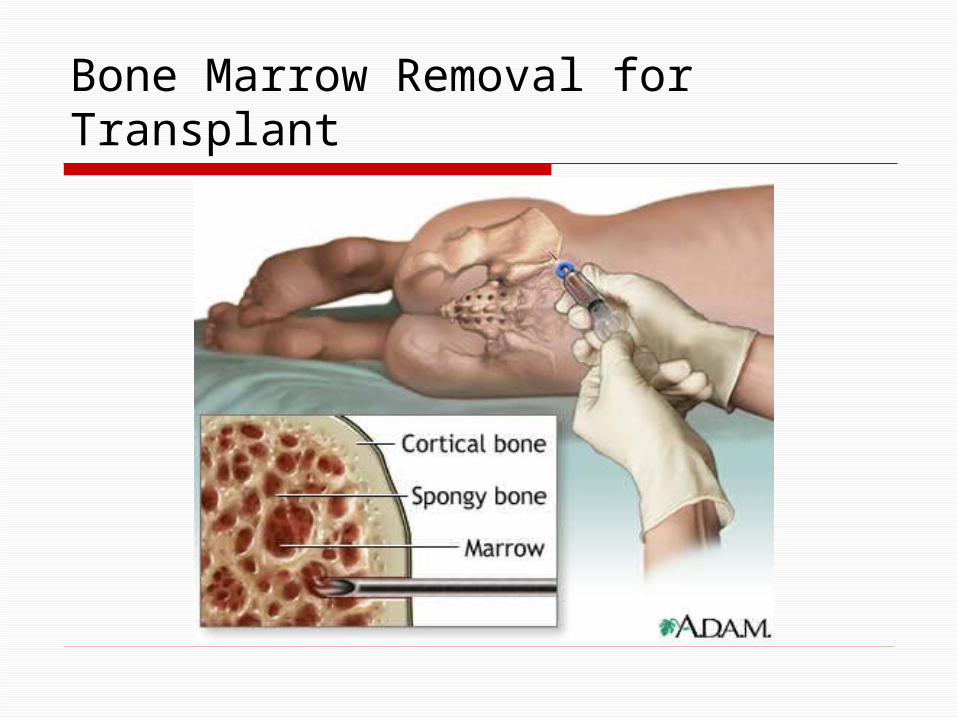
Bone Marrow Removal for Transplant

Platelets are fragments of megakaryocytes (found in bone marrow)
Platelets function in the clotting mechanism by forming a temporary plug that helps seal breaks in blood vessels
The stem cell for platelets is the hemocytoblast
Platelets

Production of Erythrocytes: Erythropoiesis
Figure 17.5

(a) (b) (c) (d) (e)
Hemocytoblast
Myeloid stem cell Lymphoid stem cell
Myeloblast MyeloblastMyeloblast Lymphoblast
Stem cells
Committedcells
Promyelocyte PromyelocytePromyelocyte Promonocyte Prolymphocyte
Eosinophilicmyelocyte
Neutrophilicmyelocyte
Basophilicmyelocyte
Eosinophilicband cells
Neutrophilicband cells
Basophilicband cells
Develop-mentalpathway
Eosinophils NeutrophilsBasophils
Granular leukocytes
Plasma cells
Some become
Monocytes Lymphocytes
Macrophages (tissues)
Agranular leukocytes
Some become
Figure 17.11

Stem cell Developmental pathway
Hemocytoblast Megakaryoblast Promegakaryocyte Megakaryocyte Platelets
Figure 17.12
Genesis of Platelets
The sequential developmental pathway is as shown.
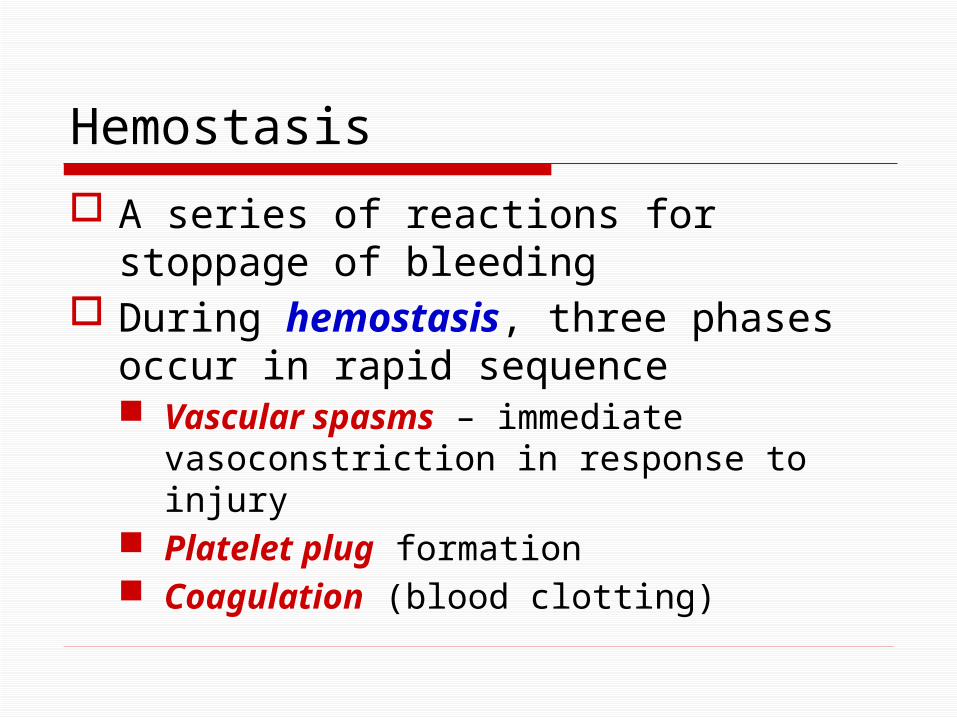
Hemostasis
A series of reactions for stoppage of bleeding
During hemostasis, three phases occur in rapid sequence Vascular spasms – immediate
vasoconstriction in response to injury Platelet plug formation Coagulation (blood clotting)

Vascular Spasm
Immediate response to injury is vasoconstriction
Factors that trigger the spasm are damaged cells, platelets and pain reflexes
As damage increases, vascular spasm increases

Platelet Plug Formation Platelets do not stick to each other or to
blood vessels when there is no damage Upon damage to blood vessel endothelium
platelets: Adhere to collagen Stick to exposed collagen fibers and form a
platelet plug Release serotonin and ADP, which attract still
more platelets The platelet plug is limited to the
immediate area of injury

A set of reactions in which blood is transformed from a liquid to a gel
Coagulation follows intrinsic and extrinsic pathways to thromboplastin
The final three steps of this series of reactions are: Prothrombin activator is formed Prothrombin is converted into
thrombin Thrombin starts the joining of fibrinogen
(plasma protein) into a fibrin mesh
Coagulation

Fibrin mesh forming in wound

Clot Retraction and Repair Clot retraction – stabilization of the
clot by squeezing serum from the fibrin strands
Repair Fibroblasts form a connective tissue
patch Endothelial cells multiply and restore the
endothelial lining of blood vessel

Cross section of healing wound-don’t pick at it!!!

Factors Limiting Clot Growth or Formation
Two homeostatic mechanisms prevent clots from becoming large Swift removal of clotting factors Stop formation of further clotting factors

Hemostasis Disorders:Thromboembolytic Conditions
Thrombus – a clot that develops and persists in an unbroken blood vessel Thrombi can block circulation, resulting in tissue
death Coronary thrombosis – thrombus in blood vessel
of the heart

Thrombus/Embolus

Hemostasis Disorders:Thromboembolytic Conditions
Embolus – a thrombus freely floating in the blood stream Pulmonary emboli can impair the ability
of the body to obtain oxygen Cerebral emboli can cause strokes
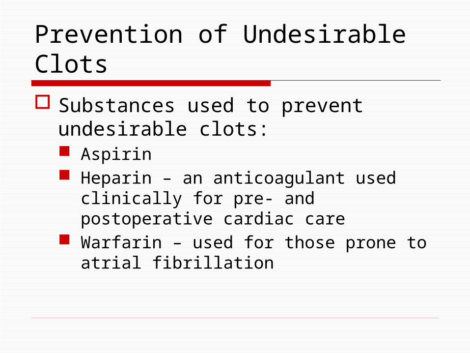
Substances used to prevent undesirable clots: Aspirin Heparin – an anticoagulant used clinically
for pre- and postoperative cardiac care Warfarin – used for those prone to atrial
fibrillation
Prevention of Undesirable Clots
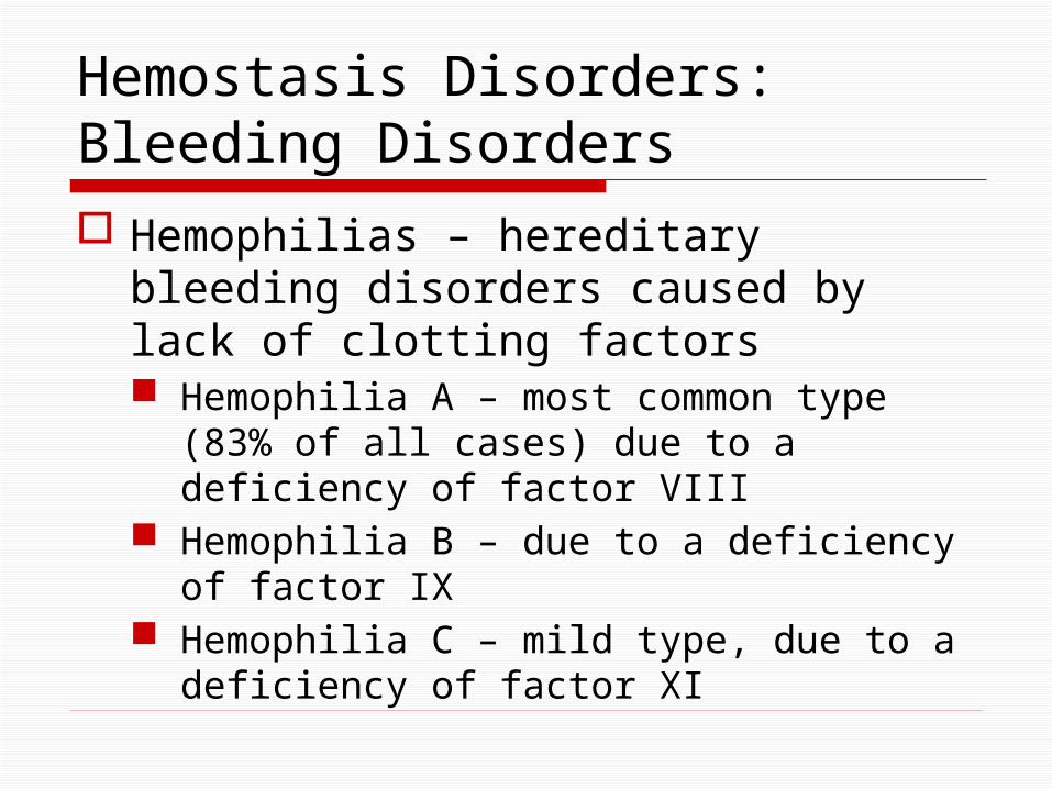
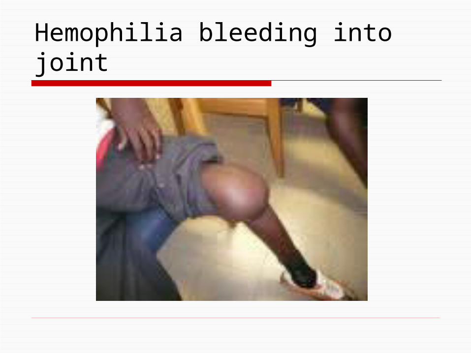
Hemophilias – hereditary bleeding disorders caused by lack of clotting factors Hemophilia A – most common type (83%
of all cases) due to a deficiency of factor VIII
Hemophilia B – due to a deficiency of factor IX
Hemophilia C – mild type, due to a deficiency of factor XI
Hemostasis Disorders: Bleeding Disorders

RBC membranes have glycoprotein antigens on their external surfaces
These antigens are: Unique to the individual Recognized as foreign if transfused
into another individual Promoters of agglutination and are
referred to as agglutinogens Presence or absence of these
antigens is used to classify blood groups
Human Blood Groups
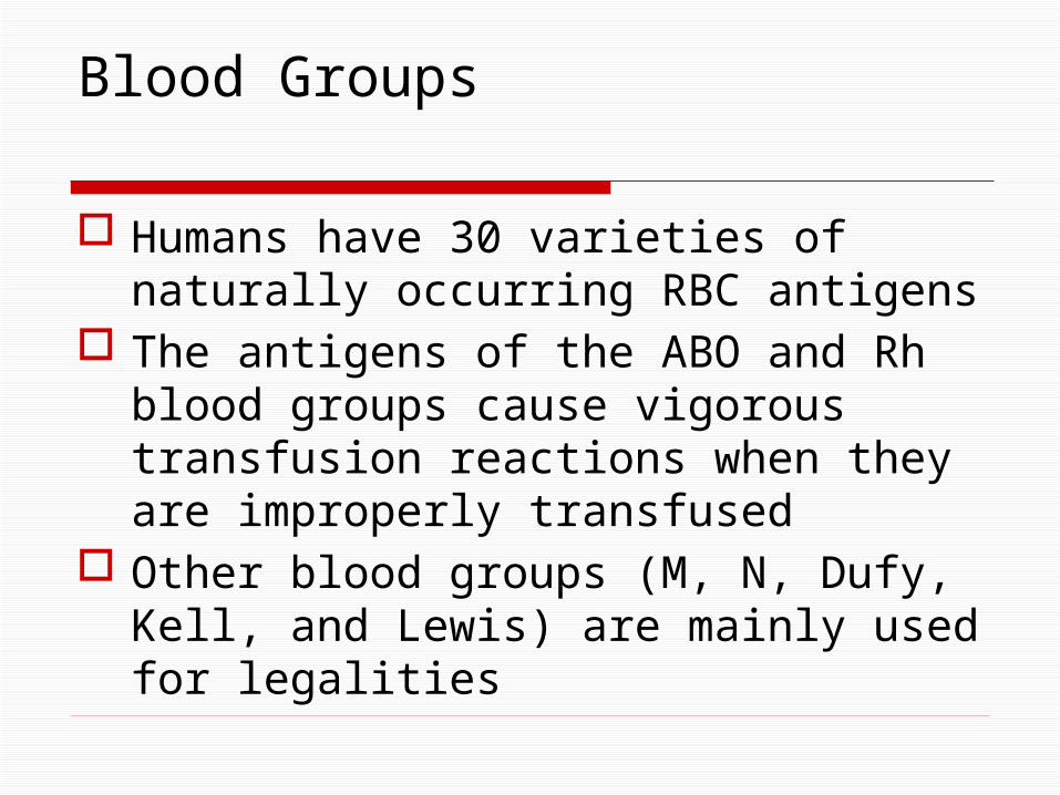
Humans have 30 varieties of naturally occurring RBC antigens
The antigens of the ABO and Rh blood groups cause vigorous transfusion reactions when they are improperly transfused
Other blood groups (M, N, Dufy, Kell, and Lewis) are mainly used for legalities
Blood Groups
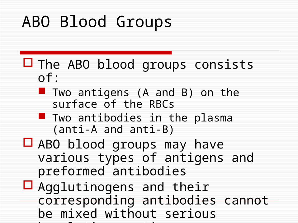
The ABO blood groups consists of: Two antigens (A and B) on the surface of
the RBCs Two antibodies in the plasma (anti-A and
anti-B) ABO blood groups may have various
types of antigens and preformed antibodies
Agglutinogens and their corresponding antibodies cannot be mixed without serious hemolytic reactions
ABO Blood Groups

ABO Blood Groups
Table 17.4

There are eight different Rh agglutinogens,
Presence of the Rh agglutinogens on RBCs is indicated as Rh+
Anti-Rh antibodies are not spontaneously formed in Rh– individuals
However, if an Rh– individual receives Rh+ blood, anti-Rh antibodies form
A second exposure to Rh+ blood will result in a typical transfusion reaction
Rh Blood Groups
Hemolytic Disease of the Newborn
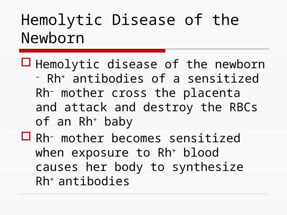
Hemolytic disease of the newborn – Rh+ antibodies of a sensitized Rh– mother cross the placenta and attack and destroy the RBCs of an Rh+ baby
Rh– mother becomes sensitized when exposure to Rh+ blood causes her body to synthesize Rh+ antibodies

Hemolytic Disease of the Newborn
The drug RhoGAM can prevent the Rh– mother from becoming sensitized
Treatment of hemolytic disease of the newborn involves pre-birth transfusions and exchange transfusions after birth
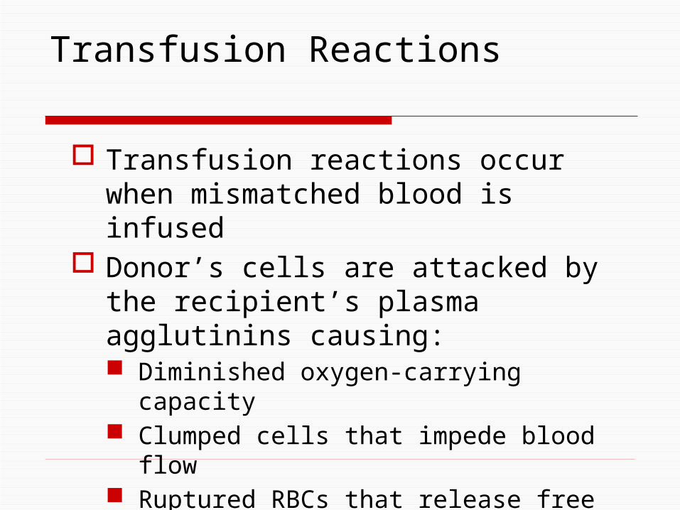
Transfusion reactions occur when mismatched blood is infused
Donor’s cells are attacked by the recipient’s plasma agglutinins causing: Diminished oxygen-carrying capacity Clumped cells that impede blood flow Ruptured RBCs that release free
hemoglobin into the bloodstream that causes kidney failure
Transfusion Reactions

Blood type being tested
RBC agglutinogens Serum Reaction
Anti-A Anti-B
AB A and B + +
B B – +
A A + –
O None – –
Blood Typing