Chapter 13 Chlorophyll Fluorescence Applications in ...guide to the use of Chl fluorescence for...
16
277 1 Preface Chlorophyll (Chl) fluorescence has become one of the most common and useful techniques in photosynthetic field research. Its non-invasiveness, sensitivity and the wide availability of reliable instruments, also makes it a convenient and suitable technique in microalgal bio- technology to monitor a culture’s photosynthetic per- formance. Experimentally, homogenous microalgal cultures in suspension have also been ideal objects in photosynthetic studies. For the purpose of this book we summarised results of experiments since the 1990s that have pioneered the practical use of Chl fluores- cence methods to monitor the physiological status of fast-growing microalgal mass cultures, optimising and estimating biomass productivity or finding marker pro- cesses of certain compound synthesis. In their biomass, microalgae produce various valuable bioactive substances such as pigments, polyunsaturated fatty acids, antioxidants, essential amino acids or immunologically-effective, virostatic and cytostatic compounds. Therefore, microalgae are cultivated com- mercially for biomass as food and feed additives, as a source for pharmacology and cosmetics, or, on a small-scale, for research of diagnostic products. 2 Historical Overview of Using Chl Fluorescence in Microalgal Mass Cultures Microalgal cultures, outdoors, are exposed to changes in environmental conditions, particularly irradiance. To cope with variable light intensity in combination with other stresses during the day, quickly-growing microal- gae have developed fast and prompt regulation mecha- nisms, usually operating on a time-scale of seconds to minutes. Outdoor culture performance can be monitored through the assay of dry weight, or photosynthesis mea- surements carried out in the laboratory, but the time required to complete these measurements is rather long. In algal biotechnology warning signals must be recognized as soon as possible in order to prevent a significant reduc- tion in daily productivity, or to avoid situations which, in a few days, may culminate in culture loss. Since unfa- vourable environmental conditions and their synergisms affect the function of Photosystem II (PSII), directly or indirectly, chlorophyll fluorescence represents a useful tool in microalgal biotechnology – giving rapid evidence of stress affecting the photosynthetic activity of the cul- ture and certain quantification on productivity. In the 1990s, Chl fluorescence measurements were employed to examine the photosynthetic performance of microalgal mass cultures (e.g. Vonshak et al. 1994; Torzillo et al. 1996). In particular, questions were stud- ied associated with the relationship of fluorescence- based measures of PSII photobiochemical activity as a means to estimate primary productivity (Genty et al. 1989; Chapter 13 Chlorophyll Fluorescence Applications in Microalgal Mass Cultures Jiří Masojídek, Avigad Vonshak, and Giuseppe Torzillo J. Masojídek (*) Institute of Microbiology, Academy of Science, Opatovický mlýn, CZ-37981 Třeboň, Czech Republic and Institute of Physical Biology, University of South Bohemia, Zámek 136, CZ-37333 Nové Hrady, Czech Republic e-mail: [email protected] A. Vonshak Ben Gurion University J. Blaustein Institutes for Desert Research, Sede Boqer 84990, Israel G. Torzillo Istituto per lo Studio degli Ecosistemi, CNR, Via Madonna del Piano 10, 50019 Sesto Fiorentino (Firenze), Italy D.J. Suggett et al. (eds.), Chlorophyll a Fluorescence in Aquatic Sciences: Methods and Applications, Developments in Applied Phycology 4, DOI 10.1007/978-90-481-9268-7_13, © Springer Science+Business Media B.V. 2011
Transcript of Chapter 13 Chlorophyll Fluorescence Applications in ...guide to the use of Chl fluorescence for...
277
1 Preface
Chlorophyll (Chl) fluorescence has become one of the most common and useful techniques in photosynthetic field research. Its non-invasiveness, sensitivity and the wide availability of reliable instruments, also makes it a convenient and suitable technique in microalgal bio-technology to monitor a culture’s photosynthetic per-formance. Experimentally, homogenous microalgal cultures in suspension have also been ideal objects in photosynthetic studies. For the purpose of this book we summarised results of experiments since the 1990s that have pioneered the practical use of Chl fluores-cence methods to monitor the physiological status of fast-growing microalgal mass cultures, optimising and estimating biomass productivity or finding marker pro-cesses of certain compound synthesis.
In their biomass, microalgae produce various valuable bioactive substances such as pigments, polyunsaturated fatty acids, antioxidants, essential amino acids or immunologically-effective, virostatic and cytostatic compounds. Therefore, microalgae are cultivated com-
mercially for biomass as food and feed additives, as a source for pharmacology and cosmetics, or, on a small-scale, for research of diagnostic products.
2 Historical Overview of Using Chl Fluorescence in Microalgal Mass Cultures
Microalgal cultures, outdoors, are exposed to changes in environmental conditions, particularly irradiance. To cope with variable light intensity in combination with other stresses during the day, quickly-growing microal-gae have developed fast and prompt regulation mecha-nisms, usually operating on a time-scale of seconds to minutes. Outdoor culture performance can be monitored through the assay of dry weight, or photosynthesis mea-surements carried out in the laboratory, but the time required to complete these measurements is rather long. In algal biotechnology warning signals must be recognized as soon as possible in order to prevent a significant reduc-tion in daily productivity, or to avoid situations which, in a few days, may culminate in culture loss. Since unfa-vourable environmental conditions and their synergisms affect the function of Photosystem II (PSII), directly or indirectly, chlorophyll fluorescence represents a useful tool in microalgal biotechnology – giving rapid evidence of stress affecting the photosynthetic activity of the cul-ture and certain quantification on productivity.
In the 1990s, Chl fluorescence measurements were employed to examine the photosynthetic performance of microalgal mass cultures (e.g. Vonshak et al. 1994; Torzillo et al. 1996). In particular, questions were stud-ied associated with the relationship of fluorescence-based measures of PSII photobiochemical activity as a means to estimate primary productivity (Genty et al. 1989;
Chapter 13Chlorophyll Fluorescence Applications in Microalgal Mass Cultures
Jiří Masojídek, Avigad Vonshak, and Giuseppe Torzillo
J. Masojídek (*) Institute of Microbiology, Academy of Science, Opatovický mlýn, CZ-37981 Třeboň, Czech Republicand Institute of Physical Biology, University of South Bohemia, Zámek 136, CZ-37333 Nové Hrady, Czech Republic e-mail: [email protected]
A. Vonshak Ben Gurion University J. Blaustein Institutes for Desert Research, Sede Boqer 84990, Israel
G. Torzillo Istituto per lo Studio degli Ecosistemi, CNR, Via Madonna del Piano 10, 50019 Sesto Fiorentino (Firenze), Italy
D.J. Suggett et al. (eds.), Chlorophyll a Fluorescence in Aquatic Sciences: Methods and Applications, Developments in Applied Phycology 4, DOI 10.1007/978-90-481-9268-7_13, © Springer Science+Business Media B.V. 2011

278 J. Masojídek et al.
Torzillo et al. 1996; Schreiber et al. 1998; Baker 2008; Suggett et al., Chapter 6, this volume). It was essential to determine the fluorescence quenching that results from both photochemical and non-photochemical pro-cesses (Bradbury and Baker 1984; Schreiber et al. 1986).
The aim of this chapter is to provide a simple, practical guide to the use of Chl fluorescence for algal biotechnolo-gists who wish to apply this technique in microalgal mass cultures, both in field and laboratory studies. Whilst the principles behind the measurements will be mentioned only briefly, the emphasis will be given to the applications and limitations of fluorescence technique for in situ moni-toring and its implications.
3 Microalgae Grown for Commercial Purposes and Cultivation Systems
In applied phycology, the term microalgae is usually used in its broadest sense to mean both prokaryotic cyanobacteria and eukaryotic algae – unicellular or fila-mentous photosynthetic microorganisms. Microalgae represent important CO
2 consumers and primary pro-
ducers – being the basis of the food chain in aquatic environments. Furthermore, they are one of the most efficient converters of solar energy to biomass. Dense, well-mixed mass cultures of microalgae (> 0.5 g bio-mass per litre) with sufficient nutrition and gas exchange represent artificial systems which are completely dif-ferent from optically-thin natural phytoplankton popu-lations with low biomass density, often limited by nutrient and carbon supply.
Many cultivation systems have been designed and built to grow microalgae using natural or artificial light. Two basic approaches to microalgal mass production are used: the first applies to cultivation in open reser-voirs large in area, while the second are closed vessels – photobioreactors (for review see Pulz et al. 2001; Torzillo et al. 2003; Tredici 2004). The first type – open cultivation systems – is represented by natural or artifi-cial ponds, raceways (ponds akin to race-tracks) and cascades (i.e. inclined-surface systems) (see example in Fig. 1, upper panel). In the second type – photobio-reactors (closed or semi-closed systems with natural or artificial illumination) consist of glass or transparent plastic tubes, or panels, positioned horizontally or vertically, arranged as serpentine loops, flexible coils, manifold rows or ‘fences’, in which the microalgal
suspension is continuously circulated (see example in Fig. 1, bottom panel).
In every cultivation system, several basic principles must be considered that affect productivity in mass microalgal cultures, such as: (i) culture depth, or optical cross-section (thicker culture layers would progres-sively absorb penetrating light more, rendering it unavailable for photosynthesis); (ii) turbulence where various means and techniques could be used; (iii) nutrient content and supply, including gas exchange (CO
2 supply and O
2 removal); (iv) cultivation procedure
(batch, continuous or semi-continuous, or multistage processes); (v) biomass concentration and areal den-sity; and (vi) acclimation state of microalgae (Richmond 2004; Grobbelaar 2007). The choice of a suitable cul-tivation system and the adjustment of the cultivation regime must be worked out for each individual produc-tive microalgal strain.
Myriads of microalgae have been isolated from natural habitats and are kept in numerous culture col-lections around the world. However, to date, only a few microalgal strains, mostly of aquatic origin, have been cultivated in large-scale production systems of hun-dreds to thousands of litres.
Arthrospira (Spirulina) platensis is a planktonic fila-mentous cyanobacterium composed of individual cells (about 8 µm in diameter), which grows in subtropical alkaline lakes with a temperature optimum of about 35°C. In productive cultures, Arthrospira is cultivated in shallow mixed ponds or semi-closed tubular photobio-reactors. The growth medium contains inorganic salts with a high concentration of bicarbonate, keeping the pH value between 9 and 10. This cyanobacterium is the most cultivated photosynthetic prokaryote since its biomass is widely used as a health food, feed supple-ment and as a source of fine chemicals. It contains pro-teins, polyunsaturated fatty acids, phycobiliproteins, carotenoids, polysaccharides, vitamins and minerals.
The microalga Chlorella (green alga, Chlorophyta) is a cosmopolitan genus with small globular cells (3–8 µm in diameter), including strains with a high temper-ature tolerance since some can grow between 15°C and 40°C. Chlorella grows autotrophically in an inor-ganic medium, as well as in mixotrophic and hetero-trophic conditions (for example, with an addition of acetic acid or glucose). At present, autotrophic pro-duction of Chlorella is carried out in open ponds, semi-closed tubular photobioreactors, or inclined cas-cades, since its high growth rate prevents contamination
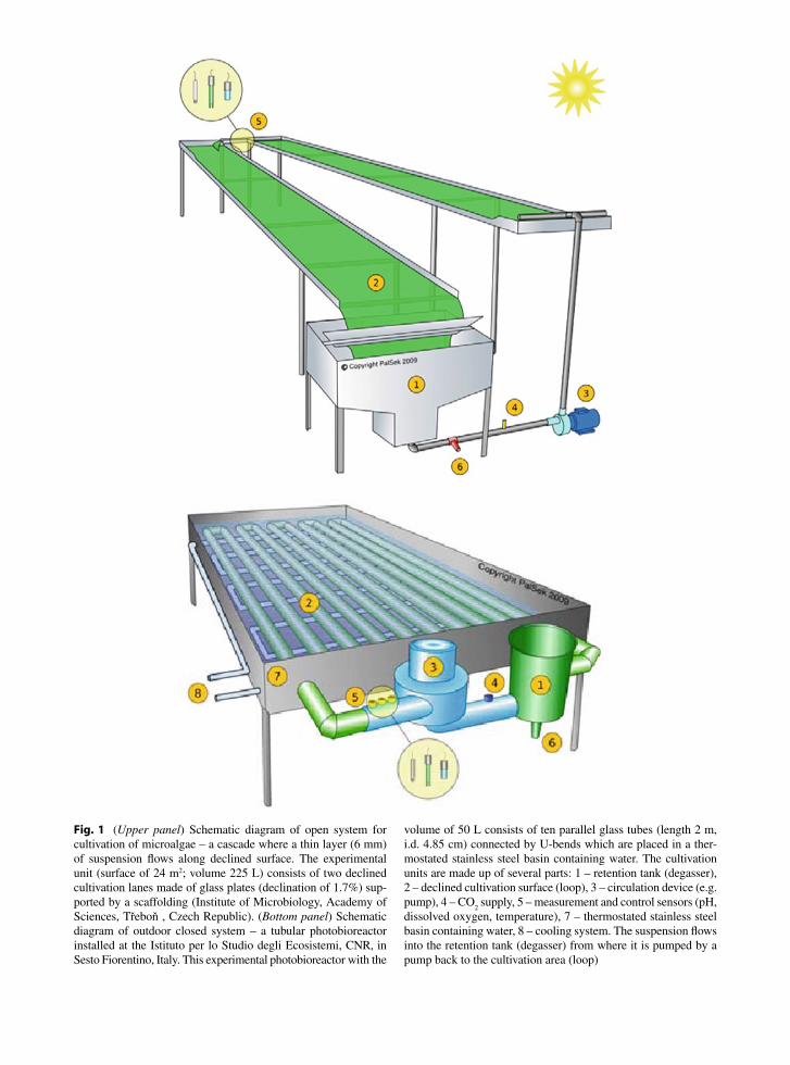
Fig. 1 (Upper panel) Schematic diagram of open system for cultivation of microalgae – a cascade where a thin layer (6 mm) of suspension flows along declined surface. The experimental unit (surface of 24 m2; volume 225 L) consists of two declined cultivation lanes made of glass plates (declination of 1.7%) sup-ported by a scaffolding (Institute of Microbiology, Academy of Sciences, Třeboň , Czech Republic). (Bottom panel) Schematic diagram of outdoor closed system – a tubular photobioreactor installed at the Istituto per lo Studio degli Ecosistemi, CNR, in Sesto Fiorentino, Italy. This experimental photobioreactor with the
volume of 50 L consists of ten parallel glass tubes (length 2 m, i.d. 4.85 cm) connected by U-bends which are placed in a ther-mostated stainless steel basin containing water. The cultivation units are made up of several parts: 1 – retention tank (degasser), 2 – declined cultivation surface (loop), 3 – circulation device (e.g. pump), 4 – CO
2 supply, 5 – measurement and control sensors (pH,
dissolved oxygen, temperature), 7 – thermostated stainless steel basin containing water, 8 – cooling system. The suspension flows into the retention tank (degasser) from where it is pumped by a pump back to the cultivation area (loop)

280 J. Masojídek et al.
by other microalgae. Chlorella is the most cultivated eukaryotic alga since it is widely used as a health food and feed supplement, as well as in the pharmaceutical and cosmetics industry. It contains proteins, carote-noids, some immunostimulators, polysaccharides, vitamins and minerals. The bulk of the microalgae bio-mass market is represented by Chlorella and Arthrospira with annual production of 3,000 t and 4,000 t, respectively.
Dunaliella salina (Chlorophyta) and similar hypersaline strains have biflagellated, pear-shaped cells (about 10 µm in diameter). Dunaliella produces b-carotene in high amounts, up to 12% of dry matter. This microalga is a natural source of carotenoids for some brine shrimp. The high content of b-carotene makes Dunaliella attractive to biotechnologists for large-scale production in shallow, open ponds under high solar radiation (Borowitzka 2005). About 25 t of b-carotene enriched health food is produced yearly (Ben-Amotz 2004).
Haematococcus pluvialis (Chlorophyta) is a freshwa-ter, unicellular alga with a rather complex life-cycle. A two-stage process is employed for biomass production. Under stress conditions (nutrient defi-ciency, salinity, high temperatures in combination with high irradiance), the green vegetative cells (about 10 µm diameter) produce thicker walls and change to large globular cysts (about 50 µm in diameter) with orange-red pigmentation, due to an increased deposition of astaxanthin (up to 5% of dry weight). This pigment is the important natural colorant for sal-monoid fish, shrimp, lobster and crayfish and health food market. Annual production of Haematococcus biomass is about 100 t.
4 Principles of Microalgae Mass Culturing
Microalgae belong to the fastest-growing photosynthetic organisms since their cell doubling time can be as little as several hours. The necessary cultivation require-ments for the growth of microalgal mass cultures are light, a suitable temperature and pH of the growth medium, as well as a sufficient carbon and nutrient supply. As microalgal mass cultures grow in dense suspensions, some kind of mixing is necessary to
expose cells to light evenly and to allow for an effi-cient gas exchange (CO
2 supply/O
2 removal).
Light is the most critical factor for microalgal growth. The amount of photon energy received by each cell is a combination of several factors: irradiance intensity, cell density, length of optical path (thickness of culture layer), spectral quality (light penetration), light absorption, and rate of mixing (Richmond 2004). Two basic factors have to be fulfilled for obtaining maximal photochemical efficiency and productivity: (i) cell density must be optimal, insuring that the aver-age photon irradiance per cell is close to upper end of the linear phase of the growth curve, and (ii) light-dark cycles of the cells caused by culture turbulence must be fast, close to the turnover of the photosynthetic units (Nedbal et al. 1996; Richmond 2004). The light captured by photosynthetic pigments is roughly ten times higher under full sunlight (2000 mmol photon m−2 s−1) than that required to saturate growth. In other words, as much as 90% of the photons captured by the Chl antennae can be dissipated as heat and fluores-cence. Uncritical acceptance of uncorrected photosyn-thetic efficiencies of about 10% or even higher (Pirt 1986) inevitably leads to exaggerated estimates of present and future biomass productivity. We can how-ever approach a rather more realistic maximum figure of photosynthetic efficiency (photon energy converted into biomass energy) of about 4.5% for C3 plants or microalgae by using “educated guesswork” and detailed consideration of the partial reactions involved (e.g. Boardman 1980; Benemann and Oswald 1996; Zhu et al. 2008; Walker 2009).
After irradiance, temperature is the most important parameter to control the microalgal culture growth. Some microalgal strains tolerate a broad temperature range between 15°C and 35°C (e.g. Chlorella), while Haematococcus usually requires much narrower range. However, for the majority of freshwater microalgae the optimum temperature range is within 25–30°C.
Basically, two cultivation regimes are used for the growth of microalgal cultures. In the batch regime, the culture is inoculated and at a certain point of growth it is harvested. In the continuous regime, the culture is harvested continuously according to its growth rate and fresh medium is added to replace nutrients. In practice, semi-continuous or semi-batch regimes are often adopted; i.e. where a part of the culture is harvested at regular intervals, usually every day.

28113 Chlorophyll Fluorescence Applications in Microalgal Mass Cultures
4.1 Culture Maintenance
Successful cultivation requires continuous monitoring of physico-chemical parameters, i.e. irradiance, pH, temperature, dissolved oxygen concentration, and nutrient status. The basic biological method used is a microscopic examination to detect morphological changes and contamination by other microorganisms. CO
2 serves as the main carbon source and is added on
demand. Nutrient status can be followed by monitoring the concentration of nitrogen, using it as a measure for adding the proportional amounts of other nutrients. In the mass cultivation of microalgae, monocultures are usually required for biomass exploitation; contaminants often represent a substantial limitation to large-scale production in microalgae cultures. Thus in some cases, e.g. for the cultivation of Haematococcus, the use of a closed system becomes mandatory.
Sufficient degassing of the microalgal suspension is necessary in order to prevent the accumulation of oxygen in the culture that can increase photorespiration and promote photoinhibition of photosynthesis resulting in a decline in growth, particularly when the suspension is cultivated in a closed system (Torzillo et al. 1998). On the other hand, excessive mixing can cause hydro-dynamic stresses on the cells, and consequently reduce productivity.
Culture growth may be estimated as changes of optical density (O.D.) at 750 nm, or in biomass dry weight in time. Specific growth rate of culture is obtained with the following equation, m = (ln X
2–ln
X1)/(t
2–t
1) expressed in [h−1 or day−1].
Biomass productivity is expressed as the areal or volumetric yield per unit time, i.e. in [g m−2 day−1] or in [g L−1 day−1], respectively. The highest m is obtained when all requirements for cell growth are optimal and light is saturating (low biomass concentration). Highest productivity, in contrast, is achieved when cells are light-limited (dense cultures), the growth rate of which being about a half of the maximum (Richmond 2004).
Methods of biophysical and biochemical moni-toring of activity generally reflect the status of the cells’ photosynthetic apparatus and are used to adjust the appropriate cultivation conditions for the production of biomass or certain compounds. For example, photosynthetic oxygen production and Chl fluorescence yield are considered as reliable and
sensitive indicators of photosynthetic activity in microalgal cultures.
5 Interpretation of Chl Fluorescence Parameters in Microalgae Mass Cultures
Chl fluorescence measurements make it possible to fol-low the energy distribution between the photochemical and non-photochemical processes in photosynthesis. Different fluorescence-based parameters are described in the literature for the yields of photochemical energy conversion in PSII, which are complementary with the yields of non-photochemical energy dissipation.
Microalgae mass cultures are well-mixed suspen-sions representing averaged cell population, dense and homogenous which represent a completely different experimental object as compared to optically-thin phy-toplankton populations with chlorophyll concentration lower by several orders of magnitude, or static plant leaves where fluorescence signals are mostly emitted by a surface cell layer. In microalgal cultures, Chl fluo-rescence reflects the performance of photochemical processes in the photosynthetic apparatus, especially PSII, and subsequently its biomass productivity. The contribution of the PSI emission in the total signal is mostly small and for practical purposes is often neglected. A careful approach is required when mea-suring fluorescence and evaluating data in cyanobacte-ria since the fluorescence emission of numerous PSI complexes and phycobilisomes as well as state transi-tion effect contribute significantly to the total signal, which affects the correct determination of certain parameters (Ting and Owens 1992; Büchel and Wilhelm 1993; Schreiber et al. 1995).
At present, basically two Chl fluorescence approaches are used to monitor photosynthetic effi-ciency in microalgal mass cultures: rapid fluorescence induction or relaxation kinetics (Fig. 2) and the saturation-pulse method (Fig. 3).
At first, and for a long time, starting with the experi-ments of Kautsky in the 1930s, the most common way of measuring Chl fluorescence was based on the ‘con-ventional’ principle using dark-adapted samples. Fluorescence is excited by multi- or single-turnover light (e.g. produced usually by red LED; l
max = 660 nm

282 J. Masojídek et al.
which is absorbed by chlorophyll) and fluorescence emission is detected at wavelengths beyond 690–710 nm with the help of a suitable photosensor (photomultiplier or photodiode). The rapid Chl fluorescence induction kinetics (the so-called Kautsky curve) of all oxygenic photosynthetic organisms shows a polyphasic rise (Chl fluorescence transient) between the initial (F
0) and the
maximum (Fm) fluorescence during the first second of
illumination (Neubauer and Schreiber 1987; Schreiber and Neubauer 1987). The phases on the curve were des-ignated as O, J, I and P using a logarithmic time scale
(Strasser et al. 1995; Fig. 2a in this chapter). Since the 1990s, the theory has been elaborated to become the so-called ‘JIP-test’ (Govindjee 1995; Strasser and Strasser 1995; Strasser et al. 1995; Srivastava et al. 1999). The current understanding of the OJIP transient rise is that it reflects the filling-up (i.e. reduction) of the electron acceptor pool of PSII (Ph, Q
A, Q
B and PQ pool) in a
four-photon process (Strasser et al. 2004). The inflec-tion J (2 ms) represents the double-reduction of electron carriers Ph, Q
A, and Q
B, and because of a limitation in
electron acceptance by QB, this step usually occurs when
cells are exposed to excessive light that increases the degree of reduction of the PQ pool. The step I (30 ms) is connected to a three-electron reduction in the PSII elec-tron carriers to different redox states (e.g. Ph Q
A−Q
B2−
,
Ph-QAQ
B2− or Ph−Q
A−Q
B−) of the reaction centre complex
which reduces the PQ pool and probably reflects the heterogeneity of the PSII centres, the Q
A-fast-reducing
and slow-reducing ones. The drop in the induction curve of microalgal cells beyond the P step indicates that the PQ pool is being re-oxidized due to the demand of reduction equivalents from the Calvin-Benson cycle.
Time [ms]10-1 100 101 102 103
10-1 100 101 102 103 104
Flu
ore
scen
ce in
du
ctio
n [
r.u
.]
0800 h
O
J
a
b
IP=FmHaematococcus
F0
1200 h
1400 h
1800 h
Time [ ms ]
Flu
ore
scen
ce in
du
ctio
n [
r.u
.] Haematococcus
1800 h
0800 h1200 h1400 h
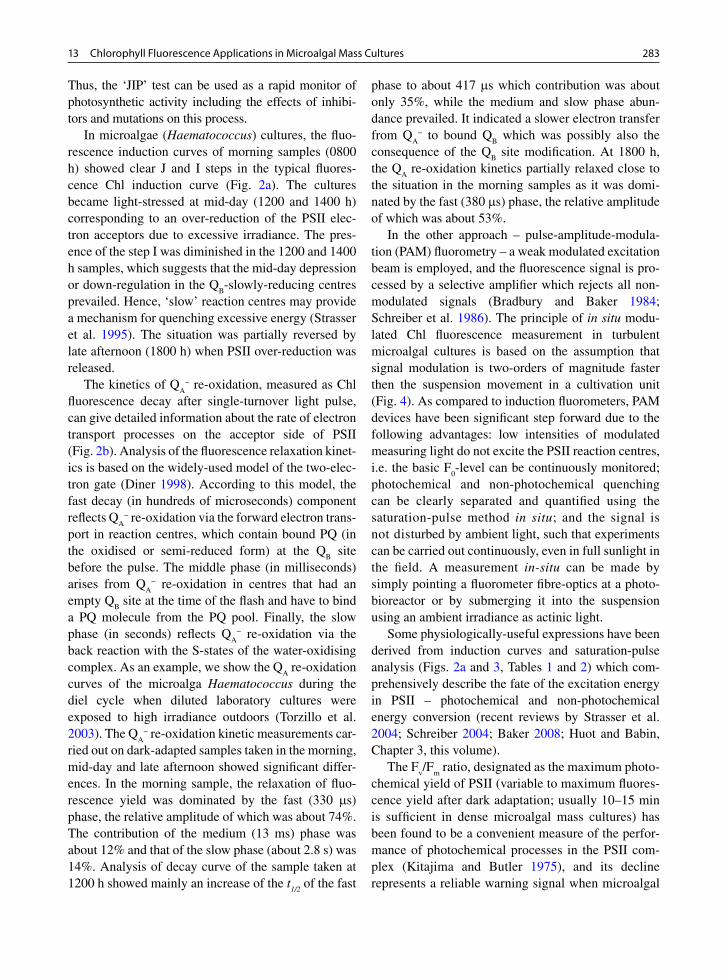
Fig. 2 Rapid Chl fluorescence induction kinetics measured in an outdoor culture of the alga Haematococcus at various periods of the diel cycle (0800, 1200, 1400 and 1800 h). Rapid fluores-cence induction curves normalised to F
0 level (panel a).
Intermediate steps of J and I represent various reduction states of the electron carriers of the PSII complex. Q
A reoxidation kinet-
ics after single-turnover light pulse was measured in an outdoor culture of the alga Haematococcus at various periods of the diel cycle (0800, 1200, 1400 and 1800 h) (panel b)
Fig. 3 Fluorescence quenching analysis using saturation-pulse method. The sample (microalgae culture) is illuminated with actinic light (AL) and a series of saturating pulses (SP) in order to reach the steady state F¢ and F
m¢ level. The minimum and
maximum fluorescence levels F0 and F
m are measured in the
dark-adapted sample (10–15 min) using modulated measuring light (ML) and SP. In case of cyanobacteria, a PSII electron transport inhibitor, diuron has to be used to achieve F
m. The
parameters denoted with a prime (¢) are from the sample exposed to light. The parameters without a prime are obtained from the sample in the dark-adapted state. Weak measuring light (ML; < 0.3 mmol photons m−2 s−1) has the subsaturating intensity to PSII photochemistry; saturating light pulse (SP; >10 mmol photons m−2 s−1; 0.8 s duration)
28313 Chlorophyll Fluorescence Applications in Microalgal Mass Cultures
Thus, the ‘JIP’ test can be used as a rapid monitor of photosynthetic activity including the effects of inhibi-tors and mutations on this process.
In microalgae (Haematococcus) cultures, the fluo-rescence induction curves of morning samples (0800 h) showed clear J and I steps in the typical fluores-cence Chl induction curve (Fig. 2a). The cultures became light-stressed at mid-day (1200 and 1400 h) corresponding to an over-reduction of the PSII elec-tron acceptors due to excessive irradiance. The pres-ence of the step I was diminished in the 1200 and 1400 h samples, which suggests that the mid-day depression or down-regulation in the Q
B-slowly-reducing centres
prevailed. Hence, ‘slow’ reaction centres may provide a mechanism for quenching excessive energy (Strasser et al. 1995). The situation was partially reversed by late afternoon (1800 h) when PSII over-reduction was released.
The kinetics of QA
− re-oxidation, measured as Chl fluorescence decay after single-turnover light pulse, can give detailed information about the rate of electron transport processes on the acceptor side of PSII (Fig. 2b). Analysis of the fluorescence relaxation kinet-ics is based on the widely-used model of the two-elec-tron gate (Diner 1998). According to this model, the fast decay (in hundreds of microseconds) component reflects Q
A− re-oxidation via the forward electron trans-
port in reaction centres, which contain bound PQ (in the oxidised or semi-reduced form) at the Q
B site
before the pulse. The middle phase (in milliseconds) arises from Q
A− re-oxidation in centres that had an
empty QB site at the time of the flash and have to bind
a PQ molecule from the PQ pool. Finally, the slow phase (in seconds) reflects Q
A− re-oxidation via the
back reaction with the S-states of the water-oxidising complex. As an example, we show the Q
A re-oxidation
curves of the microalga Haematococcus during the diel cycle when diluted laboratory cultures were exposed to high irradiance outdoors (Torzillo et al. 2003). The Q
A− re-oxidation kinetic measurements car-
ried out on dark-adapted samples taken in the morning, mid-day and late afternoon showed significant differ-ences. In the morning sample, the relaxation of fluo-rescence yield was dominated by the fast (330 ms) phase, the relative amplitude of which was about 74%. The contribution of the medium (13 ms) phase was about 12% and that of the slow phase (about 2.8 s) was 14%. Analysis of decay curve of the sample taken at 1200 h showed mainly an increase of the t
1/2 of the fast
phase to about 417 ms which contribution was about only 35%, while the medium and slow phase abun-dance prevailed. It indicated a slower electron transfer from Q
A− to bound Q
B which was possibly also the
consequence of the QB site modification. At 1800 h,
the QA re-oxidation kinetics partially relaxed close to
the situation in the morning samples as it was domi-nated by the fast (380 ms) phase, the relative amplitude of which was about 53%.
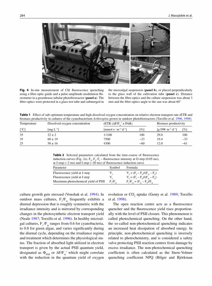
In the other approach – pulse-amplitude-modula-tion (PAM) fluorometry – a weak modulated excitation beam is employed, and the fluorescence signal is pro-cessed by a selective amplifier which rejects all non-modulated signals (Bradbury and Baker 1984; Schreiber et al. 1986). The principle of in situ modu-lated Chl fluorescence measurement in turbulent microalgal cultures is based on the assumption that signal modulation is two-orders of magnitude faster then the suspension movement in a cultivation unit (Fig. 4). As compared to induction fluorometers, PAM devices have been significant step forward due to the following advantages: low intensities of modulated measuring light do not excite the PSII reaction centres, i.e. the basic F
0-level can be continuously monitored;
photochemical and non-photochemical quenching can be clearly separated and quantified using the saturation-pulse method in situ; and the signal is not disturbed by ambient light, such that experiments can be carried out continuously, even in full sunlight in the field. A measurement in-situ can be made by simply pointing a fluorometer fibre-optics at a photo-bioreactor or by submerging it into the suspension using an ambient irradiance as actinic light.
Some physiologically-useful expressions have been derived from induction curves and saturation-pulse analysis (Figs. 2a and 3, Tables 1 and 2) which com-prehensively describe the fate of the excitation energy in PSII – photochemical and non-photochemical energy conversion (recent reviews by Strasser et al. 2004; Schreiber 2004; Baker 2008; Huot and Babin, Chapter 3, this volume).
The Fv/F
m ratio, designated as the maximum photo-
chemical yield of PSII (variable to maximum fluores-cence yield after dark adaptation; usually 10–15 min is sufficient in dense microalgal mass cultures) has been found to be a convenient measure of the perfor-mance of photochemical processes in the PSII com-plex (Kitajima and Butler 1975), and its decline represents a reliable warning signal when microalgal
284 J. Masojídek et al.
culture growth gets stressed (Vonshak et al. 1994). In outdoor mass cultures, F
v/F
m frequently exhibits a
diurnal depression that is roughly symmetric with the irradiance intensity and is mirrored by corresponding changes in the photosynthetic electron transport yield (Neale 1987; Torzillo et al. 1996). In healthy microal-gal cultures, F
v /F
m ranges from 0.6 for cyanobacteria,
to 0.8 for green algae, and varies significantly during the diurnal cycle, depending on the irradiance regime and treatment which determines the physiological sta-tus. The fraction of absorbed light utilized in electron transport is given by the actual PSII quantum yield, designated as F
PSII or DF/F
m¢ which might correlate
with the reduction in the quantum yield of oxygen
evolution or CO2 uptake (Genty et al. 1989; Torzillo
et al. 1998).The open reaction centre acts as a fluorescence
quencher and the fluorescence yield rises proportion-ally with the level of PSII closure. This phenomenon is called photochemical quenching. On the other hand, the so-called non-photochemical quenching indicates an increased heat dissipation of absorbed energy. In principle, non-photochemical quenching is inversely related to photochemistry, and is considered a safety valve protecting PSII reaction centres from damage by excess irradiance. The non-photochemical quenching coefficient is often calculated as the Stern-Volmer quenching coefficient NPQ (Bilger and Björkman
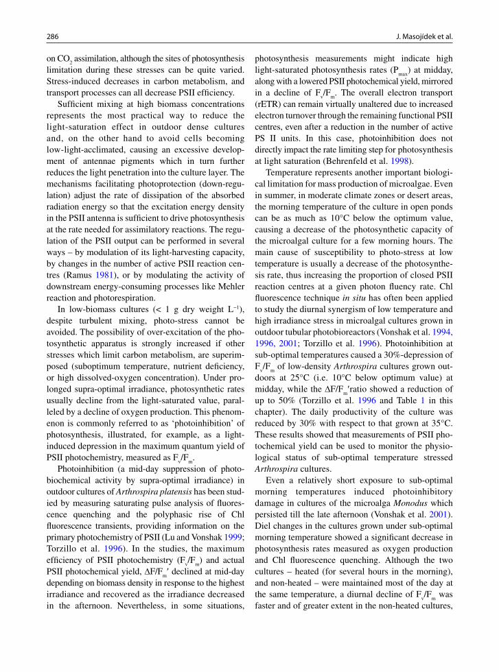
Table 2 Selected parameters calculated from the time-course of fluorescence induction curves (Fig. 2a). F
0, F
J, F
I – fluorescence intensity at O-step (0.05 ms),
at J-step (~2 ms) and I-step (~20 ms) of fluorescence induction curve
Parameter Symbol Formula
Fluorescence yield at J-step VJ
VJ = (F
J – F
0)/(F
m – F
0)
Fluorescence yield at I-step VI
VI = (F
J – F
0)/(F
m – F
0)
Maximum photochemical yield of PSII Fv/F
mF
v/F
m = (F
m – F
0)/F
m
Table 1 Effect of sub-optimum temperature and high dissolved oxygen concentration on relative electron transport rate rETR and biomass productivity in cultures of the cyanobacterium Arthrospira grown in outdoor photobioreactors (Torzillo et al. 1996, 1998)
Temperature Dissolved oxygen concentration rETR (DF/Fm¢ × PAR) Biomass productivity
[°C] [mg L−1] [mmol e− m−2 d−1] [%] [g DW m−2 d−1] [%]
35 22 ± 2 11100 100 29.0 10035 60 ± 19 7500 −33 19.4 −3325 58 ± 16 4300 −60 12.0 −61
Fig. 4 In-situ measurement of Chl fluorescence quenching using a fibre-optic guide and a pulse-amplitude-modulation flu-orometer in a greenhouse tubular photobioreactor (panel a). The fibre-optics were protected in a glass test tube and submerged in
the microalgal suspension (panel b), or placed perpendicularly to the glass wall of the cultivation tube (panel c). Distance between the fibre-optics and the culture suspension was about 3 mm and the fibre-optics angle to the sun was about 60°

28513 Chlorophyll Fluorescence Applications in Microalgal Mass Cultures
1990; Gilmore and Yamamoto 1991). Stress-induced damage of the photosynthetic apparatus is often reflected by an increase of NPQ which can compensate for a decrease of F
PSII. Compared to the qP and qN
coefficients, the FPSII
and NPQ calculation does not need the determination of F
0¢ which might be problem-
atic in the field.A relative estimate of the electron transport rate
through PSII can be obtained as the product of the actual photochemical yield of PSII and photosynthetic photon flux density, rETR = DF/F
m¢ × PPFD expressed
in mol photons m−2 s−1 (Hofstraat et al. 1994; Maxwell and Johnson 2000; Schreiber 2004). For example, pos-sible cyclic electron transport would cause deviations (Prášil et al. 1996). To translate rETR into ‘absolute’ ETR requires that light absorption specific cross sec-tion of PSII, termed a
PSII, which is often not available;
in such cases relative electron transfer rates rETR are frequently reported (Suggett et al., Chapter 6, this volume). Our experiments with Spirulina cultures cul-tivated under unfavourable conditions of sub-optimum temperature and high dissolved oxygen concentration showed a good correlation between daily sum of rETR and biomass productivity since the trends of these two parameters were similar showing the same percentage of decrease (Table 1).
Recently, saturation-pulse expressions have been updated in order to point out new parameters – Y(NO) = F¢/F
m and Y(NPQ) = F¢/F
m¢ - F¢/F
m (Klughammer
and Schreiber 2008). The validity and usefulness of the last two parameters have to be tested yet in outdoor microalgal mass cultures.
Rapid light-response curves of chlorophyll fluores-cence and photosynthetic oxygen production might be simultaneously measured in microalgal cultures at various light intensities, similar to that in phytoplank-ton populations, in a flow-through regime in a closed chamber (Masojídek et al. 2000, 2001). This can pro-vide information about the acclimation status of the photosynthetic apparatus of microalgal mass cultures and the relationship between the electron transport rate through PSII, respiration and photosynthetic oxy-gen evolution over a diel cycle, and help explain the involvement of alternate oxygen-consuming electron transfer pathways as a possible explanation for some discrepancies. Although fluorescence parameters can be measured easily, some problems might arise when they are used to predict changes in photosynthetic per-formance. In particular, questions associated with the
accurate estimation of PSII efficiency and the roles of photochemical and non-photochemical quenching as measured by fluorescence and its relationship with the rates of linear electron flux and CO
2 assimilation have
to be carefully judged.
6 Chlorophyll Fluorescence Monitoring in Microalgal Mass Cultures
Outdoor dense microalgal cultures may experience large daily variations in light intensity – in the range of one order of magnitude. Outdoor microalgal cultures in cultivation units are usually influenced by the different time-scales of light-dark regimes (Richmond 2004). The first one represents a fast, intermittent light-dark regime which is induced by turbulence of microalgal suspension. As a result, the cells in dense cultures can be shifted between full sunlight, when they are situated in the upper layer, and complete darkness, when they are at the bottom of the culture. The second type of light regime is usually directed by the circulation between the cultivation loop and degasser (dark volume) in tens of seconds to minutes. Thirdly, the slowest light-dark changes are related to diurnal changes in solar radiation.
In most cases, the photosynthetic activity of microalgae becomes saturated within 200 μmol photons m−2 s−1, that is 10% of the maximum solar irradiance. Photosynthetic activity at sub-saturating irradiance is rate-limited by light absorption and excitation energy transfer to the PSII reaction centres. Dense microalgal mass cultures are therefore predominantly grown at light limitation and, consequently, their photosynthetic performance would be more dependent on the lineal part of the P/E curve rather than on the light-saturated part (Vonshak and Torzillo 2004). It means that in a dense culture of microalgae the incident light intensity on the surface that penetrates to greater depths is uti-lized with maximum efficiency because it falls within the limited region of the growth curve.
Conversely, at over-saturating light intensities (in optically thin, low-biomass cultures), photosynthesis is limited by the interplay between electron-transfer processes and the capacity of enzymatic processes in the Calvin-Benson cycle (Sukenik et al. 1987). The rate of consumption of NADPH and ATP are major factors that determine PSII operating efficiency in many situations. Numerous environmental stresses impact
286 J. Masojídek et al.
on CO2 assimilation, although the sites of photosynthesis
limitation during these stresses can be quite varied. Stress-induced decreases in carbon metabolism, and transport processes can all decrease PSII efficiency.
Sufficient mixing at high biomass concentrations represents the most practical way to reduce the light-saturation effect in outdoor dense cultures and, on the other hand to avoid cells becoming low-light-acclimated, causing an excessive develop-ment of antennae pigments which in turn further reduces the light penetration into the culture layer. The mechanisms facilitating photoprotection (down-regu-lation) adjust the rate of dissipation of the absorbed radiation energy so that the excitation energy density in the PSII antenna is sufficient to drive photosynthesis at the rate needed for assimilatory reactions. The regu-lation of the PSII output can be performed in several ways – by modulation of its light-harvesting capacity, by changes in the number of active PSII reaction cen-tres (Ramus 1981), or by modulating the activity of downstream energy-consuming processes like Mehler reaction and photorespiration.
In low-biomass cultures (< 1 g dry weight L−1), despite turbulent mixing, photo-stress cannot be avoided. The possibility of over-excitation of the pho-tosynthetic apparatus is strongly increased if other stresses which limit carbon metabolism, are superim-posed (suboptimum temperature, nutrient deficiency, or high dissolved-oxygen concentration). Under pro-longed supra-optimal irradiance, photosynthetic rates usually decline from the light-saturated value, paral-leled by a decline of oxygen production. This phenom-enon is commonly referred to as ‘photoinhibition’ of photosynthesis, illustrated, for example, as a light-induced depression in the maximum quantum yield of PSII photochemistry, measured as F
v/F
m.
Photoinhibition (a mid-day suppression of photo-biochemical activity by supra-optimal irradiance) in outdoor cultures of Arthrospira platensis has been stud-ied by measuring saturating pulse analysis of fluores-cence quenching and the polyphasic rise of Chl fluorescence transients, providing information on the primary photochemistry of PSII (Lu and Vonshak 1999; Torzillo et al. 1996). In the studies, the maximum efficiency of PSII photochemistry (F
v/F
m) and actual
PSII photochemical yield, DF/Fm¢ declined at mid-day
depending on biomass density in response to the highest irradiance and recovered as the irradiance decreased in the afternoon. Nevertheless, in some situations,
photosynthesis measurements might indicate high light-saturated photosynthesis rates (P
max) at midday,
along with a lowered PSII photochemical yield, mirrored in a decline of F
v/F
m. The overall electron transport
(rETR) can remain virtually unaltered due to increased electron turnover through the remaining functional PSII centres, even after a reduction in the number of active PS II units. In this case, photoinhibition does not directly impact the rate limiting step for photosynthesis at light saturation (Behrenfeld et al. 1998).
Temperature represents another important biologi-cal limitation for mass production of microalgae. Even in summer, in moderate climate zones or desert areas, the morning temperature of the culture in open ponds can be as much as 10°C below the optimum value, causing a decrease of the photosynthetic capacity of the microalgal culture for a few morning hours. The main cause of susceptibility to photo-stress at low temperature is usually a decrease of the photosynthe-sis rate, thus increasing the proportion of closed PSII reaction centres at a given photon fluency rate. Chl fluorescence technique in situ has often been applied to study the diurnal synergism of low temperature and high irradiance stress in microalgal cultures grown in outdoor tubular photobioreactors (Vonshak et al. 1994, 1996, 2001; Torzillo et al. 1996). Photoinhibition at sub-optimal temperatures caused a 30%-depression of F
v/F
m of low-density Arthrospira cultures grown out-
doors at 25°C (i.e. 10°C below optimum value) at midday, while the DF/F
m¢ratio showed a reduction of
up to 50% (Torzillo et al. 1996 and Table 1 in this chapter). The daily productivity of the culture was reduced by 30% with respect to that grown at 35°C. These results showed that measurements of PSII pho-tochemical yield can be used to monitor the physio-logical status of sub-optimal temperature stressed Arthrospira cultures.
Even a relatively short exposure to sub-optimal morning temperatures induced photoinhibitory damage in cultures of the microalga Monodus which persisted till the late afternoon (Vonshak et al. 2001). Diel changes in the cultures grown under sub-optimal morning temperature showed a significant decrease in photosynthesis rates measured as oxygen production and Chl fluorescence quenching. Although the two cultures – heated (for several hours in the morning), and non-heated – were maintained most of the day at the same temperature, a diurnal decline of F
v/F
m was
faster and of greater extent in the non-heated cultures,

28713 Chlorophyll Fluorescence Applications in Microalgal Mass Cultures
reaching a midday value of 0.48, compared to 0.58 in the heated culture. The differences in photosynthetic activity between the two cultures were also reflected in bio-mass productivity which was much higher in the heated culture (by 60%) compared to the non-heated culture.
Another important environmental factor affecting microalgal cultivation represents the concentration of oxygen dissolved in the suspension, particularly in closed photobioreactors. The saturating pulse fluorescence technique has been applied to study the photoinhibition of photosynthesis in outdoor cultures of Arthrospira grown under high oxygen and/or low temperature stress in closed outdoor photobioreactors (Torzillo et al. 1998). Diurnal changes showed that when solar irradiance reached the maximum value (between 1200 and 1300 h), the maximum photochemical quantum yield of PSII in dark-adapted state, F
v/F
m and the actual photochemical
quantum yield of PSII in light-adapted state DF/Fm¢ ratios
of the Arthrospira cultures grown under high oxygen stress decreased by 35% and 60%, respectively, com-pared with the morning values. When high oxygen stress was combined with sub-optimum temperature, F
v/F
m and
DF/Fm¢ dropped even more, by 55% and 84%, respec-
tively. Photoinhibition reduced the daily productivity of the culture grown under high oxygen stress by 33%, and that of the culture grown under high oxygen-low tem-perature stress by about 60%. Changes in the biomass yield of the cultures correlated well with changes in the estimated relative electron transport rate through the PSII complex, rETR (Table 1).
The influence of unfavourable conditions (stres-sors) such as high temperature and high pH in combi-nation with high irradiance were studied in outdoor cultures of the microalga Nannochloropsis in two out-door production systems – flat panel photobioreactors and raceway ponds (Sukenik et al. 2009). The mea-surements of the Nannochloropsis photosynthetic activity using several chlorophyll fluorescence tech-niques as well as oxygen production showed that this species was able to withstand high irradiance levels. Nannochloropsis coped well with high pH conditions under physiological temperatures. However, a temper-ature rise above 32°C was detrimental and the repair processes could not keep up with the rate of damage.
As an example we present measurements of Chl fluorescence parameters related to biomass productiv-ity in outdoor Chlorella cultures of various biomass densities (Fig. 5) in outdoor thin-layer sloping cascades
0.2
0.4
0.6
0.8a
b
c
d
Fv/
Fm
[ r.
u. ]
0.2
0.4
0.6
0.8
∆F/F
m' [
r.u
. ]
Daytime [ h ]8 10 12 14 16 18
NP
Q [
r.u.
]
0.0
0.5
1.0
1.5
2.0
2.5
Biomass density [ g L−1 ]1.2-4 2.5-6.5 6.5-13.5 35-40
Pro
duct
ivity
[ g
L−1 d
−1]
0
2
4
6
2.5-6.5 g L−11.2-4 g L−1
35-40 g L−16.5-13.5 g L−1
Fig. 5 Diurnal changes in the maximum PSII photochemical yield F
v/F
m (panel a), the actual photochemical yield DF/F
m¢
(panel b), non-photochemical quenching NPQ (panel c) in correlation with biomass productivity (panel d) in Chlorella mass cultures grown at different biomass concentrations (1.2–4, 2.5–6.5, 6.5–13.5 and 35–40 g L−1) in outdoor thin-layer cascade units

288 J. Masojídek et al.
(Fig. 1). Diel changes of the Chl fluorescence parame-ters F
v/F
m, DF/F
m¢ and NPQ, suggested the interplay of
cell density vs. irradiance which resulted in correlation between the diel course of variable fluorescence param-eters and daily biomass productivity (Fig. 5a–c). In low-biomass cultures, the photochemical yield decreased significantly in the morning which was bal-anced by the NPQ increase. The results of several experiments demonstrated that a decrease of PSII pho-tochemical yield (F
v/F
m or DF/F
m¢) of about 20% at
mid-day maximum irradiance can be considered physi-ological and still compatible with high productivity (Fig. 5 in this chapter; Torzillo et al. 1996; Richmond 2000, 2004; Masojídek et al. 2003). If the mid-day val-ues of F
v/F
m or DF/F
m¢ were by 20% lower or higher
than in the morning, the cultures become either photo-inhibited or photo-limited which in both cases decreases productivity.
Similarly as biomass productivity, fluorescence measurements might indicate the induction of secondary carotenoid synthesis. The experiments with the Haematococcus cultures exposed to supra-high irradi-ance in a photobioreactor with solar concentrators showed that higher decrease of F
v/F
m and DF/F
m¢ were
counteracted by a corresponding increase of NPQ and these changes indicated faster induction of astaxanthin synthesis as compared to the ambient irradiance intensity (Masojídek et al. 2009).
7 Light Adaptation – Non-photochemical Fluorescence Quenching
Photosynthetic organisms can be exposed to rapid changes of irradiance, often in synergism with other unfavourable environmental conditions. To minimise
photoinhibition, photosynthetic organisms have evolved photoprotective mechanisms designated as short- and long-term responses (Krause 1988). These mechanisms serve to balance and optimise the light and dark photo-synthetic reactions and to preserve the functioning of the photosynthetic apparatus. The relative extent of the energy dissipation is usually quantified using the so-called non-photochemical quenching parameter NPQ (Table 3). Under most conditions, the major part of NPQ is high-energy-state dependent quenching (referred often as qE; Cosgrove and Borowitzka, this volume) and it is thought to be essential in protecting plant leaves and microalgae from photo-induced damage. Two mod-els were proposed, depending on whether the quenching is assumed to be associated with the PSII reaction centre or with the antenna. In the latter model, the process in the antennae involves the light-induced formation of the carotenoid zeaxanthin (Demmig-Adams 1990). Light dependent conversion of violaxanthin to zeaxanthin via the intermediate antheraxanthin, the so-called xan-thophyll cycle is supposed to serve as a major, short-term, light-acclimation mechanism of NPQ in higher plants. It is reversible when leaf or microalgal cell is darkened and qE relaxes within minutes. The role of xanthophylls in the thermal dissipation of surplus exci-tation energy was deduced from the linear relationship between zeaxanthin formation and the magnitude of non-photochemical quenching (Demmig et al. 1987). Unlike in higher plants, the role of the xanthophyll cycle in green algae (Chlorophyta) is ambiguous since its contribution to energy dissipation can signifi-cantly vary among species (Casper-Lindley and Björkman 1998; Masojídek et al. 1999; Jin et al. 2003). In our experiments, the xanthophyll cycle was found to be functional in various green microalgae (Chlorella, Scenedesmus, Haematococcus, Chloro-coccum, Spongiochloris); however its contribution to
Table 3 Selected parameters calculated from saturation pulse method in modulated fluorometers (Fig. 2). F
0, F
v, F
m – minimum,
variable and maximum fluorescence in dark-adapted state; F0¢,
F¢, Fv¢, F
m¢ – minimum, steady-state, variable and maximum
fluorescence in light-adapted state; PPFD – photosynthetic photon flux density. The nomenclature used is according to van Kooten and Snel (1990) – see also Cogrove and Borowitzka (Chapter 1, this volume)
Parameter Symbol Formula
Maximum photochemical yield of PSII Fv/F
mF
v/F
m = (F
m – F
0)/ F
m
Actual PSII photochemical yield FPSII
or DF/ Fm¢ F
PSII = (F
m¢ – F¢)/ F
m¢
Relative electron transport rate through PSII (rate of photochemistry) rETR rETR = FPSII
× PPFDStern-Volmer coefficient of non-photochemical quenching NPQ NPQ = (F
m–F
m¢)/F
m¢
Photochemical quenching qP qP= (Fm¢–F¢)/(F
m¢–F
0¢)
Non-photochemical quenching qN qN= (Fv–F
v¢)/F
v
28913 Chlorophyll Fluorescence Applications in Microalgal Mass Cultures
non-photochemical quenching is not as significant as in higher plants, or can vary among species (Masojídek et al. 2004b). This conclusion is supported by two facts: (i) in green algae the content of zeaxanthin nor-malized per chlorophyll was significantly lower than that reported from higher plants; and (ii) the antherax-anthin + zeaxanthin content displayed different diel kinetics than NPQ. We assume that microalgae rely on other dissipation mechanism(s), which operate along with the xanthophyll cycle-dependent quenching. In microalgae, xanthophylls probably have a preferential role as antioxidants.
In the other model, the qE quenching is located in the PSII reaction centre and is not accompanied by zeaxanthin synthesis. The quenching is associated with a reversible inactivation (quenched state) of a certain fraction of the reaction centres which is prob-ably caused by the transient over-acidification of the thylakoid lumen. Both the fluorescence quenching and PSII inactivation relax in parallel with the acti-vation of the Calvin-Benson cycle (Finazzi et al. 2004).
The second type of non-photochemical quenching, important in microalgae, represents the so-called state transition quenching (qT). This process is induced by changes in the redox state of the plastoquinone pool causing the reversible phosphorylation of antennae proteins which regulate the redistribution of light energy between PSI and PSII (Wollman 2001). The qE and qT fade out in minutes after dark-adaptation.
Processes that relax over a longer-scale (hours) are usually referred to as ‘photoinhibition’ (qI). Applied to Chl fluorescence analysis, this term generally refers to the protective processes occurring in the light-harvesting antenna as well as destructive processes in the PSII reaction centres.
Cyanobacteria which lack the xanthophyll cycle, have a significant Mehler reaction activity (O
2 uptake
by the reducing side of PSI) at light saturation which acts as a sink for electrons when PSII activity exceeds photosynthetic capacity. Recently, a photoprotective mechanism related to quenching in phycobilisomes has also been found in cyanobacteria (Kirilovsky 2007). In this mechanism, the soluble carotenoid-binding protein plays an essential role where the associated carote-noids vary among the cyanobacteria, e.g. zeaxanthin in Anacystis and Lyngbya or 3¢-hydroxyechinenone in Synechocystis and Arthrospira.
8 Major Achievements in Microalgal Mass Culture Monitoring
Since the mid-1990s, Chl fluorescence has become one of the most feasible and useful techniques in microalgal biotechnology for monitoring the photo-synthetic characteristics of a culture and subsequently estimating its biomass productivity. Results have indicated that the Chl fluorescence technique, when used in situ, is a useful tool for an immediate assess-ment of the fitness of outdoor microalgal mass cul-tures. In this way, we can elucidate the effect of changing environmental factors on the physiology of outdoor microalgal cultures. Chl fluorescence also makes it possible to control microalgal cultivation, using on-line monitoring of photobiochemical activi-ties to photo-optimise the cultivation regime (e.g. bio-mass density, turbulence, CO
2 supply). The analysis
of fast Chl fluorescence induction kinetics is used to determine the limiting photochemical processes at the molecular level.
Particularly useful photochemical expressions have been derived from analysis of Chl fluorescence quench-ing: maximal PS II quantum yield in the dark-adapted sample F
v/F
m, the effective PSII quantum yield of illu-
minated samples, DF/Fm¢, and relative electron trans-
port rate rETR, to make the correlation with biomass productivity (Torzillo et al. 1998), or eventually indi-cate bioactive compound occurrence, e.g. secondary carotenoids (Torzillo et al. 2003; Masojídek et al. 2009). Although fluorescence parameters can be measured easily, some caution is necessary to correlate them with the rates of linear electron flux and CO
2 assimilation.
9 Concluding Remarks
The increased interest in microalgal biotechnology, aimed to the production of biomass, high-value prod-ucts, or even bio-fuels, has prompted the application of on-line measurements for monitoring growth and obtaining rapid evidence of unfavourable conditions affecting the performance of outdoor cultures. For these applications the use of modulated fluorometers, which enable the actual photochemical quantum yield to be measured at a given light intensity during the day, is mandatory.

290 J. Masojídek et al.
Finally, some recommendations are necessary. (i) Though Chl fluorescence represents a rapid technique for stress detection in plants and microalgal cultures, it must always be accompanied by other physiological measurements. (ii) Indeed, it is relatively easy to gen-erate fluorescence data, thus care must always be taken to select and calculate sensible parameters. This is par-ticularly true when dealing with microalgal cultures outdoors, where growth limitations, such as light, tem-perature and other unfavourable factors can occur side by side. As long as this is kept in mind, Chl fluores-cence represents a powerful technique which allows rapid monitoring of physiological status, the use of which has been steadily increasing in both the labora-tory and field studies of microalgal cultures.
Acknowledgement The authors thank Mr. Pavel Souček for preparation of diagrams and Mr. Steve Ridgill for language corrections.
The Ministry of Education, Youth and Sports and the Czech Academy of Sciences supported this work through the project MSM6007665808 and AVOZ 50200510. Partial funding was also provided by project 522/06/1090 and 521/09/0656 of the Czech Science Foundation, and by project IAA608170601 of the Grant Agency of the Czech Academy of Sciences.
References
Baker NR (2008) Chlorophyll Fluorescence: a probe of photo-synthesis in vivo. Annu Rev Plant Biol 59:89–113
Behrenfeld MJ, Prasil O, Kolber ZS, Babin M, Falkowski PG (1998) Compensatory changes in photosystem II electron turnover rates protect photosynthesis from photoinhibition. Photosynth Res 58:259–268
Ben-Amotz A (2004) Industrial production of microalgal cell-mass and secondary products – Major industrial species: Dunaliella. In: Richmond A (ed) Handbook of microalgal mass cultures. Blackwell Science, Oxford, pp 273–280
Benemann JR, Oswald WJ (1996) Systems and economic analy-sis of microalgae ponds for conversion of CO
2 to biomass.
Final report. US DOE http://www.osti.gov/bridge/servlets/purl/493389-FXQyZ2/webviewable/493389.pdf
Bilger W, Björkman O (1990) Role of xanthophyll in photopro-tection elucidated by measurement of light-induced absor-bance changes, fluorescence and photosynthesis in leaves of Hedera canariensis. Photosynth Res 25:173–185
Boardman NK (1980) Energy from the biological conversion of solar energy. Phil Trans R Soc London A 295:477–489
Borowitzka MA (2005) Carotenoid production using microorgan-isms. In: Cohen Z, Ratledge C (eds) Single Cell Oils. AOCS Press, Champaign, IL, pp 124–137
Bradbury M, Baker NR (1984) A quantitative determination of photochemical and nonphotochemical quenching during the
slow phase of the chlorophyll fluorescence induction curve of bean leaves. Biochim Biophys Acta 765:275–81
Büchel C, Wilhelm C (1993) In vivo analysis of slow chloro-phyll fluorescence induction kinetics in algae: progress prob-lems and perspectives. Photochem Photobiol 58:137–148
Casper-Lindley C, Björkman O (1998) Fluorescence quenching in four unicellular algae with different light-harvesting and xanthophyll-cycle pigments. Photosynth Res 56:277–289
Demmig B, Winter K, Krüger A, Czygan FC (1987) Photoinhibition and zeaxanthin formation in intact leaves. Plant Physiol 84:218–224
Demmig-Adams B (1990) Carotenoids and photoprotection in plants. A role for the xanthophyll zeaxanthin. Biochim Biophys Acta 1020:1–24
Diner BA (1998) Photosynthesis: molecular biology of energy capture. Methods Enzymol 297:337–360
Finazzi G, Johnson GN, Dall’Osto L, Joliot P, Wollman F-A, Bassi R (2004) A zeaxanthin-independent nonphotochemi-cal quenching mechanism localized in the photosystem II core complex. Proc Nat Acad Sci USA 101:12375–12380
Genty B, Briantais JM, Baker NR (1989) The relationship between the quantum yield of photosynthetic electron trans-port and quenching of chlorophyll fluorescence. Biochim Biophys Acta 990:87–92
Gilmore AM, Yamamoto HY (1991) Zeaxanthin formation and energy dependent fluorescence quenching in pea chloroplasts under artificially mediated linear and cyclic electron trans-port. Plant Physiol 96:635–643
Govindjee (1995) Sixty-three years since Kautsky: chlorophyll a fluorescence. Aust J Plant Physiol 22:131–160
Grobbelaar JU (2007) Photosynthetic characteristics of Spirulina plat-ensis grown in commercial-scale open outdoor raceway ponds: what do the organisms tell us? J Appl Phycol 19:591–598
Hofstraat JW, Peeters JCH, Snel JFH, Geel C (1994) Simple determination of photosynthetic efficiency and photoinhibi-tion of Dunaliella tertiolecta by saturating pulse fluores-cence measurements. Mar Ecol Prog Ser 103:187–196
Jin ES, Yokthongwattana K, Polle JEW, Melis A (2003) Role of the reversible xanthophylls cycle in the Photosystem II dam-age and repair cycle in Dunaliella salina. Plant Physiol 132:352–364
Kitajima M, Butler WL (1975) Quenching of chlorophyll fluores-cence and primary photochemistry in chloroplasts by dibro-mothymoquinone. Biochim Biophys Acta 376:105–115
Klughammer C, Schreiber U (2008) Complementary PS II quan-tum yields calculated from simple fluorescence parameters measured by PAM fluorometry and the saturation pulse method. PAM Application Notes 1: 27–35. http://www.walz.com/e_journal/pdfs/PAN078007.pdf
Krause GH (1988) Photoinhibition of photosynthesis. An evaluation of damaging and protecting mechanisms. Physiol Plant 74: 566–574
Kirilovsky D (2007) Photoprotection in cyanobacteria: the orange carotenoid protein (OCP)-related non- photochemical-quenching mechanism. Photosynth Res 93:7–16
Lu CM, Vonshak A (1999) Photoinhibition in outdoor Spirulina platensis cultures assessed by polyphasic chloro-phyll fluorescence transients. J Appl Phycol 11:355–359
Masojídek J, Torzillo G, Koblížek M, Kopecký J, Bernardini P, Sacchi A, Komenda J (1999) Photoadaptation of two mem-bers of the Chlorophyta (Scenedesmus and Chlorella) in
29113 Chlorophyll Fluorescence Applications in Microalgal Mass Cultures
laboratory and outdoor cultures: changes in chlorophyll fluorescence quenching and the xanthophyll cycle. Planta 209:126–135
Masojídek J, Torzillo G, Kopecký J, Koblížek M, Nidiaci L, Komenda J, Lukavská A, Sacchi A (2000) Changes in chlo-rophyll fluorescence quenching and pigment composition in the green alga Chlorococcum sp. grown under nitrogen deficiency and salinity stress. J Appl Phycol 12:417–426
Masojídek J, Grobbelaar JU, Pechar L, Koblížek M (2001) Photosystem II electron transport rate and oxygen produc-tion in natural waterblooms of freshwater cyanobacteria during the diel cycle. J Plankton Res 23:57–66
Masojídek J, Sergejevová M, Rottnerová K, Jirka V, Korečko J, Kopecký J, Zaťková I, Torzillo G, Štys D (2009) A two-stage solar photobioreactor for cultivation of microalgae based on solar concentrators. J Appl Phycol 21:55–63
Masojídek J, Koblížek M, Torzillo G (2004a) Photosynthesis in microalgae. In: Richmond A (ed) Handbook of microalgal mass cultures. Blackwell Science, Oxford, pp 20–39
Masojídek J, Kopecký J, Koblížek M, Torzillo G (2004b) The xanthophyll cycle in green algae (Chlorophyta): its role in the photosynthetic apparatus. Plant Biol 6:342–349
Masojídek J, Sergejevová M, Rottnerová K, Jirka V, Korečko J, Kopecký J, Zaťková I, Torzillo G, Štys D (2009) A two-stage solar photobioreactor for cultivation of microalgae based on solar concentrators. J Appl Phycol 21:55–63
Maxwell K, Johnson GN (2000) Chlorophyll fluorescence – a practical guide. J Exp Bot 51:659–668
Neale J (1987) Algal photoinhibition and photosynthesis in the aquatic environment. In: Kyle DJ, Osmond CB, Arntzen CJ (eds) Photoinhibition. Elsevier Science, Amsterdam, pp 39–65
Nedbal L, Tichy V, Xiong F, Grobbelaar JU (1996) Microscopic green algae and cyanobacteria in high-frequency intermittent light. J Appl Phycol 8:325–333
Neubauer C, Schreiber U (1987) The polyphasic rise of chloro-phyll fluorescence upon onset of strong continuous illumina-tion. I. Saturation characteristics and partial control by the Photosystem II acceptor side. Z Naturforsch 42c:1246–1254
Pirt SJ (1986) The thermodynamic efficiency (quantum demand) and dynamics of photosynthetic growth. New Phytol 102:3–37
Prášil O, Kolber Z, Berry JA, Falkowski PG (1996) Cyclic elec-tron flow around photosystem II in vivo. Photosynth Res 48:395–410
Pulz O, Scheibenboden K, Gross W (2001) Biotechnology with cyanobacteria and microalgae. In: Reed G (ed) Special pro-cesses: biotechnology, vol 10. Wiley-VCH, Weinheim, pp 107–136
Ramus J (1981) The capture and transduction of light energy. In: Lobban CS, Wynne MJ (eds) The biology of seaweeds. Blackwell Scientific, Oxford, pp 458–92
Richmond A (2000) Microalgal biotechnology at the turn of the millenium: a personal view. J Appl Phycol 12:441–451
Richmond A (2004) Biological principles of mass cultivation. In: Richmond A (ed) Handbook of microalgal mass cultures. Blackwell Science, Oxford, pp 125–177
Schreiber U (2004) Pulse-Amplitude-Modulation (PAM) fluorom-etry and saturation pulse method: an overview. In: Papageorgiou GC, Govindjee (eds) Chlorophyll a fluorescence: a signature of photosynthesis. Springer, Dordrecht, pp 279–319
Schreiber U, Neubauer C (1987) The polyphasic rise of chloro-phyll fluorescence upon onset of strong continuous illumina-tion: II. Partial control by the photosystem II donor side and possible ways of interpretation. Z Naturforsch 42c:1255–1264
Schreiber U, Schliwa U, Bilger W (1986) Continuous recording of photochemical and nonphotochemical fluorescence quenching with a new type of modulation fluorometer. Photosynth Res 10:51–62
Schreiber U, Endo T, Mi H, Asada K (1995) Quenching analysis of chlorophyll fluorescence by the saturation pulse method: particular aspects relating to the study of eukaryotic algae and cyanobacteria. Plant Cell Physiol 36:873–882
Schreiber U, Bilger W, Hormann H, Neubauer C (1998) Chlorophyll fluorescence as a diagnostic tool: basics and some aspects of practical relevance. In: Raghavendra AS (ed) Photosynthesis: a comprehensive treatise. Cambridge University Press, Cambridge, pp 320–334
Srivastava AM, Strasser RJ, Govindjee (1999) Greening of pea leaves: parallel measurement of 77K emission spectra, OJIP chlorophyll a fluorescence transient, period four oscillation of the initial fluorescence level, delayed light emission, and P700. Photosynthetica 37:365–392
Strasser BJ, Strasser RJ (1995) Measuring fast fluorescence transients to address environmental questions: the JIP test. In: Mathis P (ed) Photosynthesis from light to biosphere, vol 5. Kluwer, Dordrecht, pp 977–980
Strasser RJ, Srivastava A, Govindjee (1995) Polyphasic chloro-phyll a fluorescence transient in plants and cyanobacteria. Photochem Photobiol 61:33–42
Strasser RJ, Tsimili-Michael M, Srivastava A (2004) Analysis of the Chlorophyll a fluorescence transient. In: Papageorgiou GC, Govindjee (eds) Chlorophyll a fluorescence: a signa-ture of photosynthesis. Springer, Dordrecht, pp 321–362
Sukenik A, Bennet J, Falkowski PG (1987) Light-saturated pho-tosynthesis – limitation by electron transport or carbon fixa-tion? Biochim Biophys Acta 891:205–215
Sukenik A, Beardall J, Kromkamp JC , Kopecký J, Masojídek J, van Bergeijk S, Gabai S, Shaham E, Yamshon A (2009) Photosynthetic performance of outdoor Nannochloropsis mass cultures to extreme environmental conditions – assess-ment by chlorophyll fluorescence techniques. Aquat Microb Ecol 56: 297–308
Ting CS, Owens TG (1992) Limitation of the pulse-modulated technique for measuring the fluorescence characteristics of algae. Plant Physiol 100:367–373
Torzillo G, Pushparaj B, Masojidek J, Vonshak A (2003). Biological constraints in algal biotechnology. Biotechnol Bioprocess Engineering, 8:338–348
Torzillo G, Accolla P, Pinzani E, Masojídek J (1996) In situ monitoring of chlorophyll fluorescence to assess the synergistic effect of low temperature and high irradiance stresses in Spirulina cultures grown outdoors in photobioreactors. J Appl Phycol 8:283–291
Torzillo G, Bernardini P, Masojídek J (1998) On-line monitoring of chlorophyll fluorescence to assess the extent of photoinhi-bition of photosynthesis induced by high oxygen concentra-tion and low temperature and its effect on the productivity of outdoor cultures of Spirulina platensis (Cyanobacteria). J Phycol 34:504–510
Torzillo G, Goksan T, Faraloni C, Kopecky J, Masojídek J (2003) Interplay between photochemical activities and pigment
292 J. Masojídek et al.
composition in an outdoor culture of Haematococcus pluvialis during the shift from the green to red stage. J Appl Phycol 15:127–136
Tredici M (2004) Mass production of microalgae: photobioreac-tors. In: Richmond A (ed) Handbook of microalgal mass cul-tures. Blackwell Science, Oxford, pp 178–214
Van Kooten O, Snel JFH (1990) The use of chlorophyll fluores-cence nomenclature in plant stress physiology. Photosynth Res 25:147–145
Vonshak A, Torzillo G (2004) Environmental stress physiology. In: Richmond A (ed) Handbook of microalgal mass cultures. Blackwell Science, Oxford, pp 57–82
Vonshak A, Torzillo G, Tomaselli L (1994) Use of chlorophyll fluorescence to estimate the effect of photoinhibition in out-door cultures of Spirulina platensis. J Appl Phycol 6:31–34
Vonshak A, Torzillo G, Accolla P, Tomaselli L (1996) Light and oxygen stress in Spirulina platensis (cyanobacteria) grown outdoors in tubular reactors. Physiol Plant 97:175–179
Vonshak A, Torzillo G, Masojídek J, Boussiba S (2001) Sub-optimal morning temperature induces photoinhibition in dense outdoor cultures of the alga Monodus subterraneus (Eustigmatophyta). Plant Cell Environ 24:1113–1118
Walker DA (2009) Biofuels, facts, fantasy, and feasibility. J Appl Phycol 21:509–517
Wollman F-A (2001) State transitions reveal the dynamics and flexibility of the photosynthetic apparatus. The EMBO Journal 20:3623–3630
Zhu X, Long SP, Ort DR (2008) Converting solar energy into crop production. Curr Opin Biotechnol 19:153–159