Chapter 12 Nervous Tissue - Student Resources Home Page · Chapter 12 Nervous Tissue. ... 1 2 3...
53
Chapter 12 Nervous Tissue
Transcript of Chapter 12 Nervous Tissue - Student Resources Home Page · Chapter 12 Nervous Tissue. ... 1 2 3...
Chapter 12
Nervous Tissue

Regulation of Homeostasis
• endocrine and nervous system both work to maintain the “internal environment” // homeostais
– endocrine system
• communicates by means of chemical messengers (hormones) secreted into to the blood // slow
– nervous system
• employs electrical (action potentials) and chemicals (neuro-transmitters) to send messages from cell to cell // fast

Regulation of Homeostasis
• nervous system carries out its task in three basic steps:
– sense organs receive information about changes in the body and the external environment, and transmits coded messages to the spinal cord and the brain
– brain and spinal cord processes this information, relates it to past experiences, and determine what response is appropriate to the circumstances
– brain and spinal cord issue commands to muscles and gland cells to carry out such a response
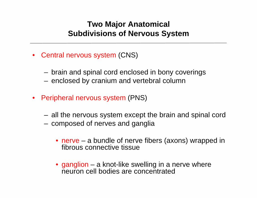
Two Major AnatomicalSubdivisions of Nervous System
• Central nervous system (CNS)
– brain and spinal cord enclosed in bony coverings– enclosed by cranium and vertebral column
• Peripheral nervous system (PNS)
– all the nervous system except the brain and spinal cord– composed of nerves and ganglia
• nerve – a bundle of nerve fibers (axons) wrapped in fibrous connective tissue
• ganglion – a knot-like swelling in a nerve where neuron cell bodies are concentrated

Subdivision of the Nervous System

Subdivisions of Nervous System

• sensory division (afferent fibers) – carries sensory signals from various receptors to the CNS
– informs the CNS of stimuli within or around the body
• somatic sensory division
– carries signals from receptors in the skin, muscles, bones, and joints
• visceral sensory division
– carries signals from the viscera of the thoracic and abdominal cavities
– heart, lungs, stomach, blood vessels, and urinary bladder
Sensory Divisions of PNS
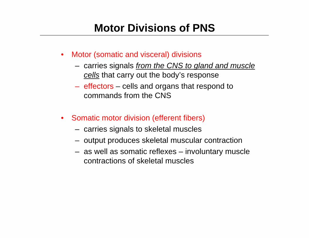
• Motor (somatic and visceral) divisions– carries signals from the CNS to gland and muscle
cells that carry out the body’s response– effectors – cells and organs that respond to
commands from the CNS
• Somatic motor division (efferent fibers)– carries signals to skeletal muscles– output produces skeletal muscular contraction – as well as somatic reflexes – involuntary muscle
contractions of skeletal muscles
Motor Divisions of PNS

• Visceral motor division (autonomic nervous system)
– carries signals to glands, cardiac muscle, and smooth muscle
– involuntary, and responses of this system and its receptors are visceral reflexes
• sympathetic division – tends to arouse body for action– accelerating heart beat and respiration, while inhibiting
digestive and urinary systems
• parasympathetic division– tends to have calming effect– slows heart rate and breathing– stimulates digestive and urinary systems
Motor Divisions of PNS

Subdivisions of Nervous System

Universal Properties of a Neuron
• excitability (irritability)– respond to environmental changes called stimuli
• conductivity– neurons respond to stimuli by producing electrical
signals that are quickly conducted to other cells at distant locations
• secretion– when electrical signal reaches end of nerve fiber,
a chemical neurotransmitter is secreted that crosses the gap and stimulates the next cell
• Note: electro-chemical communication!

Three Functional Types of Neurons
• sensory (afferent) neurons
– specialized to detect stimuli– transmit information about them to the CNS
• begin in almost every organ in the body and end in CNS• afferent – conducting signals toward CNS
• motor (efferent) neuron
– send signals out to muscles and gland cells (the effectors)• motor because most of them lead to muscles• efferent neurons conduct signals away from the CNS

Three Functional Types of Neurons
• interneurons (association) neurons
– lie entirely within the CNS
– receive signals from many neurons and carry out the integrative function
• process, store, and retrieve information and ‘make decisions’ that determine how the body will respond to stimuli
– 90% of all neurons are interneurons
– lie between, and interconnect the incoming sensory pathways, and the outgoing motor pathways of the CNS

Functional Classes of Neurons
1
23
Peripheral nervous system Central nervous system
Sensory (afferent)neurons conductsignals fromreceptors to the CNS.
Motor (efferent)neurons conductsignals from the CNSto effectors such asmuscles and glands.
Interneurons(associationneurons) areconfined tothe CNS.

• Control center of the neuron
– also called neurosoma, cell body, or perikaryon
– has a single, centrally located nucleus with large nucleolus
– cytoplasm contains mitochondria, lysosomes, a Golgi complex, numerous inclusions, and extensive rough endoplasmic reticulum and cytoskeleton
The Soma

Structure of a Neuron – The Soma
– cytoskeleton consists of dense mesh of microtubules and neurofibrils (bundles of actin filaments)
– compartmentalizes rough ER into dark staining Nisslbodies
– no centrioles – no further cell division
– Inclusions bodies – glycogen granules, lipid droplets, melanin
– Lipofuscin / inclusion bodies (golden brown pigment produced when lysosomes digest worn-out organelles)
• lipofuscin accumulates with age• wear-and-tear granules• most abundant in old neurons

• vast number of branches coming from a few thick branches from the soma
• resemble bare branches of a tree in winter
• primary site for receiving signals from other neurons
• the more dendrites the neuron has, the more information it can receive and incorporate into decision making
• provide precise pathway for the reception and processing of neural information
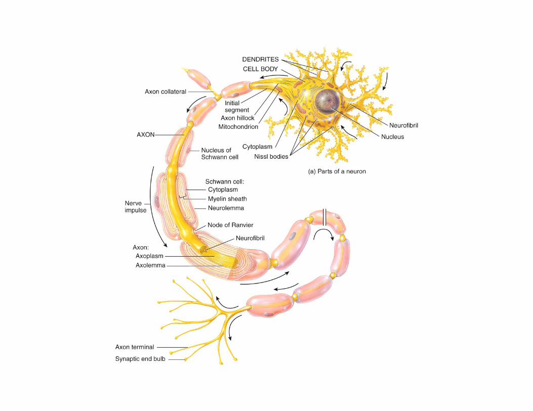
Figure 12.4a
Dendrites
SomaNucleusNucleolus
Axon
Node of Ranvier
Internodes
Synaptic knobs
Axon hillockInitial segment
Myelin sheath
Schwann cell
Axon collateral
(a)
Trigger zone:
Direction ofsignal transmission
Terminalarborization
Structure of a Neuron – The Dendrites

– Referred to as the nerve fiber
– originates from a mound on one side of the soma called the axon hillock
– cylindrical, relatively unbranchedfor most of its length
• axon collaterals – branches of axon
– branch extensively on distal end
– specialized for rapid conduction of nerve signals to points remote to the soma
– axoplasm – cytoplasm of axon Figure 12.4a
Dendrites
SomaNucleusNucleolus
Axon
Node of Ranvier
Internodes
Synaptic knobs
Axon hillockInitial segment
Myelin sheath
Schwann cell
Axon collateral
(a)
Trigger zone:
Direction ofsignal transmission
Terminalarborization
Structure of a Neuron – The Axon

– axolemma – plasma membrane of axon
– only one axon per neuron
– Schwann cells and myelin sheath enclose axon
– distal end, axon has terminal arborization – extensive complex of fine branches / like in a tree!
• synaptic knob (terminal button) – little swelling that forms a junction (synapse) with the next cell
• contains synaptic vesicles full of neurotransmitter
Structure of a Neuron – The Axon
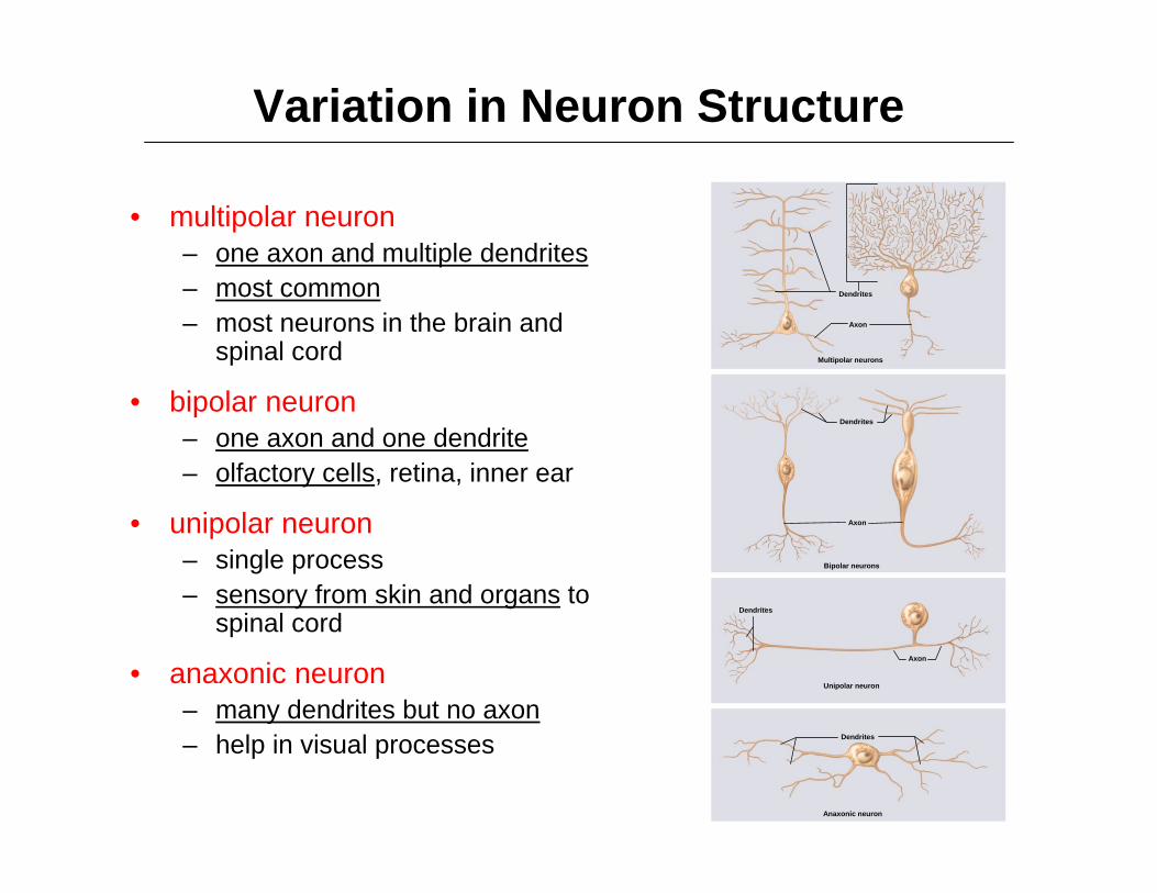
Variation in Neuron Structure
• multipolar neuron– one axon and multiple dendrites– most common– most neurons in the brain and
spinal cord
• bipolar neuron– one axon and one dendrite– olfactory cells, retina, inner ear
• unipolar neuron– single process – sensory from skin and organs to
spinal cord
• anaxonic neuron– many dendrites but no axon– help in visual processes
Dendrites
Dendrites
Dendrites
Axon
Axon
Dendrites
Axon
Unipolar neuron
Multipolar neurons
Bipolar neurons
Anaxonic neuron


Neuroglial Cells
• about a trillion (1012) neurons in the nervous system (note: approximately 100 trillion in organism!)
• neuroglia outnumber the neurons by as much as 50 to 1
• neuroglia (glial cells)
– support and protect the neurons /// bind neurons together and form framework for nervous tissue
– in fetus, guide migrating neurons to their destination
– if the surface of a mature neuron is not in synaptic contact with another neuron then non-synaptic surface is covered by glial cells
• prevents neurons from touching each other • gives precision to conduction pathways

Six Types of Neuroglial Cells (1 of 3)
• four types occur only in CNS
– oligodendrocytes• form myelin sheaths in CNS• each arm-like process wraps around a nerve fiber
forming an insulating layer that speeds up signal conduction
– ependymal cells• lines internal cavities of the brain• cuboidal epithelium with cilia on apical surface• secretes and circulates cerebrospinal fluid (CSF)
– clear liquid that bathes the CNS– microglia
• small, wandering macrophages• formed by white blood cell called monocytes• thought to perform a complete checkup on the brain
tissue several times a day• wander in search of cellular debris to phagocytize

Six Types of Neuroglial Cells (2 of 3)
– Astrocytes
• most abundant glial cell in CNS
• cover entire brain surface and most nonsynaptic regions of the neurons in the gray matter of the CNS
• diverse functions
– form a supportive framework of nervous tissue
– have extensions (perivascular feet) that contact blood capillaries that stimulate them to form a tight seal called the blood-brain barrier

Six Types of Neuroglial Cells (2 of 3)
• Astrocyte / diverse functions (cont.)
– convert blood glucose to lactate and supply this to the neurons for nourishment
– nerve growth factors secreted by astrocytes promote neuron growth and synapse formation
– communicate electrically with neurons and may influence synaptic signaling
– regulate chemical composition of tissue fluid by absorbing excess neurotransmitters and ions
– astrocytosis or sclerosis – when neuron is damaged, astrocytes form hardened scar tissue and fill space formerly occupied by the neuron

Six Types of Neuroglial Cells (3 of 3)
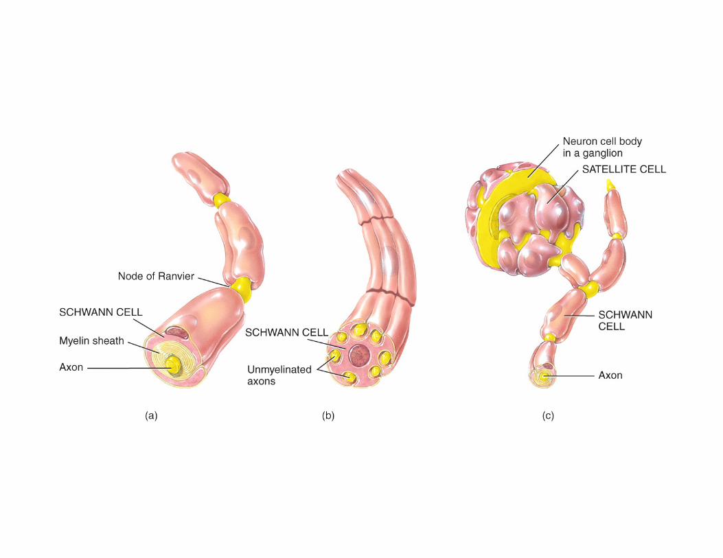
• Two types occur only in PNS
– Schwann cells• envelope nerve fibers in PNS• wind repeatedly around a nerve fiber• produces a myelin sheath similar to the ones
produced by oligodendrocytes in CNS• assist in the regeneration of damaged fibers
– Satellite cells• surround the neurosomas in ganglia of the PNS• provide electrical insulation around the soma• regulate the chemical environment of the neurons

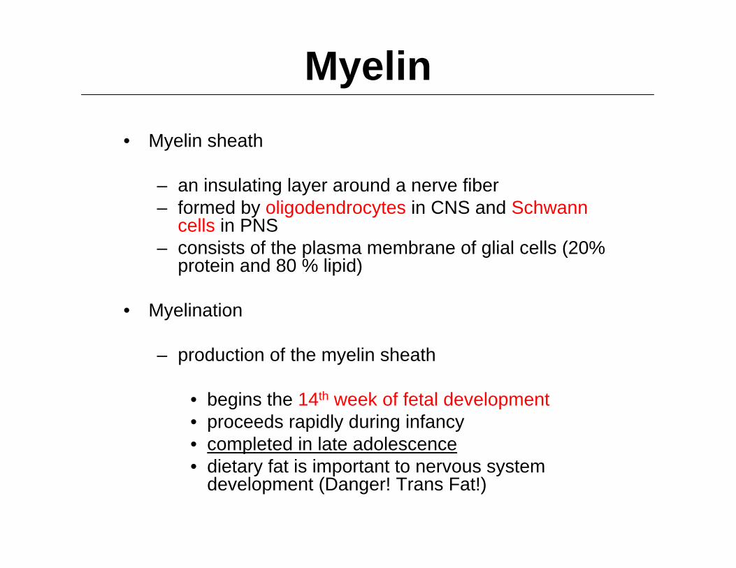
Myelin• Myelin sheath
– an insulating layer around a nerve fiber– formed by oligodendrocytes in CNS and Schwann
cells in PNS– consists of the plasma membrane of glial cells (20%
protein and 80 % lipid)
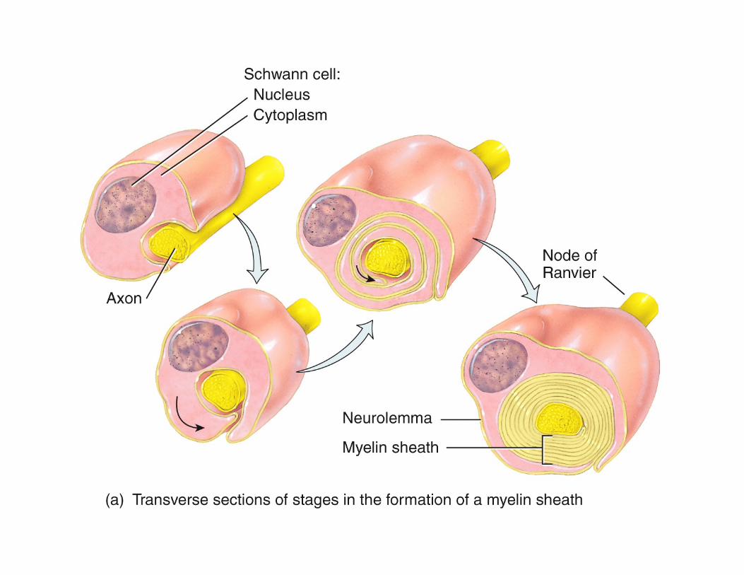
• Myelination
– production of the myelin sheath
• begins the 14th week of fetal development• proceeds rapidly during infancy• completed in late adolescence• dietary fat is important to nervous system
development (Danger! Trans Fat!)

Myelin in PNS– Schwann cell spirals repeatedly around a single
nerve fiber
– lays down as many as a hundred layers of its own membrane
– no cytoplasm between the membranes
– neurilemma – thick outermost coil of myelin sheath
• contains nucleus and most of its cytoplasm• external to neurilemma is basal lamina and a thin
layer of fibrous connective tissue – endoneurium
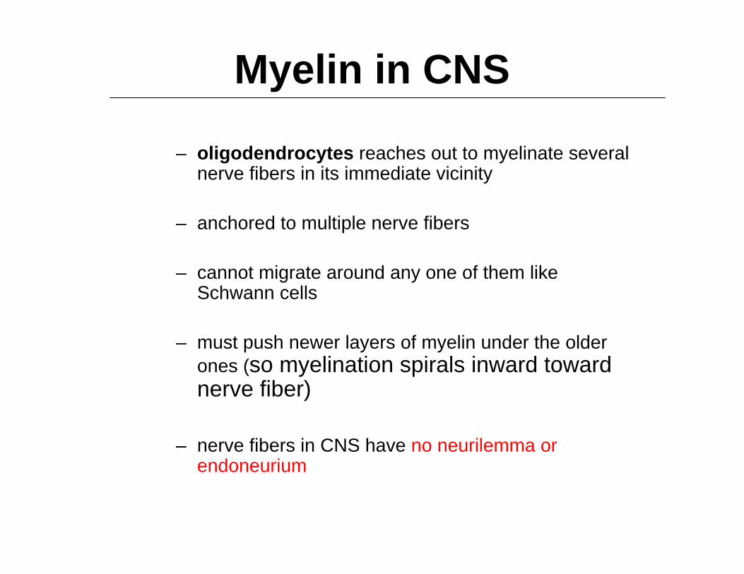
Myelin in CNS
– oligodendrocytes reaches out to myelinate several nerve fibers in its immediate vicinity
– anchored to multiple nerve fibers
– cannot migrate around any one of them like Schwann cells
– must push newer layers of myelin under the older ones (so myelination spirals inward toward nerve fiber)
– nerve fibers in CNS have no neurilemma or endoneurium

Myelin• Many Schwann cells and oligodendrocytes
are needed to cover one nerve fiber
• Myelin sheath is segmented– nodes of Ranvier – gap between segments
– internodes – myelin covered segments from one gap to the next
– initial segment – short section of nerve fiber between the axon hillock and the first glial cell
– trigger zone – the axon hillock and the first segment of neurolemma /// play an important role in initiating a nerve signal
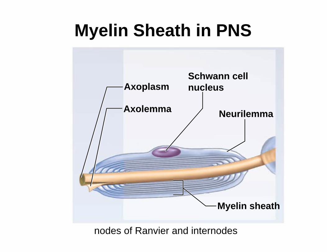
Myelin Sheath in PNS
nodes of Ranvier and internodes
Myelin sheath
Axolemma
Axoplasm
Neurilemma
Schwann cellnucleus

Myelination in CNS
Oligodendrocyte
Nerve fiber
Myelin

Myelination in PNSCopyright © The McGraw-Hill Companies, Inc. Permission required for reproduction or display.
Axon
Myelin sheath
Schwann cell
Nucleus
Basal lamina
Neurilemma
Endoneurium
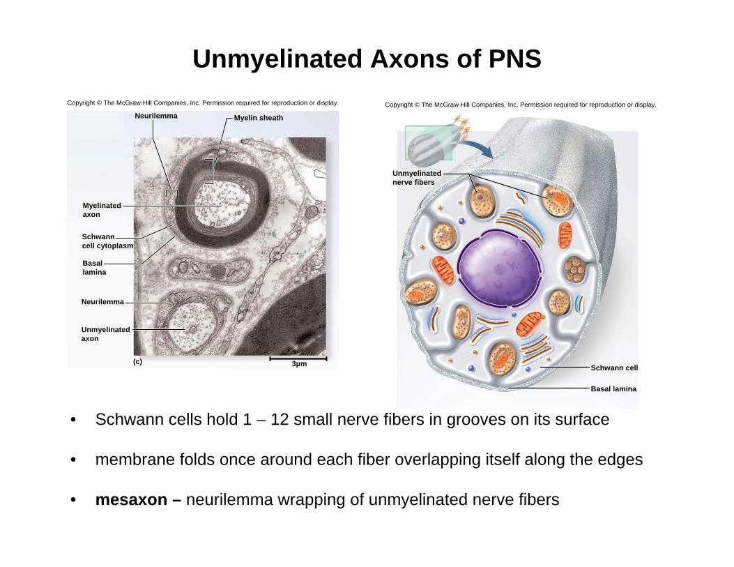
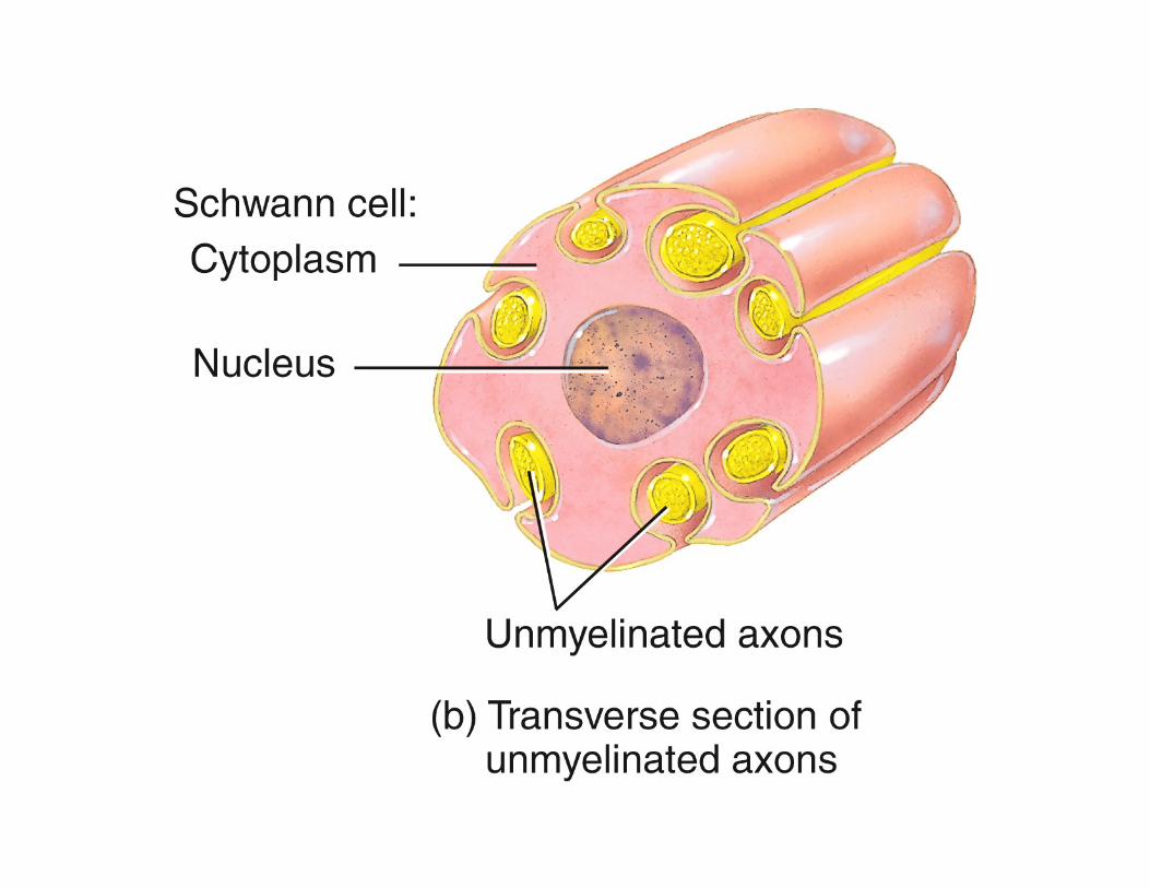
Unmyelinated Axons of PNS
• Schwann cells hold 1 – 12 small nerve fibers in grooves on its surface
• membrane folds once around each fiber overlapping itself along the edges
• mesaxon – neurilemma wrapping of unmyelinated nerve fibers
Copyright © The McGraw-Hill Companies, Inc. Permission required for reproduction or display.
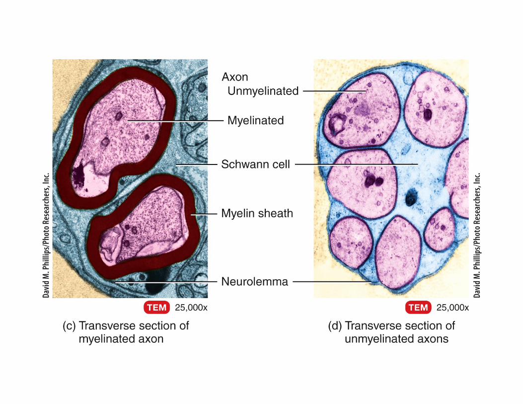
(c)
Myelin sheathNeurilemma
Neurilemma
3µm
Myelinatedaxon
Schwanncell cytoplasm
Basallamina
Unmyelinatedaxon
Copyright © The McGraw-Hill Companies, Inc. Permission required for reproduction or display.
Schwann cell
Basal lamina
Unmyelinatednerve fibers

Conduction Speed of Nerve Fibers
• speed at which a nerve signal travels along a nerve fiber depends on two factors
– diameter of fiber– presence or absence of myelin / amount of myelination– Temperature (lower speed when cooled)
• signal conduction occurs along the surface of a fiber
– larger fibers have more surface area and conduct signals more rapidly
– myelin further speeds signal conduction
– Speeds ranging from 0.5 to 130 meters per second (1 to 290 miles per hour)

Conduction Speed of Nerve Fibers
• conduction speed
– small, unmyelinated fibers - 0.5 - 2.0 m/sec
– small, myelinated fibers - 3 - 15.0 m/sec
– large, myelinated fibers - up to 120 m/sec
– slow signals supply the stomach and dilate pupil where speed is less of an issue
– fast signals supply skeletal muscles and transport sensorysignals for vision and balance

Regeneration of Peripheral Nerve Fibers (not neurons)
• regeneration of a damaged peripheral nerve fiber can occur if:
– soma is intact / not damaged– at least some of neurilemma remains
• fiber distal to the injury cannot survive and it degenerates
– macrophages clean up tissue debris at the point of injury and beyond
• soma swells, ER breaks up, and nucleus moves off center
– due to loss of nerve growth factor from neuron’s target cell

Regeneration of Peripheral Nerve Fibers (not neurons)
• axon stump sprouts multiple growth processes // severed distal end continues to degenerate
• regeneration tube – formed by Schwann cells, basal lamina, and the neurilemmanear the injury
– regeneration tube guides the growing sprout back to the original target cells /// re-establishes synaptic contact
• nucleus returns to normal shape
• Note: regeneration of damaged nerve fibers in the CNS may not occur
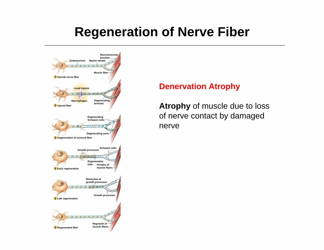
Regeneration of Nerve Fiber
Denervation Atrophy
Atrophy of muscle due to loss of nerve contact by damaged nerve
Endoneurium Myelin sheath
Local trauma
Muscle fiber
Macrophages
Growth processes
Degenerating axon
Schwann cells
Growth processes
Normal nerve fiber
Injured fiber
Degeneration of severed fiber
Early regeneration
Late regeneration
Regenerated fiber
1
2
3
4
5
6
DegeneratingSchwann cells
Neuromuscularjunction
Degeneratingterminal
Regenerationtube Atrophy of
muscle fibers
Retraction ofgrowth processes
Regrowth ofmuscle fibers

Electrophysiology of Neurons
• Santiago Ramony Cajal
• Established the neuron doctrine
– nervous pathway is not a continuous ‘wire’ or tube– Nervous pathway a series of cells separated by
gaps called synapses.
• Neuron doctrine brought up two key questions:
– how does a neuron generate a electrical signal?– how does it transmit a meaningful message to the
next cell?
Nerve Growth Factor
• nerve growth factor (NGF)
– a protein secreted by a gland, muscle, and glial cells and picked up by the axon terminals of the neurons.
– prevents apoptosis (programmed cell death) in growing neurons
– enables growing neurons to make contact with their target cells
• isolated by Rita Levi-Montalcini in 1950s
• won Nobel Prize in 1986 with Stanley Cohen
• use of growth factors is now a vibrant field of research


![12 [chapter 12 nervous tissue]](https://static.fdocuments.us/doc/165x107/5a6496047f8b9a27568b6f5d/12-chapter-12-nervous-tissue.jpg)