Chapter 10: Sensory Physiology - Las Positas...
51
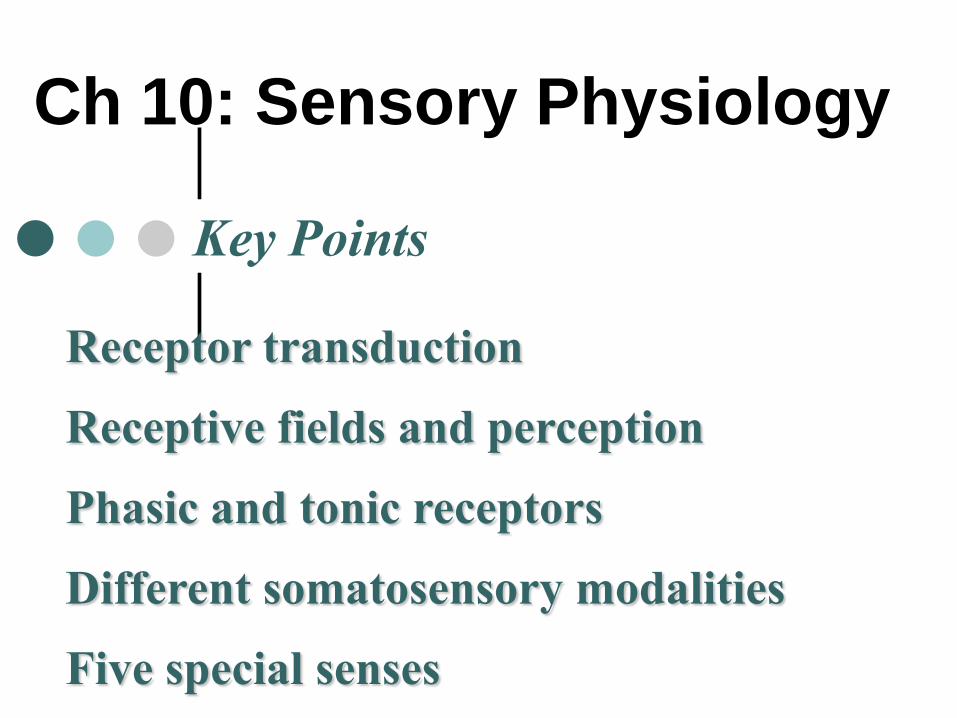
Ch 10: Sensory Physiology Receptor transduction Receptive fields and perception Phasic and tonic receptors Different somatosensory modalities Five special senses Key Points
-
Upload
nguyenlien -
Category
Documents
-
view
218 -
download
0
Transcript of Chapter 10: Sensory Physiology - Las Positas...
Ch 10: Sensory Physiology
Receptor transduction
Receptive fields and perception
Phasic and tonic receptors
Different somatosensory modalities
Five special senses
Key Points

Classification of Sensory
System by structural Complexity
Somatic (= general)
senses
1. Touch
2. Temperature
3. Nociception
4. Itch
5. Proprioception
Special senses
1.
2.
3.
4.
5.

Sensory Receptors - Overview
are transducers → convert stimuli into graded potential (receptor potential)
are of various complexity
react to particular forms of stimuli
Chemoreceptors
_____________
_____________
_____________
Fig 10-1

Sensory Transduction
Converts Stimulus into graded potential = receptor potential. How
is stimulus energy transduced?
Receptor potential in neuron above threshold AP
Receptor potential in non-neural receptors changes influence on NT release

Receptive Fields
Each 1° sensory neuron picks up
information from a receptive field
Often convergence onto 2° sensory
neuron summation of multiple stimuli
Size of receptive field determines
sensitivity to stimulus Two point
discrimination test (see lab)
Fig 10-2
Fig 10-3

Sensory
Pathway
Stimulus
Sensory receptor (= transducer)
_______________________
CNS
Integration, perception

Sensory Info
Pathways and
CNS Integration
Fig 10-4
Somato-
sensory cortex
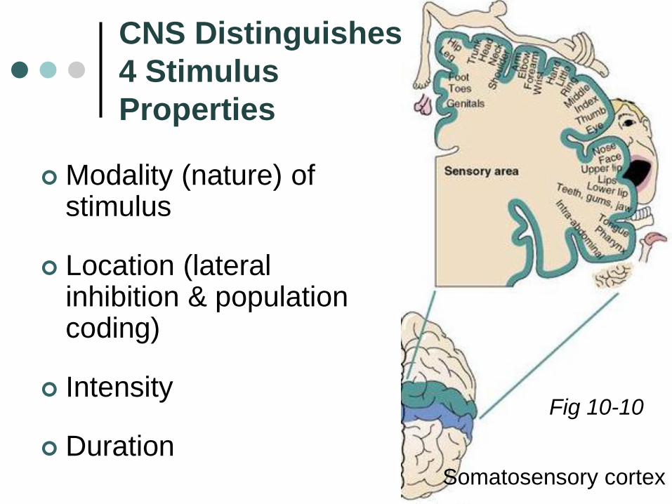
CNS Distinguishes
4 Stimulus
Properties
Modality (nature) of stimulus
Location (lateral inhibition & population coding)
Intensity
Duration
Fig 10-10
Somatosensory cortex
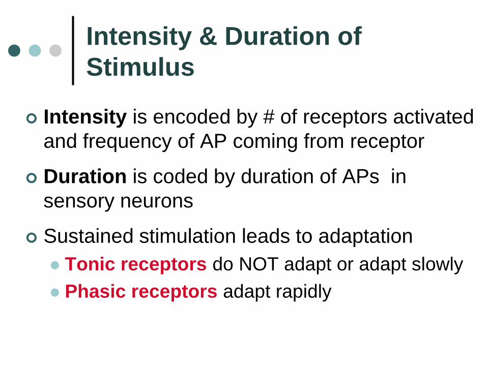
Intensity & Duration of
Stimulus
Intensity is encoded by # of receptors activated
and frequency of AP coming from receptor
Duration is coded by duration of APs in
sensory neurons
Sustained stimulation leads to adaptation
Tonic receptors do NOT adapt or adapt slowly
Phasic receptors adapt rapidly

Tonic Receptors vs. Phasic Receptors
Slow or no adaptation
Continuous signal
transmission for
duration of stimulus
Monitoring of
parameters that must
be continually
evaluated, e.g.:
barorecptors
__________ ?
Rapid adaptation
Cease firing if strength
of a continuous
stimulus remains
constant
Allow body to ignore
constant unimportant
information, e.g.:
___________?
Fig 10-8

Somatic Senses
Primary sensory neuronsfrom receptor to spinal cord or medulla
Secondary sensory neurons always cross over (in spinal cord or medulla) thalamus
Tertiary sensory neurons somatosensory cortex (post central gyrus)
Fig 10-9

Touch Receptors
Free or encapsulated dendritic endings
In skin and deep organs. e.g.:
Pacinian corpuscles
concentric layers of c.t. large receptive
field detect vibration
opens mechanically
gated ion channel
rapid adaptation
receptor type?
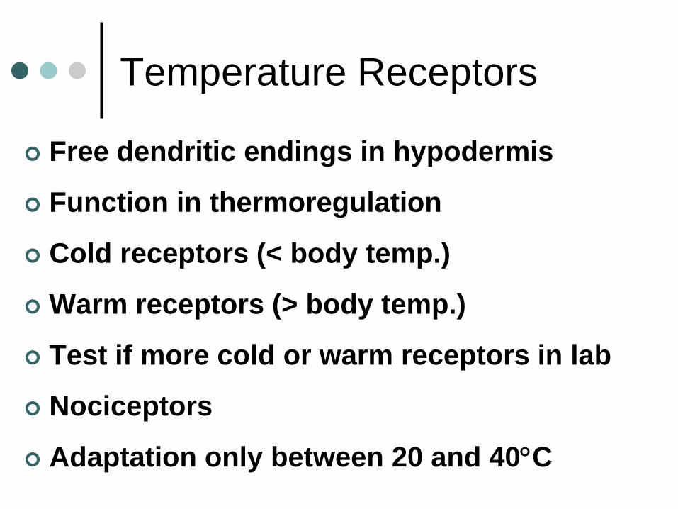
Temperature Receptors
Free dendritic endings in hypodermis
Function in thermoregulation
Cold receptors (< body temp.)
Warm receptors (> body temp.)
Test if more cold or warm receptors in lab
Nociceptors
Adaptation only between 20 and 40C

Nociceptors
Free dendritic endings
Activation by strong, noxious stimuli - Function?
3 categories: Mechanical Thermal (menthol and cold / capsaicin and hot) Chemical (includes chemicals from injured tissues)
May activate 2 different pathways: Reflexive protective – integrated in spinal cord Ascending to cortex (pain or pruritus)
Fast (A) vs. slow pain (C) (review axon diameter, myelination)
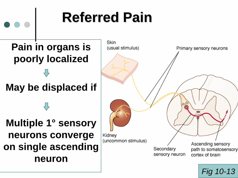
Referred Pain
Pain in organs is
poorly localized
May be displaced if
Multiple 1° sensory
neurons converge
on single ascending
neuron
Fig 10-13

Chemoreception: Smell and Taste
2 of the five special senses
Very old (bacteria use to sense environment)
Olfaction
Olfactory epithelium has 1,000 differentodorant receptors
Bipolar neurons continuously divide!
G-protein – cAMP mediated
Brain uses “population coding” to discriminate 1,000s of odors
Fig 10-14
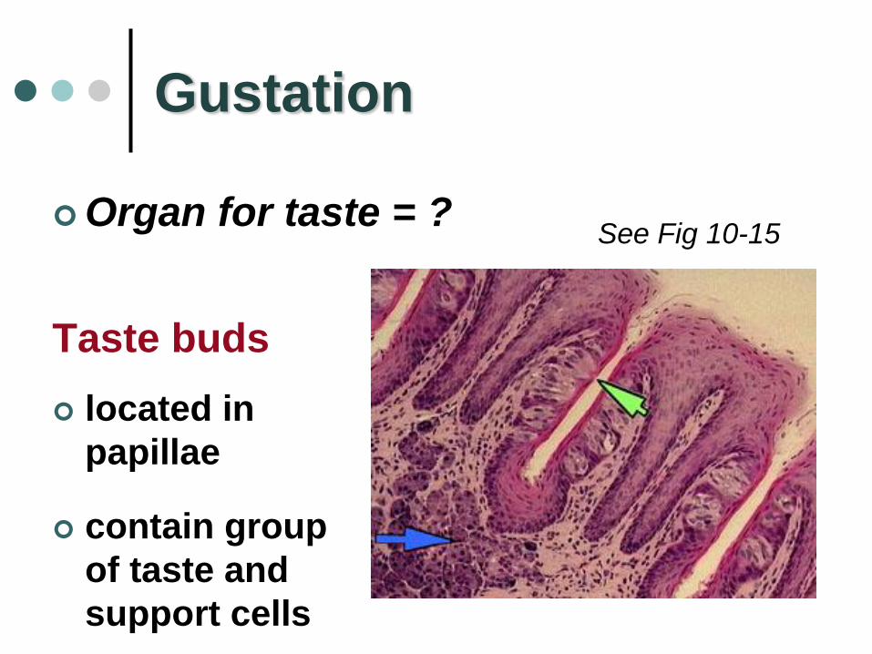
Gustation
Organ for taste = ?
Taste buds
located in
papillae
contain group
of taste and
support cells
See Fig 10-15

sour
Phasic receptors
______ adaptation!
Taste Buds: Five taste sensations
5) __________?
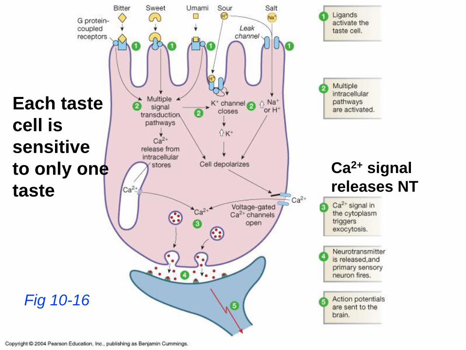
Fig 10-16
Each taste
cell is
sensitive
to only one
taste
Ca2+ signal
releases NT

Sour and Salt Ligands
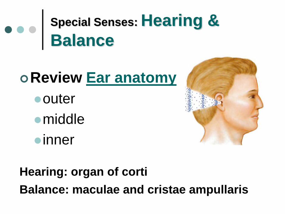
Special Senses: Hearing &
Balance
Review Ear anatomy
outer
middle
inner
Hearing: organ of corti
Balance: maculae and cristae ampullaris

Sound
transmission
Sound waves
Tympanic membrane vibrations
Ossicles transmit & amplify vibration
Via oval window to perilymph then endolymph

cont.
Vibrations in endolymph stimulate sets of receptor cells
NT release of receptor cell stimulates nearby sensory neuron
Impulse to auditory cortex of temporal lobe via ________________ nerve

Hearing Transduction
= multi-step process:
air waves mechanical vibrations fluid
waves chemical signals APs
At rest ~ 10% of
ion channels
open
More ion
channels open:
Excitation
All channels
closed:
Inhibition
Fig 10-21

Signal Transduction cont.
At restExcitiation
Inhibition
Interpretation of Sound Waves:
Pitch Perception
Sound wave frequency expressed in Hertz
(Hz) = wavelength / sec
Human can hear between 20 and 20,000 Hz
High pitch = high frequency
Low pitch = low frequency
Tone = pure sound of 1 frequency (e.g.
tuning fork)

Basilar Membrane
Pitch perception is function of basilar membrane
BM stiff near oval window
BM more flexible near distal end
Brain translates location on membrane into pitch of sound
→Temporal aspects of frequency are transformed into spatial coding
→ Spatial coding is preserved in auditory cortex
Compare to Fig 10-22

Interpretation of Sound Waves:
Loudness perception
Rate at which APs are fired loudness
Sound Intensity Measurement:
Decibel Scale (dB) starts at 0 and is logarithmic
130 dB pain threshold
> 80 dB frequently or prolonged ?
Examples: noisy restaurant ~ 70 dB rock concert ~ 120 dB

2 (3) types of Hearing Loss
Conduction deafness
External or middle ear
Many possible etiologies
Otitis media, otosclerosis etc….
Sensorineural deafness (+ central)
Damage to neural structures (from receptors to
cortical cells)
Most common: gradual loss of receptor cells

Equilibrium = State of Balance
Utricle and saccule(otolith organs) with maculae (sensory receptors) for linear acceleration and head position
Semicircular canals and ampullae with cristae ampullaris (sensory receptors) for rotational acceleration
Important besides inner ear: input from vision & stretch receptors in muscle

Otolith Organs of Maculae
Maculae and Crista ampullarisreceptors similar to organ of corti receptors
However: gravity & acceleration provide force to move stereo cilia Fig 10-25

Motion Sickness
= Equilibrium disorder
Due to sensory input
mismatch
Example?
Antimotion drugs (e.g.: Dramamine):
Depression of vestibular inputs
Not in book

Vestibular
Nystagmus
= Reflex movement via input from
semicircular canals & cristae ampullaris
As you rotate
eyes slowly drift in opposite direction (due to backflow
of endolymph)
then rapid eye movement in direction of rotation to
establish new fixation point
Continues until endolymph comes to rest
Sudden stop ?

Vision
Review eye anatomy
especially:
Path of light through eyeball
Cellular layers of retina
Intrinsic eye muscles
Blind spot and fovea centralis

Vision Process can be Divided
into Three Steps
1. Light enters eye, is focused by
lens onto retina
2. Photoreceptors
transduce light
energy into
electrical signal
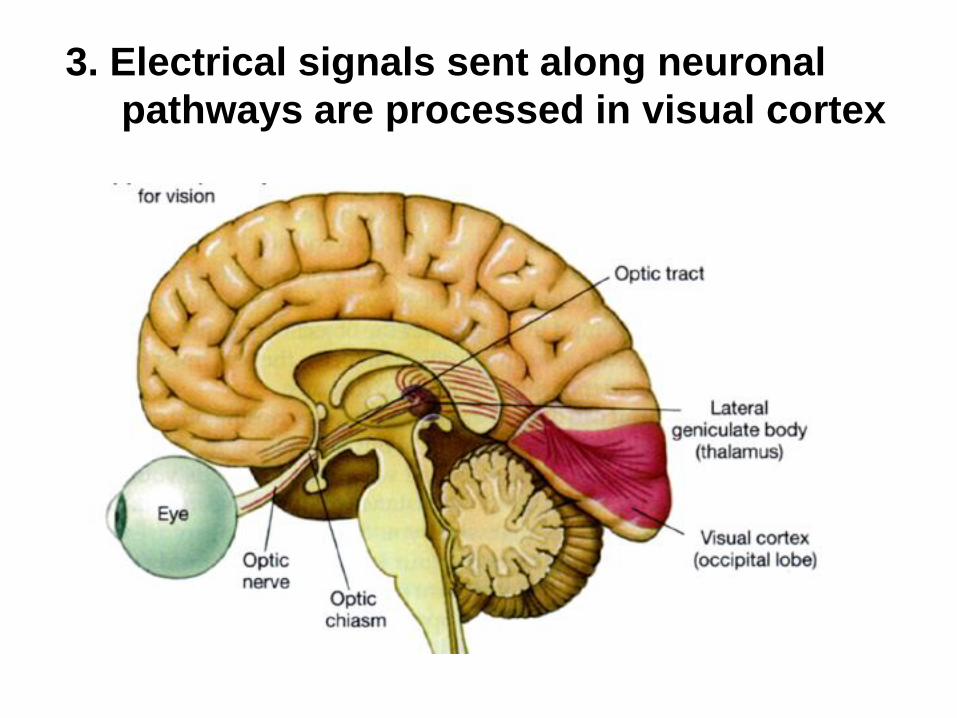
3. Electrical signals sent along neuronal
pathways are processed in visual cortex

Light Modification Pre-Retina
• Amount of light is changed by altering
pupil aperture from 1.5 – 8 mm
• Pupillary constriction due to ?
• Dilation ?
• Pupillary reflex is
consensual

The Lens
Light is focused by
changing lens shape
Refraction: Due to different
densities, light waves are
bent, or refracted when
passing from one
medium into another

Accommodation: Light is focused (to keep
objects in focus) by changing lens shape
Lens attached to ciliary muscle via
suspensory ligament (= zonulas)
Ciliary muscle contracts
. . . . . .
Lens bulges upFig 10-31
See also Fig 10-32

Vision Problems
Presbyopia (loss of accommodation)
Myopia (near-sightedness)
Hyperopia (far-sightedness)
Astigmatism (asymmetry of cornea
and/or lens)Test of visual acuity in lab

Compare to Fig 10-33

Sensory Transduction Converts
Stimuli into Graded Potentials
Stimulus energy is transduced into a
membrane potential change.
How can you create an excitatory or
inhibitory signal?
Important concept from 1st part of chapter:
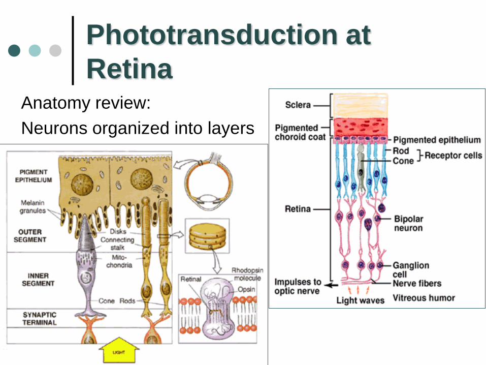
Phototransduction at
RetinaAnatomy review:
Neurons organized into layers

Light = Electromagnetic Energy
Wavelength
for visible
light: =?
Some
animals
can see UV
and IR
waves

Photo-
Receptors
Rods Monochromatic
night time vision
1 pigment (Rhodopsin)
Most numerous except in fovea
Cones
High acuity vision & daytime color vision
3 pigments
Fig 10-38
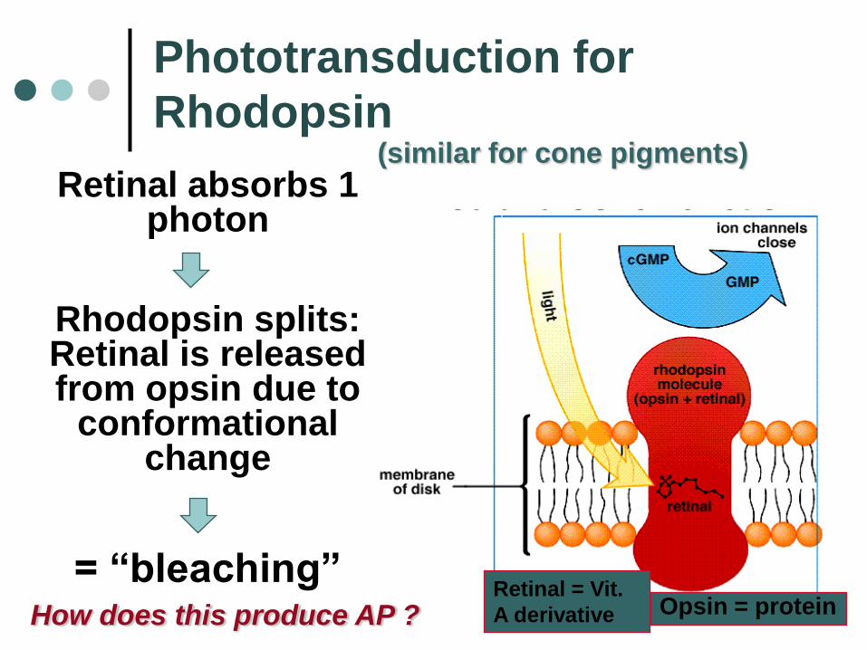
Phototransduction for
Rhodopsin
Retinal absorbs 1 photon
Rhodopsin splits: Retinal is released from opsin due to
conformational change
= “bleaching”Opsin = protein
Retinal = Vit.
A derivativeHow does this produce AP ?
(similar for cone pigments)

No Light:
Rhodopsin inactive
Cells have membrane
potential of ~ - 40
mV (what does that
mean, what is it due
to ?)
Continuous (= tonic)
NT release to
adjacent bipolar
cells
Fig 10-39

Light:Rhodopsin splits
Activation of transducin
( = __? - protein)
2nd messenger cascade
decreases cGMP
levels
Na+ channels close ?
NT release decreases

120 Mio rods
6 Mio cones
only 1.2 Mio axons enter optic nerve mechanism?
Visual processing
in visual cortex
Optic nerves enter
brain at optic
chiasma: some
fibers cross sides
right side visual
field to left side
brain
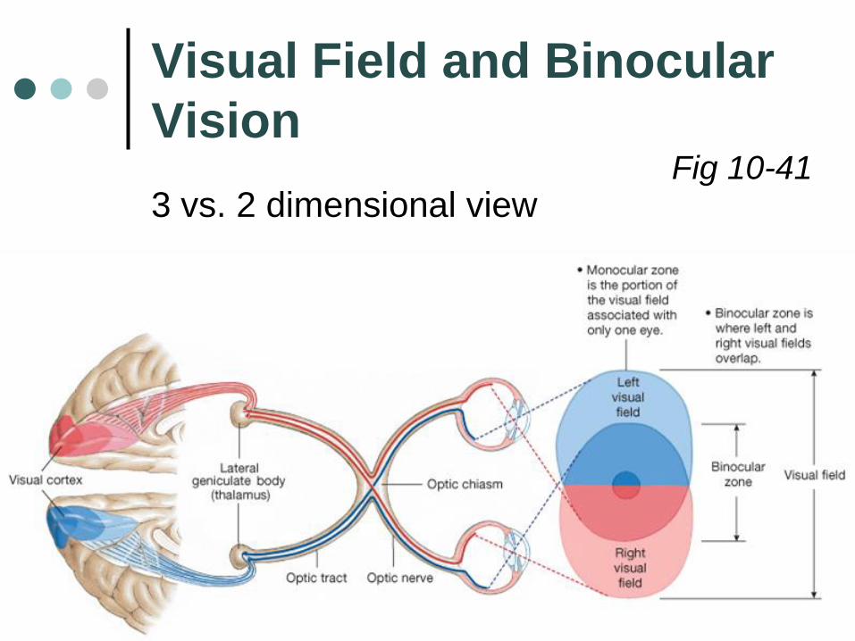
Visual Field and Binocular
Vision
3 vs. 2 dimensional viewFig 10-41