CHAPTER 10: DIGESTIVE SYSTEMnursingnsn.weebly.com/uploads/2/4/1/3/24135302/chapter_11... · CHAPTER...
42
1 K D UNIT 5/CHAP.2: INPUT AND OUT PUT OF BODY REQUIRED COMPONENTS // NSNM/ 2013-2014 CHAPTER 10: DIGESTIVE SYSTEM At the end of this chapter, student will be able to: a) Describe the general functions of the digestive system, and name its major divisions. b) Explain the difference between mechanical and chemical digestion, and name the end products of digestion. c) Describe the structure and functions of the teeth and tongue. d) Explain the functions of saliva. e) Describe the location and function of the pharynx and esophagus. f) Describe the structure and function of each of the four layers of the alimentary tube. g) Describe the location, structure, and function of the stomach, small intestine, liver, gallbladder, and pancreas. h) Describe absorption in the small intestine. i) Describe the location, structure and functions of the large intestine. j) Explain the functions of the normal flora of the colon. 10.1 INTRODUCTION TO THE DIGESTIVE. The digestive system contributes to homeostasis by breaking down food into forms that can be absorbed and used by body cells. It also absorbs water, vitamins, and minerals, and eliminates wastes from the body. The food we eat contains a variety of nutrients, which are used for building new body tissues and repairing damaged tissues. Food is also vital to life because it is our only source of chemical energy. However, most of the food we eat consists of molecules that are too large to be used by body cells. Therefore, foods must be broken down into molecules that are small enough to enter body cells, a process known as digestion. The organs involved in the breakdown of food collectively called the digestive system are the focus of this chapter. 10.2 FUNCTIONS OF THE DIGESTIVE SYSTEM Food provides the raw materials or nutrients that cells use to reproduce and to build new tissue. The energy needed for cell reproduction and tissue building is released from food in the process of cell respiration. The function of the digestive system is to change the complex
-
Upload
vuongquynh -
Category
Documents
-
view
225 -
download
5
Transcript of CHAPTER 10: DIGESTIVE SYSTEMnursingnsn.weebly.com/uploads/2/4/1/3/24135302/chapter_11... · CHAPTER...

1 K
D UNIT 5/CHAP.2: INPUT AND OUT PUT OF BODY REQUIRED COMPONENTS //NSNM/ 2013-2014
CHAPTER 10: DIGESTIVE SYSTEM
At the end of this chapter, student will be able to:
a) Describe the general functions of the digestive system, and name its major divisions.
b) Explain the difference between mechanical and chemical digestion, and name the end
products of digestion.
c) Describe the structure and functions of the teeth and tongue.
d) Explain the functions of saliva.
e) Describe the location and function of the pharynx and esophagus.
f) Describe the structure and function of each of the four layers of the alimentary tube.
g) Describe the location, structure, and function of the stomach, small intestine, liver,
gallbladder, and pancreas.
h) Describe absorption in the small intestine.
i) Describe the location, structure and functions of the large intestine.
j) Explain the functions of the normal flora of the colon.
10.1 INTRODUCTION TO THE DIGESTIVE.
The digestive system contributes to homeostasis by breaking down food into forms that can
be absorbed and used by body cells. It also absorbs water, vitamins, and minerals, and
eliminates wastes from the body. The food we eat contains a variety of nutrients, which are
used for building new body tissues and repairing damaged tissues. Food is also vital to life
because it is our only source of chemical energy. However, most of the food we eat consists
of molecules that are too large to be used by body cells. Therefore, foods must be broken
down into molecules that are small enough to enter body cells, a process known as digestion.
The organs involved in the breakdown of food collectively called the digestive system are
the focus of this chapter.
10.2 FUNCTIONS OF THE DIGESTIVE SYSTEM
Food provides the raw materials or nutrients that cells use to reproduce and to build new
tissue. The energy needed for cell reproduction and tissue building is released from food in
the process of cell respiration. The function of the digestive system is to change the complex

2 K
D UNIT 5/CHAP.2: INPUT AND OUT PUT OF BODY REQUIRED COMPONENTS //NSNM/ 2013-2014
organic nutrient molecules into simple organic and inorganic molecules that can then be
absorbed into the blood or lymph to be transported to cells.
Overall, the digestive system performs six basic processes:
1. Ingestion. This process involves taking foods and liquids into the mouth (eating).
2. Secretion. Each day, cells within the walls of the GI tract and accessory digestive organs
secrete a total of about 7 liters of water, acid, buffers, and enzymes into the lumen (interior
space) of the tract.
3. Mixing and propulsion. Alternating contractions and relaxations of smooth muscle in the
walls of the GI tract mix food and secretions and propel them toward the anus. This
capability of the GI tract to mix and move material along its length is called motility.
4. Digestion. Mechanical and chemical processes break down ingested food into small
molecules.
In mechanical digestion the teeth cut and grind food before it is swallowed, and then smooth
muscles of the stomach and small intestine churn the food. As a result, food molecules
become dissolved and thoroughly mixed with digestive enzymes.
In chemical digestion the large carbohydrate, lipid, protein, and nucleic acid molecules in
food are split into smaller molecules by hydrolysis. Digestive enzymes produced by the
salivary glands, tongue, stomach, pancreas, and small intestine catalyze these catabolic
reactions.
A few substances in food can be absorbed without chemical digestion. These include
vitamins, ions, cholesterol, and water.
5. Absorption. Is the entrance of ingested and secreted fluids, ions, and the products of
digestion into the epithelial cells lining the lumen of the GI tract is called absorption. The
absorbed substances pass into blood or lymph and circulate to cells throughout the body.
6. Defecation /Elimination. Here the wastes, indigestible substances, bacteria, cells sloughed
from the lining of the GI tract, and digested materials that were not absorbed in their journey
through the digestive tract leave the body through the anus. The eliminated material is termed
feces.
10.3 DIVISIONS OF THE DIGESTIVE SYSTEM

3 K
D UNIT 5/CHAP.2: INPUT AND OUT PUT OF BODY REQUIRED COMPONENTS //NSNM/ 2013-2014
Figure: Digestive system. Trace the path of food from the mouth to the anus. The large
intestine consists of the cecum, the colon (composed of the ascending, transverse,
descending, and sigmoid colon), the rectum, and the anal canal. Note also the location of the
accessory organs of digestion: the pancreas, the liver, and the gallbladder.
Two groups of organs compose the digestive system, the gastrointestinal (GI) tract and the
accessory digestive organs.

4 K
D UNIT 5/CHAP.2: INPUT AND OUT PUT OF BODY REQUIRED COMPONENTS //NSNM/ 2013-2014
The gastrointestinal (GI) tract, or alimentary canal is a continuous tube that extends from
the mouth to the anus through the thoracic and abdominopelvic cavities.
Organs of the gastrointestinal tract include the mouth, most of the pharynx, esophagus,
stomach, small intestine, and large intestine. The length of the GI tract is about 5–7 meters
in a living person. It is longer in a cadaver (about 7–9 meters).
The accessory digestive organs include the teeth, tongue, salivary glands, liver,
gallbladder, and pancreas.
Teeth aid in the physical breakdown of food, and the tongue assists in chewing and
swallowing.
The other accessory digestive organs, however, never come into direct contact with food.
They produce or store secretions that flow into the GI tract through ducts; these secretions aid
in the chemical breakdown of food.
The GI tract contains food from the time it is eaten until it is digested and absorbed or
eliminated. Muscular contractions in the wall of the GI tract physically break down the food
by churning it and propel the food along the tract, from the esophagus to the anus. The
contractions also help to dissolve foods by mixing them with fluids secreted into the tract.
Enzymes secreted by accessory digestive organs and cells that line the tract break down the
food chemically.
10.4 TYPES OF DIGESTION
The food we eat is broken down in two complementary processes:
Mechanical digestion is the physical breaking up of food into smaller pieces. Chewing is an
example of this. As food is broken up, more of its surface area is exposed for the action of
digestive enzymes.
The work of the digestive enzymes is the chemical digestion of broken- up food particles, in
which complex chemical molecules are changed into much simpler chemicals that the body
can utilize. Such enzymes are specific with respect to the fat, protein, or carbohydrate food
molecules each can digest. For example, protein-digesting enzymes work only on proteins,
not on carbohydrates or fats. Each enzyme is produced by a particular digestive organ and
functions at a specific site. However, the enzyme’s site of action may or may not be its site of
production.

5 K
D UNIT 5/CHAP.2: INPUT AND OUT PUT OF BODY REQUIRED COMPONENTS //NSNM/ 2013-2014
10.5 END PRODUCTS OF DIGESTION
The three types of complex organic molecules found in food are carbohydrates, proteins, and
fats. Each of these complex molecules is digested to a much more simple substance that the
body can then use.
Carbohydrates, such as starches and disaccharides, are digested to monosaccharides such as
glucose, fructose, and galactose.
Proteins are digested to amino acids, and
Fats are digested to fatty acids and glycerol.
Also part of food, and released during digestion, are vitamins, minerals, and water.
10.6 GENERAL STRUCTURAL LAYERS OF THE ALIMENTARY TUBE
The wall of the GI tract from the lower esophagus to the anal canal has the same basic, four-
layered arrangement of tissues. The four layers of the tract, from deep to superficial, are the
mucosa, submucosa, muscularis, and serosa.
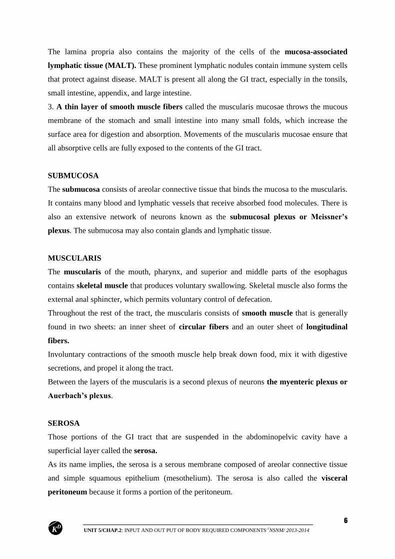
MUCOSA
The mucosa, or inner lining of the GI tract, is a mucous membrane.
It is composed of:
(1) a layer of epithelium in direct contact with the contents of the GI tract,
(2) a layer of connective tissue called the lamina propria, and
(3) a thin layer of smooth muscle (muscularis mucosae).
1. The epithelium in the mouth, pharynx, esophagus, and anal canal is mainly nonkeratinized
stratified squamous epithelium that serves a protective function.
Simple columnar epithelium, which functions in secretion and absorption, lines the stomach
and intestines.
Located among the epithelial cells are exocrine cells that secrete mucus and fluid into the
lumen of the tract, and several types of endocrine cells, collectively called enteroendocrine
cells, that secrete hormones.
2. The lamina propria is areolar connective tissue containing many blood and lymphatic
vessels, which are the routes by which nutrients absorbed into the GI tract reach the other
tissues of the body.
6 K
D UNIT 5/CHAP.2: INPUT AND OUT PUT OF BODY REQUIRED COMPONENTS //NSNM/ 2013-2014
The lamina propria also contains the majority of the cells of the mucosa-associated
lymphatic tissue (MALT). These prominent lymphatic nodules contain immune system cells
that protect against disease. MALT is present all along the GI tract, especially in the tonsils,
small intestine, appendix, and large intestine.
3. A thin layer of smooth muscle fibers called the muscularis mucosae throws the mucous
membrane of the stomach and small intestine into many small folds, which increase the
surface area for digestion and absorption. Movements of the muscularis mucosae ensure that
all absorptive cells are fully exposed to the contents of the GI tract.
SUBMUCOSA
The submucosa consists of areolar connective tissue that binds the mucosa to the muscularis.
It contains many blood and lymphatic vessels that receive absorbed food molecules. There is
also an extensive network of neurons known as the submucosal plexus or Meissner’s
plexus. The submucosa may also contain glands and lymphatic tissue.
MUSCULARIS
The muscularis of the mouth, pharynx, and superior and middle parts of the esophagus
contains skeletal muscle that produces voluntary swallowing. Skeletal muscle also forms the
external anal sphincter, which permits voluntary control of defecation.
Throughout the rest of the tract, the muscularis consists of smooth muscle that is generally
found in two sheets: an inner sheet of circular fibers and an outer sheet of longitudinal
fibers.
Involuntary contractions of the smooth muscle help break down food, mix it with digestive
secretions, and propel it along the tract.
Between the layers of the muscularis is a second plexus of neurons the myenteric plexus or
Auerbach’s plexus.
SEROSA
Those portions of the GI tract that are suspended in the abdominopelvic cavity have a
superficial layer called the serosa.
As its name implies, the serosa is a serous membrane composed of areolar connective tissue
and simple squamous epithelium (mesothelium). The serosa is also called the visceral
peritoneum because it forms a portion of the peritoneum.

7 K
D UNIT 5/CHAP.2: INPUT AND OUT PUT OF BODY REQUIRED COMPONENTS //NSNM/ 2013-2014
The esophagus lacks a serosa; instead only a single layer of areolar connective tissue called
the adventitia forms the superficial layer of this organ.
10.7 DETAILS ABOUT PERTONEUM
The peritoneum is the largest serous membrane of the body; it consists of a layer of simple
squamous epithelium (mesothelium) with an underlying supporting layer of areolar
connective tissue.
The peritoneum is divided into the parietal peritoneum, which lines the wall of the
abdominopelvic cavity, and the visceral peritoneum, which covers some of the organs in the
cavity and is their serosa
The thin space containing lubricating serous fluid that is between the parietal and visceral
portions of the peritoneum is called the peritoneal cavity. In certain diseases, the peritoneal
cavity may become distended by the accumulation of several liters of fluid, a condition called
ascites.
Some organs lie on the posterior abdominal wall and are covered by peritoneum only on their

8 K
D UNIT 5/CHAP.2: INPUT AND OUT PUT OF BODY REQUIRED COMPONENTS //NSNM/ 2013-2014
anterior surfaces; they are not in the peritoneal cavity. Such organs, including the kidneys,
ascending and descending colons of the large intestine, duodenum of the small intestine, and
pancreas, are said to be retroperitoneal.
Unlike the pericardium and pleurae, which smoothly cover the heart and lungs, the
peritoneum contains large folds that weave between the viscera. The folds bind the organs to
one another and to the walls of the abdominal cavity. They also contain blood vessels,
lymphatic vessels, and nerves that supply the abdominal organs.
There are five major peritoneal folds: the greater omentum, falciform ligament, lesser
omentum, mesentery, and mesocolon.
The greater omentum (omentum: fat skin), the largest peritoneal fold, drapes over the
transverse colon and coils of the small intestine like a fatty apron. The greater omentum is a
double sheet that folds back on itself, giving it a total of four layers. From attachments along
the stomach and duodenum, the greater omentum extends downward anterior to the small
intestine, then turns and extends upward and attaches to the transverse colon. The greater
omentum normally contains a considerable amount of adipose tissue. Its adipose tissue
content can greatly expand with weight gain. The many lymph nodes of the greater omentum
contribute macrophages and antibody producing plasma cells that help combat and contain
infections of the GI tract.
2. The falciform ligament attaches the liver to the anterior abdominal wall and diaphragm.
The liver is the only digestive organ that is attached to the anterior abdominal wall.
3. The lesser omentum arises as an anterior fold in the serosa of the stomach and duodenum,
and it suspends the stomach and duodenum from the liver. It is the pathway for blood vessels
entering the liver and contains the hepatic portal vein, common hepatic artery, and common
bile duct, along with some lymph nodes.
4. The mesentery ( middle), binds the jejunum and ileum of the small intestine to the
posterior abdominal wall. It extends from the posterior abdominal wall to wrap around the
small intestine and then returns to its origin, forming a double-layered structure. Between the
two layers are blood and lymphatic vessels and lymph nodes.
5. Two separate folds of peritoneum, called the mesocolon bind the transverse colon
(transverse mesocolon) and sigmoid colon (sigmoid mesocolon) of the large intestine to the

9 K
D UNIT 5/CHAP.2: INPUT AND OUT PUT OF BODY REQUIRED COMPONENTS //NSNM/ 2013-2014
posterior abdominal wall . It also carries blood and lymphatic vessels to the intestines.
Together, the mesentery and mesocolon hold the intestines loosely in place, allowing
movement as muscular contractions mix and move the luminal contents along the GI tract.
Neural innervations of GI tract

10 K
D UNIT 5/CHAP.2: INPUT AND OUT PUT OF BODY REQUIRED COMPONENTS //NSNM/ 2013-2014
The gastrointestinal tract is regulated by an intrinsic set of nerves known as the enteric
nervous system and by an extrinsic set of nerves that are part of the autonomic nervous
system.
ENTERIC NERVOUS SYSTEM
It consists of about 100 million neurons that extend from the esophagus to the anus. The
neurons of the ENS are arranged into two plexuses: the myenteric plexus and submucosal
plexus.
The myenteric plexus (myo- muscle), or plexus of Auerbach, is located between the
longitudinal and circular smooth muscle layers of the muscularis.
The submucosal plexus, or plexus of Meissner, is found within the submucosa.
The plexuses of the ENS consist of motor neurons, interneurons, and sensory neurons.
Because the motor neurons of the myenteric plexus supply the longitudinal and circular
smooth muscle layers of the muscularis, this plexus mostly controls GI tract motility
(movement), particularly the frequency and strength of contraction of the muscularis.
The motor neurons of the submucosal plexus supply the secretory cells of the mucosal
epithelium, controlling the secretions of the organs of the GI tract.
The interneurons of the ENS interconnect the neurons of the myenteric and submucosal
plexuses.

11 K
D UNIT 5/CHAP.2: INPUT AND OUT PUT OF BODY REQUIRED COMPONENTS //NSNM/ 2013-2014
The sensory neurons of the ENS supply the mucosal epithelium. Some of these sensory
neurons function as chemoreceptors, receptors that are activated by the presence of certain
chemicals in food located in the lumen of a GI organ.
Other sensory neurons function as stretch receptors, receptors that are activated when food
distends (stretches) the wall of a GI organ.
AUTONOMIC NERVOUS SYSTEM
Although the neurons of the ENS can function independently, they are subject to regulation
by the neurons of the autonomic nervous system.
The vagus (X) nerves supply parasympathetic fibers to most parts of the GI tract, with the
exception of the last half of the large intestine, which is supplied with parasympathetic fibers
from the sacral spinal cord.
The parasympathetic nerves that supply the GI tract form neural connections with the ENS.
Parasympathetic preganglionic neurons of the vagus or pelvic splanchnic nerves synapse with
parasympathetic postganglionic neurons located in the myenteric and submucosal plexuses.
Some of the parasympathetic postganglionic neurons in turn synapse with neurons in the
ENS; others directly innervate smooth muscle and glands within the wall of the GI tract. In
general, stimulation of the parasympathetic nerves that innervate the GI tract causes an
increase in GI secretion and motility by increasing the activity of ENS neurons.
Sympathetic nerves that supply the GI tract arise from the thoracic and upper lumbar regions
of the spinal cord. Like the parasympathetic nerves, these sympathetic nerves form neural
connections with the ENS. Sympathetic postganglionic neurons synapse with neurons located
in the myenteric plexus and the submucosal plexus. In general, the sympathetic nerves that
supply the GI tract cause a decrease in GI secretion and motility by inhibiting the neurons
of the ENS. Emotions such as anger, fear, and anxiety may slow digestion because they
stimulate the sympathetic nerves that supply the GI tract.
GASTROINTESTINAL REFLEX PATHWAYS
Many neurons of the ENS are components of GI (gastrointestinal) reflex pathways that
regulate GI secretion and motility in response to stimuli present in the lumen of the GI tract.
The initial components of a typical GI reflex pathway are sensory receptors (such as
chemoreceptors and stretch receptors) that are associated with the sensory neurons of the
ENS.

12 K
D UNIT 5/CHAP.2: INPUT AND OUT PUT OF BODY REQUIRED COMPONENTS //NSNM/ 2013-2014
The axons of these sensory neurons can synapse with other neurons located in the ENS, CNS,
or ANS, informing these regions about the nature of the contents and the degree of distension
(stretching) of the GI tract. The neurons of the ENS, CNS, or ANS subsequently activate or
inhibit GI glands and smooth muscle, altering GI secretion and motility.
MOUTH:
The mouth, also referred to as the oral or buccal cavity, is formed by the cheeks, hard and
soft palates, and tongue.
The cheeks form the lateral walls of the oral cavity. They are covered externally by skin and
internally by a mucous membrane. Buccinator muscles and connective tissue lie between the
skin and mucous membranes of the cheeks.
The lips or labia (fleshy borders) are fleshy folds surrounding the opening of the mouth.
They contain the orbicularis oris muscle and are covered externally by skin and internally by
a mucous membrane. During chewing, contraction of the buccinators muscles in the cheeks
and orbicularis oris muscle in the lips helps keep food between the upper and lower teeth.
These muscles also assist in speech.
The oral vestibule of the oral cavity is a space bounded externally by the cheeks and lips
and internally by the gums and teeth. The oral cavity proper is a space that extends from the
gums and teeth to the fauces, the opening between the oral cavity and the oropharynx(throat).
The palate is a wall or septum that separates the oral cavity from the nasal cavity, forming
the roof of the mouth. This important structure makes it possible to chew and breathe at the
same time.
The hard palate the anterior portion of the roof of the mouth is formed by the maxillae and
palatine bones and is covered by a mucous membrane; it forms a bony partition between the
oral and nasal cavities.
The soft palate, which forms the posterior portion of the roof of the mouth, is an arch-shaped
muscular partition between the oropharynx and nasopharynx that is lined with mucous
membrane.
Hanging from the free border of the soft palate is a conical muscular process called the
uvula.
During swallowing, the soft palate and uvula are drawn superiorly, closing off the
nasopharynx and preventing swallowed foods and liquids from entering the nasal cavity.

13 K
D UNIT 5/CHAP.2: INPUT AND OUT PUT OF BODY REQUIRED COMPONENTS //NSNM/ 2013-2014
Lateral to the base of the uvula are two muscular folds that run down the lateral sides of the
soft palate: Anteriorly, the palatoglossal arch extends to the side of the base of the tongue;
posteriorly, the palatopharyngeal arch extends to the side of the pharynx.
The palatine tonsils are situated between the arches, and the lingual tonsils are situated at
the base of the tongue. At the posterior border of the soft palate, the mouth opens into the
oropharynx through the fauces
SALIVARY GLANDS
A salivary gland is a gland that releases a secretion called saliva into the oral cavity.
Ordinarily, just enough saliva is secreted to keep the mucous membranes of the mouth and
pharynx moist and to cleanse the mouth and teeth. When food enters the mouth, however,
secretion of saliva increases, and it lubricates, dissolves, and begins the chemical breakdown
of the food. The mucous membrane of the mouth and tongue contains many small salivary
glands that open directly, or indirectly via short ducts, to the oral cavity. These glands
include labial, buccal, and palatal glands in the lips, cheeks, and palate, respectively, and
lingual glands in the tongue, all of which make a small contribution to saliva.

14 K
D UNIT 5/CHAP.2: INPUT AND OUT PUT OF BODY REQUIRED COMPONENTS //NSNM/ 2013-2014
However, most saliva is secreted by the major salivary glands, which lie beyond the oral
mucosa, into ducts that lead to the oral cavity. There are three pairs of major salivary
glands: the parotid, submandibular, and sublingual glands
The parotid glands are located inferior and anterior to the ears, between the skin and the
masseter muscle. Each secretes saliva into the oral cavity via a parotid duct.
The submandibular glands are found in the floor of the mouth; they are medial and partly
inferior to the body of the mandible. Their ducts are the submandibular ducts.
The sublingual glands are beneath the tongue and superior to the submandibular glands.
Their ducts are the lesser sublingual ducts.
Figure: The salivary glands shown in left lateral view.
Composition and Functions of Saliva
Chemically, saliva is 99.5% water and 0.5% solutes. Among the solutes are ions, including
sodium, potassium, chloride, bicarbonate, and phosphate. Also present are some dissolved
gases and various organic substances, including urea and uric acid, mucus, immunoglobulin
A, the bacteriolytic enzyme lysozyme and salivary amylase, a digestive enzyme that acts on starch.

15 K
D UNIT 5/CHAP.2: INPUT AND OUT PUT OF BODY REQUIRED COMPONENTS //NSNM/ 2013-2014
The water in saliva provides a medium for dissolving foods so that they can be tasted by
gustatory receptors and so that digestive reactions can begin.
Chloride ions in the saliva activate salivary amylase, an enzyme that starts the breakdown of
starch.
Bicarbonate and phosphate ions buffer acidic foods that enter the mouth, so saliva is only
slightly acidic (pH 6.35–6.85).
Salivary glands help remove waste molecules from the body, which accounts for the
presence of urea and uric acid in saliva.
Mucus lubricates food so it can be moved around easily in the mouth, formed into a ball, and
swallowed.
Immunoglobulin A (IgA) prevents attachment of microbes so they cannot penetrate the
epithelium, and the enzyme lysozyme kills bacteria.
Salivation
The secretion of saliva, called salivation is controlled by the autonomic nervous system.
Amounts of saliva secreted daily vary considerably but average 1000–1500 mL
Normally, parasympathetic stimulation promotes continuous secretion of a moderate
amount of saliva, which keeps the mucous membranes moist and lubricates the movements of
the tongue and lips during speech.
The saliva is then swallowed and helps moisten the esophagus. Eventually, most components
of saliva are reabsorbed, which prevents fluid loss.
Sympathetic stimulation dominates during stress, resulting in dryness of the mouth.
If the body becomes dehydrated, the salivary glands stop secreting saliva to conserve water;
the resulting dryness in the mouth contributes to the sensation of thirst.
The feel and taste of food also are stimulators of salivary gland secretions.
The smell, sight, sound, or thought of food may also stimulate secretion of saliva.
The tongue is an accessory digestive organ composed of skeletal muscle covered with
mucous membrane. Together with its associated muscles, it forms the floor of the oral cavity.
The tongue is divided into symmetrical lateral halves by a median septum that extends its
entire length, and it is attached inferiorly to the hyoid bone, styloid process of the temporal
bone, and mandible.
Each half of the tongue consists of an identical complement of extrinsic and intrinsic
muscles.
16 K
D UNIT 5/CHAP.2: INPUT AND OUT PUT OF BODY REQUIRED COMPONENTS //NSNM/ 2013-2014
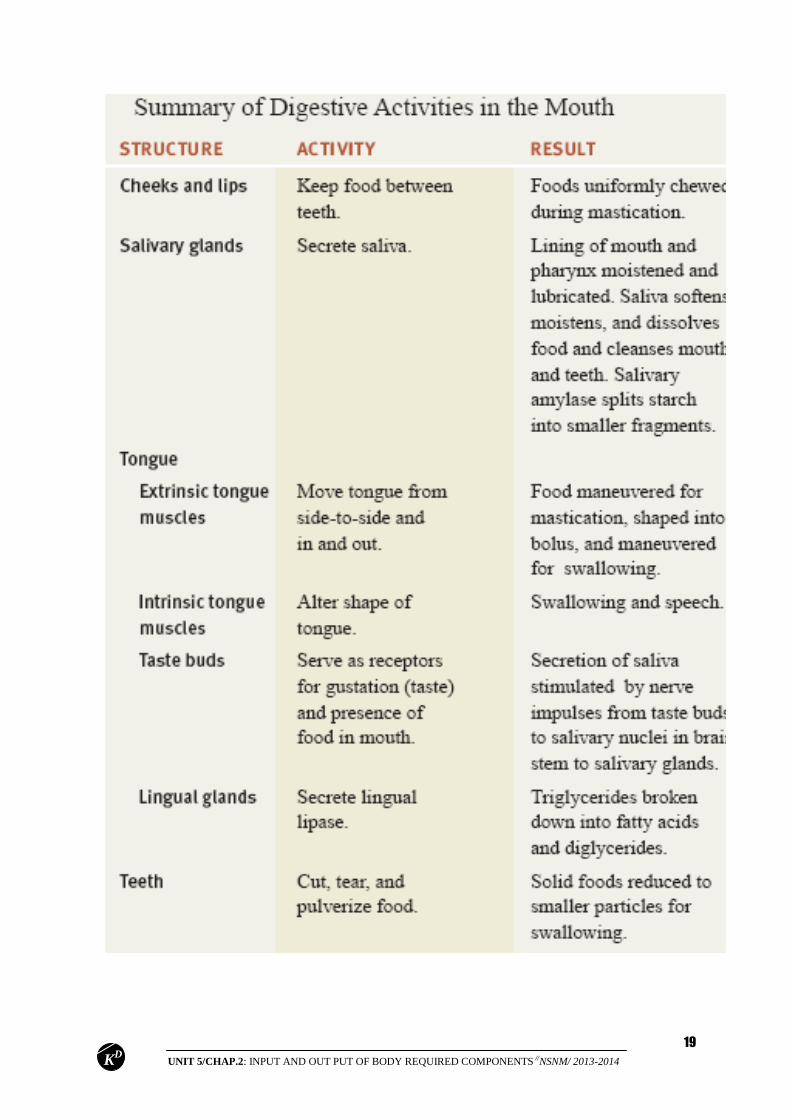
The extrinsic muscles of the tongue, which include the hyoglossus, genioglossus, and
styloglossus move the tongue from side to side and in and out to maneuver food for chewing,
shape the food into a rounded mass, and force the food to the back of the mouth for
swallowing. They also form the floor of the mouth and hold the tongue in position.
The intrinsic muscles alter the shape and size of the tongue for speech and swallowing. The
intrinsic muscles include the longitudinalis superior, longitudinalis inferior, transversus
linguae, and verticalis linguae muscles.
The dorsum (upper surface) and lateral surfaces of the tongue are covered with papillae
which contain taste buds, the receptors for gestation (taste). Some papillae lack taste buds,
but they contain receptors for touch and increase friction between the tongue and food,
making it easier for the tongue to move food in the oral cavity.
Lingual glands of the tongue secrete both mucus and a watery serous fluid that contains the
enzyme lingual lipase, which acts on triglycerides.
TEETH
The teeth, or dentes, are accessory digestive organs located in sockets of the alveolar
processes of the mandible and maxillae.
The alveolar processes are covered by the gingivae, or gums, which extend slightly into each
socket. The sockets are lined by the periodontal ligament or membrane (odont- tooth), that
anchors the teeth to the socket walls.
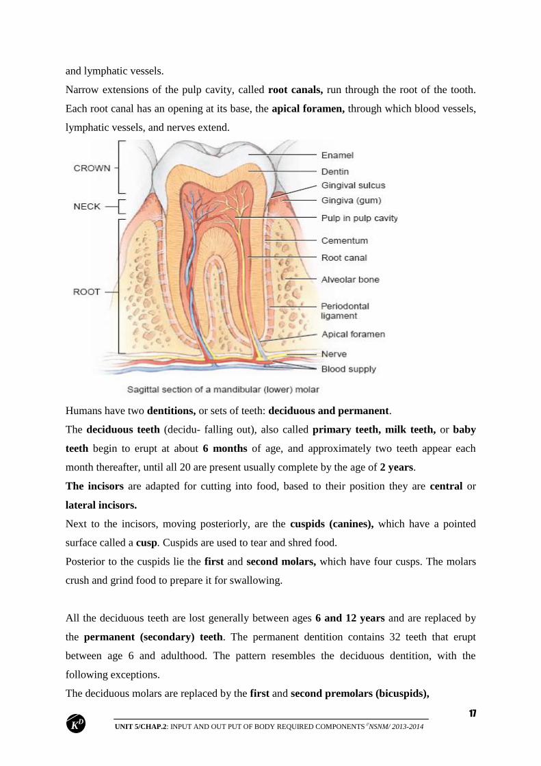
A typical tooth has three major external regions: the crown, root, and neck.
The crown is the visible portion above the level of the gums.
The root, they are one to three roots embedded in the socket
Theneck is the constricted junction of the crown and root near the gum line.
Internally, dentin forms the majority of the tooth. Dentin gives the tooth its basic shape and
rigidity. The dentin of the crown is covered by enamel, which consists primarily of calcium
phosphate and calcium carbonate. Enamel serves to protect the tooth from the wear and tear
of chewing. It also protects against acids that concerned with the treatment of abnormal
conditions of the tissues immediately surrounding the teeth, such as gingivitis.
The dentin of the root is covered by cementum, another bonelike substance, which attaches
the root to the periodontal ligament.
The dentin of a tooth encloses a space. The enlarged part of the space, the pulp cavity, lies
within the crown and is filled with pulp, a connective tissue containing blood vessels, nerves,
17 K
D UNIT 5/CHAP.2: INPUT AND OUT PUT OF BODY REQUIRED COMPONENTS //NSNM/ 2013-2014
and lymphatic vessels.
Narrow extensions of the pulp cavity, called root canals, run through the root of the tooth.
Each root canal has an opening at its base, the apical foramen, through which blood vessels,
lymphatic vessels, and nerves extend.
Humans have two dentitions, or sets of teeth: deciduous and permanent.
The deciduous teeth (decidu- falling out), also called primary teeth, milk teeth, or baby
teeth begin to erupt at about 6 months of age, and approximately two teeth appear each
month thereafter, until all 20 are present usually complete by the age of 2 years.
The incisors are adapted for cutting into food, based to their position they are central or
lateral incisors.
Next to the incisors, moving posteriorly, are the cuspids (canines), which have a pointed
surface called a cusp. Cuspids are used to tear and shred food.
Posterior to the cuspids lie the first and second molars, which have four cusps. The molars
crush and grind food to prepare it for swallowing.
All the deciduous teeth are lost generally between ages 6 and 12 years and are replaced by
the permanent (secondary) teeth. The permanent dentition contains 32 teeth that erupt
between age 6 and adulthood. The pattern resembles the deciduous dentition, with the
following exceptions.
The deciduous molars are replaced by the first and second premolars (bicuspids),

18 K
D UNIT 5/CHAP.2: INPUT AND OUT PUT OF BODY REQUIRED COMPONENTS //NSNM/ 2013-2014
The permanent molars, which erupt into the mouth posterior to the premolars, do not
replace any deciduous teeth and erupt as the jaw grows to accommodate them.The first
molars at age 6 (six-year molars), the second molars at age 12 (twelve-year molars), and the
third molars (wisdom teeth) after age 17 or not at all.
Often the human jaw does not have enough room posterior to the second molars to
accommodate the eruption of the third molars. In this case, the third molars remain embedded
in the alveolar bone and are said to be impacted. They often cause pressure and pain and
must be removed surgically.
19 K
D UNIT 5/CHAP.2: INPUT AND OUT PUT OF BODY REQUIRED COMPONENTS //NSNM/ 2013-2014

20 K
D UNIT 5/CHAP.2: INPUT AND OUT PUT OF BODY REQUIRED COMPONENTS //NSNM/ 2013-2014
PHARYNX
When food is first swallowed, it passes from the mouth into the pharynx, a funnel-shaped
tube that extends from the internal nares to the esophagus posteriorly and to the larynx
anteriorly. The pharynx is composed of skeletal muscle and lined by mucous membrane, and
as described in the preceding chapter is divided into three parts: the nasopharynx, the
oropharynx, and the laryngopharynx. The nasopharynx functions only in respiration, but both
the oropharynx and laryngopharynx have digestive as well as respiratory functions.
Swallowed food passes from the mouth into the oropharynx and laryngopharynx; the
muscular contractions of these areas help propel food into the esophagus and then into the
stomach. No digestion takes place in the pharynx. Its only related function is swallowing, the
mechanical movement of food. When the bolus of food is pushed backward by the tongue,
the constrictor muscles of the pharynx contract as part of the swallowing reflex. The reflex
center for swallowing is in the medulla, which coordinates the many actions that take place:
constriction of the pharynx, cessation of breathing, elevation of the soft palate to block the
nasopharynx, elevation of the larynx and closure of the epiglottis, and peristalsis of the
esophagus.
.
ESOPHAGUS
The esophagus is a collapsible muscular tube, about 25 cm (10 in.) long, that lies posterior
to the trachea. The esophagus begins at the inferior end of the laryngopharynx and passes
through the mediastinum anterior to the vertebral column. Then it pierces the diaphragm
through an opening called the esophageal hiatus, and ends in the superior portion of the
stomach. Sometimes, part of the stomach protrudes above the diaphragm through the
esophageal hiatus. This condition, termed a hiatus hernia.
At each end of the esophagus, the muscularis becomes slightly more prominent and forms
two sphincters, the upper esophageal sphincter (UES), which consists of skeletal muscle,
and the lower esophageal sphincter (LES), which consists of mooth muscle.
The upper esophageal sphincter regulates the movement of food from the pharynx into the
esophagus;

21 K
D UNIT 5/CHAP.2: INPUT AND OUT PUT OF BODY REQUIRED COMPONENTS //NSNM/ 2013-2014
The lower esophageal sphincter regulates the movement of food from the esophagus into the
stomach, that means the LES relaxes to permit food to enter the stomach, and then contracts
to prevent the backup of stomach contents.
The movement of food from the mouth into the stomach is achieved by the act of swallowing,
or deglutition. Deglutition is facilitated by the secretion of saliva and mucus and involves the
mouth, pharynx, and esophagus. Swallowing occurs in three stages:
(1) the voluntary stage, in which the bolus is passed into the oropharynx;
(2) the pharyngeal stage, the involuntary passage of the bolus through the pharynx into the
esophagus; and
(3) the esophageal stage, the involuntary passage of the bolus through the esophagus into the
stomach.

22 K
D UNIT 5/CHAP.2: INPUT AND OUT PUT OF BODY REQUIRED COMPONENTS //NSNM/ 2013-2014
STOMACH
The stomach is located in the upper left quadrant of the abdominal cavity, to the left of the
liver and in front of the spleen. Although part of the alimentary tube, the stomach is not a
tube, but rather a sac that extends from the esophagus to the small intestine. Because it is a
sac, the stomach is a reservoir for food, so that digestion proceeds gradually and we do not
have to eat constantly. Both mechanical and chemical digestion takes place in the stomach.
The cardiac orifice is the opening of the esophagus, and the fundus is the portion above the
level of this opening. The
body of the stomach is
the large central portion,
bounded laterally by the
greater curvature and

23 K
D UNIT 5/CHAP.2: INPUT AND OUT PUT OF BODY REQUIRED COMPONENTS //NSNM/ 2013-2014
medially by the lesser curvature. The pylorus is adjacent to the duodenum of the small
intestine, and the pyloric sphincter surrounds the junction of the two organs. The fundus
and body are mainly storage areas, whereas most digestion takes place in the pylorus.
When the stomach is empty, the mucosa appears wrinkled or folded. These folds are called
rugae; they flatten out as the stomach is filled and permit expansion of the lining without
tearing it. The gastric pits are the glands of the stomach and consist of several types of cells;
their collective secretions are called gastric juice. These cells are:
Mucous cells secrete mucus, which coats the stomach lining and helps prevent erosion by the
gastric juice.
Chief cells secrete pepsinogen, an inactive form of the enzyme pepsin.
Parietal cells produce hydrochloric acid (HCl); these cells have enzymes called proton
pumps, which secrete H+ ions into the stomach cavity. The H+ ions unite with Cl– ions that
have diffused from the parietal cells to form HCl in the lumen of the stomach. HCl converts
pepsinogen to pepsin, which then begins the digestion of proteins to polypeptides, and also
gives gastric juice its pH of 1 to 2. This very acidic pH is necessary for pepsin to function
and also kills most microorganisms that enter the stomach. The parietal cells also secrete
intrinsic factor, which is necessary for the absorption of vitamin B12.
Enteroendocrine cells called G cells secrete the hormone gastrin. Gastric juice is secreted in
small amounts at the sight or smell of food. This is a parasympathetic response that
ensuresthat some gastric juice will be present in the stomach when food arrives. The presence
of food in the stomach causes the G cells to secrete gastrin, a hormone that stimulates the
secretion of greater amounts of gastric juice.
The external muscle layer of the stomach consists of three layers of smooth muscle: circular,
longitudinal, and oblique layers. These three layers are innervated by the mesenteric
plexuses of the enteric nervous system. Stimulatory impulses are carried from the CNS by the
vagus nerves (10th cranial) and provide for very efficient mechanical digestion to change
food into a thick liquid called chyme. The pyloric sphincter is usually contracted when the
stomach is churning food; it relaxes at intervals to permit small amounts of chyme to pass
into the duodenum. This sphincter then contracts again to prevent the backup of intestinal
contents into the stomach

24 K
D UNIT 5/CHAP.2: INPUT AND OUT PUT OF BODY REQUIRED COMPONENTS //NSNM/ 2013-2014
o

25 K
D UNIT 5/CHAP.2: INPUT AND OUT PUT OF BODY REQUIRED COMPONENTS //NSNM/ 2013-2014
SMALL INTESTINE
The small intestine is about 1 inch (2.5 cm) in diameter and approximately 20 feet (6 m)
long and extends from the stomach to the cecum of the large intestine. Within the abdominal
cavity, the large intestine encircles the coils of the small intestine.
The small intestine has the following parts: duodenum, jejunum, ileum.
The duodenum is the first 10 inches (25 cm) of the small intestine. The common bile duct
enters the duodenum at the ampulla of Vater (or hepatopancreatic ampulla).
The jejunum is about 8 feet long, and the ileum is about 11 feet in length.
Digestion is completed in the small intestine, and the end products of digestion are absorbed
into the blood and lymph. The mucosa has simple columnar epithelium that includes cells
with microvilli and goblet cells that secrete mucus. Enteroendocrine cells secrete the
hormones of the small intestine. Lymph nodules called Peyer’s patches are especially
abundant in the ileum to destroy absorbed pathogens. The external muscle layer has the
typical circular and longitudinal smooth muscle layers that mix the chyme with digestive
secretions and propel the chyme toward the colon. Stimulatory impulses to the enteric nerves
of these muscle layers are carried by the vagus nerves. The waves of peristalsis, however, can
take place without stimulation by the central nervous system; the enteric nervous system can

26 K
D UNIT 5/CHAP.2: INPUT AND OUT PUT OF BODY REQUIRED COMPONENTS //NSNM/ 2013-2014
function independently and promote normal peristalsis. There are three sources of digestive
secretions that function within the small intestine: the liver, the pancreas, and the small
intestine itself. We will return to the small intestine after considering these other organs.
LARGE INTESTINE
The large intestine, also called
the colon, is approximately 2.5
inches (6.3 cm) in diameter and 5
feet (1.5 m) in length. It extends
from the ileum of the small
intestine to the anus, the terminal
opening.The cecum is the first
portion, and at its junction with

27 K
D UNIT 5/CHAP.2: INPUT AND OUT PUT OF BODY REQUIRED COMPONENTS //NSNM/ 2013-2014
the ileum is the ileocecal valve, which is not a sphincter but serves the same purpose. After
undigested food (which is now mostly cellulose) and water pass from the ileum into the
cecum, closure of the ileocecal valve prevents the backflow of fecal material. Attached to the
cecum is the appendix, a small, dead-end tube with abundant lymphatic tissue. The
remainder of the colon consists of the ascending, transverse, and descending colon, which
encircle the small intestine; the sigmoid colon, which turns medially and downward; the
rectum; and the anal canal. The rectum is about 6 inches long, and the anal canal is the last
inch of the colon that surrounds the anus.
No digestion takes place in the colon. The only secretion of the colonic mucosa is mucus,
which lubricates the passage of fecal material. The longitudinal smooth muscle layer of the
colon is in three bands called taeniae coli. The rest of the colon is “gathered” to fit these
bands. This gives the colon a puckered appearance; the puckers or pockets are called
haustra, which provide for more surface area within the colon. The functions of the colon
are the absorption of water, minerals, and vitamins and the elimination of undigestible
material. About 80% of the water that enters the colon is absorbed (400 to 800 mL per day).
Positive and negative ions are also absorbed. The vitamins absorbed are those produced
by the normal flora, the trillions of bacteria that live in the colon. Vitamin K is produced
and absorbed in amounts usually sufficient to meet a person’s daily need. Other vitamins
produced in smaller amounts include riboflavin, thiamin, biotin, and folic acid.
ELIMINATION OF FECES
Feces consist of cellulose and other undigestible material, dead and living bacteria, and water.
Elimination of feces is accomplished by the defecation reflex, a spinal cord reflex that may
be controlled voluntarily. The rectum is usually empty until peristalsis of the colon pushes
feces into it. These waves of peristalsis tend to occur after eating, especially when food enters
the duodenum. The wall of the rectum is stretched by the entry of feces, and this is the
stimulus for the defecation reflex. Stretch receptors in the smooth muscle layer of the rectum
generate sensory impulses that travel to the sacral spinal cord. The returning motor impulses
cause the smooth muscle of the rectum to contract.
Surrounding the anus is the internal anal sphincter, which is made of smooth muscle. As
part of the reflex, this sphincter relaxes, permitting defecation to take place. The external
anal sphincter is made of skeletal muscle and surrounds the internal anal sphincter.

28 K
D UNIT 5/CHAP.2: INPUT AND OUT PUT OF BODY REQUIRED COMPONENTS //NSNM/ 2013-2014
If defecation must be delayed, the external sphincter may be voluntarily contracted to close
the anus. The awareness of the need to defecate passes as the stretch receptors of the rectum
adapt. These receptors will be stimulated again when the next wave of peristalsis reaches the
rectum.
LIVER
The liver consists of two large lobes, right and left, and fills the upper right and center of the
abdominal cavity, just below the diaphragm. The structural unit of the liver is the liver
lobule, a roughly hexagonal column of liver cells (hepatocytes). Between adjacent lobules are
branches of the hepatic artery and portal vein. The capillaries of a lobule are sinusoids,
large and very permeable vessels between the rows of liver cells. The sinusoids receive blood
from both the hepatic artery and portal vein, and it is with this mixture of blood that the liver
cells carry out their functions. The hepatic artery brings oxygenated blood, and the portal vein
brings blood from the digestive organs and spleen. Each lobule has a central vein. The
central veins of all the lobules unite to form the hepatic veins, which take blood out of the
liver to the inferior vena cava. The cells of the liver have many functions, but their only
digestive function is the production of bile. Bile enters the small bile ducts, called bile
canaliculi, on the liver cells, which unite to form larger ducts and finally merge to form the
hepatic duct, which takes bile out of the liver. The hepatic duct unites with the cystic duct
of the gallbladder to form the common bile duct, which takes bile to the duodenum. Bile is
mostly water and has an excretory function in that it carries bilirubin and excess cholesterol
to the intestines for elimination in feces. The digestive function of bile is accomplished by
bile salts, which emulsify fats in the small intestine. Emulsification means that large fat
globules are broken into smaller globules. This is mechanical, not chemical, digestion; the fat
is still fat but now has more surface area to facilitate chemical digestion. Production of bile
is stimulated by the hormone secretin, which is produced by the duodenum when food
enters the small intestine.
OTHER FUNCTIONS OF LIVER
The liver is a remarkable organ, and only the brain is capable of a greater variety of
functions. The liver cells (hepatocytes) produce many enzymes that catalyze many different
chemical reactions. These reactions are the functions of the liver. As blood flows through the
29 K
D UNIT 5/CHAP.2: INPUT AND OUT PUT OF BODY REQUIRED COMPONENTS //NSNM/ 2013-2014
sinusoids (capillaries) of the liver materials are removed by the liver cells, and the products of
the liver cells are secreted into the blood.
1. Carbohydrate metabolism: the liver regulates the blood glucose level. Excess glucose is
converted to glycogen (glycogenesis) when blood glucose is high; the hormones insulin and
cortisol facilitate this process. During hypoglycemia or stress situations, glycogen is
converted back to glucose (glycogenolysis) to raise the blood glucose level. Epinephrine and
glucagon are the hormones that facilitate this process. The liver also changes other
monosaccharides to glucose. Fructose and galactose, for example, are end products of the
digestion of sucrose and lactose. Because most cells, however, cannot readily use fructose
and galactose as energy sources, they are converted by the liver to glucose, which is easily
used by cells.
2 Amino acid metabolism: The liver regulates blood levels of amino acids based on tissue
needs for protein synthesis. Of the 20 different amino acids needed for the production of
human proteins, the liver is able to synthesize 12, called the nonessential amino acids. The
chemical process by which this is done is called transamination, the transfer of an amino
group (NH2) from an amino acid present in excess to a free carbon chain that forms a
complete, new amino acid molecule. The other eight amino acids, which the liver cannot
synthesize, are called the essential amino acids. In this case, “essential” means that the
amino acids must be supplied by our food, because the liver cannot manufacture them.
Similarly, “non-essential” means that the amino acids do not have to be supplied in our food
because the liver can make them. All 20 amino acids are required in order to make our body
proteins. Excess amino acids, those not needed right away for protein synthesis, cannot be
stored. However, they do serve another useful purpose. By the process of deamination,
which also occurs in the liver, the NH2 group is removed from an amino acid, and the
remaining carbon chain may be converted to a simple carbohydrate molecule or to fat. Thus,
excess amino acids are utilized for energy production: either for immediate energy or for the
potential energy stored as fat in adipose tissue. The NH2 groups that were detached from the
original amino acids are combined to form urea, a waste product that will be removed from
the blood by the kidneys and excreted in urine.
3. Lipid metabolism: The liver forms lipoproteins, which are molecules of lipids and
proteins, for the transport of fats in the blood to other tissues. The liver also synthesizes

30 K
D UNIT 5/CHAP.2: INPUT AND OUT PUT OF BODY REQUIRED COMPONENTS //NSNM/ 2013-2014
cholesterol and excretes excess cholesterol into bile to be eliminated in feces. Fatty acids
are a potential source of energy, but in order to be used in cell respiration they must be
broken down to smaller molecules. In the process of beta-oxidation, the long carbon chains
of fatty acids are split into two-carbon molecules called acetyl groups, which are simple
carbohydrates. These acetyl groups may be used by the liver cells to produce ATP or may be
combined to form ketones to be transported in the blood to other cells. These other cells then
use the ketones to produce ATP in cell respiration.
4. Synthesis of plasma proteins: The liver synthesizes many of the proteins that circulate in
the blood. Albumin, the most abundant plasma protein, helps maintain blood volume by
pulling tissue fluid into capillaries. The clotting factors are also produced by the liver.
These, as you recall, include prothrombin, fibrinogen, and Factor 8, which circulate in the
blood until needed in the chemical clotting mechanism. The liver also synthesizes alpha and
beta globulins, which are proteins that serve as carriers for other molecules, such as fats, in
the blood.
5. Formation of bilirubin: This is another familiar function: The liver contains fixed
macrophages that phagocytize old red blood cells (RBCs). Bilirubin is then formed from the
heme portion of the hemoglobin. The liver also removes from the blood the bilirubin formed
in the spleen and red bone marrow and excretes it into bile to be eliminated in feces.
6. Phagocytosis by Kupffer cells: The fixed macrophages of the liver are called Kupffer
cells (or stellate reticuloendothelial cells). Besides destroying old RBCs, Kupffer cells
phagocytize pathogens or other foreign material that circulate through the liver. Many of the
bacteria that get to the liver come from the colon. These bacteria are part of the normal flora
of the colon but would be very harmful elsewhere in the body. The bacteria that enter the
blood with the water absorbed by the colon are carried to the liver by way of portal
circulation. The Kupffer cells in the liver phagocytize and destroy these bacteria, removing
them from the blood before the blood returns to the heart.
7. Storage: The liver stores the fat-soluble vitamins A, D, E, and K, and the water-soluble
vitamin B12.Up to a 6- to 12-month supply of vitamins A and D may be stored, and beef or
chicken liver is an excellent dietary source of these vitamins. Also stored by the liver are the
minerals iron and copper. You already know that iron is needed for hemoglobin and

31 K
D UNIT 5/CHAP.2: INPUT AND OUT PUT OF BODY REQUIRED COMPONENTS //NSNM/ 2013-2014
myoglobin and enables these proteins to bond to oxygen. Copper (as well as iron) is part of
some of the proteins needed for cell respiration, and is part of some of the enzymes necessary
for hemoglobin synthesis.
8. Detoxification: The liver is capable of synthesizing enzymes that will detoxify harmful
substances, that is, change them to less harmful ones. Alcohol, for example, is changed to
acetate, which is a two carbon molecule (an acetyl group) that can be used in cell respiration.
Medications are all potentially toxic, but the liver produces enzymes that break them down
or change them. When given in a proper dosage, a medication exerts its therapeutic effect but
is then changed to less active substances that are usually excreted by the kidneys. An
overdose of a drug means that there is too much of it for the liver to detoxify in a given time,
and the drug will remain in the body with possibly harmful effects. This is why alcohol
should never be consumed when taking medication. Such a combination may cause the
liver’s detoxification ability to be overworked and ineffective, with the result that both the
alcohol and the medication will remain toxic for a longer time. Barbiturates taken as sleeping
pills after consumption of alcohol have too often proved fatal for just this reason. Ammonia is
a toxic substance produced by the bacteria in the colon. Because it is soluble in water, some
ammonia is absorbed into the blood, but it is carried first to the liver by portal circulation.
The liver converts ammonia to urea, a less toxic substance, before the ammonia can circulate
and damage other organs, especially the brain. The urea formed is excreted by the kidneys.
GALLBLADDER
The gallbladder is a sac about 3 to 4 inches (7.5 to 10 cm) long located on the undersurface
of the right lobe of the liver. Bile in the hepatic duct of the liver flows through the cystic duct
into the gallbladder ; which stores bile until it is needed in the small intestine. The gallbladder
also concentrates bile by absorbing water .When fatty foods enter the duodenum, the
enteroendocrine cells of the duodenal mucosa secrete the hormone cholecystokinin. This
hormone stimulates contraction of the smooth muscle in the wall of the gallbladder, which
forces bile into the cystic duct, then into the common bile duct, and on into the duodenum.

32 K
D UNIT 5/CHAP.2: INPUT AND OUT PUT OF BODY REQUIRED COMPONENTS //NSNM/ 2013-2014
PANCREAS
The pancreas is located in the upper left abdominal quadrant between the curve of the
duodenum and the spleen and is about 6 inches (15 cm) in length.
The endocrine functions of the pancreas were discussed in the Chapter (endocrine system),
so only the exocrine functions will be considered here.

33 K
D UNIT 5/CHAP.2: INPUT AND OUT PUT OF BODY REQUIRED COMPONENTS //NSNM/ 2013-2014
The exocrine glands of the pancreas are called acini (singular: acinus). They produce
enzymes that are involved in the digestion of all three types of complex food molecules. The
pancreatic enzyme amylase digests starch to maltose. Lipase converts emulsified fats to fatty
acids and glycerol. The emulsifying or fat-separating action of bile salts increases the surface
area of fats so that lipase works effectively. Trypsinogen is an inactive enzyme that is
changed to active trypsin in the duodenum. Trypsin digests polypeptides to shorter chains of
amino acids. The pancreatic enzyme juice is carried by small ducts that unite to form
larger ducts, then finally the main pancreatic duct. An accessory duct may also be present.
The main pancreatic duct emerges from the medial side of the pancreas and joins the
common bile duct to the duodenum. The pancreas also produces a bicarbonate juice
(containing sodium bicarbonate), which is alkaline. Because the gastric juice that enters the
duodenum is very acidic, it must be neutralized to prevent damage to the duodenal mucosa.
This neutralizing is accomplished by the sodium bicarbonate in pancreatic juice, and the
pH of the duodenal chyme is raised to about 7.5. Secretion of pancreatic juice is stimulated
by the hormones secretin and cholecystokinin, which are produced by the duodenal mucosa
when chyme enters the small intestine. Secretin stimulates the production of bicarbonate
juice by the pancreas, and cholecystokinin stimulates the secretion of the pancreatic
enzymes.
ABSORPTION
Most absorption of the end products of digestion takes place in the small intestine (although
the stomach does absorb water and alcohol). The process of absorption requires a large
surface area, which is provided by several structural modifications of the small intestine.
Plica circulares, or circular folds, are macroscopic folds of the mucosa and submucosa,
somewhat like accordion pleats. The mucosa is further folded into projections called villi,
which give the inner surface of the intestine a velvet like appearance. Each columnar cell of
the villi also has microvilli on its free surface. Microvilli are microscopic folds of the cell
membrane, and are collectively called the brush border. The absorption of nutrients takes
place from the lumen of the intestine into the vessels within the villi. Notice that within each
villus is a capillary network and a lacteal, which is a dead-end lymph capillary. Water-
soluble nutrients are absorbed into the blood in the capillary networks. Monosaccharides,
amino acids, positive ions, and the water-soluble vitamins (vitamin C and the B
vitamins) are absorbed by active transport.

34 K
D UNIT 5/CHAP.2: INPUT AND OUT PUT OF BODY REQUIRED COMPONENTS //NSNM/ 2013-2014
Negative ions may be absorbed by either passive or active transport mechanisms. Water is
absorbed by osmosis following the absorption of minerals, especially sodium. Certain
nutrients have additional special requirements for their absorption: For example, vitamin
B12 requires the intrinsic factor produced by the parietal cells of the gastric mucosa, and the
efficient absorption of calcium ions requires parathyroid hormone and vitamin D. Fat-
soluble nutrients are absorbed into the lymph in the lacteals of the villi. Bile salts are
necessary for the efficient absorption of fatty acids and the fat-soluble vitamins (A, D, E,
and K). Once absorbed, fatty acids are recombined with glycerol to form triglycerides. These
triglycerides then form globules that include cholesterol and protein; these lipid–protein
complexes are called chylomicrons.
In the form of chylomicrons, most absorbed fat is transported by the lymph and eventually
enters the blood in the left subclavian vein. Blood from the capillary networks in the villi
does not return directly to the heart but first travels through the portal vein to the liver. This
pathway enables the liver to regulate the blood levels of glucose and amino acids, store
certain vitamins, and remove potential poisons from the blood.
REGULATION OF DIGESTIVE SECRETIONS
Secretion Nervous Regulation Chemical Regulation
Saliva Presence of food in mouth
or sight of food;
parasympathetic impulses
along 7th and 9th cranial
nerves
None
Gastric juice Sight or smell of food;
parasympathetic impulses
along 10th cranial nerves
Gastrin produced by the G
cells of the gastric mucosa
when food is present in the
stomach
Bile:
Secretion by
the liver
Contraction of
None Secretin produced by the
enteroendocrine cells of the
duodenum when chyme enters
None Cholecystokinin—produced
by the enteroendocrine cells of

35 K
D UNIT 5/CHAP.2: INPUT AND OUT PUT OF BODY REQUIRED COMPONENTS //NSNM/ 2013-2014
the gallbladder the duodenum when chyme
enters
Enzyme pancreatic juice None Cholecystokinin from the
duodenum
Bicarbonate pancreatic juice None Secretin from the duodenum
Intestinal juice Presence of chyme in the
duodenum; parasympathetic
impulses along 10th cranial
nerves
None

36 K
D UNIT 5/CHAP.2: INPUT AND OUT PUT OF BODY REQUIRED COMPONENTS //NSNM/ 2013-2014
37 K
D UNIT 5/CHAP.2: INPUT AND OUT PUT OF BODY REQUIRED COMPONENTS //NSNM/ 2013-2014
AGING AND THE DIGESTIVE SYSTEM
Many changes can be expected in the aging digestive system. The sense of taste becomes less
acute, less saliva is produced, and there is greater likelihood of periodontal disease and loss
of teeth. Secretions are reduced throughout the digestive system, and the effectiveness of
peristalsis diminishes. Indigestion may become more frequent, especially if the LES loses its
tone, and there is a greater chance of esophageal damage. In the colon, diverticula may form;
these are bubble- like outpouchings of the weakened wall of the colon that may be
asymptomatic or become infected. Intestinal obstruction, of the large or small bowel, occurs
with greater frequency among the elderly. Sluggish peristalsis contributes to constipation,
which in turn may contribute to the formation of hemorrhoids. The risk of oral cancer or
colon cancer also increases with age. The liver usually continues to function adequately even
well into old age, unless damaged by pathogens such as the hepatitis viruses or by toxins such
as alcohol.There is a greater tendency for gallstones to form, perhaps necessitating removal
of the gallbladder. Inflammation of the gallbladder (cholecystitis) is also more frequent in
older adults. In the absence of specific diseases, the pancreas usually functions well, although
acute pancreatitis of unknown cause is somewhat more likely.

38 K
D UNIT 5/CHAP.2: INPUT AND OUT PUT OF BODY REQUIRED COMPONENTS //NSNM/ 2013-2014
o Applications to the nursing care
1° DISORDERS OF THE STOMACH
Vomiting is the expulsion of stomach and intestinal contents through the esophagus and
mouth.
Stimuli include irritation of the stomach, motion sickness, food poisoning, or diseases such as
meningitis. The vomiting center is in the medulla, which coordinates the simultaneous
contraction of the diaphragm and the abdominal muscles. This squeezes the stomach and
upper intestine, expelling their contents. As part of the reflex, the lower esophageal sphincter
relaxes, and the glottis closes. If the glottis fails to close, as may happen in alcohol or drug
intoxication, aspiration of vomitus may occur and result in fatal obstruction of the respiratory
passages.
Pyloric stenosis means that the opening of the pyloric sphincter is narrowed, and emptying
of the stomach is impaired. This is most often a congenital disorder caused by hypertrophy of
the pyloric sphincter. For reasons unknown, this condition is more common in male infants
than in female infants. When the stomach does not empty efficiently, its internal pressure
increases. Vomiting relieves the pressure; this is a classic symptom of pyloric stenosis.
Correcting this condition requires surgery to widen the opening in the sphincter.
A gastric ulcer is an erosion of the mucosa of the stomach. Because the normal stomach
lining is adapted to resist the corrosive action of gastric juice, ulcer formation is the result of
oversecretion of HCl or undersecretion of mucus. As erosion reaches the submucosa, small
blood vessels are ruptured and bleed. If vomiting occurs, the vomitus has a “coffee-ground”
appearance due to the presence of blood acted on by gastric juice. A more serious
complication is perforation of the stomach wall, with leakage of gastric contents into the
abdominal cavity, and peritonitis.
The bacterium called Helicobacter pylori is the cause of most gastric ulcers. For many
patients, a few weeks of antibiotic therapy to eradicate this bacterium has produced rapid
healing of their ulcers. This bacterium also seems to be responsible for virtually all cases of
stomach cancer. The medications that decrease the secretion of HCl are useful for ulcer
patients not helped by antibiotics.
39 K
D UNIT 5/CHAP.2: INPUT AND OUT PUT OF BODY REQUIRED COMPONENTS //NSNM/ 2013-2014
2° GALLSTONES
One of the functions of the gallbladder is to concentrate bile by absorbing water. If the bile
contains a high concentration of cholesterol, absorption of water may lead to precipitation
and the formation of cholesterol crystals. These crystals are gallstones. If the gallstones are
small, they will pass through the cystic duct and common bile duct to the duodenum without
causing symptoms. If large, however, the gallstones cannot pass out of the gallbladder, and
may cause mild to severe pain that often radiates to the right shoulder. Obstructive jaundice
may occur if bile backs up into the liver and bilirubin is reabsorbed into the blood. Several
treatments are available for gallstones. Medications that dissolve gallstones work slowly,
over the course of several months, and are useful if biliary obstruction is not severe.
An instrument that generates shock waves (called a lithotripter) may be used to pulverize the
stones into smaller pieces that may easily pass into the duodenum; this procedure is called
lithotripsy. Surgery to remove the gallbladder (cholecystectomy) is required in some cases.
The hepatic duct is then connected directly to the common bile duct, and dilute bile flows
into the duodenum. Following such surgery, the patient should avoid meals high in fats.
3° DISORDERS OF THE INTESTINES
Duodenal ulcers are erosions of the duodenal wall caused by the gastric juice that enters
from the stomach. The most serious consequences are bleeding and perforation. Paralytic
ileus is the cessation of contraction of the smooth muscle layer of the intestine. This is a
possible complication of abdominal surgery, but it may also be the result of peritonitis or
inflammation elsewhere in the abdominal cavity. In the absence of peristalsis, intestinal
obstruction may occur. Bowel movements cease, and vomiting occurs to relieve the pressure
within the alimentary tube. Treatment involves suctioning the intestinal contents to eliminate
any obstruction and to allow the intestine to regain its normal motility.
Lactose intolerance is the inability to digest lactose because of deficiency of the enzyme
lactase. Lactase deficiency may be congenital, a consequence of prematurity, or acquired
later in life. The delayed form is quite common among people of African or Asian ancestry,
and in part is genetic. When lactose, or milk sugar, is not digested, it undergoes fermentation
in the intestine. Symptoms include diarrhea, abdominal pain, bloating, and flatulence (gas
formation).

40 K
D UNIT 5/CHAP.2: INPUT AND OUT PUT OF BODY REQUIRED COMPONENTS //NSNM/ 2013-2014
Salmonella food poisoning is caused by bacteria in the genus Salmonella. These are part of
the intestinal flora of animals, and animal foods such as meat and eggs may be sources of
infection. These bacteria are not normal for people, and they cause the intestines to secrete
large amounts of fluid. Symptoms include diarrhea, abdominal cramps, and vomiting and
usually last only a few days. For elderly or debilitated people, however, salmonella food
poisoning may be very serious or even fatal.
Diverticula are small outpouchings through weakened areas of the intestinal wall. They are
more likely to occur in the colon than in the small intestine and may exist for years without
causing any symptoms. The presence of diverticula is called diverticulosis. Inflammation of
diverticula is called diverticulitis, which is usually the result of entrapment of feces and
bacteria. Symptoms include abdominal pain and tenderness and fever. If uncomplicated,
diverticulitis may be treated with antibiotics and modifications in diet. The most serious
complication is perforation of diverticula, allowing fecal material into the abdominal cavity,
causing peritonitis. A diet high in fiber is believed to be an important aspect of prevention, to
provide bulk in the colon and prevent weakening of its wall.
4° INFANT BOTULISM
Botulism is most often acquired from food. When the spores of the botulism bacteria are in
an anaerobic (without oxygen) environment such as a can of food, they germinate into active
bacteria that produce a neurotoxin. If people ingest food containing this toxin, they will
develop the paralysis that is characteristic of botulism. For infants less than 1 year of age,
however, ingestion of just the bacterial spores may be harmful. The infant’s stomach does not
produce much HCl, so ingested botulism spores may not be destroyed. Of equal importance,
the infant’s normal colon flora is not yet established. Without the normal population of colon
bacteria to provide competition, spores of the botulism bacteria may germinate and produce
their toxin. An affected infant becomes lethargic and weak; paralysis may progress slowly or
rapidly.
Treatment (antitoxin) is available, but may be delayed if botulism is not suspected. Many
cases of infant botulism have been traced to honey that was found to contain botulism spores.
Such spores are not harmful to older children and adults, who have a normal colon flora that
prevents the botulism bacteria from becoming established.
5° FIBERS

41 K
D UNIT 5/CHAP.2: INPUT AND OUT PUT OF BODY REQUIRED COMPONENTS //NSNM/ 2013-2014
Fiber is a term we use to refer to the organic materials in the cell walls of plants. These are
mainly cellulose and pectins. The role of dietary fiber and possible benefits that a high-fiber
diet may provide are currently the focus of much research. It is important to differentiate
what is known from what is, at present, merely speculation. Many studies have shown that
populations (large groups of people, especially those of different cultures) who consume
high-fiber diets tend to have a lower frequency of certain diseases.
These include diverticulitis, colon cancer, coronary artery disease, diabetes, and
hypertension. Such diseases are much more common among populations whose diets are low
in vegetables, fruits, and whole grains, and high in meat, dairy products, and processed foods.
In contrast, a 2005 study showed no protective effect of fiber against colon cancer. What we
can say for sure is that fiber may not be the only dietary or environmental factor involved.
Claims that high-fiber diets directly lower blood levels of cholesterol and fats are not
supported by definitive clinical or experimental studies. One possible explanation may be that
a person whose diet consists largely of high-fiber foods simply eats less of the foods high in
cholesterol and fats, and this is the reason for that person’s lower blood levels of fats and
cholesterol. Should people try to make great changes in their diets? Probably not, not if they
are careful to limit fat intake and to include significant quantities of vegetables and fruits.
Besides the possible benefits of fiber, unprocessed plant foods provide important amounts of
vitamins and minerals.
6° HEPATITIS
Hepatitis is inflammation of the liver caused by any of several viruses. The most common of
these hepatitis viruses have been designated A, B, and C, although there are others.
Symptoms of hepatitis include anorexia, nausea, fatigue, and possibly jaundice.
Severity of disease ranges from very mild (even asymptomatic) to fatal. The three hepatitis
viruses have different modes of transmission and different consequencesfor affected people.
Hepatitis A is an intestinal virus that is spread by the fecal–oral route. Food contaminated by
the hands of people with mild cases is the usual vehicle of transmission, although shellfish
harvested from water contaminated with human sewage are another possible source of this
virus. Hepatitis A is most often mild, recovery provides lifelong immunity, and the carrier
42 K
D UNIT 5/CHAP.2: INPUT AND OUT PUT OF BODY REQUIRED COMPONENTS //NSNM/ 2013-2014
state is not known to occur. A vaccine is available, but people who have been exposed to
hepatitis A may receive gamma globulin by injection to prevent the disease.
Hepatitis B is contracted by exposure to the body fluids of an infected person; these fluids
include blood and semen. Hepatitis B may be severe or even fatal, and approximately 10% of
those who recover become carriers of the virus. Possible consequences of the carrier state are
chronic hepatitis progressing to cirrhosis or primary liver cancer. Of equal importance,
carriers are sources of the virus for others, especially their sexual partners. A vaccine is
available for hepatitis B, and healthcare workers who have contact with blood, even just
occasional contact, should receive it. Other potential recipients of the vaccine are the sexual
partners of carriers. Pediatricians now consider this vaccine one of the standard ones for
infants.
Hepatitis C virus is also present in body fluids and is spread by blood or mucous membrane
contact. Most people develop chronic disease, but many may remain asymptomatic for years
after being infected. With active disease the virus may cause liver failure. The only therapy
then is a liver transplant. It is important for healthcare personnel, and their patients, to know
that these types of hepatitis are not spread by blood transfusions. Donated blood is tested for
all three viruses.