CHAPTER - 1 WHITE SPOT SYNDROME VIRUS: A...
18
CHAPTER - 1 WHITE SPOT SYNDROME VIRUS: A REVIEW
Transcript of CHAPTER - 1 WHITE SPOT SYNDROME VIRUS: A...
CHAPTER - 1
WHITE SPOT SYNDROME VIRUS: A REVIEW
CHAPTER-l
WHITE SPOT SYNDROME VIRUS: A REVIEW
1.1 History
The virus has a history starting from a decade. It was first reported in 1992 during
the mass mortality of shrimps (Penaeus japonicus and P.monodon) in Taiwan (Chen
1992, Wang etall995, Lo and Kou 1998). Subsequently, the disease was noticed during
1993 in Japan (Nakano 1994, Takahashi et al., 1994) and china. It appeared in Korea and
Thailand during 1994 (Wongteerasupaya et al., 1995, Huang et al., 1994, Chanratchakool
1996, ASCC 1995, Nash 1995, Kasornchandra et al.,1995, 1997) Several disastrous
outbreaks of white spot syndrome disease have also occurred in the Gulf of Mexico and
on the South eastern coast of US (Lightner et al., 1997, Lo et al., 1999, Wang et al.,
1999, Lightner 1999, Lightner & Redman, 1993). In India the initial outbreak was in
Andrapradesh and Tamilnadu during Nov- Dec 1994 (Anon, 1995, Krishna et al., 1997).
During Feb-Mar 1995 it spread to Orissa and later to West Bengal. Along the West coast
the disease appeared during 1995 in Goa, Kamataka (Shankar and Mohan 1998,
Karunasagar et al., 1997) and Kerala. In Kerala the first reported outbreak occurred in
1995 at Model Shrimp Farm and Training Centre Poyya, Trichur. Subsequently it spread
throughout Kerala. (Sen et al., 1999, Jasmin and Mary 2000). The White Spot Virus was
first isolated accidentally while studying Yellow Head Virus (Wongteerasupaya et al.,
1995).
1.2 Definition
The disease is caused by a rod shaped bacilliform virus generally termed as White
Spot Syndrome Virus. The generally accepted name of the pathogen are Baculoviral
hypodermal and mesodermal haematopoietic necrosis virus (HHNBV), Shrimp explosive
epidemic disease (SEED) in China in 1993-94 (Cai et al., 1995), China virus disease/
Chinese baculovirus (CBV) ( Tapay et al., 1997, Lu et al., 1997), Rod shaped nuclear
virus, Systemic ectodermal and Mesodermal baculovirus (SEMBV) (Takahashi et al.,
1
1996, Wongteerasupaya, 1995), Red disease (RD), White spot disease (WSD), White
spot baculovirus (WSBV) and White spot syndrome virus (WSSV). Studies indicate that
these viruses are identical although slight differences may exist among them causing the
same disease with clinical manifestation (Wongteerasupaya et aI., 1996, Kasomchandra
et ai" 1998, Nadala & Loh, 1998, Park et aI" 1998, Lo et aI" 1999, Wang et aI" 1999).
1.3 Classification
Due to the morphology, size, site of assembly and nucleic acid of the virus, it has
been proposed to be a member of the genes Non- occluded baculovirus, subfamily
Nudibaculovirinae and family Baculoviridae. In 1995, the International Committee on
Taxonomy of viruses (ICTV) deleted the genus Non-occluded baculovirus and the
subfamily Nudibaculovirinae and left the viruses previously in this classification as
unassinged invertebrate viruses. (Murphy et al " 1995). Analysis of a 12kbp fragment of
the 200kbp geneome of white spot syndrome virus of shrimp (WSSV) revealed that the
virus resembled baculoviruses in morphology and pathology. Eight open reading frames
were apparent including genes for the large and small subunits of ribonucleotide
reductases phylogenetic analysis showed that these genes did not share an immediate
common ancestor with the corresponding baculovirus genes. The data suggested that
WSSV is either a member of a novel genus in the family baculoviridae, or a possible
representative of the family. The name Whispovirus (a siglum for White spot) were
proposed (Van Hulten, 1999).
1.4 Geographical distribution
It emerged during the early 1990s in Taiwan (Chen 1992) and has caused a
serious ongoing epizootic in the shrimp growing countries of Asia, including China,
India, Thailand, Japan, Korea, Indonesia, Malaysia, Vietnam, Philippines, Australia
(Inouye et ai" 1994, 1996, Momoyama et aI" 1994, Nakano et aI" 1994, Takahashi et
ai" 1994, Chen 1995, Flegel et ai" 1995, Huang et ai" 1995, Wang et ai" 1995,
Wongteerasupaya et ai" 1995, Kimura et aI., 1996, Mohan et ai" 1998, Magbanua et al
" 2000, Flegel 1996, Edgerton 1996). Many disastrous outbreaks occurred in Gulf of
Mexico and South eastern coast of the United States (Lightner et ai" 1997, Lo et ai"
2
1999, Wang et al 1999) Aquaculture 1999 available at
\\'WW.aphis. usda. gov/vs/aqua/wss.html.)
1.5 Species affected
Almost all the species of penaeid shrimp are susceptible to White Spot Syndrome
Virus (WSSV) infection. The major species naturally infected by the virus include
Penaeus monodon, P. chinensis, P. indicus, Ppencillates, P japonicus (Inouye et aI., 1994,
1996, Nakano et aI., 1994, Takahashi et aI., 1994, Chou et al., 1995, 1998, Flegel et
al.,1995, Huang et aI., 1995, Wang et al., 1995, Wongteerasupaya et aI., 1995, 1996,
Chang et al.,1996, Kimura et aI., 1996, Lo et aI., 1996, Kasomchandra et aI., 1998,
Mohan et al., 1998, Nunan et al., 1998, Park et al., 1998). Mortality due to WSSV have
also been observed in P. setiferus from the State of Texas and South Carolina in the USA
(Lightner et aI., 1997, Lo et aI., 1999, Wang et aI., 1999). Other penaeid prawns infected
with WSSV include Metapenaeus ensis, P.aztecus, P.duorarum, P.merguiensis,
P.semisulcatus, P.stylirostris, P.vannamei and Trachypenaeus curvirostris (Cai et aI.,
1995, Lightner et al., 1997, 1998, Nunan and Lightner 1997, Tapay et al., 1997, Chang
et al., 1998 (c), Nunan et al., 1998, Wang et aI., 1998, Wang et aI., 1999). Non penaeid
species infected include Exopalaemon orientalis, Macrobrachium rosenbergii,
Orconectes punctimanus and Procambarus sp (Richrnan et al., 1997, Chang et al., 1998
(c), Peng et aI., 1998, Wang et aI., 1998).
1.6 Structure of WSSV
Envelope: The morphology of the negatively stained intact WSSV virions was
non occluded, largely rod shaped to somewhat elliptical, with an average size of 110-
130nm in diameter and 260-350 nm in length. Each viral particle has a long tail like
envelope extension at one extremity. The envelop was clearly trilaminar, consisting of 2
electron opaque layers separated by 1 electron lucent layer (Huang et al., 2001, Wang et
ai.,1999, Durand et aI., 1997, Wang et aI., 2000(b), Wongteerasupaya et al., 1995,
Nadala et aI., 1998, Inouye et aI., 1994, Adams and Mc. Clintock 1991).
3
Capsid: They were cylindrical in shape with one end flat and the other end
pointed. The capsid measured 244 +/- 28nm by 80+/-11 nm, the extended nucleocapsid
showed a pattern of electron opaque bands (18nm) alternating with electron transparent
bands 3nm arranged perpendicular to the long axis of the nucleocapsid. (Hameed et ai.,
1998, Wang et al., 1999, Durand et al., 1997, Wang et al., 2000(b), Takahashi et
al.,1994, Hang et ai., 2001). Around 15 conspicuous vertical helices located along the
long axis, of the rod shaped nucleocapsid core were also evident. Each helix with in the
nucleocapsid has 2 parallel striations composed of 14 globular capsomers or sub units
each of which are 8nm in diameter. The size of each helix and striation is 19 x 80 and 8 x
80 respectively. The spacing between each helix is 7nm, while the two striations with
each helix is 3nm apart. (Huang et al., 2001).
Genome: A double stranded circular DNA molecule longer than 150kbp (Wang
et aI., 1995), 305kbp (Zhang et al., 200 1 (b), 200Kbp (Yang et ai., 1997).
Characterization and partial cloning of the genomic DNA of the baculovirus from
P.japonicus was carried out (Arimoto, 1995). The genomic variations among
geographical isolates of White Spot Syndrome virus using restriction analysis and
southern blot hybridization was carried out and found that only slight variation exist
between them (Wang et al., 2000(a). The WSSV genomic DNA was sequenced (Zhang et
aI., 2001(b), Yang et ai., 2001) and several genes encoding for the basic proteins have
been identified (Zhang et ai., 2001(b), Van Hulten et ai., 2000). Initially the virus was
thought to have only 3 structural proteins such as 27, 22 and 18KDa (Hameed et ai.,
1998), and later four proteins such as 19, 23.5, 27.5 and 75Kda (Nadala et ai., 1998,
Nadala and Loh (1998) 28, 26, 24, and 19Kda (Van Halten et al., 2000,2002), 19,23,
25 Kda (Wang et al., 2000). Later around 13 consistent protein bands ranging from 16 K
Da to 190 K Da were identified (Huang et al., 2001). The morphogenesis of WSSV have
been described by several researchers (Durand et al., 1997( a), Wang et al., 1997,
Takahashi et al., 1994, Wang et al., 2000, Wang et al., 1999).
4
1.7 Clinical signs
General clinical si%ns of the uisease are reuuish uisco\oration with white s-pots on
the exoskeleton and epidermis with muscle opacity, lethargy, surfacing frequently, loss of
balance, reduced feeding and preening activity, molting inhibition (in certain cases) and
reddening ofuropod, telson, and periopods (Takahashi et aI., 1994, Nakano et aI., 1994,
Chen and Kou 1994, Rajan et al., 2000, Kasornchandra et aI., 1994, Hameed et aI., 1998,
Momoyama et aI., 1994, Chou et aI., 1995, Wang et aI., 1995, Lightner 1996, Peng et aI.,
1998). A preliminary study on the developing mechanism of the characteristic white
spots on the shell in P. monodon was carried out (Wang et aI., 1996). One of the features
of this virus is the transformation of latent to patent stage. It has been noticed that the
latent stage persists for longer - months together, and the transformation of latent to
patent takes within hours under stressful conditions. Stresses could be crowding, high
temperature, oxygen depletion, ammonia toxicity at high pH, hydrogen sulphide, very
high and very low salinity and even periopod excision. (Peng and Lo, 1998, Peng et aI.,
1997, Kasomchandra et aI., 1998, Kou and Lu, 1997, Hameed et aI., 1998).
1.8 Mode of transmission
Mode of transmission of the virus can be through various ways. It has been
noticed that frozen products exported from Asian countries contained infectious virus
particles. Principally this is transmitted through water and natural feed (Rajan et aI.,
2000, Nakano et al., 1994, Shankar and Mohan 1998, Chou et al., 1995, Andres Soto et
al., 2001). The presence of WSSV was detected in frozen commodity shrimp imported to
US (Overstreet et al., 1998). Massive transmission is through death and disintegration of
the infected animals. Meanwhile vertical transmission also has been demonstrated.
(Mohan et aI., 1997, Tsai et aI., 1999, Lo et aI., 1997 Bootland et aI., 1991). Various lab
experiments to study the mode of transmission of WSSV have been carried out
(Supamattaya et aI., 1998, Chang et al., 2001, Kanchanaphum et aI., 1998).
1.9 Carriers/ reservoirs
The virus has a wide range of potential hosts (Flegel, 1997). It infects not only
several species of penaeid shrimp including those cultivated in the Western hemisphere
5
(Lu et al., 1997(b) and also a wide range of other decapods including crabs and other
related crustaceans (Chen et aI., 2000). In Taiwan, (Peng et aI., 1998, Chang et aI., 1998
(c), and Wang et aI., 1998, Wang et al., 1997(b) polymerase chain reaction (PCR)
analysis along with detailed histology including TEM and in situ hybridization,
confirmed that many of the suspected carriers are indeed infected. Some carriers have
been shown to transmit the virus to P.monodon. These carriers include penaeid shrimps,
other shrimps, crabs, lobsters, copepods and insect larvae. Certain prawns such as
Metapenaeus dobsoni, Parapenaeopsis styli/era, Solenocera indica, Squilla mantis and
certain crabs like Charybdis annulata, C.cruciata, Macropthalmus sulcatus, Gelasimus
marionis nitidus, Metopograpsus messor were also detected as the new hosts of WSSV
(Hossain et al., 2001). Similar studies in Thailand have confirmed that local crabs can be
carriers. One of the studies by Supamattaya et al., 1998, showed that the swimming crab
Portunus pe/agicus and the mud crab, Scylla serrata could be infected with white spot
disease virus by injection or feeding. Moreover, these crabs subsequently showed typical
white spot viral disease histopathology by light and electron microscopy. Leisons were
positive by in situ hybridization with a DNA probe specific for white spot disease virus
(Wongteerasupaya 1996). Two fresh water crabs (Paratelphusa hydrodomous and
P.pulvinata) were foung to be hosts for WSSV (Hameed et al., 2001). Rajendran et al.,
1999, conducted experimental studies on the southeast coast of India by injecting or
feeding white spot virus obtained from infected P. monodon to five species of shrimp
(P.monodon, P.indicus, P.semisulcatus, Metapenaeus monocerus and M dobsonii), 2
species of freshwater prawns (Macrobrachium rosenbergii and M idella), four species of
crab (s. serrata, s.tranquebarica, Metapograpsus sp and Sesarma sp) and 3 species of
lobster (Panulirus homarus, P.ornatus, and P.polyphagus). All species examined were
susceptible to the virus. Experimental infections in the shrimp had the same clinical signs
and histopathological characteristics as in naturally infected P.monodon. A cumulative
mortality of 100% was observed with in 5 to 7 days in shrimp injected with white spot
disease virus and 7 to 9 days in shrimp fed with infected tissue. Two species of mud crab
(s. serrata and S. tranquebarica) survived the infection for 30 days without any clinical
symptoms. All 3 species of lobster survived the infection for 70 days without clinical
symptoms. However, bioassay and histological studies revealed that crabs, prawns and
6
lobsters may act as asymptomatic carriers/ reservoir hosts of white spot disease virus.
This is the first report with evidence of the carrier/ reservoir capacity of these hosts
through histological and bioassay evidence. Experimental infection with WSSV in the
cray fishes Cherax quandricarinatus and Pacifastacus leniusculus revealed it as a
potential host ofWSSV (Shi et al., 2000, Jiravanichpaisal et al., 2001). An investigation
to check artemia as a possible vector for WSSV proved that it cannot transmit the disease
and so cannot be considered as a vector (Hameed et al., 2002) The tolerance of fresh
water prawn Macrobrachium rosenbergii to WSSV was also studied ( Hameed et al.,
2000).
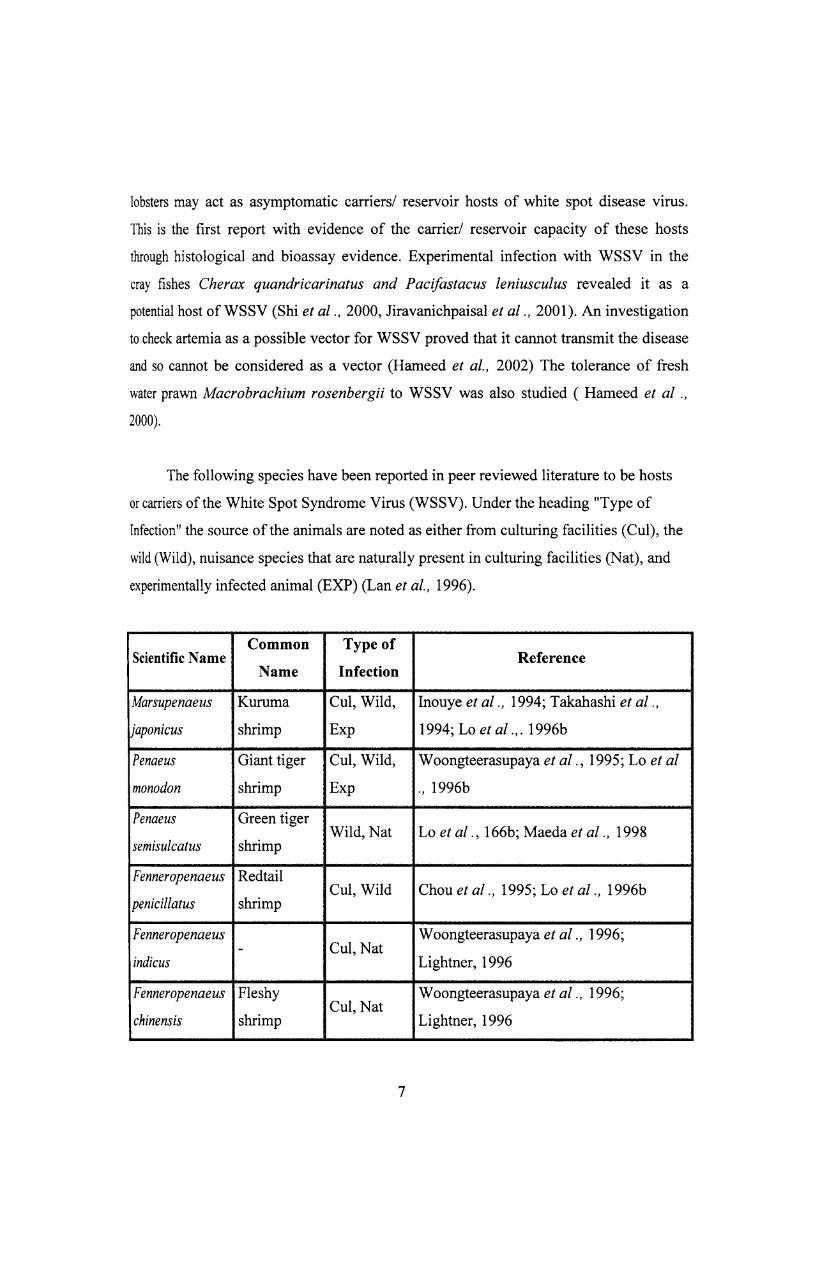
The following species have been reported in peer reviewed literature to be hosts
or carriers of the White Spot Syndrome Virus (WSSV). Under the heading "Type of
Infection" the source of the animals are noted as either from culturing facilities (CuI), the
wild (Wild), nuisance species that are naturally present in culturing facilities (Nat), and
experimentally infected animal (EXP) (Lan et al., 1996).
Common Type of Scientific Name Reference
Name Infection
Marsupenaeus Kuruma CuI, Wild, Inouye et al., 1994; Takahashi et al.,
japonicus shrimp Exp 1994; Lo et al.,. 1996b
Penaeus Giant tiger CuI, Wild, Woongteerasupaya et al., 1995; Lo et al
monodon shrimp Exp "' 1996b
Penaeus Green tiger Wild, Nat Lo et al ., 166b; Maeda et al., 1998
semisulcatus shrimp
Fenneropenaeus Redtail CuI, Wild Chou et al., 1995; Lo et al., 1996b
penicillatus shrimp
Fenneropenaeus CuI, Nat
Woongteerasupaya et al., 1996; -
indicus Lightner, 1996
Fenneropenaeus Fleshy CuI, Nat
Woongteerasupaya et al., 1996;
chinensis shrimp Lightner, 1996
7
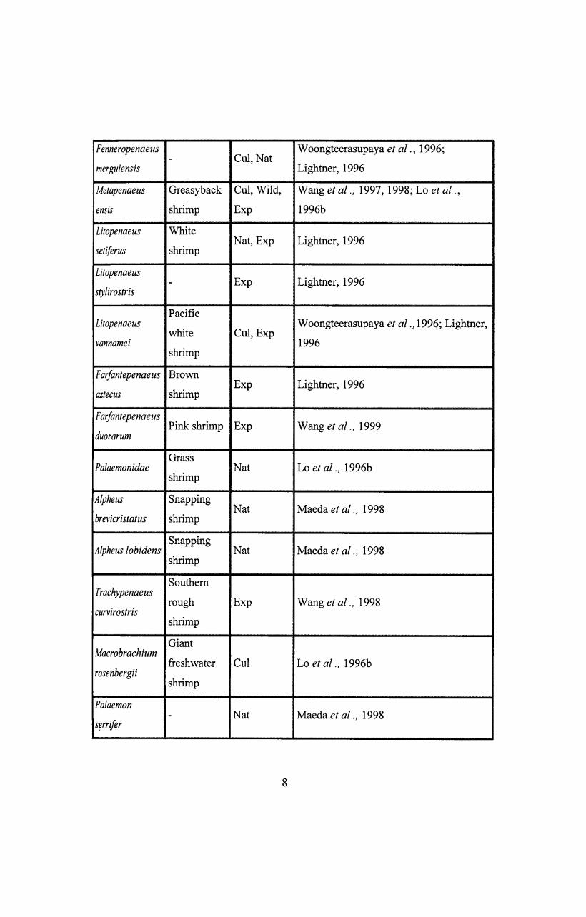
Fenneropenaeus CuI, Nat
Woongteerasupaya et al ., 1996; -
merguiensis Lightner, 1996
Metapenaeus Greasyback CuI, Wild, Wang et al., 1997, 1998; Lo et al.,
ensis shrimp Exp 1996b
Litopenaeus White
setiferus shrimp Nat, Exp Lightner, 1996
Litopenaeus Exp Lightner, 1996 -
stylirostris
Pacific Litopenaeus
white CuI, Exp Woongteerasupaya et al., 1996; Lightner,
vannamei 1996 shrimp
FarJantepenaeus Brown Exp Lightner, 1996
aztecus shrimp
FarJantepenaeus Pink shrimp
duorarum Exp Wang et al., 1999
Grass Palaemonidae Nat Lo et al., 1996b
shrimp
Alpheus Snapping Nat Maeda et al., 1998
brevicristatus shrimp
Alpheus lobidens Snapping
Nat Maeda et al., 1998 shrimp
Southern Trachypenaeus
curvirostris rough Exp Wang et al., 1998
shrimp
Giant Macrobrachium
freshwater CuI Lo et al., 1996b rosenbergii
shrimp
Palaemon - Nat Maeda et al., 1998
s~rrifer
8
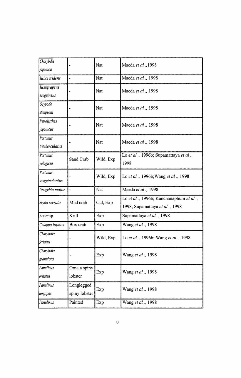
Charybdis Nat Maeda et al., 1998 -
japonica
Helice tridens - Nat Maeda et al., 1998
Hemigrapsus Nat Maeda et al., 1998 -
sanguine us
Ocypode Nat Maeda et al., 1998 -
stimpsoni
Petrolisthes - Nat Maeda et al., 1998
japonicus
Portunus - Nat Maeda et al., 1998
trituberculatus
Portunus Lo et al., 1996b; Supamattaya et al., Sand Crab Wild, Exp
pe/agicus 1998
Portunus - Wild, Exp Lo et al., 1996b;Wang et al., 1998
sanguinolentus
Upogebia major - Nat Maeda et al., 1998
Scylla serrata Mud crab CuI, Exp Lo et al., 1996b; Kanchanaphum et al.,
1998; Supamattaya et al., 1998
Acetes sp. Krill Exp Supamattaya et al., 1998
Ca/appa lophos Box crab Exp Wang et a/., 1998 . Charybdis
Wild, Exp Lo et a/., 1996b; Wang et al., 1998 -feriatus
Charybdis Exp Wang et al., 1998 -
granulata
Panulirus Omata spiny Exp Wang et al., 1998
ornatus lobster
Panulirus Longlegged
longipes spiny lobster Exp Wang et al., 1998
Panulirus Painted Exp Wang et al., 1998
9
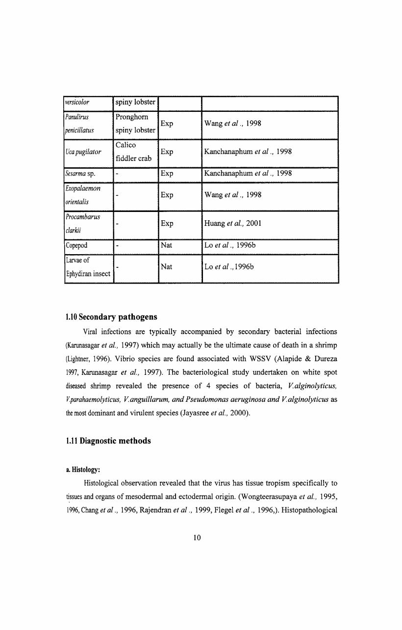
versicolor spiny lobster
Panulirus Pronghorn
penicillatus spiny lobster Exp Wang et al., 1998
Calico Uca pugilator
fiddler crab Exp Kanchanaphum et al., 1998
Sesarma sp. - Exp Kanchanaphum et al., 1998
Exopalaemon Exp Wang et al., 1998 -
orientalis
Procambarus - Exp Huang et al., 2001
clarkii
Copepod - Nat Lo et al., 1996b
Larvae of - Nat Lo et al., 1996b
Ephydiran insect
1.10 Secondary pathogens
Viral infections are typically accompanied by secondary bacterial infections
(Karunasagar et al., 1997) which may actually be the ultimate cause of death in a shrimp
(Lightner, 1996). Vibrio species are found associated with WSSV (Alapide & Dureza
1997, Karunasagar et al., 1997). The bacteriological study undertaken on white spot
diseased shrimp revealed the presence of 4 species of bacteria, Valginolyticus,
V.parahaemolyticus, Vanguillarum, and Pseudomonas aeruginosa and Valginolyticus as
the most dominant and virulent species (Jayasree et al., 2000).
1.11 Diagnostic methods
a. Histology:
Histological observation revealed that the virus has tissue tropism specifically to
tissues and organs of mesodermal and ectodermal origin. (Wongteerasupaya et al., 1995,
1996, Chang et al., 1996, Rajendran et al., 1999, Flegel et al., 1996,). Histopathological
10
condition includes severe nuclear hypertrophy, cellular degeneration, multi focal necrosis
and hemocytic encapsulation in the infected tissues. (Wongteerasupaya et al., 1995, 1996,
Chang et al., 1996, Rajendran et al., 1999, Lo et al., 1997, Karunasagar et al., 1997,
Flegel et al., 1996(b), Mohan et al., 1998, 1997, Lo et al(a)., 1996, Sudha et al., 1998,
Wang et al., 1997(a). Tissues of ectodermal and mesodermal origin such as sub cuticular
shell epithelium, gill epithelium, sub cuticular stomach epithelium, connective tissue,
haematopoietic tissue, antennal gland and nervous tissue are severely infected by the
virus. Development of intercellular hypertrophy observed in cells in the necrotic tissue
was different in different stages of the viral infection. Eosinophilic intranuclear inclusions
surrounded by marginated basophilic chromatin were found in the early stage. It was
followed by enlargement of the eosinophilic intracellular inclusions and finally the
swollen nuclei were filled with a prominent pale basophilic inclusion, which occupied
most of the cytoplasm of the infected cell (Kasornchandra et al., 1998).
b. Electron microscopy:
Electron microscopic examination revealed the presence of double walled
enveloped, non-occluded rod shaped virions. Complete virus is typically characterized by
an apical envelope extension. The nucleocapsid displays a superficially segmented
appearance. Each segment seems to be formed of sub units, which are arranged in 2
parallel rows. The cylinder representing the nucleocapsid is closed at one extremity by a
smaller segment those forms a slightly rounded end while the opposite extremity is
squared. Different views on the pattern of morphogenesis exist (Durand et al., 1996,
Huang et al., 1995, Huang et al., 2001, Inouye et al., 1994, Takahashi et al., 1994,
Wongteerasupaya, 1995).
c. DNA based diagnostics:
DNA hybridization probes for the white spot disease virus have been developed
by several laboratories (Chang et al., 1996, Durand et al., 1996). The primers for
detection of this virus by PCR technology have also been developed,
(a) Fl 5' ACTACT AACTTCAGCCTATCTAG3',
RI 5'TAATGCGGGTGTAATGTTCTTACG3',
11
F25' GTAACTGCCCCTTCCATCTCC3',
R2S' TACGGCAGCTGCTGCACCTTGT3'( Lo et al., 1996) where primers Fl and RI
amplify a 1447 bp fragment on the WSSV genome while F2 and R2 amplify a 941 bp
fragment internal to the 1447bp fragment. Kasornchandra et al ., 1998 developed another
pnmer,
(b) Fl 5'TCACATCGAGAGACCTCTGTAC3'
RI 5' TCT AGGACGGACGGACT ATGGCAA3' Which amplifies a 520bp fragment.
Amplified DNA of viral isolates from Thailand, Indonesia, Malaysia, China, Taiwan and
Japan.
Yet another primer developed by Thakahashi et al., 1996 is,
(c) Fl 5'GACAGAGATATGCAGGCCAA3'
RI5'ACCAGTGTTTCGTCATGGAG3'
Various other primers have been developed for the detection of WSSV (Wang et al.,
1996 (a), Nunan and Lightner 1997, Marielle et al., 2000, Karunasagar et al.,
(unpublished), Vijayan et al., (unpublished). Two commercial kits are available in India
marketed by Mangalore Biotech (P) Ltd, Mangalore and Bangalore Genei (P) Ltd,
Bangalore.
Several methods are available for the detection of white spot disease virus, which
include peR (Kim et al., 1998, Nunan et al., 1998, Peng et al., 1998, Hsu et al., 1999, Lo
et al., 1996 (b), Kaitpathomchai et al., 2001, Tang & Lightner, 2000, Tan et al., 2001
Otta et al., 1999), in situ hybridization (Chang et al., 1996, Durand et al., 1996,
Wongteerasupaya et al., 1996, Chang et al., 1998, Chang et al., 1996, Tsai et al., 1999)
dot blot hybridization (Wongteerasupaya et al., 1996, Hameed et al., 1998) and ELISA
(Harneed et al., 1998). A non-stop, single tube, semi-nested PCR technique for grading
the severity ofWSSV was also put in use (Kiatpathomchai et al., 2001). Quantification of
White spot syndrome virus DNA through a competitive polymerase chain reaction was
also done (Tang & Lightner, 2000). Tapay et al., 1999 developed primers for PCR based
on the sequence of a cloned fragment of the white spot disease virus genome and used the
primers to detect white spot disease virus from both experimentally and naturally infected
shrimp. They developed one step and two step PCR protocol as a very sensitive and
12
specific alternative protocol to Western blot assay for the detection of white spot disease
virus. A sensitive immunodot assay for WSSV was developed using the specific rabbit
polyc1onal antiserum developed from a truncated version of the WSSV 27.5 KDa
envelope protein (You et ai., 2002, Zhang et al., 2001). A dot blot nitrocellulose enzyme
immunoassay has been developed against WSSV (Nadala and Loh, 2000). Westernblot
(Nadala et al., 1997 , Bruce et ai., 1993), dot blot (Chang et al., 1998 (b), Southern blot
hybridization (Wang et al., 2000 (b)) Monoclonal antibodies (Zang et al., 1999, Zhan et
ai., 1999, Poulos et al., 2001, Shih et al., 2001, Liu et al., 2001,Anil et al., 2002) were
also used as diagnostic tools. An immunoassay with recombinant antigen of WSSV was
also carried out. Primary shrimp cell culture was also used for the study on WSSV
(Kasomchandra & Boonyaratpalin, 1998).
1.12 Management
Prevention and control of WSSV infection:
The major routes of infection are the infected water and carrier shrimp (Flagel et
al., 1995).
The best immediate approach to manage this virus is to implement a package of
preventive measures. These include pond preparation by disinfection and elimination of
potential viral carriers, the use of filters at the inlets to remove potential carriers, the
refusal to use fresh feed inputs, disinfection of ponds before discharge, and cessation of
water exchange for 4 days after a discharge. Monitoring of brood stock, post larvae and
pond reared shrimp using DNA probes. The most effective disinfection agent appears to
be chlorine at approximately 30 ppm. However, since the virus does not seem to remain
infections for more than a few days when free in seawater a simple process of storage can
remove this threat, so long as no carriers are present. Implementing this package will
require a good deal of cooperation on the part of the shrimp farmers.
Although no treatment are known that will rescue infected shrimp, work
originating at NICA has indicated that some medicinal plant extracts may be effective in
preventing YHV infection in aquarium trials (Direkbusarakom & Ruangpan, 1998).
13
Further tests are underway to confirm these results and to try to determine the mechanism
of protection. In addition there are indications that various nutrient supplements (eg.
Vitamin- C, HUF A's, Astaxan ) may improve chances of escaping from the virus. There
are still claims that various bacterial amendments can be used to prevent YHV infections,
if they are used continuously. It appears that the most effective disinfectant for WSSV is
formalin ( Pratanpipat et at., 1996). It is effective at 70 ppm (or even as little as 20 ppm
in aquarium tests) preventing transmission through water. This level may not directly
harm the plankton bloom and a consequent drop in DO. The situation is such that the
treatment may not deal with the carrier status of the virus and application of 70 ppm of
formalin at 6 hourly intervals is apparently required to prevent the transmission by
cohabitation.
The post larvae are strongly implicated as the possible route of WSSV
transmission to grow out systems. However, there are ways to block this route effectively.
It is shown that the impact of WSSV & MBV can be substantially reduced or essentially
eliminate by simply washing nauplei, with or without disinfectant after they are harvested
from spawning tanks (Chen, 1992). The practice of feeding fresh crab to brood stock
animals should also be stopped. If these measures are combined with prior brood stock
screening with a DNA probe, PL assay with either DNA probe or by way of diagnostic
peR, before stocking, it should be possible to close this route completely.
Mohan and Shankar, 1997 are of the opinion that the endodermal cells are not
affected and the infected shrimp may not shed the virus along with faces as it happens in
the case of monodon baculovirus (MBV) which is found only in midgut and
hepatopancrease. Death and disintegration of a WSSV infected shrimp appears to
contribute significantly to the viral load in the water. Removal of dead and moribund
shrimps in practical as an important management tool in shrimp farms of Thailand to
minimize the viral load in the water.
Karunasagar et aI., (1996) reported the use of an immunostimulant developed by
them as 'Aquastim' containing yeast glucan and a bacterial product. According to them,
14
the use of 'Aquastim' is perfectly environment friendly technology unlike the technology
of using chlorine and other anti microbial chemicals. However, regular application and
good water quality management would be important for successful cultivation. In
corporation of vitamin C in the feed can be recommended because this has been shown to
enhance the immune response in shrimp.
Rao, 1996 proposed the following management techniques for successful
cultivation of prawns, which include Pond preparation as the major task. As a
precautionary measure to present the virulence of shrimp virus, an antiviral herbal
powder made by fairly pulverizing the sun dried complex plant of Phyllanthus niruri, a
herb belonging to Euphorbiaceae family has been reported. The antiviral herbal powder
should be administrated in four doses during the culture period in monthly intervals. The
single dose should be 1-2 g/Kg feed for 3 days.
In case of white spot disease infected farms the disease can be cured by adopting
following procedures.
Continuous or daily draining of bottom water and pumping in fresh water till the
problem is solved. Make sure that there will be no abnormal or sudden change of vital
parameters. If change in inevitable, it should be gradual. Step should be taken to ensure
proper phytoplankton management. Administer anti-viral powder made from Phyllanthus
ninuri at the dose of2 tp 3 g/Kg feed /day for 4 to 5 days. The use of Phyllanthus spp and
Clinicanthus mutants for shrimp viral disease cure was supported by Dr. Boonsirm
Withyachumnarnkul, Department of Anatomy, Machidol University, Bangkok, Thailand
and the group - c.P. One litre / acre of a standard iodophore to inactivate virus and to
oxidise NH3 has been recommended.
The technique known as SLC-URINUM therapy is carried out by mixing SLC
URINUM with the supplementary pelleted feed to the shrimp which was kept at room
temperature for about 2-3 hrs (Chondar, 1996).
15
Yaligar and Pai, (1996) described the use of Calotropis gigantea to manage
WSSV in shrimp ponds.
Anti viral activity has been reported in shrimp for lipopolysacchrides
(Newman,1999), peptidoglycans from several species of bacteria such as
Bifidobacterium (Itami et al., 1998), Bacillus (Takahashi et al., 1998) and glucans from
Schizophyllan commune and Saccharomyces cereviseae (Song et al., 1997) all were
reported to have a protective effect on shrimp against WSSV. Meanwhile a suI fated
polysaccharide fucoidan has been found to be active on enveloped virus (Takahashi et al.,
1998).
For the management of WSSV rapid diagnostic technique for the avoidance,
determination of carriers, reservoirs of infection, development of specific pathogens free
(SPF) or resistant brood stock have to be given top priority. Another management
strategy shall be individual spawning of females and separate rearing of the resulting
larvae. Further, a set of similar non-destructive quantitative measures of shrimp health,
which can be used as part of early warning system, has to be developed. Perhaps studies
on crustacean immunity (eg. types of haemocytes, enzyme activities, haemolymph factors
etc.) will give some of the needed tools. Immunostimulants, probiotics and vaccines as
desired to replace antibiotics along with good management factors can be incorporated in
the management regime.
1.13 Conclusion
Since its emergence as the most important single pathogen in shrimp culture
systems WSSV has been given utmost importance among researchers. Thanks to their
efforts, so much is known now about the pathogen but still inadequate to have a fool
proof management strategy. Considering this requirement, the present work was
undertaken on three aspects such as,
16
I. Demonstration of the WSSV by histological and electron microscopic preparations
and unraveling its morphogenesis,
2. Development ofhyperimmune polyc1onal antiserum and
3. Development of a vaccine.
It is hoped that the achievements in the three fronts shall contribute to develop an
appropriate strategy for the management of WSSV.
17