CHAPTER 1 Qualitative Knowledge Models in Functional ...The queries present, in tabular format,...
24
& CHAPTER 1 Qualitative Knowledge Models in Functional Genomics and Proteomics MOR PELEG, IRENE S. GABASHVILI, and RUSS B. ALTMAN 1.1. INTRODUCTION Predicting pathological phenotypes based on genetic mutations remains a fundamental and unsolved issue. When a gene is mutated, the molecular function- ality of the gene product may be affected and many cellular processes may go awry. Basic molecular functions occur in networks of interactions and events that produce subsequent cellular and physiological functions. Most knowledge of these interactions is represented diffusely in the published literature, Excel lists, and specialized relational databases and so it is difficult to assess our state of under- standing at any moment. Thus it would be very useful to systematically store knowl- edge in data structures that allow the knowledge to be evaluated and examined in detail by scientists as well as computer algorithms. Our goal is to develop technol- ogy for representing qualitative, noisy, and sparse biological results in support of the eventual goal of fully accurate quantitative models. In a recent paper, we described an ontology that we developed for modeling bio- logical processes [1]. Ontologies provide consistent definitions and interpretations of concepts in a domain of interest (e.g., biology) and enable software applications to share and reuse the knowledge consistently [2]. Ontologies can be used to perform logical inference over the set of concepts to provide for generalization and expla- nation facilities [3]. Our biological process ontology combines and extends two existing components: a workflow model and a biomedical ontology, both described in the methods and tools section. Our resulting framework possesses the following properties: (1) it allows qualitative modeling of structural and functional aspects of a biological system, (2) it includes biological and medical concept models to allow for querying biomedical information using biomedical abstractions, (3) it allows 1 Genomics and Proteomics Engineering in Medicine and Biology. Edited by Metin Akay Copyright # 2007 the Institute of Electrical and Electronics Engineers, Inc.
Transcript of CHAPTER 1 Qualitative Knowledge Models in Functional ...The queries present, in tabular format,...
&CHAPTER 1
Qualitative Knowledge Models inFunctional Genomics andProteomics
MOR PELEG, IRENE S. GABASHVILI, and RUSS B. ALTMAN
1.1. INTRODUCTION
Predicting pathological phenotypes based on genetic mutations remains a
fundamental and unsolved issue. When a gene is mutated, the molecular function-
ality of the gene product may be affected and many cellular processes may go
awry. Basic molecular functions occur in networks of interactions and events that
produce subsequent cellular and physiological functions. Most knowledge of
these interactions is represented diffusely in the published literature, Excel lists,
and specialized relational databases and so it is difficult to assess our state of under-
standing at any moment. Thus it would be very useful to systematically store knowl-
edge in data structures that allow the knowledge to be evaluated and examined in
detail by scientists as well as computer algorithms. Our goal is to develop technol-
ogy for representing qualitative, noisy, and sparse biological results in support of the
eventual goal of fully accurate quantitative models.
In a recent paper, we described an ontology that we developed for modeling bio-
logical processes [1]. Ontologies provide consistent definitions and interpretations
of concepts in a domain of interest (e.g., biology) and enable software applications
to share and reuse the knowledge consistently [2]. Ontologies can be used to perform
logical inference over the set of concepts to provide for generalization and expla-
nation facilities [3]. Our biological process ontology combines and extends two
existing components: a workflow model and a biomedical ontology, both described
in the methods and tools section. Our resulting framework possesses the following
properties: (1) it allows qualitative modeling of structural and functional aspects of a
biological system, (2) it includes biological and medical concept models to allow for
querying biomedical information using biomedical abstractions, (3) it allows
1
Genomics and Proteomics Engineering in Medicine and Biology. Edited by Metin AkayCopyright # 2007 the Institute of Electrical and Electronics Engineers, Inc.

hierarchical models to manage the complexity of the representation, (4) it has a
sound logical basis for automatic verification, and (5) it has an intuitive, graphical
representation.
Our application domain is disease related to transfer ribonucleic acid (tRNA).
Transfer RNA constitutes a good test bed because there exists rich literature on
tRNA molecular structure as well as the diseases that result from abnormal struc-
tures in mitochondria (many of which affect neural processes). The main role of
tRNA molecules is to be part of the machinery for the translation of the genetic
message, encoded in messenger RNA (mRNA), into a protein. This process
employs over 20 different tRNA molecules, each specific for one amino acid and
for a particular triplet of nucleotides in mRNA (codon) [4]. Several steps take
place before a tRNA molecule can participate in translation. After a gene coding
for tRNA is transcribed, the RNA product is folded and processed to become a
tRNA molecule. The tRNA molecules are covalently linked (acylated) with an
amino acid to form amino-acylated tRNA (aa-tRNA). The aa-tRNA molecules
can then bind with translation factors to form complexes that may participate in
the translation process. There are three kinds of complexes that participate in trans-
lation: (i) an initiation complex is formed by exhibiting tRNA mimicry release
factors that bind to the stop codon in the mRNA template or by a misfunctioning
tRNA complexed with guanidine triphosphate (GTP) and elongation factor
causing abnormal termination, and (iii) a ternary complex is formed by binding
elongating aa-tRNAs (tRNAs that are acylated to amino acids other than formyl-
methionine) with GTP and the elongation factor EF-tu. During the translation
process, tRNA molecules recognize the mRNA codons one by one, as the mRNA
molecule moves through the cellular machine for protein synthesis: the ribosome.
In 1964, Watson introduced the classical two-site model, which was the accepted
model until 1984 [5]. In this model, the ribosome has two regions for tRNA
binding, so-called aminoacyl (A) site and peptidyl (P) site. According to this
model, initiation starts from the P site, but during the normal cycle of elongation,
each tRNA enters the ribosome from the A site and proceeds to the P site before
exiting into the cell’s cytoplasm. Currently, it is hypothesized that the ribosome
has at least three regions for tRNA binding: the A and P sites and an exit site
(E site) through which the tRNA exits the ribosome into the cell’s cytoplasm [6].
Protein synthesis is terminated when a stop codon is reached at the ribosomal A
site and recognized by a specific termination complex, probably involving factors
mimicking tRNA. Premature termination (e.g., due to a mutation in tRNA) can
also be observed [7].
When aa-tRNA molecules bind to the A site, they normally recognize and bind to
matching mRNA codons—a process known as reading. The tRNA mutations can
cause abnormal reading that leads to mutated protein products of translation.
Types of abnormal reading include (1) misreading, where tRNA with nonmatching
amino acid binds to the ribosome’s A site; (2) frame shifting, where tRNA that
causes frame shifting (e.g., binds to four nucleotides of the mRNA at the A site) par-
ticipates in elongation; and (3) halting, where tRNA that cause premature termin-
ation (e.g., tRNA that is not acetylated with an amino acid) binds to the A site.
2 QUALITATIVE KNOWLEDGE MODELS IN FUNCTIONAL GENOMICS AND PROTEOMICS

These three types of errors, along with the inability to bind to the A site or destruc-
tion by cellular enzymes due to misfolding, can create complex changes in protein
profiles of cells. This can affect all molecular partners of produced proteins in the
chain of events connecting genotype to phenotype and produce a variety of pheno-
types. Mutations in human tRNA molecules have been implicated in a wide range of
disorders, including myopathies, encephalopathies, cardiopathies, diabetes, growth
retardation, and aging [8]. Development of models that consolidate and integrate
our understanding of the molecular foundations for these diseases, based on avail-
able structural, biochemical, and physiological knowledge, is therefore urgently
needed.
In a recent paper [9], we discussed an application of our biological process ontol-
ogy to genomics and proteomics. This chapter extends the section on general com-
puter science theories, including Petri Nets, ontologies, and information systems
modeling methodologies, as well as extends the section on biological sources of
information and discusses the compatibility of our outputs with popular databases
and modeling environments.
The chapter is organized as follows. Section 1.2 describes the components we
used to develop the framework and the knowledge sources for our model. Section
1.3 discusses our modeling approach and demonstrates our knowledge model and
the way in which information can be viewed and queried using the process of trans-
lation as examples. We conclude with a discussion and conclusion.
1.2. METHODS AND TOOLS
1.2.1. Component Ontologies
Our framework combines and extends two existing components: The workflow
model and biomedical ontology. The workflow model [10] consists of a process
model and an organizational (participants/role) model. The process model can rep-
resent ordering of processes (e.g., protein translation) and the structural components
that participate in them (e.g., protein). Processes may be of low granularity (high-
level processes) or of high granularity (low-level processes). High-level processes
are nested to control the complexity of the presentation for human inspection.
The participants/role model represents the relationships among participants (e.g.,
an EF-tu is a member of the elongation factors collection in prokaryotes) and the
roles that participants play in the modeled processes (e.g., EF-tu has enzymic func-
tion: GTPase). We used the workflow model as a biological process model by
mapping workflow activities to biological processes, organizational units to bio-
molecular complexes, humans (individuals) to their biopolymers and networks of
events, and roles to biological processes and functions.
A significant advantage of the workflow model is that it can map to Petri Nets
[11], a mathematical model that represents concurrent systems, which allows veri-
fication of formal properties as well as qualitative simulation [12]. A Petri Net is
represented by a directed, bipartite graph in which nodes are either places or
1.2. METHODS AND TOOLS 3

transitions, where places represent conditions (e.g., parasite in the bloodstream) and
transitions represent activities (e.g., invasion of host erythrocytes). Tokens that are
placed on places define the state of the Petri Net (marking). A token that resides in a
place signifies that the condition that the place represents is true. A Petri Net can be
executed in the following way. When all the places with arcs to a transition have a
token, the transition is enabled, and may fire, by removing a token from each input
place and adding a token to each place pointed to by the transition. High-level Petri
Nets, used in this work, include extensions that allow modeling of time, data, and
hierarchies.
For the biomedical ontology, we combine the Transparent Access to Multiple
Biological Information Sources (TAMBIS) [13] with the Unified Medical Language
System (UMLS) [14]. TAMBIS is an ontology for describing data to be obtained
from bioinformatics sources. It describes biological entities at the molecular level.
UMLS describes clinical and medical entities. It is a publicly available federation
of biomedical controlled terminologies and includes a semantic network with 134
semantic types that provides a consistent categorization of thousands of biomedical
concepts. The 2002AA edition of the UMLS Metathesaurus includes 776,940 con-
cepts and 2.1 million concept names in over 60 different biomedical source vocabul-
aries. We augmented these two core terminological models [1] to represent
mutations and their effects on biomolecular structures, biochemical functions, cellu-
lar processes, and clinical phenotypes. The extensions include classes for represent-
ing (1) mutations and alleles and their relationship to sequence components, (2) a
nucleic acid three-dimensional structure linked to secondary and primary structural
blocks, and (3) a set of composition operators, based on the nomenclature of com-
position relationships, due to Odell [15].
Odell introduced a nomenclature of six kinds of composition. We are using three
of these composition relationships in our model. The relationship between a biomo-
lecular complex (e.g., ternary complex) and its parts (e.g., GTP, EF-tu, aa-tRNA) is
a component–integral object composition. This relationship defines a configuration
of parts within a whole. A configuration requires the parts to bear a particular func-
tional or structural relationship to one another as well as to the object they constitute.
The relationship between an individual molecule (e.g., tRNA) and its domains (e.g.,
D domain, T domain) is a place–area composition. This relationship defines a con-
figuration of parts, where parts are the same kind of thing as the whole and the parts
cannot be separated from the whole. Member–bunch composition groups together
molecules into collections when the collection members share similar functionality
(e.g., elongation factors) or cellular location (e.g., membrane proteins). We have
not found the other three composition relationships due to Odell to be relevant for
our model.
We implemented our framework using the Protege-2000 knowledge-modeling
tool [16]. We used Protege’s axiom language (PAL) to define queries in a subset
of first-order predicate logic written in the Knowledge Interchange Format syntax.
The queries present, in tabular format, relationships among processes and structural
components as well as the relationship between a defective process or clinical phe-
notype and the mutation that is causing it.
4 QUALITATIVE KNOWLEDGE MODELS IN FUNCTIONAL GENOMICS AND PROTEOMICS
1.2.2. Translation into Petri Nets
We manually translated the tRNA workflow model into corresponding Petri Nets,
according to mapping defined by others [12]. The Petri Net models that we used
were high-level Petri Nets that allow the representation of hierarchy and data. Hier-
archies enable expanding a transition in a given Petri Net to an entire Petri Net, as is
done in expanding workflow high-level processes into a net of lower level processes.
We upgraded the derived Petri Nets to Colored Petri Nets (CPNs) by:
1. Defining color sets for tRNA molecules (mutated and normal), mRNA mol-
ecules, and nucleotides that comprise the mRNA sequence and initiating the
Petri Nets with an initial marking of colored tokens
2. Adding guards on transitions that relate to different types of tRNA molecules
(e.g., fMet-tRNA vs. elongating tRNA molecules)
3. Defining mRNA sequences that serve as the template for translation
We used the Woflan Petri Net verification tool [17] to verify that the Petri Nets
are bounded (i.e., no accumulation of an infinite amount of tokens) and live (i.e.,
deadlocks do not exist). To accommodate limitations in the Woflan tool, which
does not support colored Petri Nets, we manually made several minor changes to
the Petri Nets before verifying them. We simulated the Petri Nets to study the
dynamic aspects of the translation process using the Design CPN tool [18], which
has since been replaced by CPN Tools.
1.2.3. Sources of Biological Data
We gathered information from databases and published literature in order to develop
the tRNA example considered in this work. We identified data sources with infor-
mation pertaining to tRNA sequence, structure, modifications, mutations, and
disease associations. The databases that we used were:
. Compilation of mammalian mitochondrial tRNA genes [19], aimed at defining
typical as well as consensus primary and secondary structural features of mam-
malian mitochondrial tRNAs (http://mamit-trna.u-strasbg.fr/)
. Compilation of tRNA sequences and sequences of tRNA genes [20] (http://www.uni-bayreuth.de/departments/biochemie/sprinzl/trna/)
. The Comparative RNA website (http://www.rna.icmb.utexas.edu/), which
provides a modeling environment for sequence and secondary-structure com-
parisons [21]
. Structural Classifications of RNA (SCOR, http://scor.lbl.gov/scor.html) [22]
. The RNA Modification Database (http://medlib.med.utah.edu/RNAmods),
which provides literature and data on nucleotide modifications in RNA [23]
. A database on tRNA genes and molecules in mitochondria and photosynthetic
eukaryotes (http://www.ba.itb.cnr.it/PLMItRNA/) [8]
1.2. METHODS AND TOOLS 5

. Online Mendelian Inheritance in Man (OMIM) (http://www.ncbi.nlm.nih.
gov/omim/), which catalogs human genes and genetic disorders [24]
. BioCyc (http://metacyc.org/), a collection of genome and metabolic pathway
databases which describes pathways, reactions, and enzymes of a variety of
organisms [25]
. Entrez, the life sciences search engine, which provides views for a variety of
genomes, complete chromosomes, contiged sequence maps, and integrated
genetic and physical maps (http://www.ncbi.nlm.nih.gov/gquery/gquery.fcgi?itool ¼ toolbar) [26]
. MITOMAP, A human mitochondrial genome database [27] (http://www.
mitomap.org/)
. The UniProt/Swiss-Prot Protein Knowledgebase, which gives access to
wealthy annotations and publicly available resources of protein information
(http://us.expasy.org/sprot/sprot-top.html)
In addition, we used microarrays [28] and mass spectral data [29], providing
information on proteins involved in tRNA processing or affected by tRNA
mutations.
1.3. MODELING APPROACH AND RESULTS
Our model represents data using process diagrams and participant/role diagrams.
Appendix A on our website (http://mis.hevra.haifa.ac.il/�morpeleg/NewProcess
Model/Malaria_PN_Example_Files.html) presents the number of processes,
participants, roles, and links that we used in our model. The most granular
thing that we represented was at the level of a single nucleotide (e.g., GTP).
The biggest molecule that we represented was the ribosome. We chose our
levels of granularity in a way that considers the translation process under the
assumption of a perfect ribosome; we only considered errors in translation that
are due to tRNA. This assumption also influenced our design of the translation
process model. This design follows individual tRNA molecules throughout the
translation process and therefore represents the translocation of tRNA molecules
from the P to the E site and from the A to the P site as distinct processes that
occur in parallel. The level of detail in which we represented the model led us
to consider questions such as (1) “Can tRNA bind the A site before previously
bound tRNA molecule is released from the E site?” and (2) “Can fMet tRNA
form a ternary complex?”
1.3.1. Representing Mutations
Variation in gene products (protein or RNA) can result from mutations in the nucleo-
tide sequence of a gene, leading to altered (1) translation, (2) splicing, (3) posttran-
scriptional end processing, or (4) interactions with other cellular components
coparticipating in biological processes. In addition, variation can result from a
6 QUALITATIVE KNOWLEDGE MODELS IN FUNCTIONAL GENOMICS AND PROTEOMICS

normal sequence that is translated improperly by abnormal tRNA molecules.
Thus, we must be able to represent variation not only in DNA sequences
(genome) but also in RNA and protein. Therefore, in our ontology, every sequence
component (of a nucleic acid or protein) may be associated with multiple alleles.
Each allele may have mutations that are either pathogenic (associated with abnormal
functions) or neutral. A mutation is classified as a substitution, insertion, or
deletion [30].
1.3.2. Representing Nucleic Acid Structure
The TAMBIS terminology did not focus on three-dimensional structure. We
extended the TAMBIS ontology by specifying tertiary-structure components of
nucleic acids. A nucleic acid tertiary-structure component is composed of interact-
ing segments of nucleic acid secondary-structure components. We added three
types of nucleic acid secondary-structure components: nucleic acid helix, nucleic
acid loop, and nucleic acid unpaired strand. Figure 1.1 shows the tertiary-structure
components of tRNA (acceptor domain, D domain, T domain, variable loop, and
anticodon domain). Also shown is the nucleic acid tertiary-structure component
frame that corresponds to the tRNA acceptor domain. The division of tRNA into
structural domains, the numbering of nucleotides of the generic tRNA molecule,
and the sequence-to-structure correspondence was done according to conventional
rules [20].
FIGURE 1.1. Tertiary-structure components. Normal tRNA is composed of five nucleic acid
tertiary-structure components. One of these components (tRNA acceptor domain) is shown in
the middle frame. Each nucleic acid tertiary-structure component is composed of segments of
nucleic acid secondary-structure components. The nucleic acid unpaired strand of the tRNA
acceptor domain, which is a kind of nucleic acid secondary-structure component, is shown on
the right.
1.3. MODELING APPROACH AND RESULTS 7

FIG
UR
E1.2
.
8

1.3.3. Representing Molecular Complexes
Biological function can be associated with different levels of molecular structure. In
some cases a function can be associated with a domain (of a protein or nucleic acid).
In other cases, a function is associated with individual molecules or with molecular
complexes. Sometimes, a function is not specifically mapped to a molecular struc-
ture but is attributed to collections of molecules that are located in a particular cel-
lular compartment. In addition, biologists define collections of molecules that share
a common function (e.g., termination factors). The participant/role representation of
our framework represents molecular structures that participate in processes as well
as composition and generalization relationships among participants (molecules).
In our tRNA example, we are using three kinds of these composition relation-
ships: (1) component–integral object composition, (2) member–bunch compo-
sition, and (3) place–area composition. Figure 1.2 shows examples of these
relationships. Generalization (is-a) relationships are used to relate subclasses of par-
ticipants to their superclasses. For example, terminator tRNA, nonterminating
tRNA, and fMet tRNA are subclasses of the tRNA class.
1.3.4. Representing Abnormal Functions and Processes
In addition to representing relationships among process participants, our framework
can represent the roles that participants have in a modeled system. We distinguish
two types of roles: molecular-level functional roles (e.g., a role in translation) and
roles in clinical disorders (e.g., the cause of cardiomyopathy). Each role is specified
using a function/process code taken from the TAMBIS ontology. To represent
dysfunctional molecular-level roles, we use an attribute, called role_present, which
signifies whether the role is present or absent or this information is unknown. For
example, Figure 1.2 shows that three mutations of tRNA that exhibit the role of
misreading. The figure also shows tRNA mutations that have roles in the cardio-
myopathy disorder. Cardiomyopathy is one of the concepts from the clinical
ontology, discussed later in this section.
FIGURE 1.2. Part of participant/role diagram showing molecules involved in translation
and roles they fulfill. Individual molecules are shown as rectangles (e.g., tRNA). They are
linked to domains (e.g., D domain) using dashed connectors. Biomolecular complexes are
shown as hexagons (e.g., ternary complex) and linked to their component molecules using
arrowhead connectors. Collections of molecules that share similar function or cellular
location are shown as triangles (e.g., elongation factors) and are linked to the participants
that belong to them using connectors with round heads. Generalization relationships are
shown as dotted lines (e.g., fMet-tRNA is-a tRNA). Functional roles are shown as ellipses
that are linked to the participants that exhibit those roles. Clinical disorders that are associated
with mutated participants are shown as diamonds (e.g., cardiomyopathy) and are linked to the
participants that exhibit roles in these disorders. The insert shows the details of the misreading
role. It is specified as a translation role (TAMBIS class) that is not present (role_present ¼
false). Also shown are some of the participants that perform the misreading role.
1.3. MODELING APPROACH AND RESULTS 9

FIG
UR
E1
.3.
10

Processes are represented using the process model component of our framework.
We augmented the workflow model with elements taken from the object process
methodology (OPM) [31] to create a graphical representation of the relationships
between a process and the static components that participate in it, as shown in
Figure 1.3. We used different connectors to connect a process to its input sources,
output sources, and participants that do not serve as substrates or products (e.g., cat-
alysts such as amino acid synthetase). We added a fourth type of connector that links
a process to a chemical that inhibits the process (e.g., borrelidin). Figures 1.3
through 1.6 present details of the translation process and the processes leading to
it. The figures show the normal process as well as processes that result in abnormal
translation. We have considered only tRNA-related failures of translation. Detailed
explanation of each process diagram is given in the legends. Figures 1.4 and 1.5
present the details of the translation process, depicted in Figure 1.3. Figure 1.4 pre-
sents the translation process according to the classical two-site model [5]. Figure 1.5
presents a recent model of the translation process [32]. The details of the process of
tRNA binding to the A site, of Figure 1.5, are shown in Figure 1.6.
The processes normal reading, misreading, frame shifting, and halting, shown in
Figure 1.6, all have a process code of binding, since in all of them tRNA binds to
ribosome that has occupied E and P sites.
The types of arrows that connect molecules to a process define their role as sub-
strates, products, inhibitors, activators, or molecules that participate without chan-
ging their overall state in the framework (e.g., enzyme). The logical relationships
among participants are specified in a formal expression language. For example,
double-clicking on the misreading process, shown in Figure 1.6, shows its partici-
pants, which are specified as
(Shine–Delgarno in E XOR tRNA0 in E) AND tRNA1 in P AND(tRNA2 that can bind to incorrect codon in ternarycomplex XOR tRNA that has altered flexibility internary complex) AND tRNA2 in A AND EF-tu AND GDP
FIGURE 1.3. Process diagram showing processes leading to translation. Ellipses represent
activities. Ellipses with bold contours represent high-level processes, whereas ellipses
without bold contours represent low-level processes (that are not further expanded). The
dark rounded rectangles represent routing activities for representing logical relationships
among component activities of a process diagram. The router (checkpoint) labeled XOR rep-
resents a XOR split that signifies that the two processes that it connects to are mutually exclu-
sive. A XOR join connects the three processes shown in the middle of the diagram to the
translation process. Dotted arrows that link two activities to each other represent order
relationships. Participants are shown as light rectangles. Arrows that point from a participant
toward a process specify that the participant is a substrate. Arrows that point in the opposite
direction specify products. Connectors that connect participants (e.g., amino acid synthetase)
to processes and have a circle head represent participation that does not change the state of the
participant. Inhibitors (e.g., tobramycin) are linked to processes via a dashed connector. The
details of the translation process are shown in Figures 1.4 and 1.5.
1.3. MODELING APPROACH AND RESULTS 11
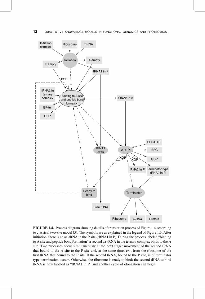
FIGURE 1.4. Process diagram showing details of translation process of Figure 1.4 according
to classical two-site model [5]. The symbols are as explained in the legend of Figure 1.3. After
initiation, there is an aa-tRNA in the P site (tRNA1 in P). During the process labeled “binding
to A site and peptide bond formation” a second aa-tRNA in the ternary complex binds to the A
site. Two processes occur simultaneously at the next stage: movement of the second tRNA
that bound to the A site to the P site and, at the same time, exit from the ribosome of the
first tRNA that bound to the P site. If the second tRNA, bound to the P site, is of terminator
type, termination occurs. Otherwise, the ribosome is ready to bind; the second tRNA to bind
tRNA is now labeled as “tRNA1 in P” and another cycle of elongation can begin.
12 QUALITATIVE KNOWLEDGE MODELS IN FUNCTIONAL GENOMICS AND PROTEOMICS

FIGURE 1.5. Process diagram showing details of the translation process of Figure 1.4
according to model of Connell and Nierahus [32]. The details of the process labeled
“binding of tRNA to A site” are shown in Figure 1.6. After initiation, shine dalgarno is
placed at the E site, and the first tRNA (tRNA1) is placed at the P site. Next, tRNA2 transi-
ently binds to the A site. This step is followed by three activities which are done concurrently:
(1) exit from the E site of either Shine–Delgarno or tRNA0 bound to the E site (at later stages
of the elongation process), (2) binding to the A site followed by peptide bond formation, and
(3) a routing activity (marked by an unlabeled round-corner square). The routing activity is
needed for correspondence with the CPN that simulates the translation process, which
needs to distinguish among the tRNA molecules that are bound to each of the three sites.
At the next stage, tRNA2 at the A site shifts to the P site and at the same time, tRNA1 at
the P site shifts to the E site. If tRNA2 bound to the P site is of terminator type, termination
occurs. Otherwise, the ribosome is ready to bind; the second tRNA to bind is now labeled as
“bound tRNA1,” and the first tRNA to bind is labeled as “bound tRNA0,” and another cycle of
elongation can begin.
1.3. MODELING APPROACH AND RESULTS 13

FIG
UR
E1
.6.
Pro
cess
dia
gra
msh
ow
ing
no
rmal
and
abn
orm
alre
adin
gp
roce
sses
.S
tart
ing
fro
ma
rib
oso
me
wit
htR
NA
inb
oth
Ean
dP
site
s,fo
ur
alte
rnat
ive
pro
cess
esca
nle
adto
rib
oso
me
wit
hth
eA
site
occ
up
ied
wit
htR
NA
:n
orm
alre
adin
g,m
isre
adin
g,fr
ame
shif
tin
g,an
dh
alti
ng
.S
ym
bo
lsar
e
asex
pla
ined
inth
ele
gen
do
fF
igu
re1
.3.
Th
ein
sert
sho
ws
the
pro
cess
cod
e(b
ind
ing
)o
fth
ere
adin
gp
roce
ss,
tak
enfr
om
the
TA
MB
ISo
nto
log
y.
14
1.3.5. Representing High-Level Clinical Phenotypes
Our clinical ontology relies on the UMLS but does not include all of the concepts of
the Metathesaurus. Instead, we are building our clinical ontology by importing con-
cepts as we need them. We add clinical concepts to the clinical ontology by creating
them as subclasses of the semantic types defined by the semantic network. Each
concept has a concept name and a concept code that come from the Metathesaurus
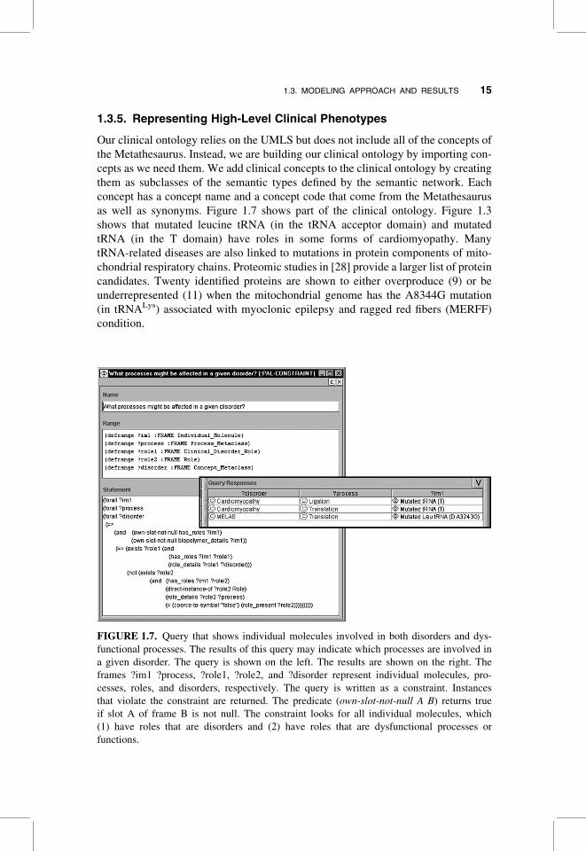
as well as synonyms. Figure 1.7 shows part of the clinical ontology. Figure 1.3
shows that mutated leucine tRNA (in the tRNA acceptor domain) and mutated
tRNA (in the T domain) have roles in some forms of cardiomyopathy. Many
tRNA-related diseases are also linked to mutations in protein components of mito-
chondrial respiratory chains. Proteomic studies in [28] provide a larger list of protein
candidates. Twenty identified proteins are shown to either overproduce (9) or be
underrepresented (11) when the mitochondrial genome has the A8344G mutation
(in tRNALys) associated with myoclonic epilepsy and ragged red fibers (MERFF)
condition.
FIGURE 1.7. Query that shows individual molecules involved in both disorders and dys-
functional processes. The results of this query may indicate which processes are involved in
a given disorder. The query is shown on the left. The results are shown on the right. The
frames ?im1 ?process, ?role1, ?role2, and ?disorder represent individual molecules, pro-
cesses, roles, and disorders, respectively. The query is written as a constraint. Instances
that violate the constraint are returned. The predicate (own-slot-not-null A B) returns true
if slot A of frame B is not null. The constraint looks for all individual molecules, which
(1) have roles that are disorders and (2) have roles that are dysfunctional processes or
functions.
1.3. MODELING APPROACH AND RESULTS 15

1.3.6. Representing Levels of Evidence for Modeled Facts
Different facts that are represented in our framework are supported by varying
degrees of evidence. It is important to allow users to know what support different
facts have, especially in cases of conflicting information. We therefore added a cat-
egorization of evidence according to the type of experimentation by which facts
were established. The categorization includes broad categories, such as “in vivo,”
“in vitro,” “in situ,” “in culture,” “inferred from other species,” and “speculative.”
Facts, such as the existence of a biomolecule or its involvement in a process are
tagged with the evidence categories.
1.3.7. Querying the Model
Using PAL we composed first-order logic queries that represent in tabular form
relationships among processes and structural components. Table 1.1 shows a
TABLE 1.1 Types of Biological Queries and Motivating Biological Examples
Query Type Example Derived Answer from Model
1. Alleles
1.1 Alleles that have
roles in dysfunctional
processes and/or
disorders
Alleles that have roles in
both dysfunctional pro-
cesses and disorders
Mutated tRNA (T) causes cardi-
omyopathy and has roles in
amino acylationþ halted
translation
Mutated Leu tRNA (D) causes
mitochondrial myopathy
encephalopathy lactocidosis
stroke (MELAS) and has a role
in misreading
2. Roles
2.1 Individual molecules
or biocomplexes that have
the same role
Scoped to cellular
location, same substrates
and products, same bio-
logical process (partici-
pation), or to same (or
different) inhibitor
Individual molecules that
have the same set of
roles
Individual molecules that
have a role in a dys-
functional process
Individual molecules that
have a role in a disorder
Mutated tRNA (anticodon) and
mutated tRNA (acceptor) both
have only the role of misreading
Incorrect translation: mutated
tRNA (anticodon), mutated
tRNA (T), mutated Pro tRNA
(anticodon U34mU), mutated
Leu tRNA (D A3243G), mutated
tRNA (acceptor)
Incorrect ligation: mutated
tRNA (T)
Incorrect processing: tRNA
precursor with mutated 30 end
Cardiomyopathy: mutated tRNA
(T), mutated Leu tRNA
(acceptor)
MELAS: mutated Leu tRNA (D)
(continued )
16 QUALITATIVE KNOWLEDGE MODELS IN FUNCTIONAL GENOMICS AND PROTEOMICS

TABLE 1.1 Continued
Query Type Example Derived Answer from Model
3. Reaction (functional model)
3.1 All atomic
activities that share
the same substrates
(products, inhibitors)
What atomic activities
have the same sub-
strates and products?
None in the modeled system
4. Biological process
4.1 All activities of a
certain kind of biological
process, according to the
TAMBIS classification
hierarchy (scoped to
cellular location
substrates)
All activities that are a
kind of binding and
involve binding of
tRNA
Formation of ternary complex,
formation of initiation com-
plex, formation of termination
complex, binding to A site,
normal reading, misreading,
halting, frame shifting
4.2 All activities that are
inhibited by inhibitor x
Activities inhibited by
tobramycin and
mupirocin
Amino acid acylation
4.3 What processes
might be affected in a
given disorder?
What processes might be
affected in a given
disorder?
Amino acid acylation and trans-
lation (reading) are affected in
cardiomyopathy
Translation (reading) is affected
in cardiomyopathy
5. Reachability
5.1 If an activity is
inhibited what other
activities can take place?
Is it a deadlock?
Inhibiting “normal read-
ing” (no supply of
normal tRNA): what
activities may take
place?
Directly in XOR: misreading,
frame shifting, halting
5.2 If an activity is
inhibited, can we still
get to a specified state?
If we inhibit “formation of
ternary complex,” can
we reach a state where
the activity “termin-
ation” is enabled?
Yes. For example, the firing
sequence t1t2t4t1t2t5t6t7t8t10t11
t12t13 of Figure 1.8
5.3 Does an inhibitor
inhibit an entire
high-level process?
Does tobramycin inhibit
the translation process?
Yes. It inhibits the process
“formation of initiation
complex” which is essential to
take place before translation
5.4 Establish a marking,
find reachability
Elongating tRNA is a
substrate. What path-
ways will be taken?
Amino acid acylation, followed
by formation of ternary
complex, followed by
translation
6. Temporal/dynamic aspects
6.1 What other
processes occur in
parallel to process X?
What processes occur in
parallel to “binding to A
site” (Fig. 1.6)
“Shine–Delgarno exits”
XOR “tRNA1 exits”
1.3. MODELING APPROACH AND RESULTS 17

FIGURE 1.8. Colored Petri Net that corresponds to Figure 1.5 showing current three-site
model of translation. Squares represent transitions, corresponding to workflow processes.
Ellipses represent places, corresponding to conditions that are true after a workflow process
has terminated. Text to the top left of places indicates their allowed token type, which can be
tRNA or mRNA. The values of tokens of tRNA type used in this figure are Shine_
Delgarno, Initiator_tRNA, Terminator_tRNA, Terminator, and Lys_Causing_Halting. Other
token types that we use in our model (not shown) represent other mutations of tRNA molecules.
The values of tokens of mRNA type are always “normal.” Text below places specifies initial
placement of tokens in those places. Text above transitions indicates guarding conditions,
which refer to token types. Text on connectors indicates token variables that flow on those con-
nectors. The variables used are a, b, and c for tRNA tokens and m for mRNA tokens. Transitions
are also labeled t6, . . . , t15, in correspondence with query 5.2 of Table 1.1.
18 QUALITATIVE KNOWLEDGE MODELS IN FUNCTIONAL GENOMICS AND PROTEOMICS
summary of all the query types that we composed. They are grouped into six
categories that concern (1) alleles, (2) functional roles and roles in disorder pheno-
types, (3) reactions and their participants, (4) biological processes, (5) ability to
reach a certain state of a modeled system, and (6) temporal/dynamic aspects of a
modeled system. Queries that were especially interesting to us were (1) finding
mutations that cause molecular-level processes and functions to be dysfunctional,
(2) finding mutations that cause clinical disorders, and (3) finding processes that
might be affected in a given disorder. Figure 1.7 shows the query and query
results for the third query.
1.3.8. Simulating the Model
As shown in Figures 1.4 and 1.5, we created two different models of the translation
process: a historical model and a current model. When we translated the workflow
models into the corresponding Petri Nets, we were able to test predictions of these
two models by showing that under certain concentrations of reactants the different
models resulted in different dynamic behavior which produced different translation
products. For example, when the mRNA contained a sequence of Asn–Leu–Asn (or
in general, aa1–aa2–aa1) and the system was initialized with a low concentration of
Asn-tRNA, then protein translation proceeded in the classical two-site model but
was halted in the current three-site model, which required Asn-tRNA and Leu-
tRNA to be bound to the ribosome while a second Asn-tRNA bound the A site.
The Petri Net that corresponds to the workflow model of Figure 1.5 is shown in
Figure 1.8. The tRNA mutations were represented as colored tokens, belonging to
the tRNA color set (see Fig. 1.8), and mRNA molecules were represented as
tokens belonging to the mRNA color set.
The Petri nets derived from our workflow model can also be used for educational
purposes. They can demonstrate (1) concurrent execution of low-level processes
within the translation process (e.g., tRNA molecules that were incorporated into syn-
thesized proteins can be amino acylated and used again in the translation process),
(2) introduction of mutations into synthesized proteins, and (3) the affect of certain
dysfunctional components on pools of reactants (e.g., nonmutated tRNAs).
1.4. DISCUSSION
Deducing molecular mechanisms of disease based on molecular models is a very dif-
ficult problem. Even more complicated is the task of correlating genotypic variation
to clinical phenotypes. A review by Florentz and Sissler [33] shows that, despite the
accumulation of information about the positions of a large number of mutations
within mitochondrial tRNAs, it is not possible to identify simple basic patterns
for use in predicting the pathogenicity of new mutations. The multifaceted nature
of effects produced by tRNA mutations is apparent from recent proteomics
studies [29] and is emphasized in current reviews [34, 35]. The authors conclude
that it is critical to examine not only the affected tRNA but also its interactions,
or relationships, with other compartmental components. These arguments
1.4. DISCUSSION 19
emphasize the importance of a knowledge model able to integrate practical infor-
mation at multiple levels of detail and from multiple experimental sources.
The knowledge framework presented here links genetic sequence, structure, and
local behavior to high-level biological processes (such as disease). The model pro-
vides a mechanism for integrating data from multiple sources. In our tRNA example,
we integrated information from structural biology, genetics and genomics, molecu-
lar biology, proteomics, and clinical science. The information can be presented
graphically as process diagrams or participant/role diagrams. The frames that rep-
resent participants, roles, processes, and relationships among them contain citations
to the original data sources.
Our model has several advantages, in addition to its ability to integrate data from
different sources. First, we can define queries that create views of the model in a
tabular format. The queries extract useful relationships among structures, sequences,
roles, processes, and clinical phenotypes. Second, our model can be mapped in a
straightforward manner to Petri Nets. We developed software that automatically
translates our biological process model into Petri Net formalisms and formats
used by various Petri Net tools [36]. We have used available tools to qualitatively
simulate a modeled system and to verify its boundedness and liveness and to
answer a set of biological questions that we defined [36]. Boundedness assumes
that there is no infinite accumulation of tokens in any system state. In our
example, this corresponds to concentration of tRNA and mRNA molecules in a
cell. Liveness ensures that all Petri Net transitions (which correspond to workflow
activities) can be traversed (enabled).
A disadvantage of our model is its need for manual data entry. Natural language
processing techniques are not able to automatically parse scientific papers into the
semantic structure of our ontology. The effort required to enter data into our
model is considerable. The entry of a substantial set of data about all relevant cel-
lular reactions and processes would require a major distributed effort by investi-
gators trained in knowledge representation and biology.
1.5. CONCLUSION
One of the ultimate goals of proteomics and genomics engineering is to develop a
model of the real cell, of its program responsible for different behaviors in
various intra- and extracellular environments. Our long-term goal is to develop a
robust knowledge framework that is detailed enough to represent the phenotypic
effects of genomic mutations. The results presented here are a first step in which
we demonstrate that the knowledge model developed in another context (malaria
invasion biology) is capable of capturing a qualitative model of tRNA function.
We have presented a graphical knowledge model for linking genetic sequence poly-
morphisms to their structural, functional, and dynamic/behavioral consequences,
including disease phenotypes. We have shown that the resulting qualitative model
can be queried (1) to represent the compositional properties of the molecular ensem-
bles, (2) to represent the ways in which abnormal processes can result from
20 QUALITATIVE KNOWLEDGE MODELS IN FUNCTIONAL GENOMICS AND PROTEOMICS
structural variants, and (3) to represent the molecular details associated with high-
level physiological and clinical phenomena. By translating the workflow represen-
tation into Petri Nets we were able to verify boundedness and liveness. Using simu-
lation tools, we showed that the Petri Nets derived from the historic and current
views of the translation process yield different dynamic behavior.
ACKNOWLEDGMENTS
The work was funded by the Burroughs-Wellcome Fund and by National Institutes of Health
grants LM-05652 and LM-06422.
REFERENCES
1. M. Peleg, I. Yeh, and R. B. Altman, “Modeling biological processes using Workflow and
Petri Net models,” Bioinformatics, 18: 825–837, 2002.
2. T. R. Gruber, “Toward principles for the design of ontologies used for knowledge
sharing,” Int. J. Human-Computer Stud., 43: 907–928, 1995.
3. S. Schulze-Kremer, “Ontologies for molecular biology,” paper presented at the Proceed-
ings of the Third Pacific Symposium on Biocomputing, Hawaii, 1998.
4. M. Ibba, C. Stathopoulos, and D. Soll, “Protein synthesis: Twenty three amino acids and
counting,” Curr Biol., 11: R563–565, 2001.
5. K. H. Nierahus, “New aspects of the ribosomal elongation cycle,” Mol. Cell Biochem.,
61: 63–81, 1984.
6. D. N. Wilson, G. Blaha, S. R. Conell, P. V. Ivanov, H. Jenke, U. Stelzl, Y. Teraoka, and K. H.
Nierahus, “Protein synthesis at atomic resolution: Mechanistics of translation in the light of
highly resolved structures for the ribosome,” Curr. Protein Peptide Sci., 3: 1–53, 2002.
7. P. J. Farabaugh and G. R. Bjork, “How translational accuracy influences reading frame
maintenance,” EMBO J., 18: 1427–1434, 1999.
8. V. Volpetti, R. Gallerani, C. D. Benedetto, S. Liuni, F. Licciulli, and L. R. Ceci,
“PLMltRNA, a database on the heterogenous genetic origin of mitochondrial tRNa
genes and tRNAs in photosynthetic eukaryotes,” Nucleic Acids Res., 31: 436–438, 2003.
9. M. Peleg, I. S. Gabashvili, and R. B. Altman, “Qualitative models of molecular function:
Linking genetic polymorphisms of tRNA to their functional sequelae,” Proc. IEEE, 90:
1875–1886, 2002.
10. L. Fisher, Workflow Handbook, published in association with the Workflow Management
Coalition, Future Strategies, Lighthouse Point, FL, 2001.
11. J. L. Peterson, Petri Net Theory and the Modeling of Systems, Prentice-Hall, Englewood
Cliffs, NJ, 1981.
12. W. M. P. v. d. Aalst, “The application of Petri Nets to workflow management,”
J. Circuits, Syst. Computers, 8: 21–66, 1998.
13. P. G. Baker, C. A. Goble, S. Bechhofer, N. W. Paton, R. Stevens, and A. Brass,
“An ontology for bioinformatics applications,” Bioinformatics, 15: 510–520, 1999.
14. C. Lindberg, “The Unified Medical Language System (UMLS) of the National Library of
Medicine,” J. Am. Med. Rec. Assoc., 61: 40–42, 1990.
REFERENCES 21

15. J. Odell, “Six different kinds of composition,” J. Object-Oriented Prog., 7: 10–15, 1994.
16. S. W. Tu and M. A. Musen, “Modeling data and knowledge in the EON guideline archi-
tecture,” paper presented at Medinfo, London, 2001.
17. H. M. W. Verbeek, T. Basten, and W. M. P. v. d. Aalst, “Diagnosing workflow processes
using Woflan,” Computer J., 44: 246–279, 2001.
18. D. CPN group at the University of Aarhus, “Design/CPN—Computer Tool for Coloured
Petri Nets,” http://www.daimi.au.dk/designCPN/, 2002.
19. M. Helm, H. Brule, D. Friede, R. Giege, D. Putz, and C. Florentz, “Search for character-
istic structural features of mammalian mitochondrial tRNAs,” RNA, 6: 1356–1379, 2000.
20. M. Sprinzl and K. S.Vassilenko, “Compilation of tRNA sequences and sequences of
tRNA genes,” Nucleic Acids Res., 33: D135–D138, 2005.
21. J. Cannone, S. Subramanian, M. N. Schnare, J. R. Collett, L. M. D’Souza, Y. Du, B. Feng,
N. Lin, L. V. Madabusi, K. M. Muller, N. Pande, Z. Shang, N. Yu, and R. R. Gutell, “The
Comparative RNA Web (CRW) site: An online database of comparative sequence and
structure information for ribosomal, intron, and other RNAs,” BMC Bioinformatics, 3:
2, 2002.
22. P. S. Klosterman, M. Tamura, S. R. Holbrook, and S. E. Brenner, “SCOR: A structural
classification of RNA database,” Nucleic Acids Res., 30: 392–394, 2002.
23. P. A. Limbach, P. F. Crain, and J. A. McCloskey, “Summary: The modified nucleosides
of RNA,” Nucleic Acids Res., 22: 2183–2196, 1994.
24. “Online Mendelian Inheritance in Man, OMIM (TM),” McKusick-Nathans Institute for
Genetic Medicine, Johns Hopkins University (Baltimore, MD) and National Center for
Biotechnology Information, National Library of Medicine (Bethesda, MD), http://www.ncbi.nlm.nih.gov/omim/, 2000.
25. P. D. Karp, C. A. Ouzounis, C. Moore-Kochlacs, L. Goldovsky, P. Kaipa, D. Ahren,
S. Tsoka, N. Darzentas, V. Kunin, and N. Lopez-Bigas, “Expansion of the BioCyc collec-
tion of pathway/genome databases to 160 genomes,” Nucleic Acids Res., 33: 6083–6089,
2005.
26. D. L. Wheeler, T. Barrett, D. A. Benson., S. H. Bryant, K. Canese, D. M. Church,
M. DiCuccio, R. Edgar, S. Federhen, W. Helmberg, D. L. Kenton, O. Khovayko, D. J.
Lipman, T. L. Madden, D. R. Maglott, J. Ostell, J. U. Pontius, K. D. Pruitt, G. D.
Schuler, L. M. Schrim, E. Sequeira, S. T. Sherry, K. Sirotkin, G. Starchenko, T. O.
Suzek, R. Tatusov, T. A. Tatusova, L. Wagner, and E. Yaschenko, “Database resources
of the National Center for Biotechnology Information,” Nucleic Acids Res., 33: D39–
D45, 2005.
27. M. C. Brandon, M. T., Lott, K. C. Nguyen, S. Spolim, S. B. Navathe, P. Baldi, and D. C.
Wallace, “MITOMAP: A human mitochondrial genome database—2004 update,”
Nucleic Acids Res., 33: D611–613, 2005.
28. W. T. Peng, M. D. Robinson, S. Mnaimneh, N. J. Krogan, G. Cagney, Q. Morris, A. P.
Davierwala, J. Grigull, X. Yang, W. Zhang, N. Mitsakakis, O. W. Ryan, N. Datta,
V. Jojic, C. Pal, V. Canadien, D. Richards, B. Beattie, L. F. Wu, S. J. Altschuler,
S. Roweis, B. J. Frey, A. Emili, J. F. Greenblatt, and T. R. Hughes, “A panoramic
view of yeast noncoding RNA processing,” Cell, 113: 919–933, 2003.
29. P. Tryoen-Toth, S. Richert, B. Sohm, M. Mine, C. Marsac, A. V. Dorsselaer, E. Leize, and
C. Florentz, “Proteomic consequences of a human mitochondrial tRNA mutation beyond
the frame of mitochondrial translation,” J. Biol. Chem., 278: 24314–24323, 2003.
22 QUALITATIVE KNOWLEDGE MODELS IN FUNCTIONAL GENOMICS AND PROTEOMICS

30. V. Giudicelli and M. P. Lefranc, “Ontology for immunogenetics: The IMGT-
ONTOLOGY,” Bioinformatics, 15: 1047–1054, 1099.
31. D. Dori, “Object-process analysis: Maintaining the balance between system structure and
behavior,” J. Logic Computation, 5: 227–249, 1995.
32. S. Connell and K. Nierahus, “Translational termination not yet at its end,” Chembiochem,
1: 250–253, 2000.
33. C. Florentz and M. Sissler, “Disease-related versus polymorphic mutations in human
mitochondrial tRNAs: Where is the difference?,” EMBO Reps., 2: 481–486, 2001.
34. H. T. Jacobs, “Disorders of mitochondrial protein synthesis,” Hum. Mol. Genet., 12:
R293–R301, 2003.
35. L. M. Wittenhagen and S. O. Kelley, “Impact of disease-related mitochondrial mutations
on tRNA structure and function,” Trends Biochem. Sci., 28: 605–611, 2003.
36. M. Peleg, D. Rubin, and R. B. Altman, “Using Petri Net tools to study properties and
dynamics of biological systems,” J. Am. Med. Inform. Assoc., 12(2): 181–199, 2005.
REFERENCES 23