Chapter 1 Protein. 1.Chemical components 2.Molecular structures 3.Biological functions...
196
Chapter 1 Protein
-
Upload
bathsheba-green -
Category
Documents
-
view
221 -
download
1
Transcript of Chapter 1 Protein. 1.Chemical components 2.Molecular structures 3.Biological functions...

Chapter 1
Protein

1. Chemical components
2. Molecular structures
3. Biological functions
4. Structure-function relationship
5. Physical and chemical properties
6. Exploration of proteins
7. Proteomics: a new frontier
Contents

Proteins are macromolecules composed of amino acids linked together through peptide bonds.
What are proteins?

• the most widely distributed biomolecules
• the most abundant biomolecules (45% of human body)
• the most complex biomolecules
• the most diversified biological functions
How are about proteins?
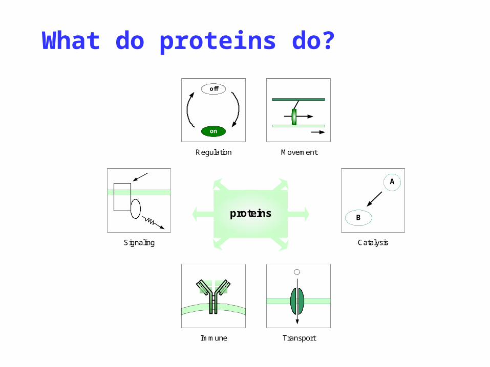
What do proteins do?
proteins
Transport
Movement
Signaling
on
off
Regulation
Immune
Catalysis
A
B
Section 1
Chemical Components of Proteins

• major elements
C (50~55%), H (~7%), O (19~20%), N (13~19%), S (~4%)
• trace elements
P, Fe, Cu, Zn, I, …
Components of proteins

• The average nitrogen content in proteins is about 16%, and proteins are the major source of N in biological systems.
• The protein quantity can be estimated.
protein in 100g sample = N per gram x 6.25 x 100

§1.1 Amino Acids
• The basic building blocks of proteins
• About 300 types of AAs in nature, but only 20 types are used for protein synthesis in biological systems.
• A amino group, a carboxyl group, a H atom and a R group are connected to a C atom.
• The C atom is an optically active center.

L-Amino acid
C H
R
-OOC
+H3N

Molecular weight
Dalton:
A unit of mass nearly equal to that of a hydrogen atom
Gly C2NO2H5 75
Ala C3NO2H7 89
Val C5NO2H11 117
Leu C6NO2H13 131
Ile C6NO2H13 131

§1.1.a Classification
• The R groups, also called side chains, make each AA unique and distinctive.
• R groups are different in their size, charge, hydrogen bonding capability and chemical reactivity.
• Aas are grouped as (1) non-polar, hydrophobic; (2) polar, neutral; (3) basic; and (4) acidic.

• R groups are non-polar, hydrophobic aliphatic or aromatic groups.
• R groups are uncharged.
• AAs are insoluble in H2O.
Non-polar and hydrophobic AAs


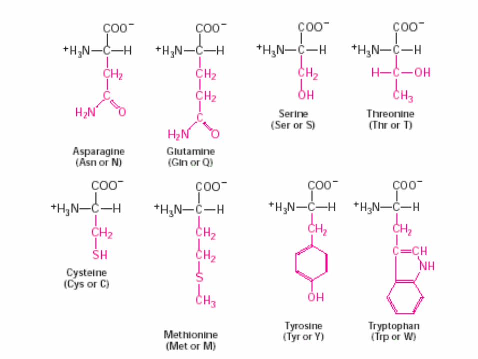
Polar and uncharged AAs
• R groups are polar: -OH, -SH, and -NH2.
• R groups are highly reactive.
• AAs are soluble in H2O, that is, hydrophilic.
• R groups have one -NH2.
• R groups are positively charged at neutral pH (=7.0).
• AAs are highly hydrophilic.
Basic AAs


Acidic AAs
• R groups have –COOH.
• R groups are negatively charged at physiological pH (=7.4).
• AAs are soluble in H2O.

Aspartic acid glutamic acid
(Asp or D) (Glu or E)

Nomenclature
Starting from the carboxyl group, and naming the rest carbon atoms sequentially in Greek letters.
-amino-propionic acid -amino--guanidinovaleric acid (alanine) (arginine)
NH3+
CH COO-CH2CH2CH2NHNH2 C
NH
CH3
NH3+
CH COO-

Special amino acids - Gly
• optically inactive
C H
H
-OOC
+H3N

Special amino acids - Pro
• Having a ring structure and imino group
CH2
CHCOO-
NH2+
CH2
CH2
CH2
CHCOO-
NH2+
CH2
CH2
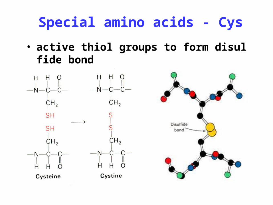
Special amino acids - Cys
• active thiol groups to form disulfide bond

§1.2 Peptide
A peptide bond is a covalent bond formed between the carboxyl group of one AA and the amino group of its next AA with the elimination of one H2O molecule.
§1.2.a Peptide and peptide bond

Peptides can be extended by adding multiple AAs through multiple peptide bonds in a sequential order.
dipeptide, tripeptide, oligopeptide, polypeptide
AAs in peptides are called as residues.

§1.2.b Biologically active peptides
Glutathione (GSH)
Glutamic acid cystein glycine

GSH peroxidase
H2O2 2GSH
2H2O
GSSG
GSH reductase
NADPH+H+
NADP+
As a reductant to protect nucleic acids and proteins from toxin by discharging free radical or H2
O2

• Peptide hormones secreted from peptidergic neurons or
– Somatostatin, Noacosapeptide, Octapeptide,
– Thyrotropin-release hormone, Antidiuretic hormone
• Neuropeptides responsible for signal transduction
– Enkephalin, Endorphin, Dynorphin, Substance P, Neuropeptide Y
Peptides

thyrotropin-release hormone
Pyroglutamic acid histidine prolinamide

Neuropeptide
name amino acid sequenceoxytocin Cys-Tyr-Ile-Gln-Asn-Cys-Pro-Leu-Gly-NH2
└──S───S──┘
Vasopresin Cys-Tyr-Phe-Gln-Asn-Cys-Pro-Arg-Gly-NH2
└──S────S──┘
Met-enkephalin Tyr- Gly-Gly-Phe-Met
Leu-enkephalin Tyr- Gly-Gly-Phe-Leu
Atrial natriuetic factor
Ser-Leu-Arg-Arg-Ser-Ser-Cys-Phe-Gly-Gly-Arg-Met-Asp-Arg-Ile-Gly-Ala-Gln-Ser-Gly-Leu-Gly-Cys-Asn-Ser-Phe-Arg-Tyr
Substance P Arg-Pro-Lys-Pro-Bln-Phe-Phe-Gly-Leu-Met-NH2
Bradykinin Arg-Pro-Pro-Gly-Phe-Ser-Pro-Phe-Arg
Section 2
Molecular Structures of Proteins

• Proteins are composed of AAs.
• Distinctive properties of proteins are determined by AA compositions, AA sequences as well as the relative positions of AAs in space.
• Proteins need well defined structures to function properly. Their structures are organized in a hierarchy format, that is, primary, secondary, tertiary and quaternary structure.
Overview

§2.1 Primary Structure
• The primary structure of proteins is defined as a linear connection of AAs along the protein chain. It is also called amino acid sequence.
• The AA sequence must be written from the N-terminus to the C-terminus.
• Peptide bonds are responsible for maintaining the primary structure.

Primary structure of insulin
• Two peptides of 21 and 30 AAs
• Two inter-chain -S-S- bonds
• One intra-chain -S-S- bond

§2.2 Secondary Structure
The secondary structure of a protein is defined as a local spatial structure of a certain peptide segment, that is, the relative positions of backbone atoms of this peptide segment.
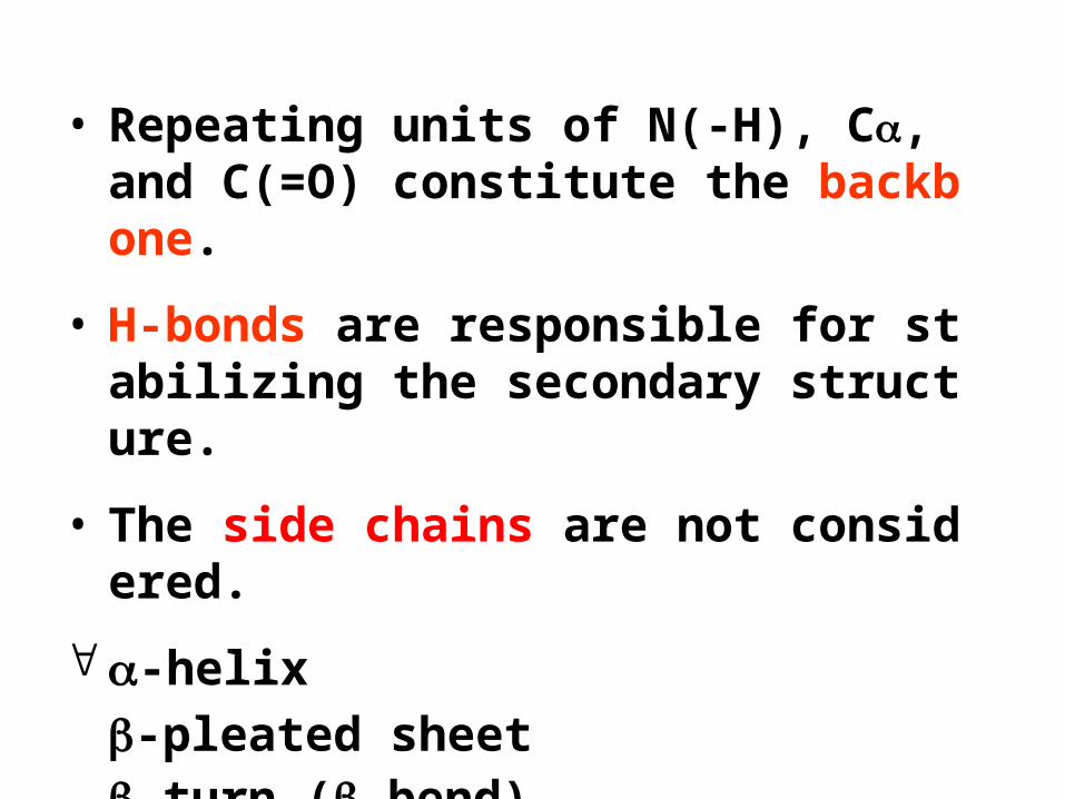
• Repeating units of N(-H), C, and C(=O) constitute the backbone.
• H-bonds are responsible for stabilizing the secondary structure.
• The side chains are not considered.
-helix
-pleated sheet-turn (-bend) random coil

Peptide unit
• Six atoms, C-C(=O)-N(-H)-C, constitute a planer peptide unit.
• The peptide unit is rigid due to the partial double bond property.
• C=O and N-H groups are in trans conformation and cannot rotate around the peptide bond.

Resonant conjugation
C
O
N
H
C
N+
H
O-
C
N
HH
0.132
0.1460.151
C
R2
0.124
R1
C
HO
C-N: 0.149nm
C=N: 0.127nm

Rotation of peptide unit
Peptide units can rotate freely around C-C and C-N bonds to form two torsion angles and .

“Beads on a string”
N-terminus
C
Peptide unit
Backbone C-terminus
Linus Carl Pauling
• b. 1901, d. 1994
• California Institute of Technology, CA
• The Nobel Prize in Chemistry (1954), “for his research into the nature of the chemical bond and its application to the elucidation of the structure of complex substances”
• The Nobel Peace Prize (1962)

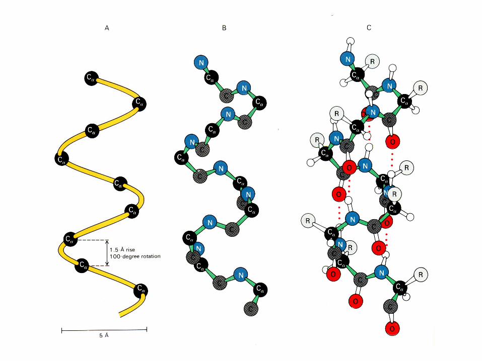
• A helical conformation is right-handed.
• 3.6 AAs per turn and a 0.15 nm vertical distance, creating a pitch of 0.54 nm.
• Side chains of AA residues protrude outward from the helical backbone.
• The hydrogen-bonds are parallel to the helical axis.
§2.2.a -helix

Left-hand versus right-hand


第n个肽键的O原子
第n+3个肽键的H原子
0. 54 nm3. 6 个残基
0. 5 nm
肽链走向
(a) (b)
C原子
O原子
N原子
H原子

The -CO group of residue n is H-bonded to the -NH group of residue (n+4).

• An extended zigzag conformation of protein backbones
• Protein backbones are arranged side-by-side through H-bonds.
• H-bonds are perpendicular to the backbone direction.
• The side chains of adjacent AAs protrude in opposite directions.
• The adjacent protein backbones can be either parallel or anti-parallel.
§2.2.b -pleated sheet


• One -turn involves four AAs. The -CO and -NH groups of the first AA are hydrogen bonded to the -NH and -CO groups of the fourth AA, respectively.
• The -turn reverses abruptly the direction of a protein backbone.
• H-bonds are perpendicular to the protein backbone.
§2.2.c -turn

§2.2.d Random coil
• There is no consistent relationship between planes.

§2.2.e Motif
• Zinc finger
HLH (helix-loop-helix)
HTH (helix-turn-helix)
Leucine zipper
When several local peptides of defined secondary structures are close enough in space, they are able to form a particular “super-secondary” structure.

§2.2.f Side chains effect
• Shape: Pro having a rigid ring (–helix disrupter)
• Size: -sheet needs AAs of small side chain. Leu, Ile, Trp, and Asn having bulky sides (hard to form –helix)
• Charge: Too many charged AAs in a short region of one peptide is hard to form –helix.

§2.3 Tertiary Structure
The tertiary structure is defined as the spatial positions of all atoms of a protein, i.e., the three-dimensional (3D) arrangement of all atoms.

Four types of interactions stabilize the protein tertiary structure.
• hydrophobic interaction
• ionic interaction
• hydrogen bond
• van der Waals interaction

§2.3.a Hydrophobic interaction
Nonpolar molecules tend to cluster together in water, that is, aqueous environment tends to squeeze nonpolar molecules together.

• A charged group is able to attract another group of opposite charges.
• The force is determined by Coulomb’s law.
§2.3.b Ionic interaction

§2.3.c Hydrogen bond
• A hydrogen atom is shared by two other atoms.
• H-donor: the atom to which H atom is more tightly attached, and the other is H-acceptor.

• An asymmetric electronic charge around an atom causes a similar asymmetry around its neighboring atoms.
• The attraction between a pair of atoms increases as they come closer, until they are repelled by van der Waals contact distance.
§2.3.d van der Waals force

Interactions stabilizing proteins

Myoglobin (Mb)
• Located in muscle to supply O2
• 1st protein in high resolution
• 153 AAs • 75% of structure i
s -helix in 8 regions.
• the interior almost entirely nonpolar residues

Ribonuclease
• A pancreatic enzyme that hydrolyzes RNA
• 124 AAs • Mainly -sheet• Highly compact a
nd nonpolar interior
• 4 disulfide bonds
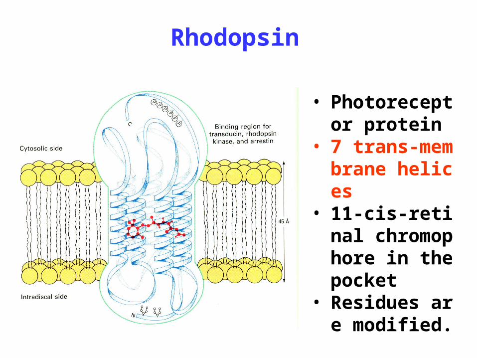
Rhodopsin
• Photoreceptor protein
• 7 trans-membrane helices
• 11-cis-retinal chromophore in the pocket
• Residues are modified.
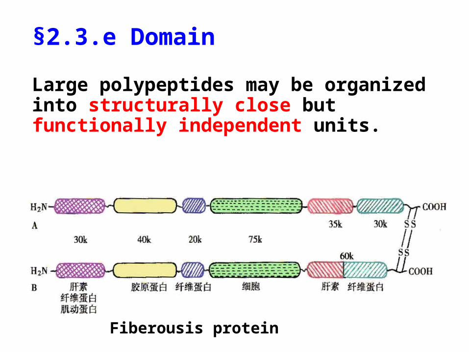
§2.3.e Domain
Large polypeptides may be organized into structurally close but functionally independent units.
Fiberousis protein

Methyl-accepting chemotaxin
• Highly conservative cytosolic domain
• Divergent periplasmic domain serving as a chemosensor
• Transducing the external singles into the cell
§2.3.f Chaperon
Chaperones are large, multisubunit proteins that promote protein foldings by providing a protective environment where polypeptides fold correctly into native conformations or quaternary structures.

• Reversibly bind to the hydrophobic portions to advance the formation of correct peptide conformations
• Bind to misfolded peptides to induce them to the proper conformations
• Assist the formation of correct disulfide bonds
How does chaperon work?

§2.4 Quaternary Structure
The quaternary structure is defined as the spatial arrangement of multiple subunits of a protein.

• Proteins need to have two or more polypeptide chains to function properly.
• Each individual peptide is called subunit.
• These subunits are associated through H-bonds, ionic interactions, and hydrophobic interactions.
• Polypeptide chains can be in dimer, trimer .., as well as homo- or hetero-form.

• O2 transporter in erythrocyte
• 2 subunits, 141 AAs 2 subunits, 146 AAs
• 4 subunits are maintained together by 8 pairs of ionic interactions.
• Each subunit contains one heme group.
• The conserved hydrophobic core stabilizes the 3D structure.
Hemoglobin(Hb)

Structure of hemoglobin
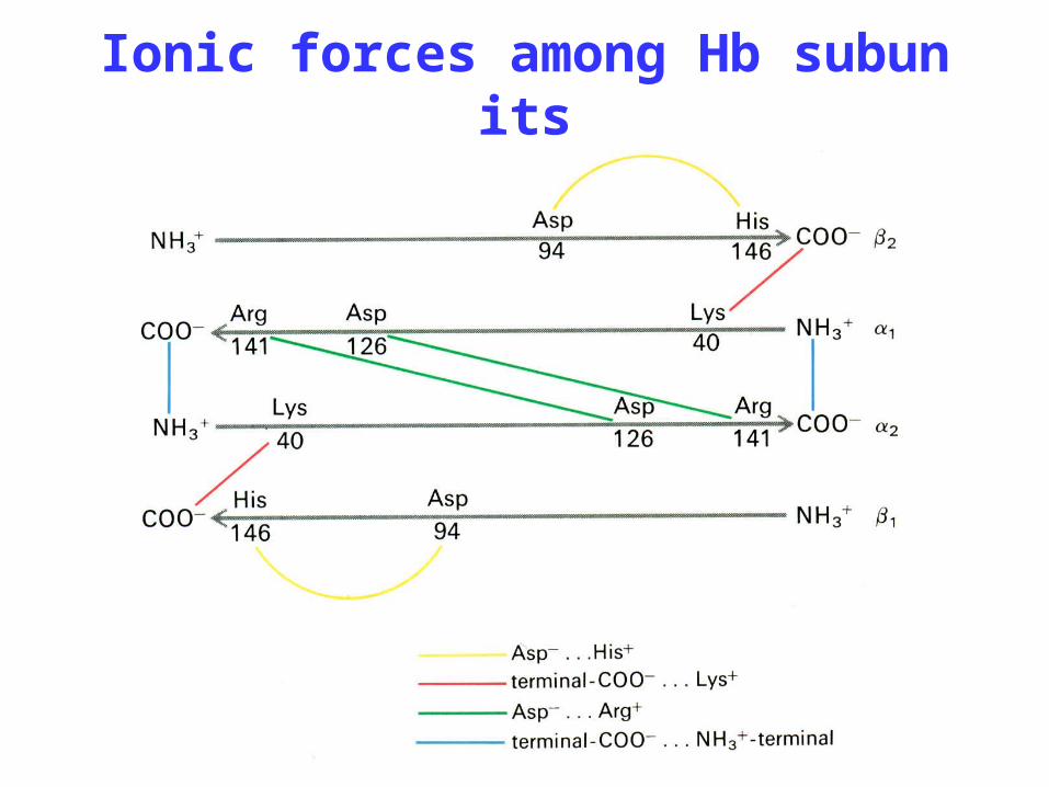
Ionic forces among Hb subunits

From primary to quaternary structure

• Constituents
simple protein
conjugated protein = protein + prosthetic groups
Prosthetic group is non-protein part, binding to protein by covalent bond. This group can be carbohydrates, lipids, nucleic acids, phosphates, pigments, or metal ions.
§2.5 Protein classification

• Classification based on the overall shape
• Globular protein:
long/short < 10 , soluble in water; including enzymes, transportors, receptors, regulators, …
• Fibrous protein:
highly elongated; insoluble in water; including collage, elastin, α-keratin, …
Section 3
Biological Functions of Proteins

proteins
Transport
Movement
Signaling
on
off
Regulation
Immune
Catalysis
A
B

• Hb can bind O2 reversibly, just like Mb.
• Both and chains are strikingly similar to that of Mb.
• Although only 24 of 146 AAs of their sequences are identical, 9 critical residues are conserved in sixty species.
• Residues on the surface are highly variable, but the nonpolar core is conserved.
§3.1 Hemoglobin

Structural similarity of Mb and Hb

Fe lies at the center of picket-fence porphyrin to form 4 coordinate bonds with 4 N atoms.
Fe-porphyrin complex
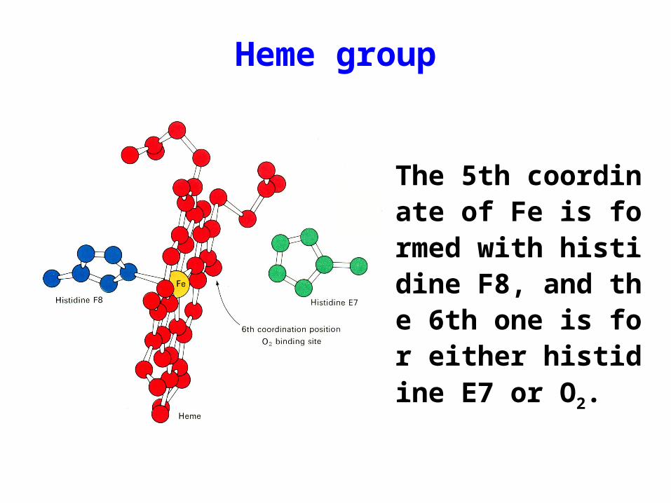
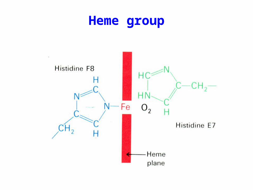
The 5th coordinate of Fe is formed with histidine F8, and the 6th one is for either histidine E7 or O2.
Heme group
Heme group

• The saturation Y is defined as the fractional occupancy of all O2-binding sites.
• Y varies with the concentration of O2 . The equilibrium constants for Hb subunits are different.
Oxygen-disassociation curve

• Hb has a lower affinity for O2 than Mb (lower P50).
• The O2–binding to the 1st subunit enhances the O2–binding to the 2nd and 3rd subunits. Such process further enhances the O2–binding to the 4th subunit significantly.
• Hb binds O2 in a positive cooperative manner, which enhances the O2 transport.
Binding behavior of Hb

Upon oxygenation, the Fe atom is moved into the porphyrin plane, leading to the formation of a strong bond with O2.
Local structural change

CO and O2 binding
Hb forces CO to bind at an angle due to steric hindrance of His E7, which weakens the binding of CO with the heme.

Conformational changes
The quaternary structure of Hb changes markedly upon oxygenation ( subunit shifts by 0.6nm and rotates by 15°).
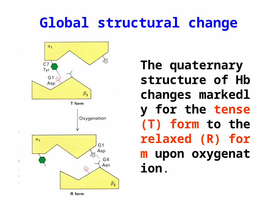
The quaternary structure of Hb changes markedly for the tense (T) form to the relaxed (R) form upon oxygenation.
Global structural change

• The behavior that the lignad-binding to one subunit causes structural changes and stimulate the further binding to other subunits is termed as allosteric effect.
• The protein is allosteric protein, and the substrate is allosteric effector.
• Allosteric effect can be influenced by activators as well as inhibitors.
Allosteric effect

Concerted versus sequential
1
non-oxygenized Hb(T conformation)
oxygenized Hb(R conformation)
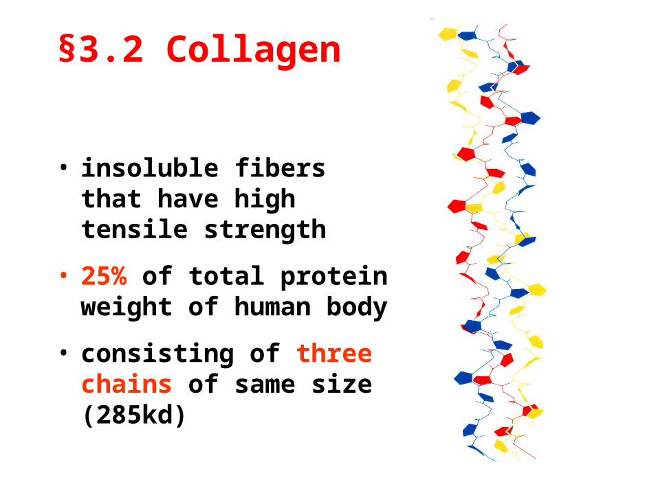
§3.2 Collagen
• insoluble fibers that have high tensile strength
• 25% of total protein weight of human body
• consisting of three chains of same size (285kd)

Collagen in different organisms
Tissue Content
Bone 88.0
Calcaneal tendon 86.0
Skin 71.9
Cornea 68.1
Cartilage 46-63
Ligament 17.0
Aorta 12-24
Liver 3.9

Unusual components
• AA componentsGly (1/3), proline (1/4), 4-hydroxyproline (1/10), 5-hydroxylysine (1%)
• AA sequences
(Gly-Pro-Y)n or (Gly-X-Hyp)n
X and Y can be any AAs.
n can be as high as a few hundreds.
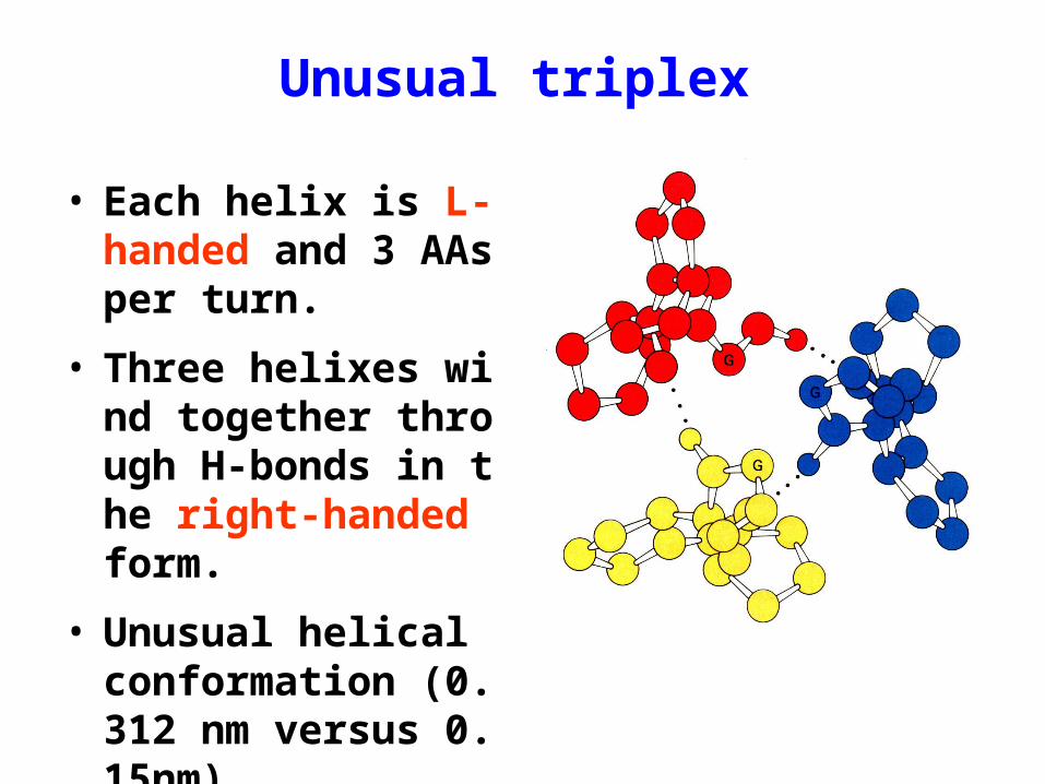
Unusual triplex
• Each helix is L-handed and 3 AAs per turn.
• Three helixes wind together through H-bonds in the right-handed form.
• Unusual helical conformation (0.312 nm versus 0.15nm)

Intermolecular cross-link
• Lys at N- and C-termini and Hly in helical regions are responsible for the cross-link.
• The linkage varies with the physiological function and the tissue age.
• 30 genes encode for collagens, and 8 post-translational modifications are needed collagen maturation.


Type of collagens


Diseases and collagen
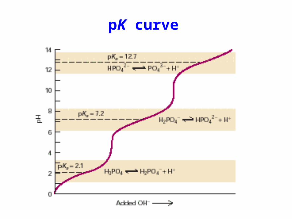
pK curve

Structure of hemoglobin

Concerted versus sequential

Model comparison
Concerted Model Sequential Model
The symmetry is essential for the subunit interaction.
Subunits can interact even if they are in different forms.
T and R forms are in equilibrium in the absence of substrate
T to R is induced by the binding of substrate.
Homotropic interaction is positive.
Homotropic interaction can be positive and negative.

Section 4
Structure-Function Relationship of Proteins

• Primary structure is the fundamental to the spatial structures and biological functions of proteins.
• For a protein of particular sequence, many conformers are possible, but only the correct one has the biological functions.
§4.1 Primary Structure and Function

1. Proteins having similar amino acid sequences demonstrate the functional similarity.
2. Proteins of incorrect structures have no proper biological functions, even their amino sequences are remained in a right order.
3. The alternation of key AAs in a protein will cause the lose of its biological functions.

Sequences of Cytochrome C
________10 ________20 ________30 ________40 ________50 ________60 ________70 ________80 ________90 _______100 ____
Human GDVEKGKKIF IMKCSQCHTV EKGGKHKTGP NLHGLFGRKT GQAPGYSYTA ANKNKGIIWG EDTLMEYLEN PKKYIPGTKM IFVGIKKKEE RADLIAYLKK ATNE
Chimpanzee GDVEKGKKIF IMKCSQCHTV EKGGKHKTGP NLHGLFGRKT GQAPGYSYTA ANKNKGIIWG EDTLMEYLEN PKKYIPGTKM IFVGIKKKEE RADLIAYLKK ATNE
Monkey GDVEKGKRIF IMKCSQCHTV EKGGKHKTGP NLHGLFGRKT GQASGFTYTE ANKNKGIIWG EDTLMEYLEN PKKYIPGTKM IFVGIKKKEE RADLIAYLKK ATNE
Macaque GDVEKGKKIF IMKCSQCHTV EKGGKHKTGP NLHGLFGRKT GQAPGYSYTA ANKNKGITWG EDTLMEYLEN PKKYIPGTKM IFVGIKKKEE RADLIAYLKK ATNE
Cow GDVEKGKKIF VQKCAQCHTV EKGGKHKTGP NLHGLFGRKT GQAPGFSYTD ANKNKGITWG EETLMEYLEN PKKYIPGTKM IFAGIKKKGE RADLIAYLKK ATNE
Dog GDVEKGKKIF VQKCAQCHTV EKGGKHKTGP NLHGLFGRKT GQAPGFSYTD ANKNKGITWG EETLMEYLEN PKKYIPGTKM IFAGIKKKGE RADLIAYLKK ATKE
Grey whale GDVEKGKKIF VQKCAQCHTV EKGGKHKTGP NLHGLFGRKT GQAVGFSYTD ANKNKGITWG EETLMEYLEN PKKYIPGTKM IFAGIKKKGE RADLIAYLKK ATNE
Horse GDVEKGKKIF VQKCAQCHTV EKGGKHKTGP NLHGLFGRKT GQAPGFSYTD ANKNKGITWK EETLMEYLEN PKKYIPGTKM IFAGIKKKTE RADLIAYLKK ATNE
• Cytochrome C is a protein which can be found in all aerobic organisms.
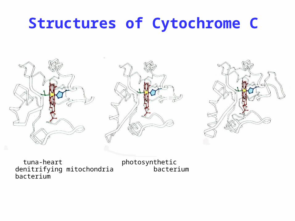
tuna-heart photosynthetic denitrifying mitochondria bacterium bacterium
Structures of Cytochrome C

1. Proteins having similar amino acid sequences demonstrate the functional similarity.
2. Proteins of incorrect structures have no proper biological functions, even their amino acid sequences are remained in a right order.
3. The alternation of key AAs in a protein will cause the lose of its biological functions.

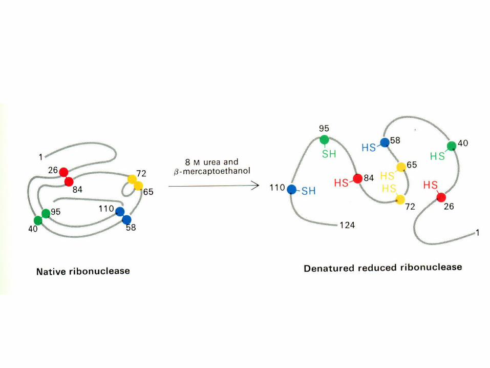
Bovine nuclease
• 124 AAs, 4 disulfide bonds (105 possibilities)

• The denatured protein remains its primary structure, but no biological function.
• Only the correct form has the enzymatic activity.

The renatured protein will restore its functions partially or fully depending upon the correctness of the refolded structure.

1. Proteins having similar amino acid sequences demonstrate the functional similarity.
2. Proteins of incorrect structures have no proper biological functions, even their amino sequences are remained in a right order.
3. The alternation of key AAs in a protein will cause the lose of its biological functions.

Sickle-cell of anemiaPatient’s symptoms:
Cough, fever and headache, a tinge of yellow in whites of eyes, visible pale mucous membrane, enlarged heart, well developed physically, anemic, much less RD cells
clinical test:
The shape of the red cells was very irregular, large number of thin, elongated, sickle-shaped and crescent-shaped forms.

• pI of sickle-cell Hb was higher than normal one by 0.23, which is equivalent to 2 to 4 net positive charges per Hb molecule. (1949, Pauling)
• 2-D electrophoresis showed only one peptide of 28 digested Hb peptides is different (1954, Ingram).
Identifying the cause
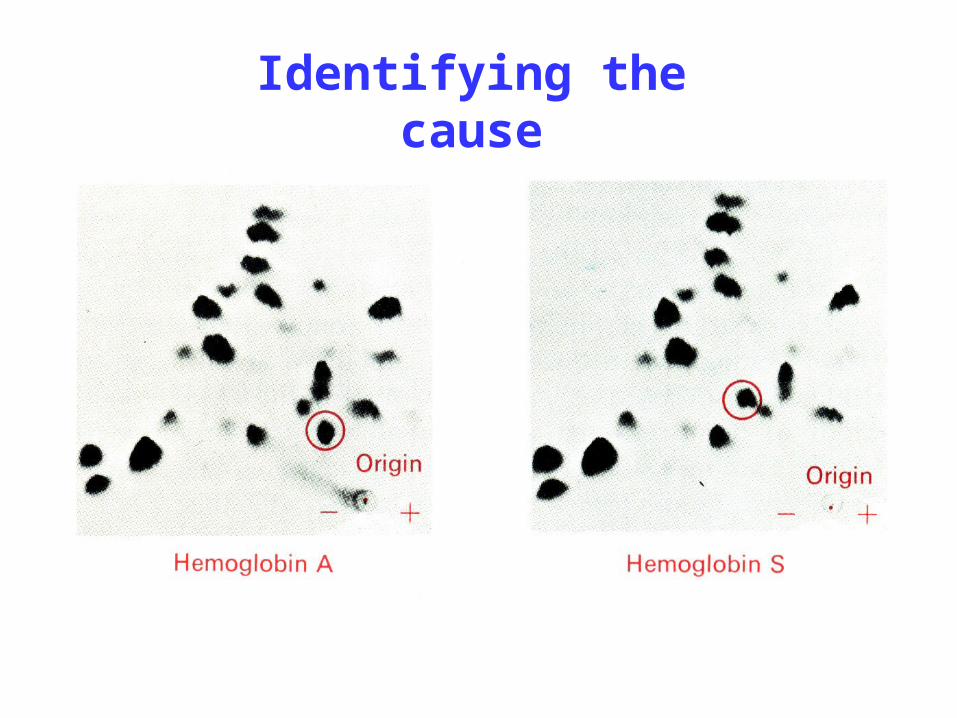
Identifying the cause

• Sequence analysis showed the difference in AA sequence.
Hb A : Val-His-Leu-Thr-Pro-Glu-Glu-Lys-
Hb S : Val-His-Leu-Thr-Pro-Val -Glu-Lys- • This is the first case of molecular diseas
e identified in history. Further studies showed that the AA variation is due to the gene mutation.
Difference in primary structure of Hb

• Proteins will experience multiple processed to become correctly folded, that is, having a correct structure.
• The incorrect protein structure may lead to function alternation or diseases.
• A particular spatial structure of a protein is strongly correlated with its specific biological functions.
§4.2 Spatial Structure and Function
• A transmissible, inheritable neural disease, destroying brain tissues by converting them to a spongy appearance
• the conformational changes of prion protein (PrP)
– PrPc: -helix, water soluble – PrPsc: -sheet, water insoluble
Mad cow disease and prion proteins

Structural changes of prion protein
PrPc PrPsc

Section 5
Physical and ChemicalProperties of Proteins
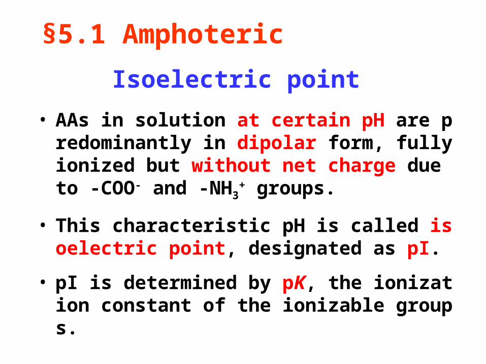
• AAs in solution at certain pH are predominantly in dipolar form, fully ionized but without net charge due to -COO- and -NH3
+ groups.
• This characteristic pH is called isoelectric point, designated as pI.
• pI is determined by pK, the ionization constant of the ionizable groups.
Isoelectric point
§5.1 Amphoteric

pH=pI
+OH-
pH>pI
+H+
+OH-
+H+
pH<pI
amphoteric
cation anion
CH
NH2
COOHR CH
NH2
COOHR
CH
NH3+
COO-R CH
NH3+
COO-R CH
NH2
COO-R CH
NH2
COO-RCH COOHR
NH3+
CH COOHR
NH3+
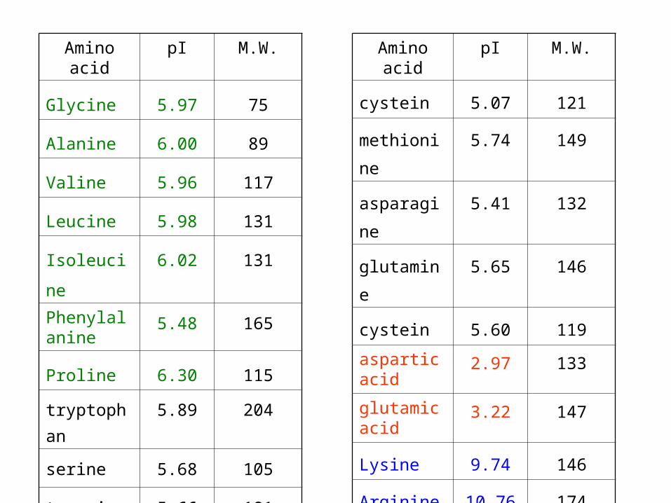
Amino acid pI M.W.
Glycine 5.97 75
Alanine 6.00 89
Valine 5.96 117
Leucine 5.98 131
Isoleucine 6.02 131
Phenylalanine
5.48 165
Proline 6.30 115
tryptophan 5.89 204
serine 5.68 105
tyrosine 5.66 181
Amino acid pI M.W.
cystein 5.07 121
methionine 5.74 149
asparagine 5.41 132
glutamine 5.65 146
cystein 5.60 119
aspartic acid
2.97 133
glutamic acid
3.22 147
Lysine 9.74 146
Arginine 10.76 174
Histidine 7.59 155
• Side-chains of a protein have many ionizable groups, making the protein either positively or negatively charged in response to the pH of the solution.
• The pH at which the protein has zero net-charge is referred to as isoelectric point (pI).

• pI of most protein is ~ 5.0, and negatively charges in body fluid (pH7.4)
• pI > 7.4: basic proteins: protamine, histone• pI < 7.4: acidic proteins: pepsin
NH3+
COO-
NH2
COO-
NH3+
COOH
+ OH-
+ H+ + H+
+ OH-
pH = pI pH > pIpH < pI
amphoteri c ani oncati on
P PP

§5.2 Colloid property
• Diameter: 1~100nm, in the range of colloid;
• Hydrophilic groups on the surface form a hydration shell;
• Hydration shell and electric repulsion make proteins stable in solution.
- - ---
------
-++
++
+
+
++ +
+
+
+
----
--
---
--
-

+
+
+
+
+
++
- --
----
-
isoelectric point (hydrophilic)
+ +
+
+
++
+ +
positively charged (hydrophobic)
---
-
----
Instable protein(deposition)
acidbase
acid base
acidbase
dehydrationdehydrationdehydration
Precipitation of protein colloid
negatively charged (hydrophobic)
positively charged (hydrophilic)
negatively charged (hydrophilic)

• The denatured proteins tend to - decrease in solubility;- increase the viscosity;- lose the biological activity;- lose crystalizability; - be susceptible to enzymatic digestion.
§5.3 Protein denaturation
The process in which a protein loses its native conformation under the treatment of denaturants is referred to as protein denaturation.

• Cause of denaturation the disruption of hydration shell and electric repulsion
• Denaturants physical: heat, ultraviolet light, violent shaking, …chemical: strong acids, bases, organic solvents, detergents, …
• Applicationssterilization, lyophilization

Renaturation
• Once the denaturants are removed, the denatured proteins tend to fold back to their native conformations partially or fully.
• The renatured proteins can restore their biological functions.

Renaturation
The denatured proteins expose their side chains or the inner part to the aqueous environment, which causes the proteins aggregated and separated out from the aqueous solution.
Protein precipitation

• When the denatured proteins become insoluble fluffy materials, heating denatured proteins will turn them into a hard solid which are not soluble even strong acids and bases are applied.
• Coagulation is an irreversible process.
Protein coagulation

§5.4 UV absorption
• Trp, Tyr, and Phe have aromatic groups of resonance double bonds.
• AAs have a strong absorption at 280nm.
• Both free and incorporated AAs show this absorption.


§5.5 Coloring reactions
• Biuret reaction: peptide bonds and Cu2+ under the heating condition to form red or purple chelates.
• Used for determine the hydrolysis of proteins since free amino acids do not react.
• Amino acids can react with ninhydrin to form a chemicals having maximal adsorption at 570 nm.
• Used for quantifying the free amino acids.

Section 6
Exploration of Protein

§6.1 Isolation and purification
• Homogenization and centrifugation
• Dialysis
• Precipitation
• Chromatography
• Electrophoresis

• Rupture the plasma membrane to release the intracellular components into the buffered solution
• Sonication, French pressure, mechanical grinding,
• Chemical reagents, lysozymes
§6.1.a Homogenization
Centrifugation
• Because of the differences in size and shape, proteins will sediment gradually under the centrifugal force until the sedimentation force and buoyant force reach the balance.
• The sedimentation behavior is described in sedimentation coefficient (S) which is proportional to the molecular weight.
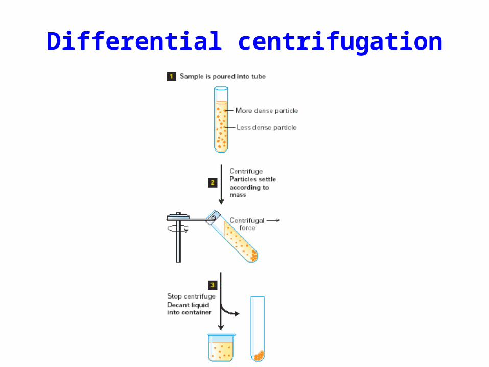
Differential centrifugation

Differential centrifugation
supernatantPellet(nuclei)
600 g,3 min
Pellet(mitochondria, chloroplasts,lysosomes, peroxisomes)
6,000 g,8 min
supernatantPellet
(plasma membrane,fragments of Golgi and ER)
40,000 g,30 min
supernatant(cytosol)
Pellet(ribosomal subunits)
100,000 g,90 min
homogenate
supernatant
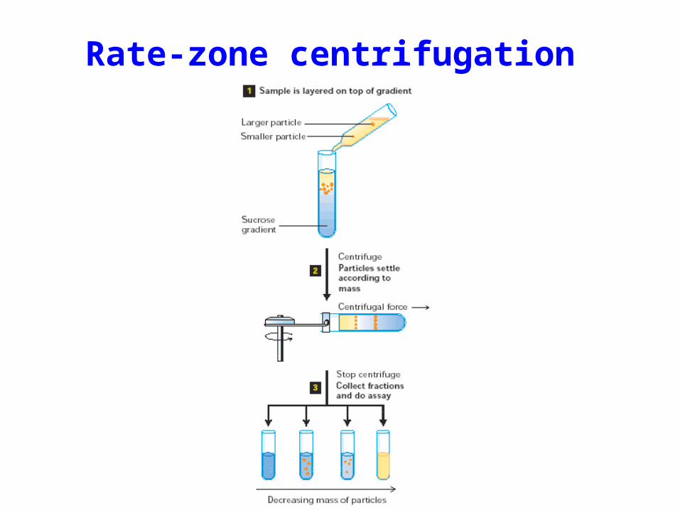
Rate-zone centrifugation
§6.1.b Dialysis
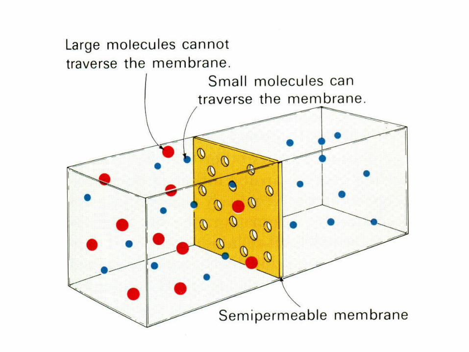
• Proteins, as macromolecules, cannot pass through the semipermeable membrane containing pores of smaller than protein dimension, thus large proteins and small molecules can be separated.
• Dialysis can be used for protein purification, desalting, and condensation.

§6.1.c Precipitation
• Adding a large quantity of salts, such as Ammonia sulfate, into the protein solution will neutralize the surface charges and the destruct the hydration shell of proteins, causing them to precipitate.
• Acetone has the similar function.
• An efficient way to concentrate proteins.

6.1.d Chromatography
When a protein solution (called as mobile phase) passes through a stationary phase, proteins can interact with the stationary phase due to the differences in size, charge, and affinity, making the different proteins flow through the stationary phase at different speeds.

OD
280
nm
El ut i on vol ume
El uti onbuff er
Protei n mi xture
Protei n 2Protei n 1
Sol i dphasecapabl eofreacti ngwi thprotei nsto beseparated
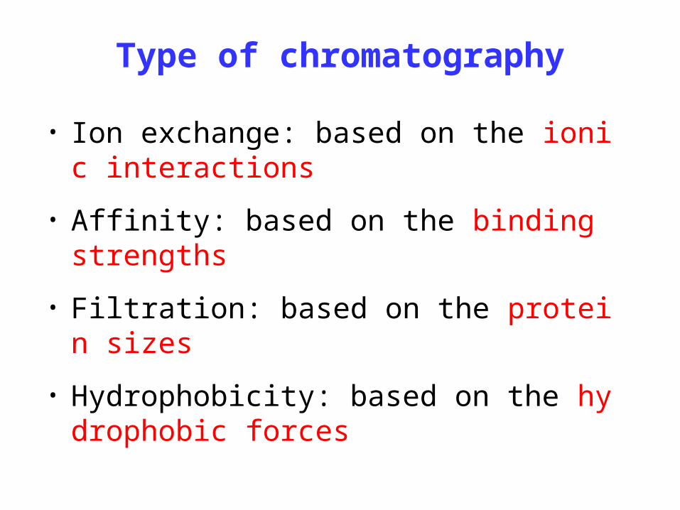
Type of chromatography
• Ion exchange: based on the ionic interactions
• Affinity: based on the binding strengths
• Filtration: based on the protein sizes
• Hydrophobicity: based on the hydrophobic forces

Ion-exchange chromatography
+
++
+
++
+
+
+
++
+
++
+
+
-
--
-
-
-
--
=
=
=
=
=
=
=
=
=
=
=
=
-
-
I oni c exchangecol umn wi thposi t i ve charge
More negati vel y chargedprotei ns bi nd to the sol i dphase t i ghtl y, and strongerel ut i on buff er i s needed toel ute them out the col umn
Less negati vel y chargedprotei ns bi nd to the sol i dphase l oosel y, and weakel uti on buff er can be used toel ute them out the col umn
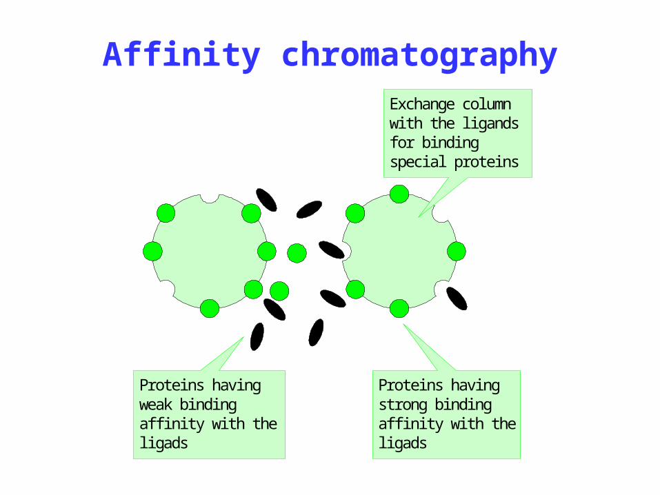
Affinity chromatographyExchange col umnwi th the l i gandsfor bi ndi ngspeci al protei ns
Protei ns havi ngstrong bi ndi ngaffi ni ty wi th thel i gads
Protei ns havi ngweak bi ndi ngaffi ni ty wi th thel i gads

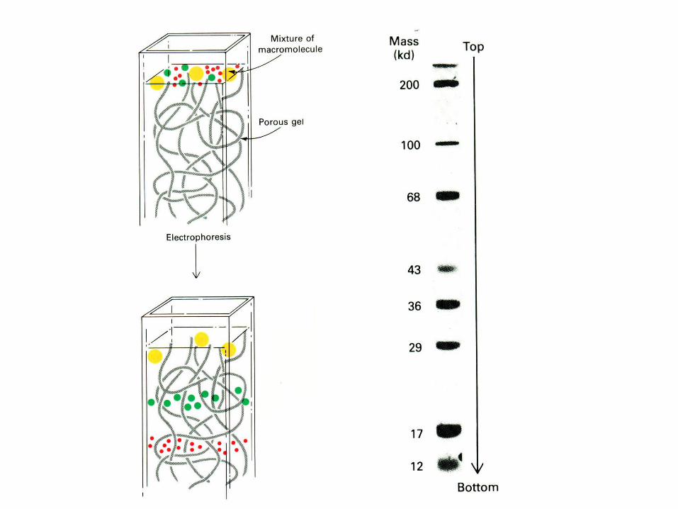
Gel filtration
Proteins are separated based on their sizes and shapes. The stationary phase is of semi-uniform pores. When the protein solution flows through porous beads, smaller proteins can enter the pores and stay there for a longer period, but larger proteins flow directly through the column, resulting in the separation of proteins. It is also called molecular sieve or size exclusion.
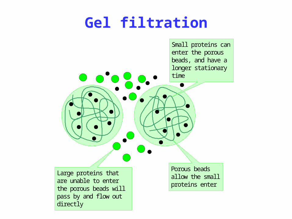
Large protei ns thatare unabl e to enterthe porous beads wi l lpass by and fl ow outdi rect l y
Smal l protei ns canenter the porousbeads, and have al onger stat i onaryti me
Porous beadsal l ow the smal lprotei ns enter
Gel filtration

Used mainly for determination of proteins
• SDS-PAGE = Sodium dodecyl sulfate polyacrylamide gel electrophoresis
• IEF = isoelectric focusing electrophoresis
• 2D = two dimensional electrophoresis
§6.2 Electrophoresis Analysis
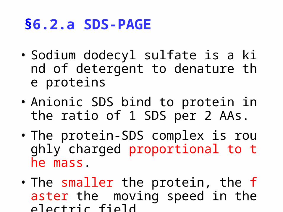
§6.2.a SDS-PAGE
• Sodium dodecyl sulfate is a kind of detergent to denature the proteins
• Anionic SDS bind to protein in the ratio of 1 SDS per 2 AAs.
• The protein-SDS complex is roughly charged proportional to the mass.
• The smaller the protein, the faster the moving speed in the electric field.
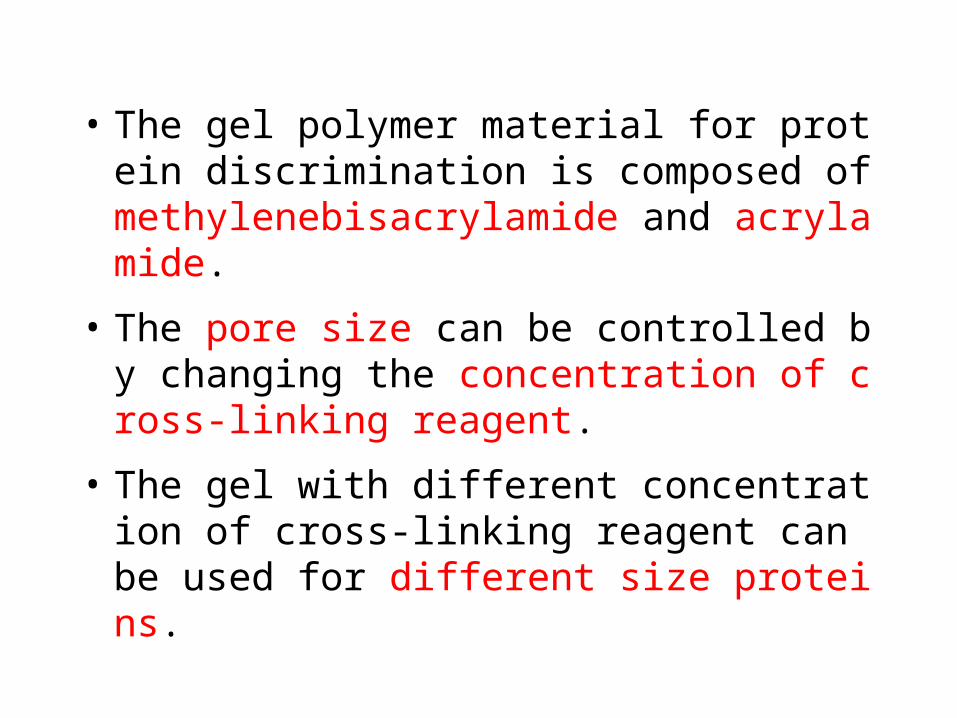
• The gel polymer material for protein discrimination is composed of methylenebisacrylamide and acrylamide.
• The pore size can be controlled by changing the concentration of cross-linking reagent.
• The gel with different concentration of cross-linking reagent can be used for different size proteins.


• Depend upon the electric properties of proteins
• The charged proteins, either positively or negatively, will migrate in the electric field.
• The proteins having net zero charges stop moving in the electric field.
§ 6.2.b Isoelectric focusing

Principle of IFE
pH 7.0
pH 3.0
Protei ns wi thdi ff erent pI
-
-
--+
-
-
-
-
-
--+
-
-
+pH 7.0
pH 3.0
-
-
--+
+
+
+
-
-
--+
+
+
+
Stabl e pHgradi entgel medi um
Protei ns ofl arge pI showzero netchanges
Protei ns ofsmal l pI showzero netchanges

§6.2.c 2D electrophoresis
• 1st dimension: isoelectric focusing electrophoresis
• 2nd dimension: SDS-PAGE
• A high throughput approach to identify proteins

§6.3 Composition and Sequence
• Composition
• Determination of terminal residues
• Edman degradation
• Sequencing strategy
• Mass spectroscopy
• Deduction form DNA sequences

§6.3.a Composition analysis
• Hydrolyzing the purified protein samples in an evacuated and sealed tube by heating it in 6 M HCl at 100°C
• Analyzing the AA components using chromatography
• (Alaa, Argb, Asnc, Aspd, … Valz)
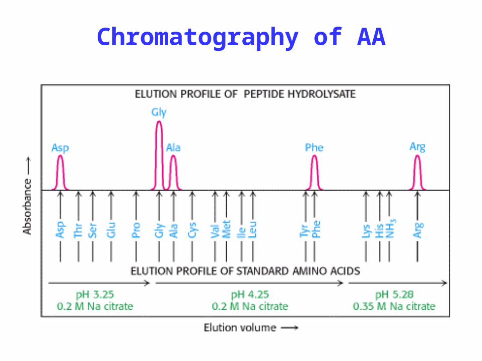
Chromatography of AA

§6.3.b Terminal residues
• The amino-terminal residue reacts with fluorodinitrobenzene or dabsyl chloride to form a stable product which can be analyzed using chromatography.
• The carboxyl-terminal residue can react with fluorodinitrobenzene or dabsyl chloride to form a stable product.

N-terminal reaction

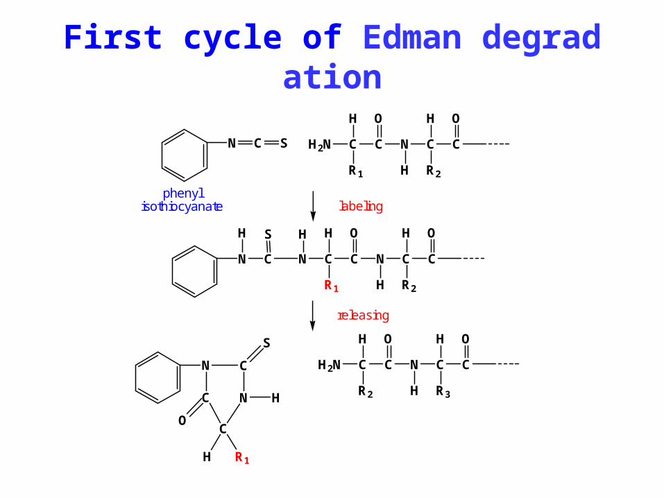
§6.3.c Edman degradation
1. Labeling the N-terminal residue with a fluorophore.
2. Cleaving the labeled residue without breaking the peptide bonds of the rest part of the peptide.
3. Determining the N-terminal residue with chromatography.
4. Repeating the same procedure to the rest peptide until the whole sequence is determined.
N C S H2N C C
H
R1
N C C
O O
H
H
R2
N C
S
H2N C C
H
R2
N C C
O O
H
H
R3C N
C
H
R1H
O
N C
S
N C C
H
R1
N C C
O O
H
H
R2
HH
labeling
releasing
phenylisothiocyanate
First cycle of Edman degradation

Edman degradation

1. Cleaving a protein into small peptides by chemical or enzymatic methods, and purify these peptides.
2. Sequencing each peptide using Edman degradation approach.
3. Overlapping peptide fragments to arrange them in a right order, and accomplishing the AA sequencing.
§6.3.d Overlapping approach
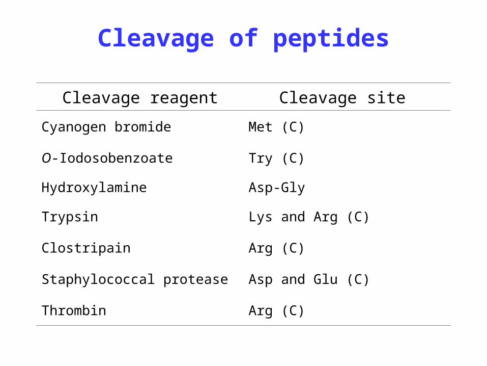
Cleavage of peptides
Cleavage reagent Cleavage site
Cyanogen bromide Met (C)
O-Iodosobenzoate Try (C)
Hydroxylamine Asp-Gly
Trypsin Lys and Arg (C)
Clostripain Arg (C)
Staphylococcal protease Asp and Glu (C)
Thrombin Arg (C)

Overlapping approach
1. Tryptic cleavage generates two peptides
Gly-Phe-Val-Glu-Arg, Val-Phe-Asp-Lys
2. Chymotryptic cleavage generates three peptides
Val-Phe, Val-Glu-Arg, Asp-Lys-Gly-Phe
3. Overlapping the sequenced peptides

Val - Phe - Asp - Lys - Gly - Phe - Val - Glu - Arg
Tryptic peptide
Chymotryptic peptide
Tryptic peptide

§6.3.e Mass spectroscopy
• Newly developed approach applied to biology and medicine areas
• Revolutionized bioanalytical technique
• Offering a fast, high accuracy, and high throughput determination for analyzing peptides and proteins.

Matrix-aided ionization
1. Deposit samples on a plate.
2. Introduce a beam of laser.
3. Ionize samples.
4. Analyze ionized molecules.
5. Determine the AA sequence.

Fragmentation of peptide
ai+1
yn+1
bi+1
ai+3
ai+2
bi+3
bi+2
xn+1
xn+3
xn+2
yn+2
yn+3
H
O
Ri
—N—C—C
H
O
Ri+1
—N—C—C
H
O
Ri+2
—N—C—C
H
O
Ri+3
—N—C—C
H
O
Ri+4
—N—C—C
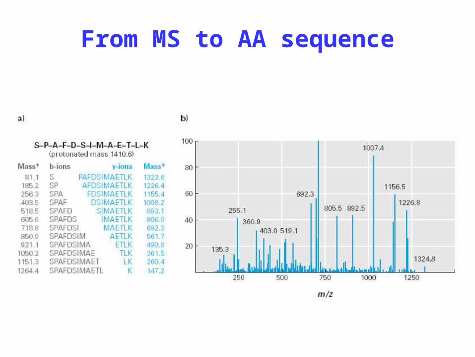
From MS to AA sequence
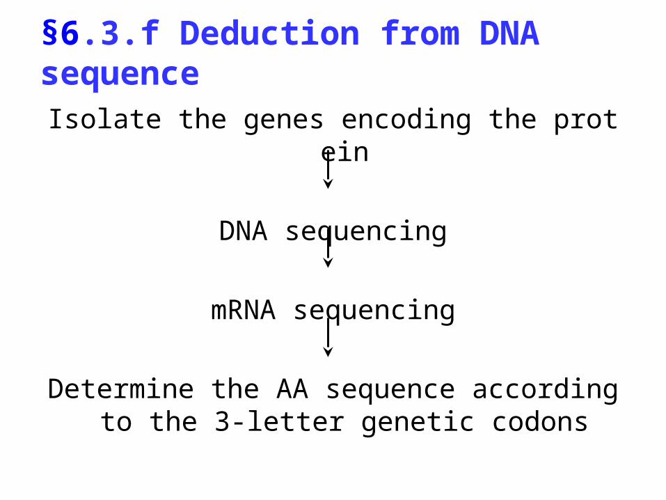
§6.3.f Deduction from DNA sequence
Isolate the genes encoding the protein
DNA sequencing
mRNA sequencing
Determine the AA sequence according to the 3-letter genetic codons
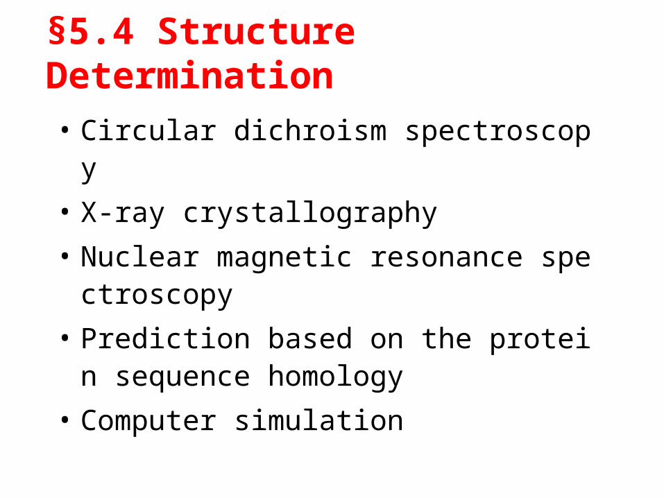
§5.4 Structure Determination
• Circular dichroism spectroscopy
• X-ray crystallography
• Nuclear magnetic resonance spectroscopy
• Prediction based on the protein sequence homology
• Computer simulation

Section 7
Proteomics: A New Frontier

Proteomics
a comprehensive knowledge about all the proteins of a cell at a specific given time.

Objectives
• Biological process – The overall process toward which this
protein contributes
• Molecular function– The biological activity the protein
accomplishes
• Cellular component– The location of protein activity

Proteomics approaches
• Separation • Sequence determination • 3D-structure • Functionality • Expression regulation • Post-translation modification

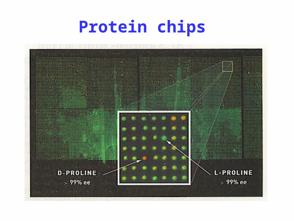
Protein chips

• Proteins synthesis, post-translation modification and maturation is a very complex process.
• Only the correct folding process leads to the correct conformation and the proper functions of proteins.
• Incorrect folding process may lead to diseases.
Incorrect conformation and diseases

Rice Yeast Bacterium
Structures of Cytochrome C
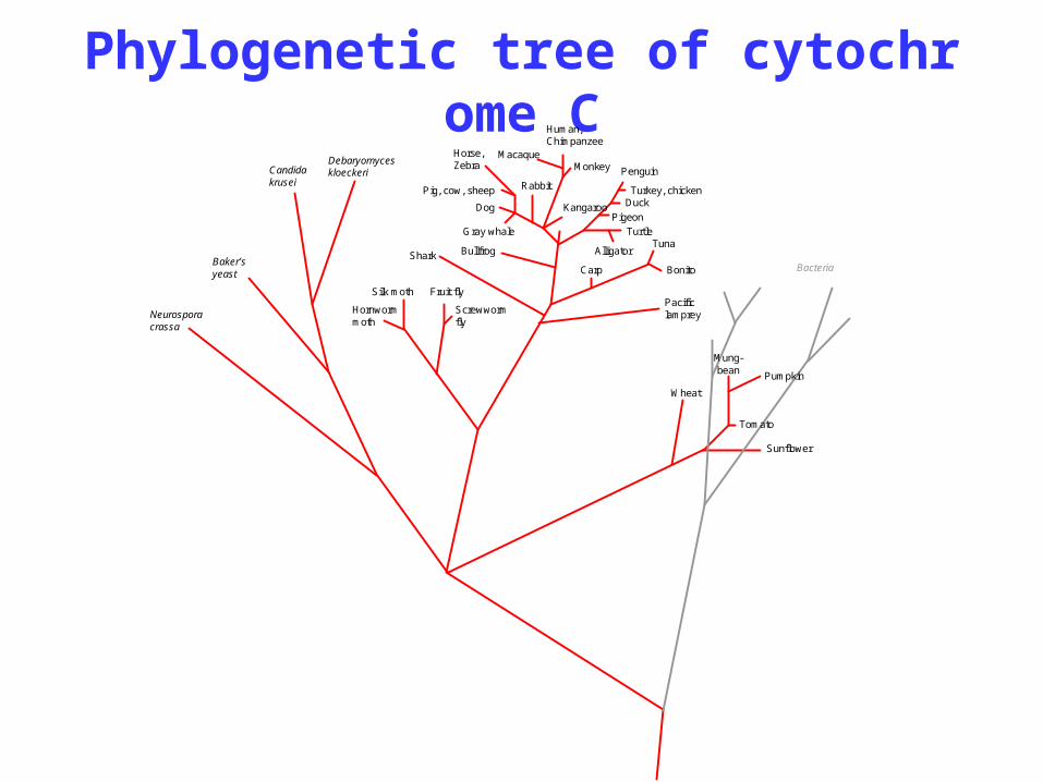
Phylogenetic tree of cytochrome CHuman,Chimpanzee
Monkey
Rabbit
Horse,Zebra
Pig, cow, sheep
Dog
Gray whale
Shark
Fruit fly
KangarooPigeon
Turkey, chickenDuck
Alligator
TurtleTuna
Sunflower
Pumpkin
Wheat
Tomato
Mung-bean
Penguin
Pacificlamprey
Macaque
Bullfrog
BonitoCarp
Silk moth
Bacteria
Screwwormfly
Neurosporacrassa
Baker’syeast
Candidakrusei
Debaryomyceskloeckeri
Hornwormmoth

Homology comparison