Chapter 1 Glucose metabolism in actinomycetes · 2016-03-08 · galactose (Type IV) (Goodfellow,...
23
Chapter 1 Glucose metabolism in actinomycetes A.M.C.R. Alves 13
Transcript of Chapter 1 Glucose metabolism in actinomycetes · 2016-03-08 · galactose (Type IV) (Goodfellow,...

Chapter 1
Glucose metabolism in actinomycetes
A.M.C.R. Alves
13

1. General aspects of actinomycetes
Actinomycetes are Gram-positive bacteria belonging to the orderActinomycetales(Lechevalier and Lechevalier, 1981), characterized by the formation of substrate andaerial mycelium on solid media, presence of spores and a high GC content of the DNA(60-70 mol%). The composition of the growth media can profoundly affect the growthand stability of substrate and aerial mycelium (Kalakoutskii and Agre, 1976). Themajority of actinomycetes are soil bacteria that play an important role in mineralizationprocesses in nature. Actinomycetes form a very important class of bacteria since theyproduce numerous natural products such as antibiotics and enzymes (Edwards, 1993).More than 50% of the known natural antibiotics produced are from actinomycetes (Berdy,1984; Miyadoh, 1993). These bacteria can be separated into different genera on the basisof morphological, physical and chemical criteria. Actinomycetes possess cell wall type Ito IV, depending on the presence of L-diaminopimelic acid (DAP) and glycine (type I),meso-DAP and glycine (type II),meso-DAP (type III), or meso-DAP, arabinose andgalactose (Type IV) (Goodfellow, 1989).
Streptomyces coelicolorA3(2) is an actinomycete with cell wall type I, belonging tothe familyStreptomycetaceae(Stackebrandt and Woese, 1981; Goodfellowet al.,1992).It is one of the best studiedStreptomycesspecies at the genetical level with a wellcharacterized chromosomal physical map, carrying more than 150 genes or gene clusters(Hopwoodet al.,1985, 1995; Kieseret al.,1992; Chater and Hopwood, 1993). Like othermembers of the orderActinomycetales, it has a complex life cycle involving three stagesof differentiation. When aerial hyphae appear and sporulation occurs, the production ofsecondary metabolites is induced (Hopwood, 1988). It is thought that the morphologicaland physiological differentiation and the onset of secondary metabolite production resultfrom common elements of regulation (Takanoet al., 1992; Bibb, 1996). Studies haveshown that mutants unable to sporulate (bald mutants) are also defective in regulation ofproduction secondary metabolites (Chater and Hopwood, 1989; Chater and Bibb, 1996;Popeet al., 1996).S. coelicolorA3(2) produces at least four secondary metabolites:actinorhodin, undecylprodigiosin, A-factor and methylenomycin (Chater and Hopwood,1989). Actinorhodin is a blue-pigmented dimeric isochromanequinone which issynthesized from an acetyl-CoA starter unit and seven malonyl-CoA extender units(Allman et al.,1981; Strohl and Connors, 1992). It is one of the best studied antibioticswith respect to its biosynthetic pathway and to its pleiotropic regulation (Allmanet al.,1981; Horinouchi and Beppu, 1984; Malpartida and Hopwood, 1984; Doull and Vining,1990; Hutchinsonet al.,1993; Fernandez-Morenoet al.,1994).
Amycolatopsis methanolicabelongs to the family ofPseudonocardiaceae(cell walltype IV) (Embleyet al., 1988; Embley and Stackebrandt, 1994). It is one of the fewmethanol-utilizing Gram-positive bacteria, employing the ribulose monophosphate
Chapter 1
14

(RuMP) cycle of formaldehyde fixation (fructose bisphosphate aldolase cleavage variant)(Hazeuet al.,1983). Because of its characteristics, this organism is a good candidate forfermentative production of aromatic amino acids and secondary metabolites derived fromthem (Dijkhuizenet al.,1985; de Boer, 1990). In recent years the main studies with thisorganism concerned the detailed characterization of the pathways involved in aromaticamino acid biosynthesis, methanol utilization, and the development of suitable plasmidvectors and of transformation systems (Dijkhuizenet al., 1993; Euverink, 1995;Vrijbloed, 1996).
Secondary metabolites are synthesized from primary metabolite precursors. Studiesof primary metabolism in these type of organisms have been very limited but wouldprovide important information about the switch between primary and secondarymetabolism and for further improvement of industrial processes for the production ofsecondary metabolites.
2. Secondary metabolites are formed by differentrearrangements from basic precursors of primary metabolism
Among Bacteria, secondary metabolites are produced by a restricted group:spore-forming bacteria, e.g. bacilli and actinomycetes (Zähner and Maas, 1972). Duringthe normal life cycle of these organisms, sporulation occurs when their growth is impairedby the supply of oxygen, or nutrients, or by other environmental factors. It is at this pointof the life cycle that secondary metabolites start to be produced (Malik, 1980; Martin andDemain, 1980).
A characteristic feature of secondary metabolism is that any given organism usuallyproduces a group of compounds belonging to the same class (Kurylowiczet al.,1976).Normally these are relatively low molecular weight compounds (Maplestoneet al.,1992;Bevanet al.,1995). Another feature of this type of metabolism is that a large number ofproducts arises from relatively few intermediates in primary metabolism (Fig. 1). Alsocombinations of different primary metabolites are often used, e.g. in case of erythromycinwhere parts of the molecule are derived from propionate,glucose, and from methyl groups(Zähner and Maas, 1972; Zähner and Anke, 1983). In contrast to primary metabolites,secondary metabolites are not essential for growth (Vining, 1992). Primary metabolitesare either building blocks for macromolecules, intermediates in reactions generatingenergy-rich compounds (ATP), coenzymes and vitamins. Secondary metabolites have nosuch vital roles in metabolism, but still may play an important role in the life cycle of theorganism (Beppu, 1992). When the organism stops growing and enters a resting phase,accumulation of primary metabolites could occur. This is potentially harmful and it has
Glucose metabolism
15

been speculated that the cells avoid this by starting to produce secondary metabolites(Malik, 1980; Vining, 1992).
The biosynthesis of secondary metabolites involves the following steps:- Uptake of nutrients into the cell and conversion into intermediates of centralmetabolism.
- Accumulation of primary metabolites and signalling molecules induces secondarymetabolite production.
- A branching-off of primary metabolites into the pathway peculiar for a specificantibiotic. Several primary metabolic pathways have been identified as sources ofprecursors for synthesis of some secondary metabolites (Fig. 1). These are: fattyacid metabolism (acetate and propionate for e.g. polyketide biosynthesis), aminoacid metabolism (e.g. serine, cysteine, valine, tyrosine), carbohydrate metabolism(hexose phosphates, phosphoglycerate, phosphoenolpyruvate, pyruvate), purineand pyrimidine metabolism (adenine mononucleotide phosphate, AMP)(Katz andDemain, 1977; Martin and Demain, 1980; Laakelet al.,1994; Bevanet al.,1995;Herrmann, 1995; Revillet al.,1995).
- The production of these secondary metabolites is regulated by pathway-specificregulatory genes that determine the onset of antibiotic production.
3. Glucose metabolism and general pathways
3.1. Sugar metabolism in actinomycetes
Although actinomycetes are able to utilize a large number of sugars for growth, untilrecent years the knowledge concerning the pathways of carbohydrate metabolism andtheir regulation was still limited (Sabateret al.,1972a, b; Angellet al.,1992; Whiteetal., 1992; Bramwellet al.,1993). During primary metabolism, the metabolic pathwaysinvolved are generally accurately tuned, to ensure the highest possible growth rate andmaximum efficiency of growth under a given set of environmental conditions. Thisbalanced growth is made possible by the involvement of a set of regulatory mechanisms,usually controlling a number of key enzymes at the levels of both their activity and theirsynthesis. For example the rate of glucose utilization may be controlled at the level ofglucose transport (e.g. the glucose-PTS system), glucose phosphorylation (glucosekinase) and at other steps in glycolysis, e.g. at the conversion of fructose-6-phosphate(F-6-P) to fructose-1,6-bisphosphate (F-1,6-P2) by the enzyme phosphofructokinase orthe conversion of phosphoenolpyruvate (PEP) plus ADP into pyruvate plus ATP by the
Chapter 1
16

a-Ketoglutarate
G6P
F6P
F1,6-P2
GAP DHAP
1,3PG
3PG
2PG
PEP
Pyruvate
Acetyl-CoA
Oxaloacetate Citrate
AconitateSuccinate
Aminoglycoside antibiotics
Macrolides and polyketidesPeptide antibiotics
Glucose
NeomycinGentamicinKanamycinTobramycinRibostamycinParomomycinStreptomycinSpectinomycin
PenicillinCephamycinGramicidin SActinomycinPolymyxinCycloserin
MethylenomycinCarbomycinEndocrocinLeucomycinCarbomycinErythromycinActinorhodinTetracyclinRifamycinStreptovaricinStreptolydiginUndecylprodigiosinCurvularinMethymycinOleandomycinSpiramycinTylosinGriseofluvin
1
2
3
4
6
7
8
9
5
10
Figure 1. Relationship between some precursors of primary metabolism and antibioticproduction. Numbers in italic indicate the enzymes catalysing each glycolytic step. 1,hexokinase; 2, glucose-6-phosphate isomerase; 3, phosphofructokinase; 4,fructose-1,6-bisphosphate aldolase; 5, triose phosphate isomerase; 6,glyceraldehyde-3-phosphate dehydrogenase; 7, phosphoglycerate kinase; 8,phosphoglycerate mutase; 9, enolase; 10, pyruvate kinase. Modified after Malik (1980).Solid arrows represent reactions of primary metabolism. Dashed arrows representpathways for several groups of secondary metabolites.
Glucose metabolism
17

enzyme pyruvate kinase. In the two actinomycetesS. coelicolor A3(2) and A.methanolica, subject of study in this thesis, the pentose phosphate pathway and theEmbden-Meyerhof-Parnas (Glycolysis) pathway are the major pathways of glucosemetabolism (chapters 2 and 4). The Entner-Doudoroff pathway additionally observed inEscherichia coli(Fraenkel, 1996), involving the reactions catalysed by the enzymes6-phosphogluconate dehydrase (EC 4.1.12) and 2-keto-3-deoxy-6-phosphogluconatealdolase (EC 4.1.2.14), was not detected (Fig. 2)
3.1.1. Glucose uptake and the PTS system
In many bacteria sugars enter the cell via an inducible or constitutive uptake system.The phosphoenolpyruvate: sugar phosphotransferase system (PTS) is known to functionin carbohydrate transport in bacteria (Reizeret al.,1994; Saieret al.,1995, 1996). Morethan 20 different carbohydrates are transported via this system which exclusively usesphosphoenolpyruvate (PEP) as phosphoryl donor in a phosphoryl transfer chain thatinvolves the two energy-coupling proteins, Enzyme I and HPr, as well as the sugarspecific, membrane-bound Enzyme II complexes. The Enzyme II permease translocatesthe sugar substrates into the cytoplasmic compartment of the bacteria concomitant withphosphorylation (Saier and Reizer, 1992; Postmaet al.,1993). Yet, actinomycete cellsgrown on sugars such as glucose or fructose show glucose and fructose kinase activity,suggesting that the transport of these sugars is not of the PTS type (Sabateret al.,1972a;Ikeda et al., 1984; Angellet al., 1992). Glucose kinase negative mutants have beenisolated and indeed were no longer able to grow on glucose (Hodgson, 1982) (chapters2 and 5).
A glucose-PTS system also has been detected in the high-GC Gram-positivebacteriumCorynebacterium glutamicum(Malin and Bourd, 1991). However, in 3Streptomycesspecies,S. coelicolorA3(2),Streptomyces lividansTK23 andStreptomycesgriseofuscusC581, only parts of a fructose-specific PTS system have been identified, buttheir physiological role remains to be determined (Titgemeyeret al.,1995).
3.1.2. Glucose repression
The apparent lack of a PTS system for glucose uptake in actinomycetes (Titgemeyeret al.,1995) also suggests that the mechanism of glucose repression in these organismsis very different from that in for instanceE. coli (Angell et al.,1992). InS. coelicolorA3(2), glucose represses the expression of many genes involved in the utilization ofalternative carbon sources (e.g. arabinose, glycerol) (Hodgson, 1982; Smith and Chater,1988; Delicet al.,1992; Angellet al.,1994; Hindle and Smith, 1994). In this case glucosekinase may mediate glucose repression via the synthesis of a metabolite that acts as a
Chapter 1
18

G6P
F6P
DHAP
F1,6-P2
GAP
Pyruvate
Glucose
TCA cycle
6PG
KDPG
Ru5P Ri5P
X5P
Glucose
Cell Membrane
1
2
3
4
56
7
910
11
8
TKTA
Figure 2. Pathways for glucose metabolism. Grey arrows indicate the enzymes of theEntner-Doudoroff pathway. Dashed arrows indicate enzymes of the pentose phosphatepathway and black arrows indicate enzymes of the glycolytic pathway.1, hexokinase; 2,glucose-6-phosphate (G6P) isomerase; 3, phosphofructokinase; 4,fructose-1,6-bisphosphate (F1,6P2) aldolase; 5, triosephosphate isomerase; 6,2-keto-3-deoxy- 6-phosphogluconate (KDPG) aldolase; 7, 6-phosphogluconate (6PG)dehydrase; 8, glucose-6-phosphate dehydrogenase; 9, 6-phosphogluconatedehydrogenase; 10, ribose-5-phosphate (Ri5P) isomerase; 11, ribulose-5-phosphate(Ru5P) epimerase; TK, transketolase; TA, transaldolase; F6P, fructose-6-phosphate;GAP, glyceraldehyde-3-phosphate; X5P, xylulose-5-phosphate.
Glucose metabolism
19

repressing signal, with glucose-6-phosphate being the best candidate. Alternatively,glucose kinase may function more directly in mediating glucose repression throughinteraction or modification of a regulatory protein that interacts with the promoter regionsof glucose repressible genes. It is also conceivable that the glycolytic flux increases whenglucose is phosphorylated and that this is an important signal in the glucose repressionpathway (Angellet al.,1992).
4. The Embden-Meyerhof-Parnas Pathway (Glycolysis):Enzymes and main points of regulation
Glycolysis is the central pathway of carbohydrate metabolism present in almost allcells (Fothergill-Gilmore and Michels, 1993). It is one of the most primitive pathwaysfor sugar degradation since enzymes belonging to this route (or to modified ones) werealso shown to be present in Archaea such asThermoproteus tenax(Siebers and Hensel,1993) and in the hyperthermophilic anaerobic bacteriumThermotoga maritima(Schröderet al., 1994). A modified pathway with ADP-linked kinases was found in thehyperthermophilic archaeonPyrococcus furiosus(Kengenet al.,1994, 1996). Glycolysisserves various functions in cellular metabolism: During the breakdown of sugarmolecules the favourable free energy of some reactions is harnessed to drive other cellularprocesses. Reducing equivalents are made available to the cell and building blocks areprovided for the synthesis of for instance amino acids, fatty acids and sterols. Theglycolytic pathway also shares some enzymes with other metabolic systems such asgluconeogenesis, the pentose phosphate pathway and the Calvin cycle (Fructose1,6-bisphosphate aldolase, phosphofructokinase, phosphoglycerate kinase,glyceraldehyde-3-phosphate dehydrogenase, triose phosphate isomerase). In order tocontrol these reactions and to adjust the glycolytic flux to the cellular needs for energyand for precursors of biosynthetic pathways, a number of control mechanisms haveevolved.
Two major control points have been identified, situated at the level of the enzymesthat catalyse irreversible steps: phosphofructokinase and pyruvate kinase (Hofmann,1976; Uyeda, 1979; Fraenkel, 1996). The activity of these enzymes can be allostericallycontrolled by different glycolytic intermediates and also by other metabolites. However,there is growing evidence that the regulation of glycolysis in actinomycetes differs insome steps from the conventional pathway present in other organisms (this thesis).
Chapter 1
20

4.1 Hexokinase
Hexokinase (EC 2.7.1.1) catalyses the first step in sugar utilization (in the absence ofa PTS system) by transfer of a phosphor group from ATP or other phosphor donors (GTP,polyphosphates (PolyPn)) to sugars, e.g. a glucose unit. Under physiological conditions,the reaction is essentially irreversible. In general, mammals and yeast hexokinases existas different isoenzymes (Fothergill-Gilmore and Michels, 1993). Normally they can beactive as monomers or dimers. The mammalian hexokinases have subunit molecularmasses of 100 kDa whereas the yeast hexokinases have a subunit size of 50 to 55 kDa(van Schaftingenet al.,1994). The best characterized hexokinases are those of the yeastSaccharomyces cerevisae(Stacheleket al., 1986). The major regulator of mammalianhexokinase is the reaction product glucose-6-phosphate (G-6-P), inhibiting enzymeactivity. Normally bacterial enzymes are smaller, with a subunit molecular mass of 30 to35 kDa. In the case ofZymomonas mobilis, two distinct hexokinases have beencharacterized, a glucose and a fructose kinase (Scopeset al.,1985). The glucose kinaseactivity is inhibited by G-6-P as in the case of mammalian enzymes. Fructose kinaseactivity is inhibited by glucose, while fructose does not show any effect on glucose kinaseactivity (Zembrzuskiet al.,1992).
In the actinomyceteActinomyces naeslundiia Poly(Pn)/ATP/GTP glucose kinase hasbeen detected (Takahashiet al., 1995). Also in organisms such asMycobacteriumtuberculosis(Hsieh et al., 1993) andPropionibacterium shermanii(Wood and Goss,1985), Poly(Pn)/ATP glucose kinases have been characterized. It has been speculated thatglucose phosphorylation was originally mediated by polyphosphates and when ATPbecame available in the environment a transition took place. This “bifunctional“ glucosekinase thus could represent an intermediate in the evolution of these enzymes (Hsiehetal., 1996b).
Recently, a new ADP-dependent glucose kinase has been detected in the archaeonP.furiosus(Kengenet al.,1995). This enzyme is a dimer as observed for glucose kinaseenzymes from bacterial sources. In the actinomyceteS. coelicolorA3(2), the gene codingfor glucose kinase enzyme (glk) has been cloned (Angellet al., 1992). Theglk generestored glucose kinase activity in glucose kinase mutants ofS. coelicolorA3(2) (Angellet al.,1994).
O
OH
H
H
H
H
OHOH
CH2OH
HOH
Glucose
O
OH
H
H
H
H
OHOH
CH2OP
HOH
Glucose-6-phosphate
ATPGTPPolyPn
ADPGDPPolyPn-1
Glucose metabolism
21

4.2. Glucose phosphate isomerase
The second step in glycolysis, the isomerization of glucose 6-phosphate to fructose6-phosphate, is catalysed by the enzyme glucose phosphate isomerase (EC 5.3.1.9).Normally this enzyme is present as a dimer of Mr 66,000 (Fothergill-Gilmore andMichels, 1993). In the case of the mouse enzyme it also contains neurotrophic activity(Gurneyet al.,1986). No information is available about the enzyme in actinomycetes.
4.3. Phosphofructokinase
4.3.1 ATP-dependent Phosphofructokinase
In most organisms, phosphofructokinase is a key regulatory enzyme of the glycolyticpathway. It catalyses the irreversible ATP-dependent phosphorylation offructose-6-phosphate (EC 2.7.1.11). This enzyme requires Mg2+ since MgATP is its truesubstrate of the enzyme. The substrate ATP can be replaced by other nucleosidetriphosphates such as ITP, GTP, UTP or CTP. F-6-P can be replaced bysedoheptulose-7-phosphate, fructose-1-phosphate, tagatose-6-phosphate orglucose-1-phosphate (Hofmann, 1976; Uyeda, 1979; Loboet al., 1991). TheATP-dependent phosphofructokinases (ATP-PFKs) are in many cases allostericallyregulated, have a high Km for their substrate fructose-6-phosphate, and a neutral pHoptimum (Hofmann, 1976; Uyeda, 1979; Fothergill-Gilmore and Michels, 1993).
ATP-PFKs can be classified in three groups according to their molecular masses(Evanset al.,1981):
Group 1 - The bacterial enzyme has normally a tetrameric structure of identicalsubunits each with a molecular mass of 35 kDa and is allosterically regulated byphosphoenolpyruvate and ADP. However, there are examples of PFKs with a tetrameric
O
OH
H
H
H
H
OHOH
CH2OP
HOH
Glucose-6-phosphate Fructose-6-phosphate
O
OH
OH
H
H
HOH
CH2OP CH2OH
Fructose-1,6-bisphosphate
O
OH
OH
H
H
HOH
CH2OP CH2OP
Fructose-6-phosphate
O
OH
OH
H
H
HOH
CH2OP CH2OHATP
ADP
Chapter 1
22

structure without allosteric regulation, as is the case inLactococcus lactis(Llanoset al.,1993). One of the most well-characterized ATP-PFK from bacterial sources is the PFK1enzyme fromE. coli (Kotzlar and Buc, 1977; Buschmeir, 1985; le Bras and Garel, 1985;Teschneret al.,1990; Johnson and Reinhart, 1992). This PFK1 is the major form of PFKin E. coliand accounts for 90% of the total PFK activity found in extracts. The remaining10% is due to a second PFK (PFK2) (Kotzlar and Buc, 1981; Guixe and Babul, 1985).The genes for PFK1 (pfkA) and for PFK2 (pfkB) have been cloned and sequenced (Daldal,1983; Hellinga and Evans, 1985), but no clear separate physiological roles for these twoenzymes have been established (Daldalet al.,1982). In fact, deletion of thepfkB genedid not give a clear difference in growth or metabolic levels of different intermediates ofglycolysis (Torres and Babul, 1991). The PFK1 enzyme fromE. coliand the PFK enzymefrom Bacillus stearothermophilusare the best studied enzymes with respect to kineticsand allosteric regulation (Blangyet al.,1968; Lauet al.,1987).
The steady-state kinetics of theE. coli PFK1 enzyme is consistent with a concertedallosteric mechanism in which two conformational states of the protein R and T are inequilibrium. The R and T states of PFK1 have the same affinity for ATP but the affinityof the R state for the substrate F-6-P is 2000 times higher (Bonne and Garel, 1992). Theeffectors ADP or GDP (activators of the PFK enzyme activity) bind preferentially to theR state and PEP (inhibitor of the PFK enzyme activity) binds to the T state.
The crystal structure of theE. coli PFK1 enzyme has been determined (Shirakiharaand Evans, 1988; Scrimer and Evans, 1990; Kundrott and Evans, 1991). Each of the 4PFK subunits is divided (Fothergill-Gilmore and Michels, 1993) in two domains. Eachactive site is in a cleft between two domains and each effector site lies between the twosubunits. It has been proposed that this enzyme (as well as other bacterial PFKs) has fouractive sites and four MgADP effector sites per tetramer. This proposal is substantiatedby analysis of the amino acid residues involved in ligand binding and catalysis(Shirakihara and Evans, 1988). The most important catalytic groups identified for theE.coli PFK1 enzyme are Asp-127, Asp-103, Asp-129, Thr-125, Arg-72 and Arg-171.Arg-162 and Arg-243 are the major contributors for the binding of F-6-P (Shirakiharaand Evans, 1988; Kundrott and Evans, 1991). During the reaction catalysed by PFK, thephosphoryl group transferred from ATP interacts with Thr-125. The replacement of theThr-125 by Ser changes the saturation by F-6-P from cooperative to hyperbolic, andabolishes the allosteric inhibition by PEP (Auzatet al.,1994b). Instead of distinct sitesfor allosteric activation and inhibition, PFK1 has only a single regulatory site, clearlyseparated from the active site (Lauet al., 1987; Lau and Fersht, 1989). Mutations inresidues Arg-21, Arg-25 and Lys-213 make the enzyme insensitive or less sensitive toboth activation by GDP and inhibition by PEP, confirming that these ligands bind at thesame site (Shirakihara and Evans, 1988).
Glucose metabolism
23

In addition to these residues, allosteric regulation involves also some residues located“between” interacting sites; these residues constitute the structural connection betweenthe active and the regulatory sites. The residue Leu-178, does not belong to the regulatoryor active site, but it is crucial for transmitting the conformational changes responsible forthe heterotropic interactions in theE. coliPFK1 (Serreet al.,1990). Also residue Glu-187(B. stearothermophilusnumbering)(Lau and Fersht, 1989), is crucial in triggering theallosteric transition. In theE. coli enzyme a change of the Glu-187 residue into Aspinduces only a minor conformational change whereas a neutral residue mutation Glu-187to Asn decreases the cooperativity of F-6-P in the presence of PEP (Lauet al.,1987; Lauand Fersht, 1989; Auzatet al.,1994a).
ATP-PFK enzymes also have been detected in actinomycetes and are described in thisthesis (chapters 4 and 5).
Group 2 - Enzymes of this group are rather similar to enzymes of group 1 but theirsubunit sizes are much larger. These enzymes are present in mammals and are composedof four identical subunits of 85 kDa (Caiet al.,1990; Fothergill-Gilmore and Michels,1993). Enzymes of this group have a greater number of effectors regulating their activitye.g. fructose-2,6-bisphosphate, citrate, ATP and 2,3-diphosphoglycerate (Hofmann,1976).
Group 3 - Enzymes of this group are octamers ("4ß4) composed of two nonidenticalsubunits with molecular masses of 112 and 118 kDa (Kriegelet al., 1991) present inyeasts. Sequence comparisons among the three classes of ATP-PFK proteins indicate thatthe yeast and mammalian enzymes have arisen as a result of gene duplication and fusionevents (Heinischet al.,1989; Fothergill-Gilmore and Michels, 1993). The activities ofthe yeast and mammalian enzymes are generally regulated by citrate, ATP andfructose-2,6-bisphosphate (F-2,6-P2). This last metabolite has been shown to play animportant role in carbohydrate metabolism in yeast cells and cells of higher organisms(Hue and Rider, 1987; Boleset al.,1996). F-2,6-P2 activates 6-phosphofructokinase andstrongly inhibits fructose-1,6-bisphosphate-1-phosphohydrolase (F-1,6bPase) increasingin this way the glycolytic flux. F-2,6-P2 is formed from fructose-6-phosphate and ATPby the enzyme 6-phosphofructo-2-kinase (6PF-2-K; EC 2.7.1.105). In mammalian cells,6PF-2-K also catalyses degradation of F-2,6-P2 to form F-6-P and inorganic phosphateby a specific fructose-2-6-bisphosphate-2-hydrolase activity (Boleset al.,1996). In thecase of the yeastS. cerevisae, separate enzymes synthesize and degrade F-2,6-P2. In liverand in plants, the level of F-2,6-P2 is a main control point of regulation in carbohydratemetabolism (Hue and Rider, 1987; Sobrinoet al.,1987; Paul and Stitt, 1993). However,in yeast the physiological importance of F-2,6-P2 is not yet clear (Nissleret al.,1984;Boleset al.,1993,1996).
Chapter 1
24
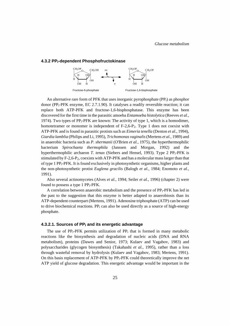
4.3.2 PPi-dependent Phosphofructokinase
An alternative rare form of PFK that uses inorganic pyrophosphate (PPi) as phosphordonor (PPi-PFK enzyme, EC 2.7.1.90). It catalyses a readily reversible reaction; it canreplace both ATP-PFK and fructose-1,6-bisphosphatase. This enzyme has beendiscovered for the first time in the parasitic amoebaEntamoeba histolytica(Reeveset al.,1974). Two types of PPi-PFK are known: The activity of type 1, which is a homodimer,homotetramer or monomer is independent of F-2,6-P2. Type 1 does not coexist withATP-PFK and is found in parasitic protists suchas Eimeria tenella(Dentonet al.,1994),Giardia lamblia(Philips and Li, 1995),Trichomonas vaginalis(Mertenset al.,1989) andin anaerobic bacteria such asP. shermanii(O'Brienet al.,1975), the hyperthermophilicbacterium Spirochaeta thermophila(Janssen and Morgan, 1992) and thehyperthermophilic archaeonT. tenax(Siebers and Hensel, 1993). Type 2 PPi-PFK isstimulated by F-2,6-P2, coexists with ATP-PFK and has a molecular mass larger than thatof type 1 PPi-PFK. It is found exclusively in photosynthetic organisms, higher plants andthe non-photosynthetic protistEuglena gracilis(Balogh et al., 1984; Enomotoet al.,1991).
Also several actinomycetes (Alveset al.,1994; Seileret al.,1996) (chapter 2) werefound to possess a type 1 PPi-PFK.
A correlation between anaerobic metabolism and the presence of PPi-PFK has led inthe past to the suggestion that this enzyme is better adapted to anaerobiosis than itsATP-dependent counterpart (Mertens, 1991). Adenosine triphosphate (ATP) can be usedto drive biochemical reactions. PPi can also be used directly as a source of high-energyphosphate.
4.3.2.1. Sources of PP i and its energetic advantage
The use of PPi-PFK permits utilization of PPi that is formed in many metabolicreactions like the biosynthesis and degradation of nucleic acids (DNA and RNAmetabolism), proteins (Dawes and Senior, 1973; Kulaev and Vagabov, 1983) andpolysaccharides (glycogen biosynthesis) (Takahashiet al., 1995), rather than a lossthrough wasteful removal by hydrolysis (Kulaev and Vagabov, 1983; Mertens, 1991).On this basis replacement of ATP-PFK by PPi-PFK could theoretically improve the netATP yield of glucose degradation. This energetic advantage would be important in the
Fructose-1,6-bisphosphate
O
OH
OH
H
H
HOH
CH2OP CH2OP
Fructose-6-phosphate
O
OH
OH
H
H
HOH
CH2OP CH2OH PPi
Pi
Glucose metabolism
25

case of fermentative metabolism. The energetic advantage discussed here rests on theassumption that the PPi used is a by-product of the biosynthesis of macromolecules. Toallow such biosynthetic reactions to occur the concentration of PPi must be kept at a lowlevel. This can be achieved by hydrolysis of PPi by the pyrophosphatase enzyme (Chenet al.,1994) or by other PPi-consuming enzymes such as the PPi-PFK enzyme (Table 1).Interestingly, organisms possessing a PPi-PFK activity contain a very low level ofpyrophosphatase activity (E. histolytica(Reeveset al.,1974),G. lamblia(Li and Philips,1995),T. vaginalis(Mertenset al., 1989),A. naeslundii(Takahashiet al., 1995),P.freudenreichii(O'Brienet al.,1975).
Another point to be discussed concerns possible alternative sources of PPi. It may begenerated as a result of metabolic cycling between glycogen and glucose-1-P through theaction of UDPG pyrophosphorylase, glycogen synthase and glycogen pyrophosphorylase(NDP-glucose synthase: NTP + glucose-1-Pà NDP-glucose + PPi) (Fig. 3).
Another possibility has been proposed for the organismsP. freudenreichiiand E.histolytica, which in addition to the PPi-PFK activity possess other unusual PPi-dependentenzymes (Mertens, 1993) (Table 1). These are phosphoenolpyruvatecarboxytransphosphorylase (EC 4.1.1.38) and pyruvate orthophosphate dikinase (PPDK,
Glucose
G6P
F6P
F-1,6-P2
PolyPn
PolyPn-1UTP
1
2
3
4
5PPi
Pi
UDP
UDPglucose
Glycogenn
Glycogenn+1
GTP/ATP
GDP/ADP
G1P
PPi
Pi
Figure 3. Proposal for PPi origin in the actinomycete A. naeslundii as a result ofmetabolic cycling between glycogen and glucose-1-P. 1, glucose kinase; 2,phosphofructokinase; 3, UDP-glucose synthase; 4, glycogen synthase; 5, glycogenpyrophosphorylase. Modified after Takahashi et al. (1995).
Chapter 1
26

E.C. 2.7.9.1). In these examples the first reaction would provide PPi and the secondreaction, towards the formation of pyruvate, would require PPi (Table 1). Finally, on theevolutionary point of view, PPi is believed to be a more ancient source of energy thanATP (Keefe and Miller, 1995). The occurrence of a PPi-dependent glycolysis inrepresentatives of the oldest eukaryotic branches (G. lamblia, T. vaginalis, E. histolytica),might be taken as experimental support for this hypothesis. This has led to the suggestionthat biochemical pathways such as glycolysis evolved from being PPi driven to ATPdriven (Kulaev and Vagabov, 1983; Wood, 1985).
4.3.2.2. Enzymes involved in synthesis and degradation ofpolyphosphate
Polyphosphate (PolyPn) has been found in volcanic condensates, deep-oceanic steamvents and in many organisms. It has been considered as a “molecular fossil”. From someof the reactions involved in PolyPn metabolism, PPi is formed (Kornberg, 1995). Table2 gives a survey of these enzyme reactions. In the bacteriaP. freudenreichiiand A.naeslundiipossessing PPi-linked glycolysis, large amounts of polyphosphate have beendetected (Clarket al.,1986; Takahashiet al.,1995). Also in these bacteria polyphosphateglucose kinase activity was present. These findings suggest that polyphosphate can beutilized in these organisms as an intracellular reservoir of energy (Takahashiet al.,1995).
Classical NTP-linked reactions Pyrophosphate-linked reactionsATP + F-6-Pà F-1,6-P2 + ADP PPi + F-6-Pßà F-1,6-P2 + ADP
ATP-phosphofructokinase PPi-phosphofructokinase
PEP + ADPà Pyruvate + ATP PEP + AMP + PPià Pyruvate + ATP +PiPyruvate kinase Pyruvate orthophosphate dikinase
PEP + GDP + CO2ßà Oxaloacetate + GTP PEP + Pi + CO2ßà Oxaloacetate + PPiphosphoenolpyruvate carboxylase phosphoenolpyruvate carboxytransphosphorylase
Acetyl-P + ADPßà Acetate + ATP Acetyl-P + Pißà Acetate + PPiAcetate kinase PPi-acetate kinase
Table 1. Pyrophosphate dependent reactions and the corresponding nucleotide-dependentreactions. Adapted from Mertens (1993).
Glucose metabolism
27

4.3.3. ADP-dependent phosphofructokinase
Recently, a third alternative form of phosphofructokinase has been detected. Thehyperthermophilic archaeon,P. furiosusemploys a ADP-PFK enzyme in its modifiedEmbden-Meyerhof pathway involving ADP-dependent kinases (Kengenet al., 1994,1996). To explain the use of ADP instead of ATP in the PFK reaction it has been suggestedthat ADP is more thermostable than ATP, and that, when the ATP level is low (after astarvation period), the organism is still able to phosphorylate glucose using ADP.
Enzyme EC number ReactionDegradation of polyphosphate (PolyPn)
Polyphosphate glucosekinase 2.7.1.63 Glucose + PolyPnà G6P + PolyPn-1
Polyphosphate:AMPphosphotransferase
PolyPn + AMPà PolyPn-1 +ADP
Endopolyphosphatase 3.6.1.10 PolyPn + H2Oà PolyPn-x + PPx
Exopolyphosphatase 3.6.1.11 PolyPn + H2Oà PolyPn-1 + Pi
Tripolyphosphatase 3.6.1.25 PPPi + H2Oà PPi + Pi
Pyrophosphatase 3.6.1.1 PPi + H2Oà 2Pi
Biosynthesis of polyphosphate (PolyPn)Polyphosphate dikinase 2.7.4.1 ATP + PolyPnßà ADP + PolyPn+1
1,3-Diphosphoglycerate:polyphosphatephosphotransferase 2.7.4.17
1,3-DPG + PolyPnßà 3-PG +PolyPn+1
Table 2. Enzymes involved in biosynthesis and degradation of polyphosphates. Adapted from vanAlebeek (1994).
Fructose-1,6-bisphosphate
O
OH
OH
H
H
HOH
CH2OP CH2OP
Fructose-6-phosphate
O
OH
OH
H
H
HOH
CH2OP CH2OHADP
AMP
Chapter 1
28

4.4. Fructose-1,6-bisphosphate aldolase
Aldolase (E.C 4.1.2.13) catalyses the reversiblecleavage of fructose-1,6-bisphosphateto triose phosphate and dihydroxyacetone phosphate (von der Ostenet al., 1989;Fothergill-Gilmore and Michels, 1993). Two different classes of aldolases can bedistinguished based on the reaction mechanisms (Alefounderet al., 1989). Class 1aldolases, but not class 2, form an intermediate with their substrate through a Schiff base.Class 2 aldolases instead require a divalent cation like Ca2+, Fe2+ or Zn2+ to stabilize theenzyme-substrate complex. Bacterial enzymes have been characterized revealingexamples of both class 1 and 2 proteins (Fothergill-Gilmore and Michels, 1993). Class 1proteins are homotetrameric enzymes with a subunit Mr of 40,000, typical for Eukarya(Horeckeret al.,1972). Bacterial representatives of class 1 enzymes have been found inE. coli (Stribling and Perham, 1973),Lactobacillus casei(London, 1974) andStaphylococcal species (Rudolphet al.,1992; Witke and Gotz, 1993). Class 2 aldolasesare dimeric proteins with identical subunits of Mr 40,000. The aldolases from yeast, asecond aldolase inE. coli (Alefounderet al.,1989) andC. glutamicum(von der Ostenetal.,1989) belong to this class. Only the crystal structures of class 1 enzymes from severalEukarya have been determined (Fothergill-Gilmore and Michels, 1993). No informationis available about this enzyme from actinomycetes.
4.5. Triosephosphate isomerase
The interconversion of dihydroxyacetone phosphate to glyceraldehyde 3-phosphateis catalysed by the enzyme triose phosphate isomerase (TIM E.C 5.3.2.1)(Fothergill-Gilmore and Michels, 1993). The enzyme does not require any cofactor. InBacteria and Eukarya analysed, TIM is a homodimeric protein with a subunit size of Mr
27,000 (Fothergill-Gilmore and Michels, 1993), but in the hyperthermophilic archaeon
Fructose-1,6-bisphosphateDihydroxyacetonphosphate
Glyceraldehyde-3-phosphate
+
O
OH
OH
H
H
HOH
CH2OP CH2OP
C C C
O
HO H
OPH
H H
C C C
OHOP
H
H
O
H H
Glyceraldehyde-3-phosphateDihydroxyacetonphosphate
C C C
O
HO H
OPH
H H
C C C
OHOP
H
H
O
H H
Glucose metabolism
29

Pyrococcus woeseiandMethanothermus fervidusTIM it is a homotetramer (Kohloffetal., 1996). It has been speculated that the tetrameric aggregation is correlated withincreased thermostability. At present, amino acid sequences of this enzyme are availablefrom representatives of most phylogenetic groups (Fothergill-Gilmore and Michels,1993). No information is available about this enzyme from actinomycetes, however.
4.6. Glyceraldehyde-phosphate dehydrogenase
The next step in the pathway, the phosphorylation of glyceraldehyde 3-phosphate to1,3-bisphosphoglycerate, is catalysed by the enzyme glyceraldehyde-phosphatedehydrogenase (GAPdh, E.C 1.2.1.12). This enzyme is a homotetramer with a subunitsize of Mr 34,000-38,000. Each of the subunits can bind NAD+ (cofactor) and the bindingcan occur cooperatively. The primary sequences of different GAPdh enzymes have beendetermined (Fothergill-Gilmore and Michels, 1993). This enzyme is the most highlyconserved enzyme from the glycolysis and the best characterized one at the structurallevel (Fothergill-Gilmore and Michels, 1993). The enzymes from Eukarya and Bacteriaare clearly homologous whereas the enzymes from Archae have been shown to be verydifferent from those of the other organisms (Fabry and Hensel, 1988; Fabryet al.,1989;Zwickl et al.,1990; Prubet al.,1993). A different origin has been proposed for the lattertype of GAPdhs (Fothergill-Gilmore and Michels, 1993). In the actinomyceteStreptomyces aureofaciens, the gene encoding GAPdh (gap) has been cloned andsequenced, showing 52% amino acid identity with bacterial and eukaryoticgap genes(Kormanecet al.,1995).
Glyceraldehyde-3-phosphate Glyceraldehyde-1,3-diphosphate
C C C
OHOP
H
H
O
H H
C C C
OH
H
OPOP
H
H
O
NAD + P+i
NADH
Chapter 1
30

4.7. 3-Phosphoglycerate kinase
Phosphoglycerate kinase (PGK) catalyses the reversible conversion of1,3-phosphoglycerate to 3-phosphoglycerate (EC 2.7.2.3). This enzyme is normally amonomer with a Mr of 44,000 (Scopes, 1973). A large number of full primary sequencesof PGK are now available. The enzymes from Eukarya, Bacteria and Archaea are quitehomologous (Fothergill-Gilmore and Michels, 1993). In the hyperthermophilicbacteriumT. maritimaPGK exists in two forms. A separate form with a subunit size of 43 kDa, andas a fusion protein (together with the TIM enzyme) with a subunit size of 70 kDa. Thisis a unique example of a bifunctional enzyme in the glycolytic pathway combining thecatalysis of two nonconsecutive reactions (Schuriget al.,1995).
4.8. Phosphoglycerate mutase
Phosphoglycerate mutase (PGM, EC 5.4.2.1) catalyses the interconversion of3-phosphoglycerate and 2-phosphoglycerate. It comprises a family of enzymes whichcatalyse reactions involving the transfer of phosphor groups among the three carbonatoms of phosphoglycerates. There are two major groups of PGM enzymes with differentkinetic properties (Fothergill-Gilmore and Michels, 1993). Group 1 type of PGM isdependent upon 2,3-diphosphoglycerate 2,3-DPG) for activity. It has been found invertebrates, yeast (Fothergill-Gilmore and Watson, 1989) and also recently in twoactinomycetesS. coelicolorA3(2) (Whiteet al.,1992) and, inA. methanolica(Alves etal., 1994) (chapter 2). Cofactor-dependent PGMs are active as monomers, dimers ortetramers depending upon the organism from which they have been isolated. Thecofactor-dependent PGM fromS. cerevisaeis the best studied PGM enzyme. The3D-structure of this tetrameric enzyme has been solved and the amino acid residuesinvolved in the active site have been assigned (Winnet al.,1981; Fothergill-Gilmore and
Glyceraldehyde-1,3-diphosphate 3-Phosphoglycerate
C C C
OH
H
OPOP
H
H
O
ADP
ATP
C C C
OH
H
OHOP
H
H
O
3-Phosphoglycerate 2-Phosphoglycerate
C C C
OH
H
OHOP
H
H
O C C C
OP
H
OHOH
H
H
O
Glucose metabolism
31
Watson, 1989). InS. coelicolorA3(2) PGM is a tetrameric enzyme and inA. methanolicaPGM1 is a monomer and PGM2 is a dimer (chapters 2 and 6).
Group 2 PGM is independent of 2,3-DPG for its activity; it occurs in higher plantsand invertebrates and is independent of any co-factors (Fothergill-Gilmore and Watson,1989; Fothergill-Gilmore and Michels, 1993).Bacillus spp. have 2,3-DPG independentPGMs which require manganese for activity (Vazquez-Leyva and Setlow, 1994). Thecofactor-independent PGMs are monomers of Mr 60,000 (Fothergill-Gilmore andMichels, 1993).
Another category of PGM is a closely related enzyme (DPG mutase/synthase EC5.4.2.4/EC 3.1.3.13) which catalyses the synthesis of 2,3-DPG and plays a role incontrolling haemoglobin oxygen affinity (Fothergill-Gilmore and Michels, 1993). Thisenzyme is considered to be an isoenzyme of the vertebrate glycolytic PGM because ofsequence similarities. DPG mutase possesses a high level of monophosphoglyceratemutase activity in addition to 2,3-DPG synthase and phosphatase activity, and it isfrequently designated as bisphosphoglycerate synthase (Fothergill-Gilmore and Watson,1989).
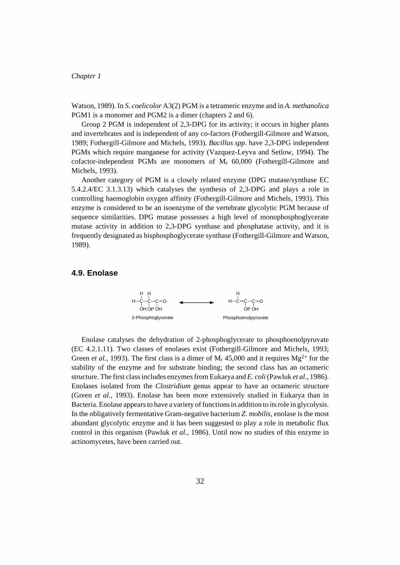
4.9. Enolase
Enolase catalyses the dehydration of 2-phosphoglycerate to phosphoenolpyruvate(EC 4.2.1.11). Two classes of enolases exist (Fothergill-Gilmore and Michels, 1993;Greenet al.,1993). The first class is a dimer of Mr 45,000 and it requires Mg2+ for thestability of the enzyme and for substrate binding; the second class has an octamericstructure. The first class includes enzymes from Eukarya andE. coli(Pawluket al.,1986).Enolases isolated from theClostridium genus appear to have an octameric structure(Greenet al., 1993). Enolase has been more extensively studied in Eukarya than inBacteria. Enolase appears to have a variety of functions in addition to its role in glycolysis.In the obligatively fermentative Gram-negative bacteriumZ. mobilis, enolase is the mostabundant glycolytic enzyme and it has been suggested to play a role in metabolic fluxcontrol in this organism (Pawluket al.,1986). Until now no studies of this enzyme inactinomycetes, have been carried out.
2-Phosphoglycerate Phosphoenolpyruvate
C C C
OP
H
OHOH
H
H
O C C C
OP OH
H
H
O
Chapter 1
32

4.10. Pyruvate kinase
Pyruvate kinase (PK, EC 2.7.1.40) is one of the major regulatory enzymes of theglycolytic pathway. It catalyses the last step of the glycolysis, the irreversible reactionfrom PEP to pyruvate (Kayne, 1973). In most bacteria this protein is a tetramer composedof identical subunits of 500 amino acid residues, although some different structures havebeen identified (Fothergill-Gilmore and Michels, 1993). Almost all PKs show positivecooperativity in binding the substrate PEP (Fothergill-Gilmore and Michels, 1993; Sakaiand Ohta, 1993). In addition, they are often regulated by a number of allosteric effectorslike glucose-6-phosphate, ribulose 5-phosphate, AMP and F-1,6-P2 (le Bras and Garel,1993). Two different mechanisms of regulation at the activity level can be found inBacteria.
E. coli andSalmonella typhimuriumeach have two differently regulated isoenzymes(Fothergill-Gilmore and Michels, 1993; Ponceet al., 1995) with different kineticproperties. InE. coli the type 1 PK isoenzyme is constitutive and allosterically activatedby F-1,6-P2. This enzyme is also susceptible to a synergistic cooperative feedbackinhibition by succinyl-CoA and ATP, and to inhibition by phosphate. The type 2 PKisoenzyme is inducible and stimulated by AMP and by some sugar monophosphates suchas ribose-5-phosphate and glucose-6-phosphate (Ponceet al.,1995). The physiologicalroles of the two pyruvate kinases and their regulation may be as follows. When cells aregrown on a glycolytic substrate, the intracellular concentration of F-1,6-P2 is high, thusstimulating the activity of isoenzyme 1. Under conditions where gluconeogenesis ispredominant, the F-1,6-P2 concentration is at such a low level that the enzyme is virtuallyinactive, whatever the concentration of PEP. However, the PEP level is such that theisoenzyme 2 can catalyse the reaction at 30-40% of the maximal rate. A decrease of thecellular energy charge would then, via the effector AMP, activate the enzyme and increasethe ATP concentration (Fothergill-Gilmore and Michels, 1993; Ponceet al.,1995).
In some protists and bacteria (E. histolytica, Bacteroides symbiosus, G. lambliaandP. freudenreichii) PK activity is absent and substituted by PPDK (Woodet al.,1977;Wood and Goss, 1985; Mertens, 1991). This enzyme differs in the use of the substratePPi instead of ADP, and the fact that it catalyses both the forward and the reverse reactions.Like the PK enzyme, it also requires the ions K+, NH4+ and Mg2+ for its activity. Thistype of activity (PPDK) is associated with other pyrophosphate-linked reactions such as
Phosphoenolpyruvate Pyruvate
C C C
OP OH
H
H
O C C C
O OHH
H
H
O
ADP
ATP
Glucose metabolism
33

Organism Gene Organization Reference400 bp
pfk pk
gap fdapgk
Bacillus megateriumgap pgk tpi
Corynebacterium glutamicumgap pgk tpi ppc
Sulfolobus solfataricuspgk gap
Thermotoga maritima tpipgk
enopgmtpipgkBacillus subtilis
Zymomonas mobilis pgkgap
Bacillus stearothermophilus pgkgap
Lactococcus lactis pfk pk ldh
Lactobacillus bulgaricus pfk pk
Escherichia coli
Sakai and Ohta, 1993
Alefounder and Perham, 1989
Schlapfer and Zuber,1992
Eikmanns, 1996;Schwinde ., 1993et al
Jones ,1995et al.
Schurig ., 1995et al
Vazquez-Levya andSetlow, 1994
Conway ., 1987,1988
et al
Davies ., 1991;Branlant ., 1989
et alet al
Llanos ., 1993et al
Branny ., 1993et al
Hellinga and Evans, 1985;Pichersky and Hess 1984
tpipfk sbp cdh
Figure 4. Examples of the organization of glycolytic genes in various bacteria.Phosphoglycerate kinase (pgk), glyceraldehyde-3-phosphate dehydrogenase (gap),triose phosphate isomerase (tpi), phosphofructokinase (pfk), pyruvate kinase (pk),enolase (eno), phosphoglycerate mutase (pgm), fructose-1,6-bisphosphate aldolase(fda), lactate dehydrogenase (ldh), periplasmic sulphate-binding protein (sbp),CDP-diglycerate hydrolase (cdh), phosphoenolpyruvate carboxylase (ppc). White barscorrespond to genes coding for non-glycolytic enzymes, that are part of a cluster ofglycolytic genes. White bar with dashed lines correspond to a possible pgk gene in B.subtilis (Vazquez-Leyva and Setlow, 1994).
Chapter 1
34

the PPi-PFK, PEP carboxytransphosphorylase and PPi-acetate kinase (EC 2.7.2.12)(Mertens, 1993) (see also section 4.3.2.1)
PK has also been characterized from different Gram-positive bacteria such asC.glutamicum(Jettenet al., 1994),Brevibacterium flavum(Ozaki and Shiio, 1969), andfrom the actinomycetesS. coelicolorA3(2) (personal communication L.V. Bystrykh) andA. methanolica(this thesis, chapter 2). In these organisms (except inS. coelicolorA3(2):dimer) the PK enzyme is a tetramer, and it is regulated at the activity level being activatedby AMP and inhibited by ATP. TheC. glutamicumenzyme differs in that it does notrequire Mg2+ ions for activity but Mn2+ or Co2+ ions (Jettenet al.,1994).
5. Organization of genes encoding glycolytic enzymes
Gene clusters encoding enzymes involved in glycolysis have been identified in severalbacteria. In some cases these clusters contain genes that code for enzymes catalysingirreversible steps in glycolysis such as ATP-PFK and PK. Alternatively genes encodingenzymes catalysing consecutive steps, such as GAPdh and PGK, may cluster. Furthervariations occur as well (Fig. 4). However, these findings show that considerable diversityexists among bacteria with respect to the transcriptional organization of their glycolyticgenes. Clustering of genes encoding glycolytic enzymes has not been reported foractinomycetes.
REFERENCES
References are listed on pages 129-140.
Glucose metabolism
35