Chapter 04 Lecture Outline - Napa Valley College 105/Mader Human Biology 14ed/chapt04...1 Chapter 04...
49
1 Chapter 04 Lecture Outline See separate PowerPoint slides for all figures and tables pre- inserted into PowerPoint without notes. Copyright © 2016 McGraw-Hill Education. Permission required for reproduction or display.
Transcript of Chapter 04 Lecture Outline - Napa Valley College 105/Mader Human Biology 14ed/chapt04...1 Chapter 04...

1
Chapter 04
Lecture Outline
See separate PowerPoint slides for all figures and tables pre-
inserted into PowerPoint without notes.
Copyright © 2016 McGraw-Hill Education. Permission required for reproduction or display.
2
Organization and Regulation
of Body Systems

3
Points to ponder
• What is a tissue? Organ? Organ system?
• What are the 4 main types of tissue?
• What do these tissues look like, how do they
function, and where are they found?
• What is the integumentary system?
• How can you prevent skin cancer?
• What is homeostasis and how is it maintained?

4
What is a tissue?
• A collection of cells of the same type that
perform a common function
• There are 4 major tissue types in the body.
1. Connective
2. Muscular
3. Nervous
4. Epithelial
4.1 Types of Tissues
5
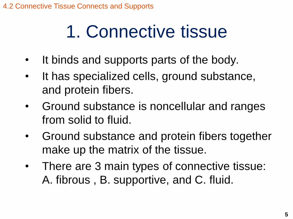
1. Connective tissue
• It binds and supports parts of the body.
• It has specialized cells, ground substance,
and protein fibers.
• Ground substance is noncellular and ranges
from solid to fluid.
• Ground substance and protein fibers together
make up the matrix of the tissue.
• There are 3 main types of connective tissue:
A. fibrous , B. supportive, and C. fluid.
4.2 Connective Tissue Connects and Supports

6
3 main types of connective tissue
A. Fibrous
B. Supportive
C. Fluid
Figure 4.4 Types of connective tissue.
4.2 Connective Tissue Connects and Supports
Types of Connective Tissue
Lymph
Contained in
Lymphatic vessels
Loose
Fibers create
loose, open
framework
Dense
Fibers are
densely packed
Cartilage
Solid yet flexible
matrix
Bone
Solid and rigid
matrix
Blood
Contained in
blood vessels
Fibrous connective tissue Fluid connective tissue Supportive connective tissue
7
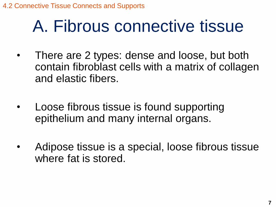
A. Fibrous connective tissue
• There are 2 types: dense and loose, but both contain fibroblast cells with a matrix of collagen and elastic fibers.
• Loose fibrous tissue is found supporting epithelium and many internal organs.
• Adipose tissue is a special, loose fibrous tissue where fat is stored.
4.2 Connective Tissue Connects and Supports

8
What does loose fibrous
connective tissue look like?
elastic fiber
collagen
fiber
fibroblast
Loose fibrous tissue
© Ed Reschke
Figures 4.1 and 4.2 Connective tissues
(components and knee).
4.2 Connective Tissue Connects and Supports
Copyright © The McGraw-Hill Companies, Inc. Permission required for reproduction or display.
Stem cell: divides to
produce other types
of cells
Collagen fiber:
unbranched, strong
but flexible
Fibroblast: divides to
produce other types
of cells
Reticular fiber:
branched, thin, and
forms network
White blood cell:
engulfs pathogens
or produces antibodies
Adipose cell:
stores fat
Ground
substance: fills
spaces between
cells and fibers
Elastic fiber:
branched and
stretchable
Blood vessel

9
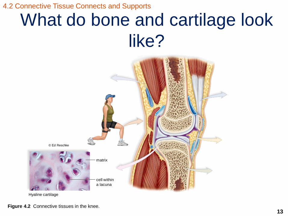
• Cells are in chambers called lacunae.
• Matrix is solid but flexible.
4.2 Connective Tissue Connects and Supports
B. Supportive connective tissue:
Cartilage

10
B. Supportive connective tissue:
Cartilage
• 3 types are distinguished by types of fibers.
1. Hyaline cartilage – fine collagen fibers
Location: Nose, ends of long bones, and fetal
skeleton
2. Elastic cartilage – more elastic fibers than cartilage
fibers
Location: Outer ear
3. Fibrocartilage – strong collagen fibers
Location: Disks between vertebrae
4.2 Connective Tissue Connects and Supports

11
B. Supportive connective tissue:
Bone • Cells are in chambers called lacunae.
• Solid and rigid matrix is made of collagen and
calcium salts.
4.2 Connective Tissue Connects and Supports

12
B. Supportive connective tissue:
Bone • 2 types are distinguished by types of fibers.
1. Compact – made of repeating circular units
called osteons which contain the hard matrix,
living cells, and blood vessels
Location: Shafts of long bones
2. Spongy – an open latticework with irregular
spaces
Location: Ends of long bones
4.2 Connective Tissue Connects and Supports
13
What do bone and cartilage look
like?
cell within
a lacuna
matrix
Hyaline cartilage
© Ed Reschke
Figure 4.2 Connective tissues in the knee.
4.2 Connective Tissue Connects and Supports

14
C. Fluid connective tissue:
Blood
• Made of a fluid matrix called plasma and cellular components that are called formed elements
• 3 formed elements: 1. Red blood cells (erythrocytes) – cells that carry
oxygen
2. White blood cells (leukocytes) – cells that fight infection
3. Platelets (thrombocytes) – pieces of cells that clot blood
4.2 Connective Tissue Connects and Supports

15
C. Fluid connective tissue:
Blood
Figure 4.3 The formed elements of blood.
4.2 Connective Tissue Connects and Supports
White blood cells
platelets
red blood cell
plasma
(surrounds
formed
elements)
16
C. Fluid connective tissue:
Lymph
• Matrix is a fluid called lymph.
• White blood cells congregate in lymph
nodes.
4.2 Connective Tissue Connects and Supports

17
2. Muscle tissue
• It allows for movement in the body.
• It is made of muscle fibers/cells and protein
fibers called actin and myosin.
• There are 3 types of muscle tissue in
humans: A. skeletal, B. smooth, and
C. cardiac.
4.3 Muscular Tissue Moves the Body

18
A. Muscle tissue - Skeletal
• Appearance: long,
cylindrical cells,
multiple nuclei,
striated fibers
• Location: attached
to bone for
movement
• Nature: voluntary
movement
Figure 4.5a. The three types of muscle tissue.
4.3 Muscular Tissue Moves the Body

19
B. Muscle tissue - Smooth
• Appearance:
spindle-shaped cell
with one nucleus,
lacks striations
• Location: walls of
hollow organs and
vessels
• Nature: involuntary
movement
Figure 4.5b. The three types of muscle tissue.
4.3 Muscular Tissue Moves the Body

20
C. Muscle tissue – Cardiac • Appearance:
branched cells with
a single nucleus,
striations with
darker striations
called intercalated
disks between cells
• Location: heart
• Nature: involuntary
movement
Figure 4.5c. The three types of muscle tissue.
4.3 Muscular Tissue Moves the Body

21
3. Nervous tissue
• It allows for communication between cells
through sensory input, integration of data, and
motor output.
• It is made of 2 major cell types: A. neurons
and B. neuroglia.
4.4 Nervous Tissue Communicates

22
A. Nervous tissue - neurons
• They are made of
dendrites, a cell
body, and an axon.
• Dendrites carry
information toward
the cell body.
• Axons carry
information away
from the cell body.
nucleus Neuron
cell body
Astrocyte Microglia
Oligodendrocyte
myelin sheath
axon
Capillary
dendrite
nucleus
cell body
axon
Micrograph of neuron
dendrite
© Ed Reschke Figure 4.6. A neuron and examples of supporting neuroglia cells.
4.4 Nervous Tissue Communicates

23
A. Nervous tissue - neuroglia
• They are a collection
of cells that support
and nourish neurons.
• They outnumber
neurons 9:1.
• Examples are
oligodendrocytes,
astrocytes, and
microglia.
axon
dendrite
nucleus
cell body
Astrocyte
Neuron
Oligodendrocyte
nucleus
cell body
dendrite
myelin sheath
Microglia
Capillary
Micrograph of neuron © Ed Reschke
4.4 Nervous Tissue Communicates
Figure 4.6. A neuron and examples of supporting neuroglia cells.

24
4. Epithelial tissue
• It is a group of cells that forms a tight, continuous network.
• It lines body cavities, covers body surfaces, and is found in glands.
• Cells are anchored by a basement membrane
on one side and free on the other side.
• It is named after the appearance of cell layers and the shape of the cells.
• There is transitional epithelium that changes in appearance in response to tension.
4.5 Epithelial Tissue Protects
25
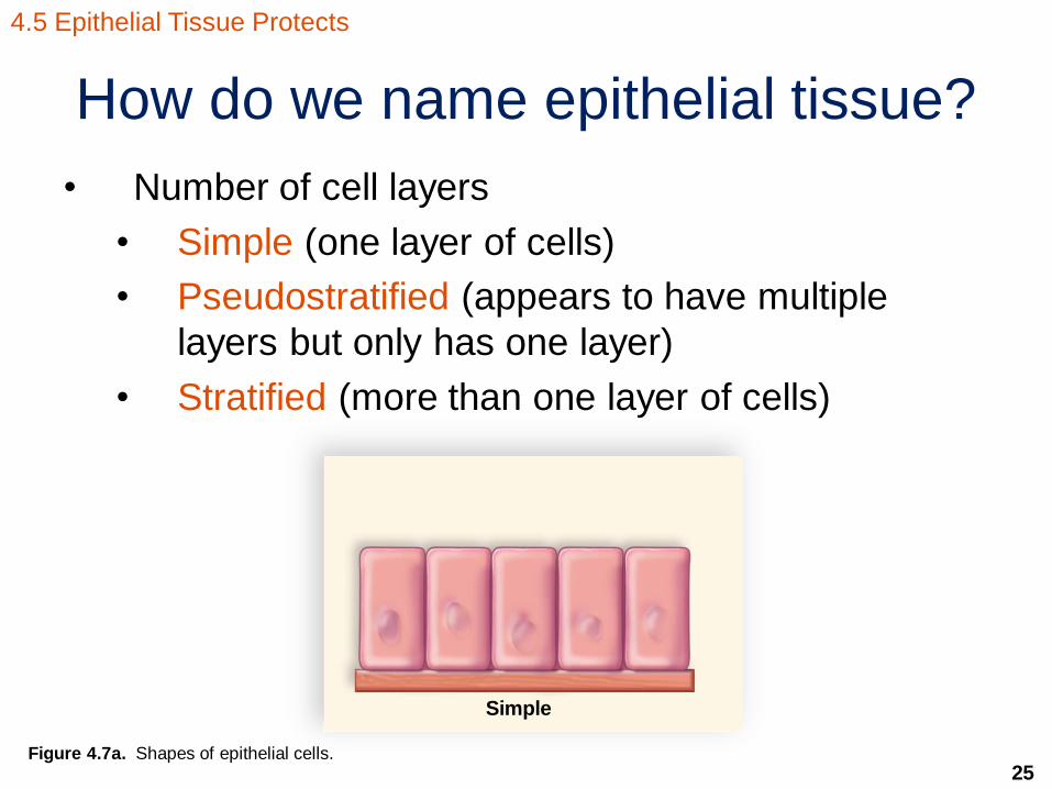
How do we name epithelial tissue?
• Number of cell layers
• Simple (one layer of cells)
• Pseudostratified (appears to have multiple
layers but only has one layer)
• Stratified (more than one layer of cells)
Figure 4.7a. Shapes of epithelial cells.
4.5 Epithelial Tissue Protects
Simple
26
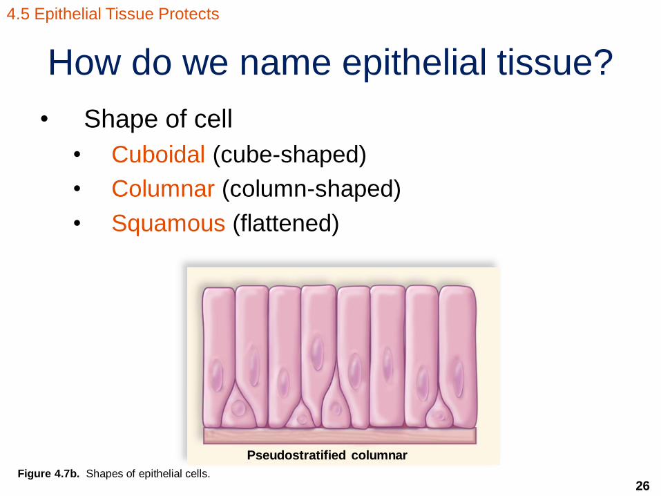
How do we name epithelial tissue?
• Shape of cell
• Cuboidal (cube-shaped)
• Columnar (column-shaped)
• Squamous (flattened)
Figure 4.7b. Shapes of epithelial cells.
4.5 Epithelial Tissue Protects
Pseudostratified columnar

27
What does epithelial tissue look like?
Simple
squamous
• Lining of lungs,
Blood vessels • protects
Simple cuboidal
• lining kidney tubules, various glands
• absorbs molecules
basement membrane basement membrane
© Ed Reschke
•
•
basement membrane basement
membrane
basement
membrane
goblet cell
secretes
mucus
goblet cell
secretes
mucus
cilia
• lining of nose,
mouth, esophagus,
anal canal, vagina • protects
Stratified squamous
• lining of trachea • sweeps impurities
towards that
Pseudostratified,
ciliated columnar
Simple columnar lining of small
intestine, oviducts absorbs nutrients
© Ed Reschke Figure 4.8. The basic types of epithelial cells.
4.5 Epithelial Tissue Protects

28
The integumentary system • It includes the skin and accessory organs such as
hair, nails, and glands.
• The skin has 2 main regions called the epidermis and the dermis.
• Under the skin there is a subcutaneous layer between the dermis and internal structures where fat is stored.
• It is important for maintaining homeostasis.
4.6 Integumentary System
29
What are the functions of the
integumentary system?
1. It protects the body from physical trauma,
invasion by pathogens, and water loss.
2. It helps regulate body temperature.
3. It allows us to be aware of our surroundings
through sensory receptors.
4. It synthesizes chemicals such as melanin and
vitamin D.
4.6 Integumentary System

30
There are 2 regions of the skin
• Epidermis
• Dermis
sweat pore
stem cells
sensory receptor
capillaries
oil gland
arrector pili muscle
free nerve endings
hair follicle
hair root
sweat gland
artery
vein
nerve
adipose tissue
Subcutaneous
layer
(hypodermis)
Dermis
Epidermis
hair shaft
Figure 4.9. Anatomy of human skin.
4.6 Integumentary System

31
The epidermis
• It is the thin, outermost layer of the skin.
• It is made of epithelial tissue.
• Cells in the uppermost layers are dead and
become filled with keratin, thus acting as a
waterproof barrier.
• Langerhans cells are a type of white blood cells
that help fight pathogens.
• Melanocytes produce melanin that lend to skin
color and protection from UV light.
• Some cells convert cholesterol to vitamin D.
4.6 Integumentary System

32
The dermis
• It is the thick, inner layer of the skin.
• It is made of dense fibrous connective tissue.
• It contains elastic and collagen fibers.
• It contains blood vessels, many sensory
receptors, and glands.
4.6 Integumentary System

33
Where are skin cells keratinized?
Figure 4.10. A light micrograph of human skin.
4.6 Integumentary System
34
What you need to know about
skin cancer?
• 2 of the 3 types that arise in the epidermis
• Basal cell carcinoma is the most common yet
least deadly form of skin cancer.
• Melanoma is the most deadly form of skin
cancer but is the least common.
4.6 Integumentary System
35
What you need to know about
skin cancer?
• What can you do to help prevent this?
• Stay out of the sun between 10 A.M. and 3 P.M.
• Wear protective clothing (tight weave, treated
sunglasses, wide-brimmed hat).
• Use sunscreen with an SPF of at least 15 that
protects from UV-A and UV-B rays.
• Do not use tanning beds.
4.6 Integumentary System

36
What might skin cancer look like?
Figure 4.11. Cancers of the skin.
4.6 Integumentary System

37
What are the accessory organs of the
skin and why are they important?
• They include nails, hair, and glands.
• Nails are derived from the epidermis and offer a protective covering.
• Hair follicles are derived from the dermis, but hair grows from epidermal cells.
• Oil glands are associated with hair and produce sebum that lubricates the hair and skin and retards bacterial growth.
• Sweat glands are derived from the dermis and help to regulate body temperature.
4.6 Integumentary System
38
Moving from tissue to organs
and organ systems
• An organ is 2 or more tissue types working
towards a particular function.
• An organ system is a combination of organs
that work together to carry out a particular
function.
4.7 Organ Systems, Body Cavities, and Membranes

39
What are the organ systems of
the human body?
Figure 4.13. Organ systems of the body.
4.7 Organ Systems, Body Cavities, and Membranes
Lymphatic and
Immune systems
Digestive system Respiratory system Urinary system
•protects body. • provides temperature
Homeostasis
•synthesizes vitamin D. •receives sensory input
Organ:Skin.
• transport system for nutrients, waste
• provides temperature,
pH, and fluid homeostasis
Organ: Heart
• defends against infectious diseases
• provides fluid
homeostasis • assists in absorption
and transport of fats
Organs: Lymphatic vessels, lymph nodes,
spleen
• ingests, digests, and
processes food • absorbs nutrients and
eliminates waste
• involved in fluid homeostasis
Organs: Oral cavity,
esophagus, stomach, small intestine, large
intestine, salivary glands,
liver, gallbladder, pancreas
• exchanges gases at
both lungs and tissues • assists in pH
homeostasis
Organs: Lungs
• excretes metabolic
wastes • provides pH and
fluids homeostasis
Organs: Kidneys, urinary bladder
Cardiovascular
systems
Integumentary
systems

40
What are the organ systems of
the human body?
Figure 4.13. Organ systems of the body.
4.7 Organ Systems, Body Cavities, and Membranes
• assists in movement
and posture
• produces heat
Organs: Muscles
• receives, processes,
and stores sensory input
• provides motor output
• coordinates organ
systems
Organs: Brain, spinal cord
• produces hormones.
• cordinate organ systems
• regulates metabolism
and stress responce
• involved fluid and
pH homeostasis
Organs: Testes, ovaries,
adrenal glands, pancreas,
thymus, thyroid, pineal gland
Reproductive system Endocrine system Nervous system Muscular system Skeletal system
• provides support
and protection
• assists in movement
• stores minerals
• produces blood cells
Organs: Bones
• produces and transports
gametes
• nurtures and gives birth to
offspring in females
Organs: Testes, penis, ovaries,
uterus, vagina

41
What are the body cavities?
Figure 4.14. Body cavities of humans.
4.7 Organ Systems, Body Cavities, and Membranes
Thoracic cavity:
contains heart,
lungs, and esophagus
Ventral
cavity
Abdominal
cavity:
Contains stomach,
liver, spleen,
pancreas,
gallbladder,
and intestines
Pelvic cavity:
contains
reproductive and other
organs
diaphragm
Vertebral
cavity:
contains
spinal cord
Dorsal
cavity
Cranial
cavity: Contains brain Thoracic cavity:
Contains esophagus,
heart, and lungs
Abdominal cavity:
contains digestive
and other organs
Pelvic cavity: contains reproductive
and other organs
peritoneum
pericardium
plurae
a.
b.

42
What about the body membranes
that line the cavities?
• Mucous membranes – line the digestive, respiratory, urinary, and reproductive systems
• Serous membranes – line the lungs, heart, and abdominal cavity and cover the internal organs; named after their location • Pleura: lungs
• Peritoneum: abdominal cavity and organs
• Pericardium: heart
4.7 Organ Systems, Body Cavities, and Membranes

43
What about the body membranes
that line the cavities?
• Synovial membranes – line the cavities of freely movable joints
• Meninges – cover the brain and spinal cord
4.7 Organ Systems, Body Cavities, and Membranes
44
What is homeostasis?
• It is the ability to maintain a relatively constant internal environment in the body.
• The nervous and endocrine systems are key in maintaining homeostasis.
• Changes from the normal tolerance limits result in illness or even death.
4.8 Homeostasis

45
All systems are important in
maintaining homeostasis
4.8 Homeostasis
Regulates and coordinates the activities of all the other systems. It responds quickly to internal and external stimuli.
Transports oxygen and nutrients to tissue cells and transports wastes away from cells. Also transports hormones secreted by the endocrine glands.
Supplies blood with nutrients and water for tissue cells. Rids the body of nondigestible remains.
Produces heat that maintains body temperature. Protects and supports internal organs.
All systems of the body contribute to maintain homeostasis. These systems in particular are especially note worthy.
Endocrine glands secrete hormones, which also regulate and coordinate the activities of other systems. Works more slowly than the nervous system.
Supplies blood with oxygen for tissue cells and rids blood of carbon dioxide. Helps regulate the acid–base balance of the blood.
Excretes nitrogenous and other wastes. Regulates water–salt balance of the blood. Helps regulate the acid–base balance of the blood.
Helps maintain blood volume by collecting excess tissue fluid and returning it via lymphatic vessels to the cardio vascular veins. Defends against disease.
Helps maintain body temperature and protects internal organs.
Nervous System
Cardiovascular System
Digestive System
Muscular System
Endocrine System
Respiratory System
Urinary System
Lymphatic System
Integumentary System
Figure 4.15.
Homeostasis by
the organ systems
of the human
body.
46
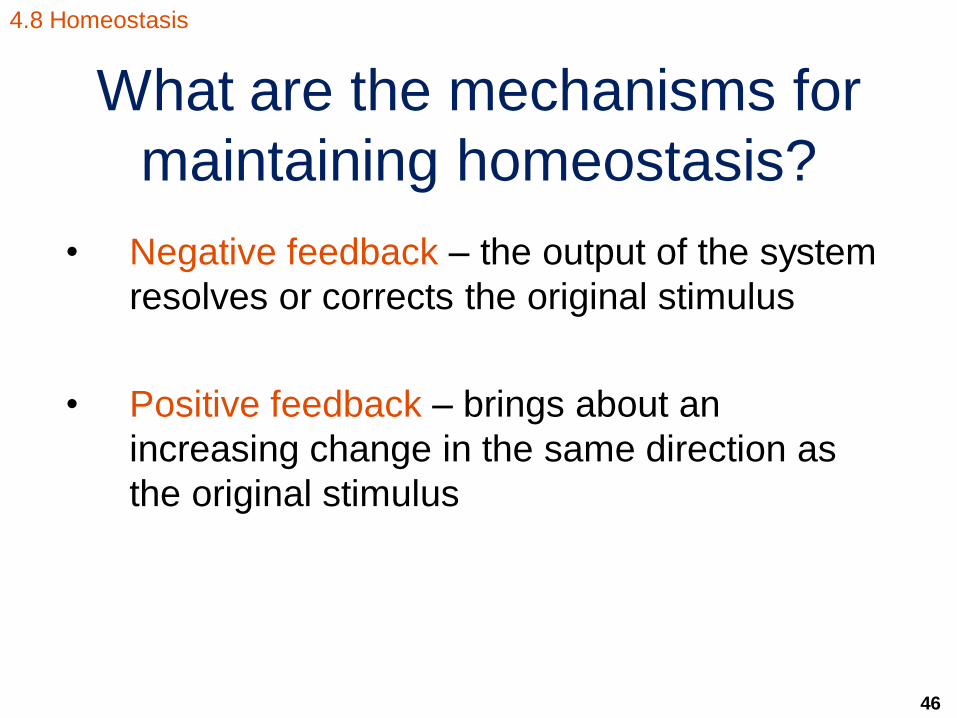
What are the mechanisms for
maintaining homeostasis?
• Negative feedback – the output of the system
resolves or corrects the original stimulus
• Positive feedback – brings about an
increasing change in the same direction as
the original stimulus
4.8 Homeostasis

47
Negative feedback • The primary
mechanism for
maintaining
homeostasis
• The output of
the system
dampens the
original stimulus
• Has 2
components
• sensor
• control center
4.8 Homeostasis
sends data to control center directs response to stimulus
Control center
negative feedback
and return to normal
stimulus
Sensor Effect
Homeostasis
Figure 4.16. Negative feedback mechanisms.
48
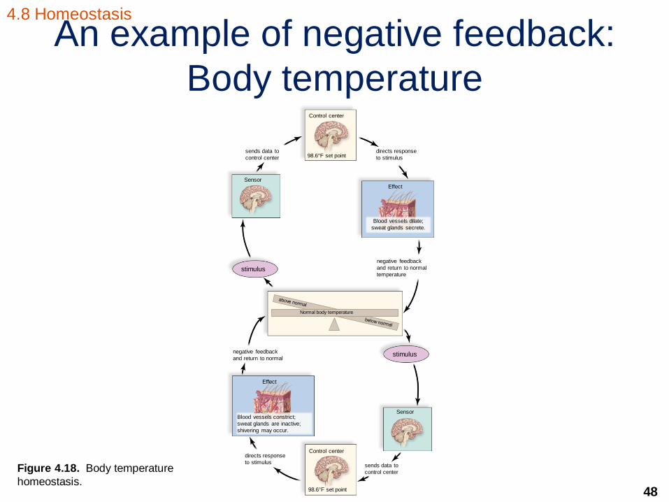
An example of negative feedback:
Body temperature
4.8 Homeostasis
Blood vessels dilate;
sweat glands secrete.
directs response
to stimulus
Control center
98.6°F set point sends data to
control center
negative feedback
and return to normal
temperature
Sensor
Effect
stimulus
stimulus negative feedback
and return to normal
Effect
Sensor
sends data to
control center
directs response
to stimulus
Blood vessels constrict;
sweat glands are inactive;
shivering may occur.
98.6°F set point
Control center
Normal body temperature
Figure 4.18. Body temperature
homeostasis.
49
Positive feedback
• A mechanism for increasing the change of the
internal environment in one direction
• An example is the secretion of oxytocin during
birth to continually increase uterine
contractions
• Can be harmful such as when a fever is too
high and continues to rise
4.8 Homeostasis

![Chapt04 Holes Lecture Animation[1]](https://static.fdocuments.us/doc/165x107/554b3a53b4c905ab378b464c/chapt04-holes-lecture-animation1.jpg)