Changes in Wnt/ -Catenin Pathway During Regulated Growth ... · Changes in Wnt/ -Catenin Pathway...
12
Changes in Wnt/-Catenin Pathway During Regulated Growth in Rat Liver Regeneration SATDARSHAN P. S. MONGA, 1 PETER PEDIADITAKIS, 1 KAREN MULE, 1 DONNA BEER STOLZ, 2 AND GEORGE K. MICHALOPOULOS 1 The wnt/-catenin pathway is important during embryo- genesis and carcinogenesis. -Catenin interaction with E- cadherin has been shown to be crucial in cell-cell adhesion. We report novel findings in the wnt pathway during rat liver regeneration after 70% partial hepatectomy using Western blot analyses, immunoprecipitation studies, and immuno- fluorescence. We found wnt-1 and -catenin proteins to be predominantly localized in hepatocytes. Immediately fol- lowing partial hepatectomy, we observed an initial increase in -catenin protein during the first 5 minutes with its trans- location to the nucleus. We show this increase to be the result of decreased degradation of -catenin (decrease in serine phosphorylated -catenin) as seen by immunopre- cipitation studies. We observed activation of -catenin deg- radation complex comprising of adenomatous polyposis coli gene product (APC) and serine-phosphorylated axin pro- tein, beginning at 5 minutes after hepatectomy, leading to its decreased levels after this time. Quantitative changes ob- served in E-cadherin protein during liver regeneration are, in general, reverse to those seen in -catenin. In addi- tion, using immunoprecipitation, we observe elevated levels of tyrosine-phosphorylated -catenin at 6 hours onward. Thus, changes in the wnt pathway during regulated growth seem to tightly regulate cytosolic -catenin levels and may be contributing to induce cell proliferation and target gene expression. Furthermore, these changes might also be in- tended to negatively regulate cell-cell adhesion for struc- tural reorganization during the process of liver regenera- tion. (HEPATOLOGY 2001;33:1098-1109.) The wnt signaling pathway, along with its components, has been shown to be crucial in directing cell fate during embry- ogenesis. 1 This pathway also regulates cell proliferation in adult tissue. 2 Appropriate regulation of this pathway is crucial for normal growth and proliferation of cells. 3,4 Aberrations in this pathway have been implicated in several human malig- nancies including, but not limited to, colorectal cancer, pros- tatic cancer, hepatocellular cancer, lung cancer, breast cancer, and melanoma. 5-14 Wnt signaling is regulated inside the cell mainly by -catenin levels, which in turn are regulated at the level of their degradation via ubiquitination. 15 It is one of the key proteins in this pathway and its deregulation is associated with molecular pathogenesis of several malignancies. Somatic mutations in the -catenin gene, its cellular redistribution, and nuclear accumulation are a few events that have been implicated in tumorigenesis and its progression by constitu- tively stimulating cell proliferation. 16,17 Distinctive sequences of events follow the activation of this signal transduction pathway. The secreted protein, wnt, binds to its receptor frizzled to inactivate glycogen synthase kinase 3 (GSK3) by hypophosphorylation 18-20 and activation of disheveled through an unknown mechanism. 2 This leads to hypophosphorylation of -catenin, the adenomatous polypo- sis coli gene product (APC), and axin. 21,22 Axin acts as a scaf- fold for the above proteins to assemble. 23 As this complex undergoes hypophosphorylation, -catenin can no longer stay bound and is released. This elevated monomeric form of -catenin binds to proteins including T-cell factor-4 (TCF-4), lymphoid enhancement factor, or pangolin 24-26 to translocate to the nucleus, where they control the transcription of target genes including c-myc and cyclin-D1, among others. 27,28 In the absence of wnt signal, GSK3, axin, APC, and -catenin are phosphorylated at specific serine and threonine residues. -Catenin is now presented to -transducing repeat-contain- ing protein for ubiquitination and degradation. 2 Thus, APC and axin are the 2 important regulatory proteins required for degradation of -catenin. Another mechanism by which in- tracytoplasmic level of -catenin is regulated is by its associ- ation with the intracellular tail of E-cadherin, an intercellular adhesion molecule. 29,30 In addition, phosphorylation of -catenin on tyrosine residues has been shown to negatively regulate cell-cell adhesion. 31 This has been studied to be an important transitional event for some tumors to become lo- cally invasive or even metastatic. 32,33 We wanted to study the wnt transduction pathway in nor- mal rat liver as well as during its regulated growth in vivo. Liver regeneration after 70% partial hepatectomy in an adult rat involves initiation of proliferation of remaining parenchy- mal cells and is a useful model for studying signaling mole- cules and other factors that are involved in cell prolifera- tion. 34-36 The remnant liver after partial hepatectomy undergoes almost complete restoration of the lost mass and function, by about 1 week’s time. 34,37-40 Cell proliferation be- gins very early during liver regeneration, peaking for hepato- cytes at 24 hours, followed by the biliary epithelium at 48 hours, Kupffer and stellate cells at 72 hours, and the sinusoi- dal endothelial cells at 96 hours. 37,41,42 Increased expression Abbreviations: GSK3, glycogen synthase kinase 3; APC, adenomatous polyposis coli gene product; TCF-4, T-cell factor 4; uPAR, urokinase receptor; HRP, horseradish peroxidase; PBS, phosphate-buffered saline. From the Departments of 1 Pathology and 2 Cell Biology and Physiology, University of Pittsburgh, School of Medicine, Pittsburgh, PA. Received September 22, 2000; accepted February 8, 2001. Supported by grants NIH CA30241 and NIH CA35373 to G.K.M. Address reprint requests to: George K. Michalopoulos, M.D., Ph.D., Professor and Chairman, Department of Pathology, University of Pittsburgh, School of Medicine, S410 Biomedical Science Tower, 200 Lothrop Street, Pittsburgh, PA 15261. E-mail: [email protected]; fax: 412-648-9846. Copyright © 2001 by the American Association for the Study of Liver Diseases. 0270-9139/01/3305-0013$35.00/0 doi:10.1053/jhep.2001.23786 1098
Transcript of Changes in Wnt/ -Catenin Pathway During Regulated Growth ... · Changes in Wnt/ -Catenin Pathway...

Changes in Wnt/�-Catenin Pathway During Regulated Growthin Rat Liver Regeneration
SATDARSHAN P. S. MONGA,1 PETER PEDIADITAKIS,1 KAREN MULE,1 DONNA BEER STOLZ,2 AND GEORGE K. MICHALOPOULOS1
The wnt/�-catenin pathway is important during embryo-genesis and carcinogenesis. �-Catenin interaction with E-cadherin has been shown to be crucial in cell-cell adhesion.We report novel findings in the wnt pathway during rat liverregeneration after 70% partial hepatectomy using Westernblot analyses, immunoprecipitation studies, and immuno-fluorescence. We found wnt-1 and �-catenin proteins to bepredominantly localized in hepatocytes. Immediately fol-lowing partial hepatectomy, we observed an initial increasein �-catenin protein during the first 5 minutes with its trans-location to the nucleus. We show this increase to be theresult of decreased degradation of �-catenin (decrease inserine phosphorylated �-catenin) as seen by immunopre-cipitation studies. We observed activation of �-catenin deg-radation complex comprising of adenomatous polyposis coligene product (APC) and serine-phosphorylated axin pro-tein, beginning at 5 minutes after hepatectomy, leading to itsdecreased levels after this time. Quantitative changes ob-served in E-cadherin protein during liver regenerationare, in general, reverse to those seen in �-catenin. In addi-tion, using immunoprecipitation, we observe elevated levelsof tyrosine-phosphorylated �-catenin at 6 hours onward.Thus, changes in the wnt pathway during regulated growthseem to tightly regulate cytosolic �-catenin levels and maybe contributing to induce cell proliferation and target geneexpression. Furthermore, these changes might also be in-tended to negatively regulate cell-cell adhesion for struc-tural reorganization during the process of liver regenera-tion. (HEPATOLOGY 2001;33:1098-1109.)
The wnt signaling pathway, along with its components, hasbeen shown to be crucial in directing cell fate during embry-ogenesis.1 This pathway also regulates cell proliferation inadult tissue.2 Appropriate regulation of this pathway is crucialfor normal growth and proliferation of cells.3,4 Aberrations inthis pathway have been implicated in several human malig-nancies including, but not limited to, colorectal cancer, pros-
tatic cancer, hepatocellular cancer, lung cancer, breast cancer,and melanoma.5-14 Wnt signaling is regulated inside the cellmainly by �-catenin levels, which in turn are regulated at thelevel of their degradation via ubiquitination.15 It is one of thekey proteins in this pathway and its deregulation is associatedwith molecular pathogenesis of several malignancies. Somaticmutations in the �-catenin gene, its cellular redistribution,and nuclear accumulation are a few events that have beenimplicated in tumorigenesis and its progression by constitu-tively stimulating cell proliferation.16,17
Distinctive sequences of events follow the activation of thissignal transduction pathway. The secreted protein, wnt, bindsto its receptor frizzled to inactivate glycogen synthase kinase3� (GSK3�) by hypophosphorylation18-20 and activation ofdisheveled through an unknown mechanism.2 This leads tohypophosphorylation of �-catenin, the adenomatous polypo-sis coli gene product (APC), and axin.21,22 Axin acts as a scaf-fold for the above proteins to assemble.23 As this complexundergoes hypophosphorylation, �-catenin can no longerstay bound and is released. This elevated monomeric form of�-catenin binds to proteins including T-cell factor-4 (TCF-4),lymphoid enhancement factor, or pangolin24-26 to translocateto the nucleus, where they control the transcription of targetgenes including c-myc and cyclin-D1, among others.27,28 Inthe absence of wnt signal, GSK3�, axin, APC, and �-cateninare phosphorylated at specific serine and threonine residues.�-Catenin is now presented to �-transducing repeat-contain-ing protein for ubiquitination and degradation.2 Thus, APCand axin are the 2 important regulatory proteins required fordegradation of �-catenin. Another mechanism by which in-tracytoplasmic level of �-catenin is regulated is by its associ-ation with the intracellular tail of E-cadherin, an intercellularadhesion molecule.29,30 In addition, phosphorylation of�-catenin on tyrosine residues has been shown to negativelyregulate cell-cell adhesion.31 This has been studied to be animportant transitional event for some tumors to become lo-cally invasive or even metastatic.32,33
We wanted to study the wnt transduction pathway in nor-mal rat liver as well as during its regulated growth in vivo.Liver regeneration after 70% partial hepatectomy in an adultrat involves initiation of proliferation of remaining parenchy-mal cells and is a useful model for studying signaling mole-cules and other factors that are involved in cell prolifera-tion.34-36 The remnant liver after partial hepatectomyundergoes almost complete restoration of the lost mass andfunction, by about 1 week’s time.34,37-40 Cell proliferation be-gins very early during liver regeneration, peaking for hepato-cytes at 24 hours, followed by the biliary epithelium at 48hours, Kupffer and stellate cells at 72 hours, and the sinusoi-dal endothelial cells at 96 hours.37,41,42 Increased expression
Abbreviations: GSK3�, glycogen synthase kinase 3�; APC, adenomatous polyposiscoli gene product; TCF-4, T-cell factor 4; uPAR, urokinase receptor; HRP, horseradishperoxidase; PBS, phosphate-buffered saline.
From the Departments of 1Pathology and 2Cell Biology and Physiology, University ofPittsburgh, School of Medicine, Pittsburgh, PA.
Received September 22, 2000; accepted February 8, 2001.Supported by grants NIH CA30241 and NIH CA35373 to G.K.M.Address reprint requests to: George K. Michalopoulos, M.D., Ph.D., Professor and
Chairman, Department of Pathology, University of Pittsburgh, School of Medicine, S410Biomedical Science Tower, 200 Lothrop Street, Pittsburgh, PA 15261. E-mail:[email protected]; fax: 412-648-9846.
Copyright © 2001 by the American Association for the Study of Liver Diseases.0270-9139/01/3305-0013$35.00/0doi:10.1053/jhep.2001.23786
1098

of immediate early genes not requiring new protein synthesisoccurs within the first few minutes of liver regeneration. Ac-tivation of 2 major transcription factor complexes that arepre-existing in the quiescent liver has been demonstrated aspart of the primary response after hepatectomy. Nuclear fac-tor-�B and signal transducer and activator of transcriptionprotein-3 activation have been shown to be important forinduction of some of the immediate early genes during liverregeneration.43,44 This induction, in its major part, involvessignaling from Kupffer cells and endothelial cells, and is de-pendent on cytokines including tumor necrosis factor � andinterleukin-6.45,46 Cytokine-independent mechanisms to in-duce cell proliferation during liver regeneration, includingthe CCAAT enhancer binding protein and phosphatase of re-generating liver-1, have also been defined.47
We examined changes in wnt signaling pathway compo-nents in a regenerating liver and provide evidence of how theadult hepatocytes in the remnant liver “activate” and “inacti-vate” this pathway to contribute toward the normal hepaticmass restoration, while keeping the growth under control. Weshow this pathway to be predominantly evident in the hepa-tocytes. We speculate that the alteration in the steady-statekinetics of �-catenin during early regeneration may, at least inpart, contribute toward activation of some known target genesof this pathway that have been demonstrated to be crucial inliver regeneration. These include c-myc, urokinase receptor(uPAR), and cyclin D1. We add this pathway to the list of thecytokine-independent “initiating mechanisms” of liver regen-eration.
MATERIALS AND METHODS
Animals and Surgery. Male Fischer 344 rats were used for the ex-periments under the strict guidelines of the Institutional Animal Useand Care Committee at the University of Pittsburgh School of Med-icine and the National Institutes of Health. Three sets of animals weresubjected to 70% partial hepatectomy as described previously.48 An-imals were allowed standard rat chow and water ad libitum, andmaintained on a 12-hour light-dark schedule, before the surgery.After metophane anesthesia, 70% hepatectomy (including the 3lobes of liver) was performed through a midline abdominal incision.For harvesting the remnant livers at 1, 5, and 15 minute time points,the incision was left open after removing the required lobes and thewound covered with sterile gauze saturated with normal saline. Forall other time points, the wound was closed employing interruptedsutures until the remaining lobes of liver were harvested under sim-ilar anesthesia. For sham surgery, a similar surgical approach wasemployed, but the xiphoid process was the only part excised. Liverswere harvested and immediately snap-frozen in liquid nitrogen. Forliver at the 0 time point, similar lobes from non manipulated animalswere excised and snap-frozen. All the frozen tissue was stored at�80°C until use.
Preparation of RNA, cDNA Probe for �-Catenin, and Northern Blot Anal-ysis. Part of the frozen tissue was used for isolating purified RNA byusing RNAzol B (Tel Test, Friendswood, TX). Twenty micrograms ofthis purified RNA was subjected to formaldehyde gel electrophoresisand transferred to GeneScreen Plus membrane (NEN Life Sciences,Boston, MA) in 10� sodium saline citrate. Rat cDNA probe wasgenerated by polymerase chain reaction using forward primer(GCGCTCCCCTCAGATGGTGTC) and reverse primer (ACGATG-GCCGGCTTGTTGC). The amplified product (0.5-kb) was analyzedby agarose gel electrophoresis, and the band was excised and sub-cloned using the TA cloning kit (Invitrogen, Carlsbad, CA). This0.5-kb insert, consisting of the unique cDNA sequence of rat �-cate-nin (corresponding to nucleotide 571-1079), was excised from theconstruct after EcoR I digestion. This was used to prepare 32P (ICN,Costa Mesa, CA)-labeled cDNA probe (2 � 108 cpm/�g) by 32P-
dCTP and the Multiprime DNA labeling kit (Amersham, Piscataway,NJ). After 30 minutes of prehybridization in Expresshyb (Clontech,Palo Alto, CA), the membrane was hybridized for 1 hour at 68°C inExpresshyb containing the denatured probe. The washings were per-formed as described in the Expresshyb protocol (Clontech) andmembrane exposed to Kodak X-omat film for 24 hours at �80°C.
Preparation of Total Cell Lysates and Different Cell Types. Whole ly-sates were prepared by homogenization of 0.4 g of stored remnantlivers from 3 sets of hepatectomies and sham surgeries in about 2.5mL of RIPA buffer (9.1 mmol/L dibasic sodium phosphate, 1.7mmol/L monobasic sodium phosphate, 150 mmol/L sodium chlo-ride, 1% Nonidet P-40, 0.5% sodium deoxycholate, 0.1% sodiumdodecyl sulfate [pH adjusted to 7.4]). These contained fresh proteaseand phosphatase inhibitor cocktails (Sigma, St. Louis, MO) in addi-tion to 1 mmol/L ethylenediaminetetraacetic acid, 1 mmol/L sodiumorthovanadate, and 0.2 mmol/L 4-2-aminoethyl benzenesulfonyl flu-oride (Sigma). Remnant livers after 70% partial hepatectomy andsham surgery were harvested at the following time points after sur-gery: 0, 1, 5, 15, 30, 60 minutes, and 3, 6, 12, 18, 24, 48, and 72hours. The concentration of the protein in the lysates was deter-mined by the bicinconinic acid protein assay with bovine serumalbumin as a standard. Concentration of the samples ranged from 25to 40 �g /�L. Aliquots of the samples were stored at �80°C until use.
Hepatocyte and biliary epithelial cells were isolated as describedbefore,49 and protein was isolated in RIPA buffer. The endothelialcells and Kupffer cells were isolated from the adult liver as describedpreviously.50 Stellate cells were isolated using the technique de-scribed previously.51,52 Twenty-five micrograms of each of the above-mentioned cell-specific protein lysates was used for Western blot.
Gel Electrophoresis and Western Blotting. All experiments were per-formed in triplicate, and the data shown in Results are representativeof all 3 sets of experiments. Two hundred micrograms of proteinfrom the cell lysates was resolved on ready gels ranging from 5% to15%, depending on the molecular weight of the target protein, usingthe mini-PROTEAN 3 electrophoresis module assembly (Biorad,Hercules, CA). Proteins were subjected to overnight electrophoretictransfer at 30 V and 90 mA in transfer buffer (25 mmol/L Tris [pH8.3], 192 mmol/L glycine, 20% methanol, and 0.025% sodium dode-cyl sulfate) to Immobilon-PVDF membranes (Millipore, Bedford,MA) using Mini Trans-Blot Electrophoretic Transfer Cell (Biorad).Blots were blocked with 5% nonfat dry instant milk in Tris-bufferedsaline–Tween (5% milk blotto) for 1 hour and incubated with pri-mary antibody in 5% milk blotto for 2 hours at room temperature orovernight at 4°C. This was followed by 2 washes for 10 minutes eachin 1% milk blotto and incubation with the horseradish peroxidase(HRP)-conjugated secondary antibody in 1% milk blotto for 1 hourat room temperature. After 4 washes each lasting 10 minutes inTris-buffered saline–Tween, the blot was subjected to fresh Super-Signal West Pico Chemiluminescent Substrate (Pierce, Rockford, IL)for 5 minutes and the blot was visualized by autoradiography. Two30-minute washes at room temperature with IgG elution buffer(Pierce) were employed for stripping the blots for reuse.
The blots were subjected to densitometric analysis after scanningthe autoradiographs using NIH Image 1.58 software. The integratedoptical density obtained from this analysis was normalized to the 0time point. This was plotted against time after hepatectomy on alinear scale using KaleidaGraph software (Synergy software). Thiswas used to analyze changes in the wnt pathway components duringliver regeneration.
Primary antibodies including anti–�-catenin (mouse), anti–E-cadherin (rabbit), anti-GSK3� (mouse), and anti-APC (rabbit) wereused at 1:200 (Santa Cruz Biotech, Santa Cruz, CA). Anti–wnt-1 andanti–TCF-4 were used at 4 �g/mL (Upstate Biotech, Lake Placid, NY).The secondary antibodies including HRP-conjugated anti-mouseand anti-rabbit were used at 1:75,000 (Chemicon, Temecula, CA).
Immunoprecipitation. The time points after partial hepatectomywere used as follows: 0, 1, 5, 30 minutes, and 6, 18, and 48 hours.Four hundred micrograms of lysate in 1-mL volume (in the presenceof protease and phosphatase inhibitors) was precleared using appro-
HEPATOLOGY Vol. 33, No. 5, 2001 MONGA ET AL. 1099

priate control IgG (normal goat) together with 20 �L of protein A/Gagarose for 30 minutes to 1 hour at 4°C (Santa Cruz). The superna-tant obtained after centrifugation (1,000g) at 4°C was incubated with5 �L (10 �g) of agarose-conjugated goat anti–�-catenin antibody(Santa Cruz) for 1 hour or overnight at 4°C. Alternatively, the super-natant was incubated with 7 �L goat anti-axin antibody (Santa Cruz)for 1 hour at 4°C employing end-over-end rotation, followed by 20�L of resuspended protein A/G agarose for 1 hour or overnight at4°C. The pellets were collected by centrifugation (1,000g) and eachwashed 4 times for 5 minutes with RIPA buffer at 4°C. The pelletswere resuspended in equal volumes of standard electrophoresis load-ing buffer with sodium dodecyl sulfate and fresh �-mercaptoethanoland boiled for 5 minutes. Twenty to 30 �L of the samples was re-solved on ready gels and transferred as described earlier. The anti-bodies used for blotting included anti-phosphoserine (mouse) at1:500 (purchased from Sigma) and anti-phosphotyrosine (mouse) at1:1,000 (purchased from Transduction Labs, Lexington, KY). TheHRP-conjugated secondary antibodies have been described else-where in this article. The blots were stripped by two 30-minutewashes at room temperature with IgG elution buffer (Pierce) andreprobed with the antibodies used for immunoprecipitation for stoi-chiometric analysis of phosphorylation.
For stoichiometric analysis, the above immunoblots were scannedand subjected to densitometry after using NIH Image 1.58 software.The integrated optical density obtained from this analysis was nor-malized to the 0 time point. Stoichiometry of serine or tyrosine phos-phorylation was represented as a ratio of integrated optical densityobtained from serine or tyrosine and �-catenin–probed blots andgraphically plotted using KaleidaGraph software (Synergy software).
Immunofluorescence Microscopy. For the normal localization stud-ies, the livers were harvested in 1 of 2 ways, depending on immuno-reactivity of antibodies under specific fixation conditions. For�-catenin staining, the livers at various time points after partial hep-atectomy were immediately frozen in liquid nitrogen. For E-cadherinlocalization, normal adult rat liver was perfused-fixed in 2% parafor-maldehyde in phosphate-buffered saline (PBS) after clearing it withPBS. Fixed liver was immersed in 2.3 mol/L sucrose in PBS overnightat 4°C, then frozen in liquid nitrogen–cooled 2-methylpentane. Liv-ers were stored at �80°C until sectioned. Livers were sectioned at 4�m at �18°C and affixed to charged Superfrost/Plus slides (Fisher,Pittsburgh, PA). Tissue was rinsed 3 times in PBS, rinsed 3 times inPBS containing 0.5% bovine serum albumin, 0.15% glycine (PBGbuffer), and blocked in 20% nonimmune goat serum in PBG bufferfor 30 minutes at room temperature. Primary antibodies includedanti–�-catenin and anti–E-cadherin (Santa Cruz). These were di-luted at 1:50 and 1:20 in PBG buffer, respectively, and added tosections for 2 hours at room temperature. Sections were washed 5times in PBG buffer, followed by application of the fluorescentlytagged secondary antibodies diluted in PBG buffer for 1 hour at roomtemperature. Secondary antibodies used for this study were goatanti-rabbit Cy3, goat anti-mouse Cy3 (Jackson ImmunoResearchLaboratories, West Grove, PA), or Alexa 488 (Molecular Probes,Eugene, OR) at a 1:3,000 (for Cy3) or 1:500 (for Alexa 488) dilution.Tissue was washed 3 times in PBG buffer and 3 times in PBS. Nucleiwere counterstained using 0.001% Hoechst dye (bis benzimide) inddH2O for 30 seconds for E-cadherin. For �-catenin, Hoechst dyewas used for normal liver after staining with goat anti-mouse Alex488 and Sytox Green nucleic acid stain (Molecular Probes) with goatanti-mouse Cy3 for the partial-hepatectomy panel. It was used at 100nmol/L in PBS for 30 seconds. Following a wash in PBS, tissue wascover-slipped using gelvatol (23 g poly[vinyl alcohol] 2000, 50 mLglycerol, 0.1% sodium azide to 100 mL PBS) and viewed on a NikonEclipse epi-fluorescence microscope. Digital images were obtainedon a Sony CCD camera using Optimas image acquisition softwarewith a frame-grabber board. Collages were prepared using AdobePhotoshop 5.0 software.
For quantitative assessment of redistribution of �-catenin to thenuclei of hepatocytes during liver regeneration, the ratio of the num-ber of hepatocytes showing nuclear localization to the total number
of hepatocytes in the same fields was calculated. Only the cells withclear hepatocyte morphology were counted. The nuclei showingfaint stains as well as nuclei showing damage caused by shearingwere excluded from the count. This corresponded well to the nuclearstaining seen by immunohistochemistry using HRP-conjugated anti-body. A total of 3 fields were counted per section. Three differentsections for each time point were used. The mean value obtained wasrounded to the nearest whole number and was representative of thepercentage of cells showing nuclear localization of �-catenin proteinat a specific time point during regeneration. This was calculated forsections at 0, 1, 5, 30 minutes, and 6 and 48 hours after partialhepatectomy.
RESULTS
Localization of Wnt-1, �-Catenin, and E-Cadherin in NormalAdult Rat Liver. To begin investigating this pathway in liverregeneration, we examined the distribution of some of thecomponents of wnt pathway in normal liver. Proteins isolatedfrom hepatocytes and biliary epithelium, stellate cells, endo-thelial cells, and Kupffer cells were examined by Westernblots and probed with anti–wnt-1 and anti–�-catenin antibod-ies. Wnt-1 protein was seen in the hepatocytes (Fig. 1A). Noother cell type showed any measurable amounts of wnt-1.�-Catenin protein was seen mainly in the hepatocytes andsome in the Kupffer cells (Fig. 1B).
We also analyzed the localization of �-catenin and E-cad-herin in the hepatocytes using immunofluorescence staining.Four-micrometer-thick sections of frozen livers fixed in ace-tone and methanol were subjected to immunofluorescencestaining techniques for �-catenin and E-cadherin, respec-tively. Our results demonstrated �-catenin labeling thehepatocyte membrane and cytoplasm (Fig. 1C). A few hepa-tocytes also exhibited �-catenin in the nucleus of the hepato-cytes (Fig. 1C, inset). E-cadherin was seen labeling the mem-brane of the hepatocytes (Fig. 1D). Some cytoplasmic labelingadjacent to the membrane was also seen. Thus, �-catenin andE-cadherin co-localize to the membrane and the cytoplasmimmediately adjacent to the membrane in the hepatocytes.Several groups have previously shown the association be-tween �-catenin and cytoplasmic domains of E-cadherin us-ing immunoprecipitation studies and Western blots.31-33
Increased �-Catenin Degradation After an Initial Increase in�-Catenin Protein During Liver Regeneration. An increase in�-catenin protein was observed at 1 to 5 minutes post–partialhepatectomy (Fig. 2A). This was followed by a decrease tobelow-normal levels in the whole liver lysate. At about 48hours, the �-catenin level returned to its physiologic state.Sham control did not show any quantitative changes in�-catenin protein during liver regeneration (Fig. 2A). Al-though a significant decrease in �-catenin protein was evidentat 15 minutes onward during liver regeneration, longer expo-sure of the blot revealed �-catenin protein to be presentthroughout liver regeneration (not shown). Its mRNA levelwas relatively low and comparable with the 0 time point until6 hours, when there was increased mRNA expression. Thisaugmentation was maintained through 48 to 72 hours afterpartial hepatectomy (Fig. 2A). The sham controls maintaineda minimal level of �-catenin expression throughout the corre-sponding time periods (Fig. 2A). Both blots were probed forglyceraldehyde-3-phosphate dehydrogenase for verifyingequal loading (not shown).
Wnt-1 protein in the whole liver lysate was reduced at 5 to15 minutes during liver regeneration and returned to its nor-mal physiologic level at about 18 hours (Fig. 2B). No quanti-
1100 MONGA ET AL. HEPATOLOGY May 2001

tative changes were seen in wnt-1 protein in the sham controls(Fig. 2B). Very little or no APC protein was seen at the 0 timepoint in the whole liver lysate. This protein began to increaseat 5 minutes and increased considerably at 15 minutesthrough 1 hour (Fig. 2B). Its levels began to decrease at about3 to 6 hours. A second peak was observed at 12 to 18 hours,and a marked decrease in the APC protein was evident at 24 to72 hours. Minimal protein levels of APC were mostly main-tained throughout the sham livers, although there was somedecrease at 30 minutes through 3 hours (Fig. 2B). No signif-
icant changes in protein levels of GSK3� and TCF-4 wereobserved in the regenerating liver (Fig. 2B). Equal loading ofthe protein was confirmed by probing the partial hepatectomyand sham blots for �-actin (Fig. 2B).
Phosphorylation of �-Catenin and Axin on Specific Serine ResiduesIs Decreased During Early Regeneration, Followed by a SubstantialIncrease at Later Time Points. Previous studies have shown thatcytoplasmic �-catenin is regulated at the level of its degrada-tion.2 A decrease in wnt signaling causes phosphorylation of�-catenin at serine residues, thus allowing it to complex with
FIG. 1. Wnt-1, �-catenin and E-cad-herin proteins localize predominantly in thehepatocytes of an adult rat liver. (A) West-ern blot demonstrates wnt-1 in hepatocytesof a resting adult liver. (B) �-Catenin is seenin hepatocytes and Kupffer cells. (C) Immu-nofluorescence staining reveals localizing of�-catenin (green) on the membranes and cy-toplasm (immediately next to the mem-brane) in the rat liver uniformly. Somehepatocytes show nuclear localization (ar-rowheads). Inset reveals nuclear localizationas depicted by the yellow color by overlay(arrowhead) of Cy3 (red) for �-catenin andSytox Green for the nuclei. Bar � 10 �m.(D) E-cadherin (red) localizes to the mem-brane (arrowheads) and the cytoplasm (im-mediate proximity to the membrane) of thehepatocytes in a normal rat liver. Bar � 10�m.
HEPATOLOGY Vol. 33, No. 5, 2001 MONGA ET AL. 1101

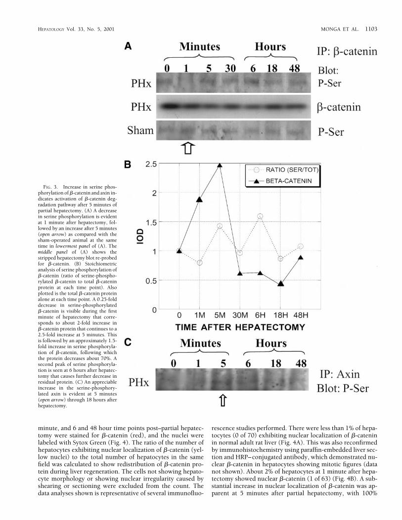
APC and axin and become degraded by ubiquitination. Wedemonstrated an initial increase in �-catenin protein at 1 to 5minutes after partial hepatectomy, followed by a substantialdecrease in this protein (Fig. 2A). No changes were observedin the mRNA levels of �-catenin during this period. To deter-mine the mechanism of the decrease in �-catenin protein, weinvestigated its phosphorylation status at serine residues. Celllysates were immunoprecipitated with anti–�-catenin anti-body and blotted with antiphosphoserine antibody. The rep-resentative immunoprecipitation studies shown in Fig. 3Ademonstrate a decrease in serine phosphorylation of �-cate-nin protein at 1 minute during liver regeneration, followed byan increase at 5 minutes onward (upper panel). The serinephosphorylation status of �-catenin remained unchanged inthe sham controls at similar time points (bottom panel). Forstoichiometric analysis of serine phosphorylation, the blotwas stripped and re-probed for �-catenin (middle panel).There was an approximately 0.25-fold decrease in serine-phosphorylated �-catenin that corresponded to the elevatedtotal �-catenin protein during this time (Fig. 3B). This wasfollowed by a 1.5-fold increase in serine phosphorylation of�-catenin that coincided with the beginning of a decrease inlevels of �-catenin protein. A second peak of serine phosphor-ylation, seen at 6 hours, matched a further decrease in thelevel of residual �-catenin protein. Thus, the peaks of serine
phosphorylation corresponded to the decreases in �-cateninprotein during liver regeneration (Fig. 3B).
Phosphorylation of axin at specific serine residues is crucialfor its successful assembly with APC, �-catenin, and GSK3�,and thus is important for degradation of �-catenin protein. Toestablish if the axin is phosphorylated at serine residues inliver in response to a decrease in wnt signaling at 5 minutesafter hepatectomy, the cell lysates were immunoprecipitatedwith anti axin antibody and blotted with anti phosphoserineantibody. The representative blot in Fig. 3C demonstratesreduced phosphorylation of axin at 1 minute, followed by asubstantial increase in serine phosphorylation of axin at 5minutes. This coincides with a reduction in wnt-1 protein at 5minutes and �-catenin at 15 minutes (Fig. 2A and 2B). This iscompatible with activation of the �-catenin destruction com-plex with increased APC protein and serine phosphorylatedaxin after 5 minutes of partial hepatectomy in the remnantlivers, resulting in �-catenin degradation.
Increased Nuclear Translocation of �-Catenin During Liver Regen-eration. We wanted to determine whether residual �-cateninwas translocating to the nucleus and, at least in part, playingrole in cell proliferation during liver regeneration. We usedimmunofluorescence to explore any increase in nuclear local-ization of �-catenin during liver regeneration. Four-microme-ter-thick frozen sections of the remnant livers at 0, 1, 5, 30
FIG. 2. Changes in wnt signalingpathway components during liverregeneration after 70% partial hepa-tectomy. (A) Elevation in �-cateninprotein at 1 to 5 minutes, fol-lowed by a significant decrease at 15minutes onward, until 48 hours inpartial-hepatectomy livers. No ap-parent change in sham-operated an-imals with comparable film-expo-sure times. The bottom 2 panels showNorthern blot analysis. An increasein �-catenin expression is seen at6 hours through 72 hours after par-tial hepatectomy. The lowest panelshows a minimal �-catenin expres-sion in sham operation. (B) Repre-sentative Western blots demonstratedecreased wnt-1 protein (upper bandin this blot corresponds to the 45-kdsize) at 5 minutes through 12 hoursthat returns to normal level at 18hours. No changes are seen in sham-operated animals. Minimal levels ofAPC protein at the 0 time point be-gins to increase at 5 minutes and iselevated until 18 hours, followed bya gradual decrease to almost physio-logic levels at 72 hours after partialhepatectomy. No changes are seen insham-operated animals. No signifi-cant changes in GSK3� or TCF-4protein levels are observed. A repre-sentative antiactin antibody–probedWestern blot confirms the equalloading of proteins from varioustime points after partial hepatectomyand sham samples.
1102 MONGA ET AL. HEPATOLOGY May 2001
minute, and 6 and 48 hour time points post–partial hepatec-tomy were stained for �-catenin (red), and the nuclei werelabeled with Sytox Green (Fig. 4). The ratio of the number ofhepatocytes exhibiting nuclear localization of �-catenin (yel-low nuclei) to the total number of hepatocytes in the samefield was calculated to show redistribution of �-catenin pro-tein during liver regeneration. The cells not showing hepato-cyte morphology or showing nuclear irregularity caused byshearing or sectioning were excluded from the count. Thedata analyses shown is representative of several immunofluo-
rescence studies performed. There were less than 1% of hepa-tocytes (0 of 70) exhibiting nuclear localization of �-cateninin normal adult rat liver (Fig. 4A). This was also reconfirmedby immunohistochemistry using paraffin-embedded liver sec-tion and HRP–conjugated antibody, which demonstrated nu-clear �-catenin in hepatocytes showing mitotic figures (datanot shown). About 2% of hepatocytes at 1 minute after hepa-tectomy showed nuclear �-catenin (1 of 63) (Fig. 4B). A sub-stantial increase in nuclear localization of �-catenin was ap-parent at 5 minutes after partial hepatectomy, with 100%
FIG. 3. Increase in serine phos-phorylation of �-catenin and axin in-dicates activation of �-catenin deg-radation pathway after 5 minutes ofpartial hepatectomy. (A) A decreasein serine phosphorylation is evidentat 1 minute after hepatectomy, fol-lowed by an increase after 5 minutes(open arrow) as compared with thesham-operated animal at the sametime in lowermost panel of (A). Themiddle panel of (A) shows thestripped hepatectomy blot re-probedfor �-catenin. (B) Stoichiometricanalysis of serine phosphorylation of�-catenin (ratio of serine-phospho-rylated �-catenin to total �-cateninprotein at each time point). Alsoplotted is the total �-catenin proteinalone at each time point. A 0.25-folddecrease in serine-phosphorylated�-catenin is visible during the firstminute of hepatectomy that corre-sponds to about 2-fold increase in�-catenin protein that continues to a2.5-fold increase at 5 minutes. Thisis followed by an approximately 1.5-fold increase in serine phosphoryla-tion of �-catenin, following whichthe protein decreases about 70%. Asecond peak of serine phosphoryla-tion is seen at 6 hours after hepatec-tomy that causes further decrease inresidual protein. (C) An appreciableincrease in the serine-phosphory-lated axin is evident at 5 minutes(open arrow) through 18 hours afterhepatectomy.
HEPATOLOGY Vol. 33, No. 5, 2001 MONGA ET AL. 1103

hepatocytes (68 of 68) depicting nuclear localization (Fig.4C). This was accompanied by a visible decrease in the cyto-plasmic staining for �-catenin in these 4-�m sections, but themembrane staining was comparable with the earlier timepoints. This redistribution was maintained at 30 minutes withabout 92% hepatocytes showing nuclear �-catenin (50 of 55),although there was a further decrease in membrane labeling ofthe hepatocytes at this time point (Fig. 4D). Nuclear andmembrane staining of hepatocyte levels was also elevated at 6hours during liver regeneration, with about 90% hepatocytes(56 of 63) still showing increased nuclear localization of�-catenin (Fig. 4E). It was still higher than normal at 18 hoursafter partial hepatectomy (data not shown). At 48 hours afterpartial hepatectomy, the distribution of �-catenin in the hepa-tocytes appeared to be approaching normal, with about 7%cells (4 of 59) exhibiting nuclear �-catenin (Fig. 4F). It ap-peared more localized to membrane and cytoplasm of the
hepatocytes at this stage, although the staining was not asintense as the normal liver.
Relative Changes in �-Catenin Tyrosine Phosphorylation and E-Cadherin Levels During Liver Regeneration. We wanted to studythe effect of quantitative changes in �-catenin protein on E-cadherin levels during liver regeneration because of theirclose association.32 We also studied the phosphorylation of�-catenin at tyrosine residues, because this event favors neg-ative regulation of cell-cell adhesion.31
A representative blot from 3 different sets of animals shownin Fig. 5A shows a decrease in E-cadherin protein at 1 minuteafter partial hepatectomy, followed by an increase at 5 min-utes onward. The protein is elevated maximally at 15 minutesthrough 1 to 3 hours (substantially more than physiologiclevels) after hepatectomy, followed by a decrease to near-nor-mal levels at 6 hours and above. Sham-operated livers did notreveal any quantitative changes (data not shown).
FIG. 4. Immunostaining demon-strates increased nuclear transloca-tion of residual �-catenin at 5 min-utes after hepatectomy (bar � 10�m). (A) �-Catenin (red) localizesto the membrane and the cytoplasmin close proximity to the membraneat the 0 time point. Only a few hepa-tocytes (�1%) show yellow nucleishowing presence of �-catenin (ar-rowheads). (B) �-Catenin at 1minute shows similar localization.(C) Almost all hepatocytes exhibitnuclear localization of �-catenin at 5minutes (arrowheads). Membranestaining is intact, but cytoplasmicstaining decreases. (D) The above re-distribution is maintained at 30 min-utes (arrowhead). (E) Increased nu-clear and membrane labeling for�-catenin is seen at 6 hours. Mostnuclei of the hepatocytes at this stageare positive for �-catenin (arrow-heads). (F) Decreased nuclear stain-ing for �-catenin is evident at 48hours. Staining is mainly membra-nous and cytoplasmic with very fewhepatocytes showing nuclear �-cate-nin (arrowheads).
1104 MONGA ET AL. HEPATOLOGY May 2001
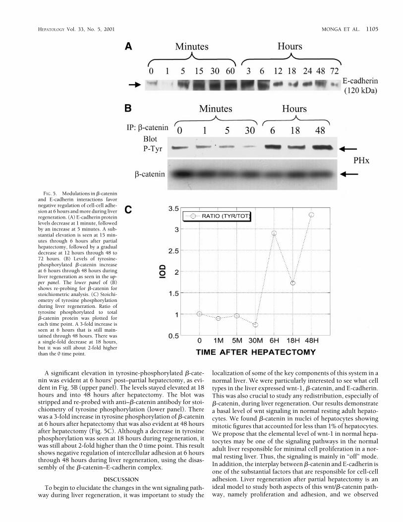
A significant elevation in tyrosine-phosphorylated �-cate-nin was evident at 6 hours’ post–partial hepatectomy, as evi-dent in Fig. 5B (upper panel). The levels stayed elevated at 18hours and into 48 hours after hepatectomy. The blot wasstripped and re-probed with anti–�-catenin antibody for stoi-chiometry of tyrosine phosphorylation (lower panel). Therewas a 3-fold increase in tyrosine phosphorylation of �-cateninat 6 hours after hepatectomy that was also evident at 48 hoursafter hepatectomy (Fig. 5C). Although a decrease in tyrosinephosphorylation was seen at 18 hours during regeneration, itwas still about 2-fold higher than the 0 time point. This resultshows negative regulation of intercellular adhesion at 6 hoursthrough 48 hours during liver regeneration, using the disas-sembly of the �-catenin–E-cadherin complex.
DISCUSSION
To begin to elucidate the changes in the wnt signaling path-way during liver regeneration, it was important to study the
localization of some of the key components of this system in anormal liver. We were particularly interested to see what celltypes in the liver expressed wnt-1, �-catenin, and E-cadherin.This was also crucial to study any redistribution, especially of�-catenin, during liver regeneration. Our results demonstratea basal level of wnt signaling in normal resting adult hepato-cytes. We found �-catenin in nuclei of hepatocytes showingmitotic figures that accounted for less than 1% of hepatocytes.We propose that the elemental level of wnt-1 in normal hepa-tocytes may be one of the signaling pathways in the normaladult liver responsible for minimal cell proliferation in a nor-mal resting liver. Thus, the signaling is mainly in “off” mode.In addition, the interplay between �-catenin and E-cadherin isone of the substantial factors that are responsible for cell-celladhesion. Liver regeneration after partial hepatectomy is anideal model to study both aspects of this wnt/�-catenin path-way, namely proliferation and adhesion, and we observed
FIG. 5. Modulations in �-cateninand E-cadherin interactions favornegative regulation of cell-cell adhe-sion at 6 hours and more during liverregeneration. (A) E-cadherin proteinlevels decrease at 1 minute, followedby an increase at 5 minutes. A sub-stantial elevation is seen at 15 min-utes through 6 hours after partialhepatectomy, followed by a gradualdecrease at 12 hours through 48 to72 hours. (B) Levels of tyrosine-phosphorylated �-catenin increaseat 6 hours through 48 hours duringliver regeneration as seen in the up-per panel. The lower panel of (B)shows re-probing for �-catenin forstoichiometric analysis. (C) Stoichi-ometry of tyrosine phosphorylationduring liver regeneration. Ratio oftyrosine phosphorylated to total�-catenin protein was plotted foreach time point. A 3-fold increase isseen at 6 hours that is still main-tained through 48 hours. There wasa single-fold decrease at 18 hours,but it was still about 2-fold higherthan the 0 time point.
HEPATOLOGY Vol. 33, No. 5, 2001 MONGA ET AL. 1105

unique changes in this pathway as the repairing and restoringprocess proceeded.
Liver regeneration after partial hepatectomy is an excellentin vivo model depicting controlled growth. Graphic represen-tation of quantitative changes in the wnt signaling pathwaycomponents from a representative group of immunoblots isdemonstrated in Fig. 6A. Increased levels of �-catenin protein(about 2.5-fold) are observed during the early minutes of liverregeneration in response to the injury induced by partial hep-atectomy. Degradation of �-catenin protein requires it to be
phosphorylated at specific serine and threonine residues, andis degraded through the ubiquitin proteasome pathway in-volving activation of a destructive machinery composed ofAPC and axin.22,23 The initial increase in �-catenin protein isshown to be caused by a decrease in its degradation as a resultof its decreased serine phosphorylation. It is thus attributableto alteration in steady-state kinetics in which the degradationpathway is inactivated as a result of a change in phosphoryla-tion status of a protein. As shown in Results, the gene expres-sion for �-catenin is not altered until 6 hours after partial
FIG. 6. Meaningful variations inwnt signaling pathway during liverregeneration and working hypothe-ses in regulated growth. (A) Graphicanalysis of quantitative changes inwnt pathway components duringliver regeneration is shown. There isan initial 2.5-fold increase in �-cate-nin protein, followed by an almost3-fold decrease. There is also a 0.5-fold decrease in wnt-1 protein as thedegradation pathway for �-catenin isactivated. Following this, there is a7-fold increase in APC protein apartfrom elevated serine-phosphory-lated �-catenin and axin. TCF-4 lev-els remain mostly unaltered duringthese events. This graph also depictsan inverse quantitative relationshipbetween E-cadherin and �-cateninproteins. An initial increase in�-catenin protein coincides with adecrease in E-cadherin levels, fol-lowed by an increase in E-cadherinprotein as �-catenin decreases. (B) Amodel of regulated growth is hy-pothesized. Modulation of wnt path-way by altered �-catenin levels (de-crease in the wnt-1 protein levelsfollowing an increase in the �-cate-nin protein) favors a possibility of afeedback-inhibitory loop in thispathway. Excess �-catenin protein[1] may activate this feedback loop,causing a decrease in wnt-1 proteinlevels [2]. This may lead to activa-tion of the degradation pathway for�-catenin through ubiquitination,thus limiting and regulating avail-ability of residual �-catenin for nu-clear translocation [3] to control thetarget gene expression and cell pro-liferation. Aberrations in this feed-back inhibition or checkpoint maylead to growth deregulation.
1106 MONGA ET AL. HEPATOLOGY May 2001

hepatectomy. The early increase in �-catenin protein is fol-lowed by a substantial decrease after 5 minutes of hepatec-tomy. It is a possibility that increased �-catenin protein mightbe triggering a response in the hepatocytes to activate its deg-radation pathway. This response in liver regeneration in-cludes an increase in APC protein levels (about 7-fold) and anincrease in serine-phosphorylated axin that is evident at 5minutes onward after partial hepatectomy. Serine-phosphory-lated �-catenin is also elevated about 1.5-fold at this time, asdescribed earlier. These events are absolutely required for de-struction of �-catenin. In addition, these events coincide withthe decrease in wnt-1 protein (0.5-fold decrease) seen at 5minutes after hepatectomy. This quantitative decrease inwnt-1 protein at this time is responsible for increased degra-dation of �-catenin. These events not only limit an increase inthe �-catenin protein, but also maintain a certain minimallevel of the protein that is required for normal cellular func-tioning. This observation is in general agreement with theavailable studies on the mechanism of �-catenin degrada-tion.4,21,24,53 All the above meaningful variations in the wntsignaling pathway components thus reveal a tight regulationof free, monomeric, and functional �-catenin in this regulatedgrowth model.
Another novel observation was an apparent inverse rela-tionship in the quantitative levels of �-catenin and E-cadherinproteins. Decrease in E-cadherin levels coincided with an ini-tial increase in �-catenin protein. This was followed by anapproximately 4-fold elevation in E-cadherin levels as �-cate-nin protein degraded, after 5 minutes of partial hepatectomy.
An initial increase in �-catenin protein and its nucleartranslocation during early liver regeneration favors its role incontributing toward initiation of cell proliferation. However,this proliferation is carefully monitored by a down-regulationof this pathway and activation of the destructive machineryfor �-catenin protein, to check or limit its excessive levels andthus to keep this growth regulated. Other workers haveshown aberrations in this pathway resulting in growth dereg-ulation seen in molecular pathogenesis of several tumors,causing sustained elevations level of �-catenin protein.2,17 Wehypothesize that �-catenin protein may regulate its own levelsthrough a mechanism of feedback inhibition in a regulatedgrowth environment such as liver regeneration (Fig. 6B). Thisfeedback-inhibitory loop appears to apply at the level of wnt-1protein, and any increase in �-catenin protein levels is nor-mally checked by a decrease in wnt-1 signaling. In liver regen-eration, after an initial increase in �-catenin protein thatmight be required to initiate target gene expression and cellproliferation, any sustained elevations in its protein levels arecontrolled by activation of its degradation pathway. Aberra-tions in this feedback loop might be responsible for sustainedelevations of both �-catenin and wnt-1 proteins, allowing�-catenin to signal constitutively and cause growth deregula-tion seen in several cancers.2,17 Further studies including gen-eration of �-catenin transgenic animals are under way to con-firm this hypothesis.
Activation of 2 transcription factors that are pre-existing inhepatic cells have been demonstrated to be part of the initiat-ing mechanism of liver regeneration after 70% partial hepa-tectomy. Nuclear translocation of nuclear factor-�B and signaltransducer and activator of transcription protein-3 have beenshown to be responsible for increased expression of severaltarget genes, including c-myc, c-fos, and Jun B, very early dur-
ing regeneration.43-46 Increased nuclear translocation of resid-ual �-catenin protein in the hepatocytes, as early as 5 minutesafter hepatectomy, may suggest its importance in initiating orpromoting target gene expression and cell proliferation. Al-though the wnt-1 protein levels begin to decrease at 5 minutesafter hepatectomy, there is an increase in serine-phosphory-lated �-catenin at this time, probably as a result of the differ-ences in their chemical kinetics. This event favors dissociationof �-catenin from GSK3�, APC, and axin and its nucleartranslocation.
Several target genes of the wnt pathway have been de-scribed.7,27,28,54 Some of these target genes, including c-myc,uPAR, and cyclin D1, have been shown to play important rolesin liver regeneration.36,55-57 C-myc expression begins to in-crease very early during liver regeneration, peaking at 1 hourto 3 hours and 12 hours’ post–partial hepatectomy, and could(at least in part) be the result of elevated nuclear �-cateninfound at these time points during the regenerating liver. Al-though elevations in the uPAR protein are observed as early as1 minute after partial hepatectomy, a positive impact of in-creased nuclear �-catenin on levels of uPAR protein at latertime points cannot be ruled out without further studies. Sim-ilarly, cyclin D1 expression in regenerating liver may be con-trolled by �-catenin. Thus, increased levels of nuclear �-cate-nin may be playing a role in directing the expression of any ofthese target genes, and further studies are under way to iden-tify specific targets important in liver regeneration.
Cellular loosening, mobility, and reorganization are crucialfor liver remodeling during liver regeneration. It has beenpreviously reported that cell-cell adhesion is affected nega-tively during liver regeneration.58 Changes in proteins in-volved in adherens junctions in liver, including vinculin and�-actinin, have been reported as early as 2 to 3 hours afterpartial hepatectomy.59 Additionally, there has been a previousreport from our laboratory concerning the activation of ma-trix metalloproteinase-2 and -7, which are responsible for di-gesting extracellular matrix at 6 to 12 hours after partialhepatectomy.60 Thus, a general negative regulation of cell ad-hesion during liver regeneration at 3 hours onward has beendefined by different mechanisms. Our present study impli-cates �-catenin and E-cadherin dissociation as one of the im-portant contributing factors for negatively regulating the he-patocyte adhesion during liver regeneration at 6 hours andonward. The novel observation of inverse relationship in thequantitative levels of �-catenin and E-cadherin proteins wasclearly evident during liver regeneration. An initial increase in�-catenin protein coincided with a significant decrease in E-cadherin levels, and as �-catenin protein decreased, a notableelevation was seen in the E-cadherin protein levels. This re-ciprocal relationship might be a compensatory event thatcould still maintain the intercellular adhesions among thehepatocytes during early regeneration. However, phosphory-lation of �-catenin at tyrosine residues at 6 hours after partialhepatectomy eliminates this compensation and becomes amajor factor for loss of the interhepatocytic contact that iscrucial for restructuring and repopulating the remnant liverinto a normally functioning and restored liver.
Acknowledgment: The authors acknowledge Dr. Bryon E.Petersen for assistance with partial hepatectomies. The au-thors thank Drs. Wendy Mars and Chandrashekar Gandhi forproviding the Western blot for the protein isolates from vari-ous cell types in the liver. The authors also thank Mark Ross
HEPATOLOGY Vol. 33, No. 5, 2001 MONGA ET AL. 1107

for his excellent assistance in immunofluorescence micros-copy.
REFERENCES
1. Cadigan KM, Nusse R. Wnt signaling: a common theme in animal devel-opment. Genes Dev 1997;11:3286-3305.
2. Peifer M, Polakis P. Wnt signaling in oncogenesis and embryogenesis—alook outside the nucleus. Science 2000;287:1606-1609.
3. Young CS, Kitamura M, Hardy S, Kitajewski J. Wnt-1 induces growth,cytosolic beta-catenin, and Tcf/Lef transcriptional activation in Rat-1fibroblasts. Mol Cell Biol 1998;18:2474-2485.
4. Papkoff J, Rubinfeld B, Schryver B, Polakis P. Wnt-1 regulates free poolsof catenins and stabilizes APC-catenin complexes. Mol Cell Biol 1996;16:2128-2134.
5. Brabletz T, Herrmann K, Jung A, Faller G, Kirchner T. Expression ofnuclear beta-catenin and c-myc is correlated with tumor size but not withproliferative activity of colorectal adenomas. Am J Pathol 2000;156:865-870.
6. Hugh TJ, Dillon SA, O’Dowd G, Getty B, Pignatelli M, Poston GJ, KinsellaAR. beta-catenin expression in primary and metastatic colorectal carci-noma. Int J Cancer 1999;82:504-511.
7. Mann B, Gelos M, Siedow A, Hanski ML, Gratchev A, Ilyas M, BodmerWF, et al. Target genes of beta-catenin-T cell-factor/lymphoid-enhancer-factor signaling in human colorectal carcinomas. Proc Natl Acad Sci U S A1999;96:1603-1608.
8. Herrmann JL, Briones F Jr, Brisbay S, Logothetis CJ, McDonnell TJ. Pros-tate carcinoma cell death resulting from inhibition of proteasome activityis independent of functional Bcl-2 and p53. Oncogene 1998;17:2889-2899.
9. Huang H, Fujii H, Sankila A, Mahler-Araujo BM, Matsuda M, CathomasG, Ohgaki H. Beta-catenin mutations are frequent in human hepatocel-lular carcinomas associated with hepatitis C virus infection. Am J Pathol1999;155:1795-1801.
10. Morita N, Uemura H, Tsumatani K, Cho M, Hirao Y, Okajima E, KonishiN, et al. E-cadherin and alpha-, beta- and gamma-catenin expression inprostate cancers: correlation with tumour invasion. Br J Cancer 1999;79:1879-1883.
11. Sommers CL, Gelmann EP, Kemler R, Cowin P, Byers SW. Alterations inbeta-catenin phosphorylation and plakoglobin expression in humanbreast cancer cells. Cancer Res 1994;54:3544-3552.
12. Tsujiuchi T, Tsutsumi M, Sasaki Y, Takahama M, Konishi Y. Differentfrequencies and patterns of beta-catenin mutations in hepatocellular car-cinomas induced by N-nitrosodiethylamine and a choline-deficientL-amino acid–defined diet in rats. Cancer Res 1999;59:3904-3907.
13. Rubinfeld B, Robbins P, El-Gamil M, Albert I, Porfiri E, Polakis P. Stabi-lization of beta-catenin by genetic defects in melanoma cell lines [Com-ments]. Science 1997;275:1790-1792.
14. Jaiswal AS, Kennedy CH, Narayan S. A correlation of APC and c-mycmRNA levels in lung cancer cell lines. Oncol Rep 1999;6:1253-1256.
15. Aberle H, Bauer A, Stappert J, Kispert A, Kemler R. beta-catenin is a targetfor the ubiquitin-proteasome pathway. EMBO J 1997;16:3797-3804.
16. Pennisi E. How a growth control path takes a wrong turn to cancer[News; Comment] [published erratum appears in Science 1998;281(5384):1809]. Science 1998;281:1438-1439, 1441.
17. Morin PJ. Beta-catenin signaling and cancer. Bioessays 1999;21:1021-1030.
18. Rubinfeld B, Albert I, Porfiri E, Fiol C, Munemitsu S, Polakis P. Bindingof GSK3beta to the APC-beta-catenin complex and regulation of complexassembly [Comments]. Science 1996;272:1023-1026.
19. Liu T, Liu X, Wang H, Moon RT, Malbon CC. Activation of rat frizzled-1promotes Wnt signaling and differentiation of mouse F9 teratocarcinomacells via pathways that require Galpha(q) and Galpha(o) function. J BiolChem 1999;274:33539-33544.
20. Hsieh JC, Rattner A, Smallwood PM, Nathans J. Biochemical character-ization of Wnt-frizzled interactions using a soluble, biologically activevertebrate Wnt protein. Proc Natl Acad Sci U S A 1999;96:3546-3551.
21. Farr GH, 3rd, Ferkey DM, Yost C, Pierce SB, Weaver C, Kimelman D.Interaction among GSK-3, GBP, axin, and APC in Xenopus axis specifi-cation. J Cell Biol 2000;148:691-702.
22. Rubinfeld B, Souza B, Albert I, Muller O, Chamberlain SH, Masiarz FR,Munemitsu S, et al. Association of the APC gene product with beta-catenin. Science 1993;262:1731-1734.
23. Kikuchi A. Roles of Axin in the Wnt signalling pathway. Cell Signal1999;11:777-788.
24. Barker N, Morin PJ, Clevers H. The Yin-Yang of TCF/beta-catenin signal-ing. Adv Cancer Res 2000;77:1-24.
25. Cho EA, Dressler GR. TCF-4 binds beta-catenin and is expressed in dis-tinct regions of the embryonic brain and limbs. Mech Dev 1998;77:9-18.
26. Huber O, Korn R, McLaughlin J, Ohsugi M, Herrmann BG, Kemler R.Nuclear localization of beta-catenin by interaction with transcriptionfactor LEF-1. Mech Dev 1996;59:3-10.
27. Tetsu O, McCormick F. Beta-catenin regulates expression of cyclin D1 incolon carcinoma cells. Nature 1999;398:422-426.
28. He TC, Sparks AB, Rago C, Hermeking H, Zawel L, da Costa LT, MorinPJ, et al. Identification of c-MYC as a target of the APC pathway [Com-ments]. Science 1998;281:1509-1512.
29. Linask KK, Knudsen KA, Gui YH. N-cadherin–catenin interaction: nec-essary component of cardiac cell compartmentalization during early ver-tebrate heart development. Dev Biol 1997;185:148-164.
30. Linask KK, Ludwig C, Han MD, Liu X, Radice GL, Knudsen KA. N-cadherin/catenin–mediated morphoregulation of somite formation. DevBiol 1998;202:85-102.
31. Roura S, Miravet S, Piedra J, Garcia de Herreros A, Dunach M. Regulationof E-cadherin/catenin association by tyrosine phosphorylation. J BiolChem 1999;274:36734-36740.
32. Kemler R. From cadherins to catenins: cytoplasmic protein interactionsand regulation of cell adhesion. Trends Genet 1993;9:317-321.
33. Jankowski JA, Bruton R, Shepherd N, Sanders DS. Cadherin and cateninbiology represent a global mechanism for epithelial cancer progression.Mol Pathol 1997;50:289-290.
34. Michalopoulos GK, DeFrances MC. Liver regeneration. Science 1997;276:60-66.
35. Stolz DB, Mars WM, Petersen BE, Kim TH, Michalopoulos GK. Growthfactor signal transduction immediately after two-thirds partial hepatec-tomy in the rat. Cancer Res 1999;59:3954-3960.
36. Panis Y, Lomri N, Emond JC. Early gene expression associated withregeneration is intact after massive hepatectomy in rats. J Surg Res 1998;79:103-108.
37. Widmann JJ, Fahimi HD. Proliferation of mononuclear phagocytes(Kupffer cells) and endothelial cells in regenerating rat liver. Am J Pathol1975;80:349-366.
38. Michalopoulos GK. HGF in liver regeneration and tumor promotion.Prog Clin Biol Res 1995;391:179-185.
39. Diehl AM, Rai RM. Liver regeneration 3: regulation of signal transductionduring liver regeneration. FASEB J 1996;10:215-227.
40. Fausto N, Webber EM: Liver Regeneration. In: Boyer J, Arias I, Fausto N,Jakoby W, Schachter D, Shafritz, eds. The Liver: Biology and Pathobiol-ogy. 3rd ed. New York: Raven, 1994:1059-1084.
41. Grisham J. A morphologic study of deoxyribonucleic acid synthesis andcell proliferation in regenerating liver; autoradiography with thymidine-H3. Cancer Res 1964;22:842-849.
42. Tanaka Y, Mak KM, Lieber CS. Immunohistochemical detection of pro-liferating lipocytes in regenerating rat liver. J Pathol 1990;160:129-134.
43. Cressman DE, Greenbaum LE, Haber BA, Taub R. Rapid activation ofpost-hepatectomy factor/nuclear factor kappa B in hepatocytes, a pri-mary response in the regenerating liver. J Biol Chem 1994;269:30429-30435.
44. Cressman DE, Diamond RH, Taub R. Rapid activation of the Stat3 tran-scription complex in liver regeneration. HEPATOLOGY 1995;21:1443-1449.
45. Cressman DE, Greenbaum LE, DeAngelis RA, Ciliberto G, Furth EE, PoliV, Taub R. Liver failure and defective hepatocyte regeneration in inter-leukin-6–deficient mice. Science 1996;274:1379-1383.
46. Yamada Y, Kirillova I, Peschon JJ, Fausto N. Initiation of liver growth bytumor necrosis factor: deficient liver regeneration in mice lacking type Itumor necrosis factor receptor. Proc Natl Acad Sci U S A 1997;94:1441-1446.
47. Taub R, Greenbaum LE, Peng Y. Transcriptional regulatory signals definecytokine-dependent and -independent pathways in liver regeneration.Semin Liver Dis 1999;19:117-127.
48. Higgins GM, Anderson, RM. Experimental pathology of the liver. I. Res-toration of the white rat following partial surgical removal. Arch Pathol1934;12:186-202.
49. Kost DP, Michalopoulos GK. Effect of 2% dimethyl sulfoxide on themitogenic properties of epidermal growth factor and hepatocyte growthfactor in primary hepatocyte culture. J Cell Physiol 1991;147:274-280.
50. Braet F, De Zanger R, Sasaoki T, Baekeland M, Janssens P, Smedsrod B,Wisse E. Assessment of a method of isolation, purification, and cultiva-tion of rat liver sinusoidal endothelial cells. Lab Invest 1994;70:944-952.
51. Gabriel A, Kuddus RH, Rao AS, Watkins WD, Gandhi CR. Superoxide-induced changes in endothelin (ET) receptors in hepatic stellate cells.J Hepatol 1998;29:614-627.
1108 MONGA ET AL. HEPATOLOGY May 2001

52. Gabriel A, Kuddus RH, Rao AS, Gandhi CR. Down-regulation of endo-thelin receptors by transforming growth factor beta1 in hepatic stellatecells. J Hepatol 1999;30:440-450.
53. Orford K, Orford CC, Byers SW. Exogenous expression of beta-cateninregulates contact inhibition, anchorage-independent growth, anoikis,and radiation-induced cell cycle arrest. J Cell Biol 1999;146:855-868.
54. Shtutman M, Zhurinsky J, Simcha I, Albanese C, D’Amico M, Pestell R,Ben-Ze’ev A. The cyclin D1 gene is a target of the beta-catenin/LEF-1pathway. Proc Natl Acad Sci U S A 1999;96:5522-5527.
55. Makino R, Hayashi K, Sugimura T. C-myc transcript is induced in ratliverat a very early stage of regeneration or by cycloheximide treatment. Na-ture 1984;310:697-698.
56. Mars WM, Liu ML, Kitson RP, Goldfarb RH, Gabauer MK, Michalopou-los GK. Immediate early detection of urokinase receptor after partial
hepatectomy and its implications for initiation of liver regeneration.HEPATOLOGY 1995;21:1695-1701.
57. Albrecht JH, Hu MY, Cerra FB. Distinct patterns of cyclin D1 regulationin models of liver regeneration and human liver. Biochem Biophys ResCommun 1995;209:648-655.
58. Kim TH, Mars WM, Stolz DB, Petersen BE, Michalopoulos GK. Extracel-lular matrix remodeling at the early stages of liver regeneration in the rat[Comments]. HEPATOLOGY 1997;26:896-904.
59. Gluck U, Rodriguez Fernandez JL, Pankov R, Ben-Ze’ev A. Regulation ofadherens junction protein expression in growth-activated 3T3 cells andin regenerating liver. Exp Cell Res 1992;202:477-486.
60. Kim TH, Mars WM, Stolz DB, Michalopoulos GK. Expression and acti-vation of pro-MMP-2 and pro-MMP-9 during rat liver regeneration.HEPATOLOGY 2000;31:75-82.
HEPATOLOGY Vol. 33, No. 5, 2001 MONGA ET AL. 1109