Changes in inner ear structure and function after drill...
7
American Journal of Clinical and Experimental Medicine 2014; 2(5): 90-96 Published online September 30, 2014 (http://www.sciencepublishinggroup.com/j/ajcem) doi: 10.11648/j.ajcem.20140205.11 ISSN: 2330-8125 (Print); ISSN: 2330-8133 (Online) Changes in inner ear structure and function after drill - induced acoustic trauma Fahd Alharbi 1 , Mohammed Rifaat Ahmed 1, 2 1 Department of Otolaryngology, Head and Neck Surgery, Faculty of Medicine, Jazan University, 2349-Almarjan-Industrial Zone Rd., Unit No. 1, Jazan, Saudi Arabia 2 Department of Otolaryngology-Head & Neck Surgery, Faculty of Medicine, Suez Canal University, Ismailia, Egypt Email address: [email protected] (F. Alharbi), [email protected] (M. R. Ahmed) To cite this article: Fahd Alharbi, Mohammed Rifaat Ahmed. Changes in Inner Ear Structure and Function after Drill - Induced Acoustic Trauma. American Journal of Clinical and Experimental Medicine. Vol. 2, No. 5, 2014, pp. 90-96. doi: 10.11648/j.ajcem.20140205.11 Abstract: Background: drilling on the intact ossicular chain produce acoustic trauma resulting in inner ear structure damage, these changes occurred in organ of corti with disruption of cytoarchitecture and cellular degeneration. Objective: To evaluate the changes in inner ear structure and function after drill - induced acoustic trauma in guinea pigs by Scanning Electron Microscope (SEM) and Otoacoustic emission (OAE). Methods and Material: An experimental study conducted on healthy pigmented guinea pigs in Otolaryngology-Head and neck Surgery Department at Jazan University, Kingdom of Saudi Arabia. Results: Thirty guinea pigs were divided into a control group (N) to document the baseline Distortion Product Otoacoustic Emissions (DPOAEs) and the normal scanning electron microscopic (SEM) morphology of the inner ear, Drill induced acoustic trauma group (D) to evaluate the effect of induced sensorineural hearing loss using micromotor drill (20,000rpm) maximum speed against the body of incus and 2weeks postoperative group (2W) to evaluate to the effect of spontaneous recovery after 2 weeks from induced sensorineural hearing loss which showed spontaneous although incomplete recovery of the DPOAEs amplitudes and SEM morphology with scar tissue replacing the damaged outer hair cells. Conclusion: Avoid touching ossicular chain when using high speed drill during ear surgery as this may cause structural and functional damage to the inner ear. Spontaneous recovery could be occurs after acoustic trauma but incomplete recovery with permanent scar formation in outer hair cells may occur. Keywords: Inner Ear, Sensorineural Hearing Loss, Otoacoustic Emission, Acoustic Trauma 1. Introduction Inadvertent drilling on the ossicular chain is one of main causes of high frequency sensorineural hearing loss (SNHL) which observed after tympanomastoid surgery by the mechanism of acoustic trauma (1,2). Dan et al 2007mentioned that drilling on the intact ossicular chain produces vibratory force that analogue with noise levels known to produce acoustic trauma resulted in inner ear structure damage (1) . That mechanism causes temporary threshold shift (usually observed at 2000 Hz and 4000 Hz after ear surgery), which resolved at the time of unpacking the ear (3, 4) . Mislav et al 1997 demonstrated permanent sensorineural hearing loss occurred if caution not taken to avoid touching an intact ossicular chain during drilling (5) . It found that touching the intact ossicular chain with burrs producing pressure conducted towards the footplate comparable to 130 to 150 dB sound pressure levels producing changes in organ of corti with disruption of cytoarchitecture and cellular degeneration occurring mostly in the outer hair cells (6) . Paparella 1982 demonstrated drilling on incus in guinea pigs with 1.4-mm diamond burr for 10 seconds result in sensorineural hearing loss. (7) The natural history of labyrinthine injury in the guinea pig is similar to that in human being (8) . This study carried out to evaluate the changes in inner ear structure and function after drill - induced acoustic trauma in guinea pigs by Scanning Electron Microscope (SEM) and Otoacoustic emission (OAE). 2. Method & Materials An experimental study conducted in Otolaryngology-Head and Neck Surgery Department, Jazan University, Saudi
-
Upload
truongtram -
Category
Documents
-
view
232 -
download
6
Transcript of Changes in inner ear structure and function after drill...

American Journal of Clinical and Experimental Medicine 2014; 2(5): 90-96 Published online September 30, 2014 (http://www.sciencepublishinggroup.com/j/ajcem) doi: 10.11648/j.ajcem.20140205.11 ISSN: 2330-8125 (Print); ISSN: 2330-8133 (Online)
Changes in inner ear structure and function after drill - induced acoustic trauma
Fahd Alharbi1, Mohammed Rifaat Ahmed
1, 2
1Department of Otolaryngology, Head and Neck Surgery, Faculty of Medicine, Jazan University, 2349-Almarjan-Industrial Zone Rd., Unit
No. 1, Jazan, Saudi Arabia 2Department of Otolaryngology-Head & Neck Surgery, Faculty of Medicine, Suez Canal University, Ismailia, Egypt
Email address: [email protected] (F. Alharbi), [email protected] (M. R. Ahmed)
To cite this article: Fahd Alharbi, Mohammed Rifaat Ahmed. Changes in Inner Ear Structure and Function after Drill - Induced Acoustic Trauma. American
Journal of Clinical and Experimental Medicine. Vol. 2, No. 5, 2014, pp. 90-96. doi: 10.11648/j.ajcem.20140205.11
Abstract: Background: drilling on the intact ossicular chain produce acoustic trauma resulting in inner ear structure damage, these changes occurred in organ of corti with disruption of cytoarchitecture and cellular degeneration. Objective: To evaluate the changes in inner ear structure and function after drill - induced acoustic trauma in guinea pigs by Scanning Electron Microscope (SEM) and Otoacoustic emission (OAE). Methods and Material: An experimental study conducted on healthy pigmented guinea pigs in Otolaryngology-Head and neck Surgery Department at Jazan University, Kingdom of Saudi Arabia. Results: Thirty guinea pigs were divided into a control group (N) to document the baseline Distortion Product Otoacoustic Emissions (DPOAEs) and the normal scanning electron microscopic (SEM) morphology of the inner ear, Drill induced acoustic trauma group (D) to evaluate the effect of induced sensorineural hearing loss using micromotor drill (20,000rpm) maximum speed against the body of incus and 2weeks postoperative group (2W) to evaluate to the effect of spontaneous recovery after 2 weeks from induced sensorineural hearing loss which showed spontaneous although incomplete recovery of the DPOAEs amplitudes and SEM morphology with scar tissue replacing the damaged outer hair cells. Conclusion: Avoid touching ossicular chain when using high speed drill during ear surgery as this may cause structural and functional damage to the inner ear. Spontaneous recovery could be occurs after acoustic trauma but incomplete recovery with permanent scar formation in outer hair cells may occur.
Keywords: Inner Ear, Sensorineural Hearing Loss, Otoacoustic Emission, Acoustic Trauma
1. Introduction
Inadvertent drilling on the ossicular chain is one of main causes of high frequency sensorineural hearing loss (SNHL) which observed after tympanomastoid surgery by the mechanism of acoustic trauma (1,2). Dan et al 2007mentioned that drilling on the intact ossicular chain produces vibratory force that analogue with noise levels known to produce acoustic trauma resulted in inner ear structure damage (1). That mechanism causes temporary threshold shift (usually observed at 2000 Hz and 4000 Hz after ear surgery), which resolved at the time of unpacking the ear (3, 4). Mislav et al 1997 demonstrated permanent sensorineural hearing loss occurred if caution not taken to avoid touching an intact ossicular chain during drilling (5).
It found that touching the intact ossicular chain with burrs producing pressure conducted towards the footplate
comparable to 130 to 150 dB sound pressure levels producing changes in organ of corti with disruption of cytoarchitecture and cellular degeneration occurring mostly in the outer hair cells (6). Paparella 1982 demonstrated drilling on incus in guinea pigs with 1.4-mm diamond burr for 10 seconds result in sensorineural hearing loss.
(7) The natural history of labyrinthine injury in the guinea pig
is similar to that in human being (8). This study carried out to evaluate the changes in inner ear
structure and function after drill - induced acoustic trauma in guinea pigs by Scanning Electron Microscope (SEM) and Otoacoustic emission (OAE).
2. Method & Materials
An experimental study conducted in Otolaryngology-Head and Neck Surgery Department, Jazan University, Saudi

91 Fahd Alharbi and Mohammed Rifaat Ahmed: Changes in Inner Ear Structure and Function after Drill - Induced Acoustic Trauma
Arabia. Healthy pigmented guinea pigs of the same age and average weight of 200 gm with basic criteria: normal eardrum documented by otoscopic examination, preserved pinna reflex and normal results in Distortion Product Otoacoustic Emissions (DPOAE). 30 guinea pigs divided into three groups: Normal group (N): 10 guinea pigs evaluated by DPOAE and SEM for organ of corti of both sides.
Drill induced acoustic trauma group (D): 10 guinea pigs immediately evaluated to see effect of induced sensorineural hearing loss using micromotor drill (20,000rpm) maximum speed against the body of incus. 2 weeks postoperative group
(2W): 10 guinea pigs evaluated to see effect of spontaneous recovery after 2 weeks from induced sensorineural hearing loss.
Distortion Product Otoacoustic Emissions (DPOAE)
Measurement: (“CELESTA 503 Oto-acoustic Emission Analyzer” from “Madsen Electronics, Denmark.).Using two pure tone stimuli, F1 and F2 (from 1 to 8 kHz) with F2/F1 ratio of 1:2 and intensity levels equal 65dB SPL. The choice of 65dB SPL as primary levels and frequency ranged from 1 to 8 kHz according to Abu Seta 2002 (9) who mentioned that 65dB gives the best response and this frequency rang found to be the least polluted by ambient noise. Also measuring input/output function failed as animals could not bear longer period of anesthesia. Consequently, only DP gram could be recorded.
Signal to noise ratio (S/N) calculated by subtract the noise floor from the corresponding amplitude. DPOAE recorded from guinea pigs contaminated by unavoidable noisy respiration anesthetized animal. This is why recording done from 1-8 k Hz and calculating the noise floor with S/N ratios. (9)
3. Surgical Steps for Drill Induced
Acoustic Trauma
Under general anesthesia, a surgical microscope with coaxial illumination used after animal anesthetized by intramuscularly injection of ketaset (ketamine hydrochloride 45 mg /kg) and intravenous thiopental 30mg/kg in ear vein. After that a post auricular infiltration by adrenaline: lidocaine 1:200,000 in the both ears performed and a post auricular incision made, then the soft tissue retracted using self-retained mastoid retractor. A small opening in the bulla made with needle and curette, exposing the ossicular chain. A 1.4-mm diamond burr at a rotation speed of 20,000 rpm held against the body of the incus for 10 sec. After surgical procedure finished (for group S), soft tissue in post auricular region sutured using absorbed sutures (chromic cat gut no 3/0) and the skin closed by interrupted non-absorbed sutures (silk 3/0).
A pressure dressing applied to the wound for 24 hours postoperative.
4. DPOAE Follow Up for All Animals
Groups
Normal group (N): DPOAE measured once as a baseline. Drill induced acoustic trauma group (D): before and
immediately after induced sensorineural hearing loss using drill-induced ossicular chain injury technique. 2weeks
postoperative group (2W): before, immediately and 2 weeks after induced sensorineural hearing loss.
5. SEM Study (Hitachi-50 Scanning
Electron Microscope)
SEM study for organ of corti (both sides) as follow: - Normal group (N): animals scarified immediately after
examination. Drill induced acoustic trauma group (D): animals
scarified immediately after drill induced acoustic trauma. 2weeks postoperative group (2W): animals scarified 2
weeks after drill induced acoustic trauma. The temporal bone (tympanic bulla) removed after death
confirmed. The bulla, the bubble of bone containing the middle ear space, opened, and excess bone removed. The cochlea fixed in glutaraldehyde 2.5% in phosphate-buffered saline, pH 7.2. The cochlea perfused through the round window after the stapes disarticulated to allow for a return path for the perfusate. The specimen placed in a large volume of fixative for 24 to 72 hours. After the fixation, the specimens transferred to saline for the initial stage of dissection.
Excess bone surrounding the cochlea removed with a surgical drill and burrs. The bony wall of the cochlea thinned with a high-speed dental drill. Complete exposure of the basilar membrane proved extremely challenging. Therefore the medial wall of the temporal bone left as a support, and only the lateral aspect of the cochlea widely exposed.
At this point, dehydration beginning. The specimen placed in 30% acetone for at least 24 hours, then in 70% acetone for at least another 24 hours. Final dissection performed with the specimen in 70% acetone. Any remaining bone fragments removed. The stria vascularis was stripped away, and Reissner’s membrane was removed. The tectorial membrane not manipulated because it retracts during dehydration. After dissection the specimen placed in 90% acetone and then in three changes of 100% acetone at intervals of at least 12 hours.
After the specimen dehydrated, it underwent critical point drying. This process involves placing the tissue in a pressure container and replacing the acetone with liquid carbon dioxide. The container warmed to greater than the critical point, which is the combination of pressure and temperature at which the carbon dioxide is transformed from a liquid to gas without a change in volume. The gas is gradually released; leaving the dry specimen with none of the distortion that would result if the acetone-saturated tissue allowed to dry directly. For most applications, scanning

American Journal of Clinical and Experimental Medicine 2014; 2(5): 90-96 92
electron microscopy requires that specimens have a conductive surface. This accomplished by placing the specimen in a vacuum chamber, in which metal or carbon atoms introduced, which deposited on the surface. In this study the specimen coated with a 200-Å layer of gold.
In the scanning electron microscope, a low-magnification image made for orientation purposes. The basilar membrane examined at a higher magnification (×500–×2000) as needed.
6. Results
Normal group (N):
Mean amplitude lowest at 1006 kHz (10.6 dB) and highest at 6060 and 8084 kHz (43.9 and 42.3 dB) respectively with tendency for amplitude to rise from low to high frequency as demonstrated in table(1).
Table 1. Distortion Product Otoacoustic Emissions (DP gram )in Normal
group (N)
S/N ratio Noise floor DP amplitude Frequency in Hz
SD X SD X SD X 5.981 8.7 .112 1.9 5.482 10.6 1006 6.019 15.5 .019 2.4 4.951 17.9 1512 5.872 20.7 .512 2.2 4.012 22.9 2011 7.510 32.8 .901 -3.5 6.701 29.3 3023 5.542 40.5 .201 -2.7 4.178 37.8 4036 4.873 47.1 .503 -3.2 3.958 43.9 6060 6.781 50.2 .809 -7.9 6.019 42.3 8084
This table shows the mean (X) and standard deviation (SD) in DP amplitude-noise floor and signal to noise ratio (S/N) in Normal group (N).
Drill induced acoustic trauma group (D):
After acoustic trauma, DPOAE mean amplitude ranged from 11.2 dB at 1006 kHz to 16.2 dB at 8084 kHz as shown in table (2).
2weeks postoperative group (2W): DPOAE mean amplitude ranged from 10.9 dB at 1006 kHz to 22.5 dB at 8084 kHz with significant improvement in all frequency demonstrated in table (3) and figure(1).
Table 2. Distortion Product Otoacoustic Emissions (DP gram )in Drill
induced acoustic trauma group (D).
S/N ratio Noise floor DP amplitude Frequency in Hz
SD X SD X SD X
9.521 9.3 .803 1.9 4.892 11.2 1006 7.637 10.9 .019 -4.5 5.934 6.4 1512 8.602 8.5 .509 -10.2 8.621 -1.7 2011 8.706 17.3 .730 -14.4 9.826 2.9 3023 7.908 18 .796 -11.3 8.019 6.7 4036 11.029 21.9 .012 -7.4 12.093 14.5 6060 9.906 24.3 .176 -8.1 6.629 16.2 8084
This table shows the mean (X) and standard deviation (SD) in DP amplitude-noise floor and signal to noise ratio (S/N) Drill induced acoustic trauma group (D).
Table 3. Distortion Product Otoacoustic Emissions (DP gram )in 2weeks
postoperative group (2W)
S/N ratio Noise floor DP amplitude Frequency in Hz
SD X SD X SD X 10.901 9.5 .112 1.4 6.809 10.9 1006 9.898 13.9 .906 -2.5 7.531 11.4 1512 12.028 16.4 .541 -4.1 5.009 12.3 2011 8.936 28.6 .807 -10.2 5.238 18.4 3023 14.806 22.5 .189 -9.6 10.089 12.9 4036 8.931 38.7 .009 -14.1 8.128 24.6 6060 9.903 30.4 .012 -7.9 6.589 22.5 8084
This table shows the mean (X) and standard deviation (SD) in DP amplitude-noise floor and signal to noise ratio (S/N) in 2weeks postoperative group (2W)
Figure 1. Shows significant improvement in 2weeks postoperative group but not yet return to normal group.
93 Fahd Alharbi and Mohammed Rifaat Ahmed: Changes in Inner Ear Structure and Function after Drill - Induced Acoustic Trauma
7. Histological Results
7.1. Grossly
The cochlea of the guinea pig is grayish yellow in color about 3X4 mm in size .It has a wide bone and tapering apex. It looks like a spiral formed of three and half turns. These turns run around a bony core that appears on longitudinal section (modiolus). The cochlear duct appeared triangular with roof made of thin membrane (Ressinner membrane) and the floor partly formed by outer part of bony spiral lamina from the modiolus and partly by basilar membrane, which carries the organ of corti on its upper surface.
7.2. SEM Results: Normal Group (N)
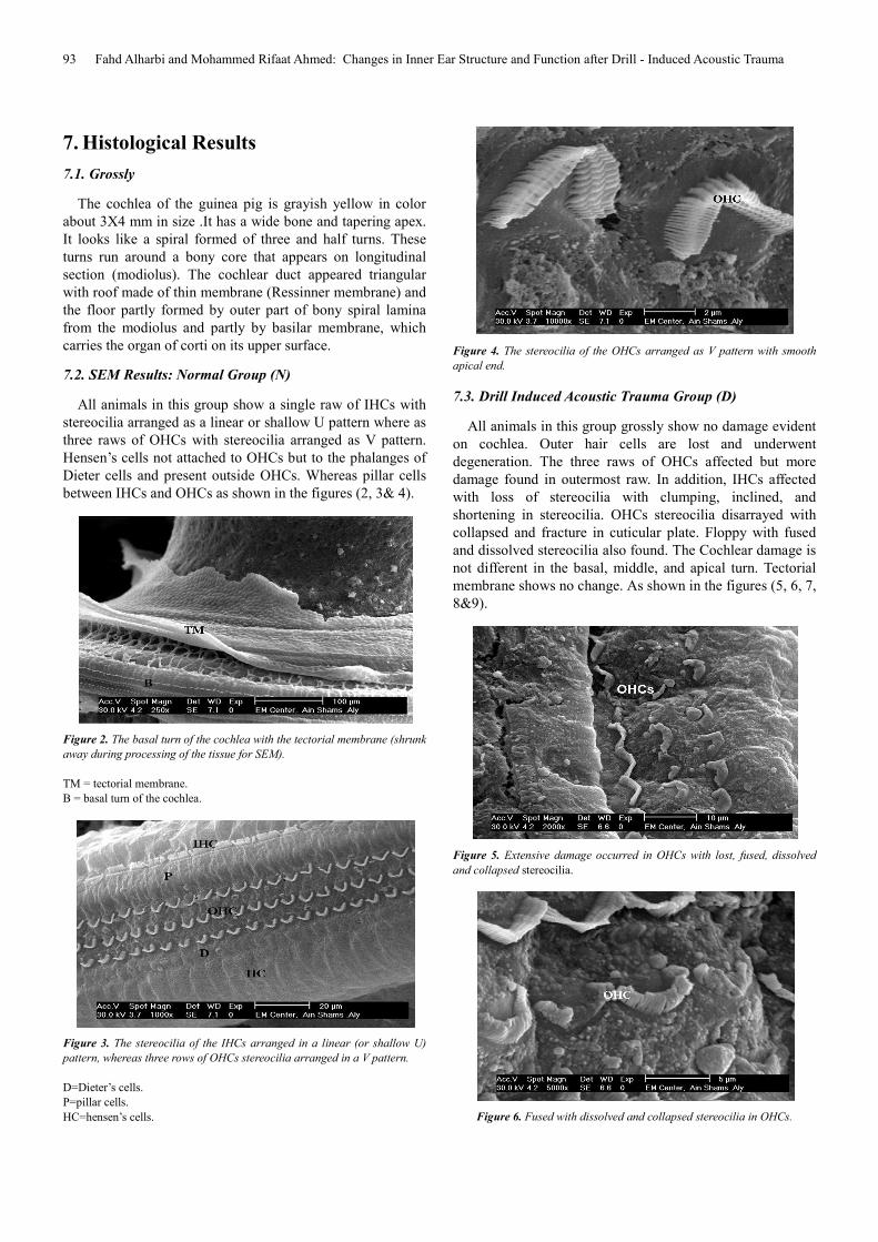
All animals in this group show a single raw of IHCs with stereocilia arranged as a linear or shallow U pattern where as three raws of OHCs with stereocilia arranged as V pattern. Hensen’s cells not attached to OHCs but to the phalanges of Dieter cells and present outside OHCs. Whereas pillar cells between IHCs and OHCs as shown in the figures (2, 3& 4).
Figure 2. The basal turn of the cochlea with the tectorial membrane (shrunk
away during processing of the tissue for SEM).
TM = tectorial membrane. B = basal turn of the cochlea.
Figure 3. The stereocilia of the IHCs arranged in a linear (or shallow U)
pattern, whereas three rows of OHCs stereocilia arranged in a V pattern.
D=Dieter’s cells. P=pillar cells. HC=hensen’s cells.
Figure 4. The stereocilia of the OHCs arranged as V pattern with smooth
apical end.
7.3. Drill Induced Acoustic Trauma Group (D)
All animals in this group grossly show no damage evident on cochlea. Outer hair cells are lost and underwent degeneration. The three raws of OHCs affected but more damage found in outermost raw. In addition, IHCs affected with loss of stereocilia with clumping, inclined, and shortening in stereocilia. OHCs stereocilia disarrayed with collapsed and fracture in cuticular plate. Floppy with fused and dissolved stereocilia also found. The Cochlear damage is not different in the basal, middle, and apical turn. Tectorial membrane shows no change. As shown in the figures (5, 6, 7, 8&9).
Figure 5. Extensive damage occurred in OHCs with lost, fused, dissolved
and collapsed stereocilia.
Figure 6. Fused with dissolved and collapsed stereocilia in OHCs.

American Journal of Clinical and Experimental Medicine 2014; 2(5): 90-96 94
Figure 7. Dissolved and collapsed OHCs stereocilia.
Figure 8. Marked OHCs sterocilia damage.
Figure 9. Showed that IHCs stereocilia affected with fused, clumping,
inclined, shortening and fracture.
7.4. 2weeks Postoperative Group (2W)
All animals in this group grossly show no damage evident on cochlea. Damage occurred in OHCs (replaced by scar formation mostly in the second and outermost raws or with disarrayed stereocilia).However, the innermost raws show completely recovery and back to normal. IHCs show a clumped stereocilia. There was difference in the extent of recovery, the middle and apical turn shows complete recovery while the basal turn shows scar formation as seen in figures (10, 11, 12&13).
Figure 10. Showed that OHCs replaced by scar formation in the outermost
row with disarrayed stereocilia.
Figure 11. Revealed disarrayed OHCs stereocilia in the outermost and
second rows.
Figure 12. Showed arrow pointed to the disarrayed OHCs stereocilia.
Figure 13. showed arrow pointed to the disarrayed OHCs stereocilia.
8. Discussion
Normal group (N): DPOAEs recorded from 1 kHz to 8 kHz based on Abu Seta 2002 who illustrated that more noise pollution if measurement below 1 kHz, also Probst et al.1994 & Moulin et al 1994 found that for 500Hz, DPOAEs presence reached only 40% at 70 dBSPL primaries. The

95 Fahd Alharbi and Mohammed Rifaat Ahmed: Changes in Inner Ear Structure and Function after Drill - Induced Acoustic Trauma
absence of DPOAEs at low frequencies attributed to an increase noise floor, mostly from animal respiration (9, 10 & 11). The choice of 65 dB SPL as primary levels and frequency range from 1 to 8 kHz based on a recent study demonstrating that 65dB gives the best response as it is the least frequency polluted by ambient noise. (10 & 11)
The use of higher level (70 dB) accompanied by more noise rejection, while the use of lower levels (50 dB) led to lower DPOAE amplitude. This contradicts with findings of Shalaby et al 1998 who measured DPOAEs at 75 dBSPL. (12)
The average DPOAE amplitudes across different frequencies in normal group (N) 29 dBSPL. The higher amplitudes of DPOAEs in guinea pigs attributed to the regular arrangement of the outer hair cells while in human cochlea the outer hair cells are irregular arranged (13). It is also possible that the larger DPOAEs recorded simply a reflection of lower total impedance offered by middle ear of the guinea pigs when compared with that of humans. (14)
The histological study for this group revealed normal ultra structure for the tectorial membrane, outer hair cells, inner hair cells, stereocilia arrangement for OHCs and IHCs. There was difference in (V) angle of stereocilia in OHCs. The narrow angle in apex of the cochlea is approximately 60 degree while it is about 120 degree in the basal turn. In addition, the separated angle between the two limbs (IHCs stereocilia) about 180 degrees. These findings are compatible with the findings of Berit 1983.
(15) Drill induced acoustic trauma group (D): The DPOAE
measurements obtained immediately after the acoustic trauma showed reduced DPOAE values. The reduction in amplitude and S/N ratio agreed with Shalaby et al 1998 who found reduction in the whole frequencies range from 500 to 8000 Hz. This difference may be due to the type of noise exposure as they use narrow band noise (500 Hz) while we use drill-induced ossicular chain injury technique. This explains why the low frequencies more affected in the work of Shalaby et al 1998. (12) Confirming to the damage effect in form of fractured, collapsed, clumping, inclined, separated and dissolved stereocilia in both OHCs and IHCs. This agrees with the findings Hamerniket al 1981 & Ye et al 1998. (16, 17)
Berit 1983 found that after acoustic trauma, fusion of the stereocilia in whole length or part with leaning in different direction and the neck of the stereocilia is the site of fracture
(15). In our study, stereocilia is the most susceptible structure for damage. This is supported by Berit 1993 (15)
. Acoustic
trauma mechanism for damage can be direct mechanical action, metabolic action and by impaired blood flow or altered permeability of cell membrane (18). IHCs changes in response to acoustic trauma explained by immediate examination of the cochlea after acoustic trauma. Also the vibration effect induced by the drill form another trauma to the inner ear with synergistic action with the noise as mentioned by Iskander et al 1991, reported that vibration affect the IHCs more than the OHCs (19).
The exact mechanism responsible for these changes is not clear as they describe in literatures whether due to noise
exposure or the effect of vibration from the drill or both. Such mechanisms include circulatory response metabolic exhaustion), increase metabolic demand and ionic changes between perilymph and endolymph (20, 21, 22, 23). Sympathetic overflow proposed to be the explanation of synergistic action of noise and vibration that leads to vascular disturbing effects can occur (24). In addition, Berit 1993 agreed that noise does not damage inner ear by single way but several types of damages (15).
2weeks postoperative group (2W):
The DPOAE results after 2 weeks from noise exposure did not return back to normal but significant improvement found. The lack of complete recovery of DPOAE after noise exposure has many possibilities. First, the hair bundle on adjacent hair cells may be misaligned and this would lead to significant reduction in DPOAE amplitude (25). A second possibility is that the nonlinearities in stiffness of stereocilia may be responsible for DPOAEs in normal ears, but these nonlinearities may be greatly reduced or absent in recovering hair cells (26). A third factor that could contribute to depression of DPOAE is the residual damage to hair cells that survived the traumatizing exposure (26). A fourth factor is the number of affiliated efferent fibers to each regenerated OHC may be changed due to improper alignment between these fibers and the regenerated OHC (27). A fifth factor is the damage of tectorial membrane as mentioned by Pyykko et al 1982 (24).
On the other hand, Sujana et al 2000 mentioned that most cases of noise induced sensorineural hearing loss show recovery within 2 weeks without treatment (28). Berit 1993 found that few days after acoustic trauma, fused and inclined stereocilia while the severe deformation of the stereocilia (giant stereocilia) appear after 18 month from the exposure (15). Dobie 1999 found that stereocilia started to return to their pre-exposure state in about 15 minutes following the mechanical stimulation (18).
Ballanger et al 1997 reported that acoustic injury to the ear has a dynamic and static phase. The dynamic phase begins during the acoustic stimulation, which results in cellular elements in the ear undergoing structure and function changes, which may be permanent or temporary. When the acoustic stimulation ceases, the structures of the ear may recover completely or partially or by scar formation which lead to static phase in which anatomical and function changes are stable. If the degree of injury is mild, the repair will occur from the cell itself, while if the degree of injury is great, the healing will happen with scar formation from migration of other cells (29).
9. Conclusions
Avoid touching ossicular chain during ear surgery as a high focused acoustic energy transmitted to inner ear causing damage in structure and deterioration of function. Spontaneous recovery could occur after acoustic trauma but may be incomplete with permanent scar formation in OHCs.

American Journal of Clinical and Experimental Medicine 2014; 2(5): 90-96 96
Individual Roles
• Fahed Alharbi: Review paper, clinical surgical work and finishing clinical assessment
• Mohamed Rifaat Ahmed: Statistical analysis, prepare discussion, results, and data collection
References
[1] Dan J ; Bibas A ; Santuli C ; Donnelly N ; Jeronimidis G ; Oconnor AF; Equivalent noise level generated by drilling onto the ossicular chain as measured by laser Doppler vibrometry: A temporal bone study .The Laryngoscope .2007, vol. 117, no. 6, pp. 1040-1045 .
[2] Domenech J .Carulla M , Traserra J :Sensorineural high-frequency hearing loss after drill-generated acoustic trauma in tympanoplasty . Arch Otorhinolaryngol. 1989;246(5):280-2.
[3] HüttenbrinkKB :Cochlear damage caused by middle ear surgeries laryygorhinootologie. 1991 Feb;70(2):66-71[Article in German] .
[4] Schick B, Schick BT ,Kochannek S , Starlinger V, Iro H : Temporary sensory hearing deficits after ear surgery--a retrospective analysis. laryygorhinootologie. 2007 Mar;86(3):200-5. Epub 2006 Nov 27[Article in German]
[5] Mislav G; Wolfgang S; Wolfgang B; Stephan R W; MalteEW:ExperimentalSensorineural Hearing Loss Following Drill-induced Ossicular Chain Injury ActaOto – Laryngologica , Volume 117, Issue 4 July 1997 , pages 497 – 500.
[6] Wolfgang S and Mislav G. “the value of methylpredinsoline in the treatment of experimental sensorineural hearing loss following drill induced ossicular chain injury in guinea pigs.” ann Otology 1999 ,41: 281-290.
[7] Paparella MM. “Acoustic trauma from bone cutting burr.” laryngoscope ,1982 ,72: 116-118.
[8] Isaacson DJ, Antonelli PJ, . “Labyrinthine fenestration in the guinea pig.” ann Otology 1999 ,41.: 281-290.
[9] Abu Seta A: The effect of noise and whole body vibration on cochlea of guinea pig, MD thesis Suez Canal University, Ismalia, Egypt. .2002.
[10] Probst R, Antonelli C, et al.. “Method and results of measurements of DPOAEs in normal and pathological ears.” Adv. Audiol1994., 4: 7-21.
[11] Moulin A and Collet L. “Distortion product otoacoustic emissions and sensorineural hearing loss.” Audiology 1994 ,33: 305-26.
[12] Shalaby A and Abdel-Maksoud A. “Effects of nose on guinea pigs.” Audiology and Histology overview Egypt J Otolaryngol. 1998,15: 55-66.
[13] Probst R, Lonsbury-Martin B.L, et al..“A Review of Otoacoustic Emissions.” Journal of the Acoustical Society of America 1991 ,89: 2027 -2067.
[14] Woodford C.W, Henderson D ,Hamernik R.P. : Static and dynamic independence of chinchilla middle ear.86 th meeting the Acoustical society of America, Los Angeles. 1973.
[15] Berit E. “stereocilia of sensory cells in normal and hearing impaired ears.” ActaOtolaryngol1983 ,42:96-105.
[16] Hamernik RP and SalviR .: “The interaction between whole body vibration and impulse noise.” J. Acoustic. Soc.Am. 1981,4: 928-34.
[17] Ye Q and RenX .“ the effect of exposure to noise in oil drilling well sites on cochlea of guinea pigs,.” Chung Hua Yu Fang Ih such Tsachih J 1998 .: 103-5.
[18] Dobie R.A : Noise-Induced Hearing Loss. In Byron J. and Bailey, (eds): Head and Neck Surgery-Otolaryngology,. Washington University, Lippincott-Raven Publishers.2nd edition 1999( p.p. 211-212).
[19] Iskander L.M and Iskander W.M, :. “cochlear changes secondary to whole body vibration in guinea pigs.” Egypt.J.Anto .1991, 14: 151-63.
[20] Axelsson A and VertsD .“Histological finding in cochlear vessels after noise.”ActaOtolaryngol1982 ,91: 237-240.
[21] BohneB.A .“Mechanism of noise damage in inner ear.”ActaOtolaryngol 1976,236: 53-8.
[22] Ryan S, and Kemp D.T, : . “The influence of contralateral stimulation on click-evoked otoacoustic emissions in humans.”British Journal of Audiology1991. 25: 391-397.
[23] Quirk W and NuttalA.L . “noise induced changes in lateral wall vessels of the cochlea in rats.” Hear.Res. 1991,52: 217-24.
[24] Pyykko I and Stark J . “sensorineural hearing loss during noise and vibration exposure.” ActaOtlolaryngol.1982 ,386: 296-300.
[25] Duckert L.G and Rubel E.W. “Morpholgical correlation of function recovery in chiken inner ear after genatmycin.” J. CONMP. Neurol. 1993, 331: 75-96.
[26] Saunders S.S and Salvi R.J. “pure tone masking patterns in adult chiken after recovery from acoustic trauma.”
[27] Acoustic.Soc.Am J.1995, 98: 1356-71.
[28] Ryals B.M, Dooling R.J.(1996): Changes in hair cell innervation and auditory senesitivity after acoustic trauma and hair cell regeneration in birds. Theme Medical publishers ;84-89.
[29] Sujana S and Micheal BA. “Dexamethason pharmacokinetics in the inner ear comparison of route of adminstration and use of faclitating agents.” Otolaryngology HNS J. 2000 ,122: 521-8.
[30] Ballanger J.J. and Johmes B. (): anatomy of inner ear. In Ballanger JJ ( ed): text book of Otorhinolaryngology, head and neck surgery,vol3. Philadelphia,Williams and wikins,15th edition1997 ( p.p 678-688).