CHALLENGES IN ACUTE PANCREATITIS: DIAGNOSIS, …
83
CHALLENGES IN ACUTE PANCREATITIS: DIAGNOSIS, ETIOLOGY AND TREATMENT Ph.D. Thesis Dóra Mosztbacher M.D. Szeged 2020
Transcript of CHALLENGES IN ACUTE PANCREATITIS: DIAGNOSIS, …

CHALLENGES IN ACUTE PANCREATITIS:
DIAGNOSIS, ETIOLOGY AND TREATMENT
Ph.D. Thesis
Dóra Mosztbacher M.D.
Szeged
2020

CHALLENGES IN ACUTE PANCREATITIS:
DIAGNOSIS, ETIOLOGY AND TREATMENT
Ph.D. Thesis
Doctoral School of Theoretical Medicine, Faculty of Medicine, University of Szeged, Szeged
Dóra Mosztbacher, M.D.
Doctoral School of Theoretical Medicine, Faculty of Medicine, University of Szeged, Szeged
First Department of Paediatrics, Faculty of Medicine, Semmelweis University, Budapest
Institute for Translational Medicine, Medical School, University of Pécs, Pécs
Supervisors:
Péter Hegyi, M.D., Ph.D., D.Sc., MAE
First Department of Medicine, Faculty of Medicine, University of Szeged, Szeged
Institute for Translational Medicine, Medical School, University of Pécs, Pécs
Andrea Párniczky, M.D., Ph.D.
Heim Pál National Institute of Paediatrics, Budapest
Institute for Translational Medicine, Medical School, University of Pécs, Pécs
Doctoral School of Theoretical Medicine, Faculty of Medicine, University of Szeged, Szeged
Szeged, 2020

- 1 -
PUBLICATIONS RELATED TO THE SUBJECT OF THE THESIS
I. Mosztbacher D, Hanák L, Farkas N, Szentesi A, Mikó A, Bajor J, et al.
Hypertriglyceridemia-induced acute pancreatitis: A prospective, multicenter,
international cohort analysis of 716 acute pancreatitis cases. Pancreatology.
2020;20(4):608-616. IF: 3.629
II. Mosztbacher D, Farkas N, Solymár M, Pár G, Bajor J, Szűcs Á, et al. Restoration of
energy level in the early phase of acute pediatric pancreatitis. World Journal of
Gastroenterology. 2017;23(6):957. IF: 3.3
III. Zsoldos F, Párniczky A, Mosztbacher D, Tóth A, Lásztity N, Hegyi P. Pain in the
early phase of pediatric pancreatitis (PINEAPPLE Trial): pre-study protocol of a
multinational prospective clinical trial. Digestion. 2016;93(2):121-6. IF: 2.088

- 2 -
SCIENTIFIC METRICS
Number of publications related to the subject of the thesis:
Cumulative impact factor of publications related to the thesis:
Q1: 2, Q2: 1, Q3: -, Q4: -
3 (2 first author)
9.017
Number of total accepted/published articles:
Cumulative impact factor of the published articles:
Q1: 11, Q2: 3, Q3: -, Q4: -
14 (4 first author)
45.799
Number of total citation by Google Scholar:
https://scholar.google.com/citations?pagesize=100&user=xvok1ZAAA
AAJ
Hirsch Index:
245
9
Number of total citation by MTM2:
https://m2.mtmt.hu/gui2/?type=authors&mode=browse&sel=10059862
&view=pubTable2
Hirsch Index:
106 independent
161 all
8

- 3 -
TABLE OF CONTENTS
I. List of abbreviations ................................................................................................... 5
II. General introduction ................................................................................................... 7
II.1. Challenges in acute pancreatitis .......................................................................... 7
II.2. Motivation for my scientific work ....................................................................... 8
III. Dose-dependent effect of hypertriglyceridemia on acute pancreatitis ........................ 9
III.1. Introduction ......................................................................................................... 9
III.2. Aims .................................................................................................................... 9
III.3. Methods ............................................................................................................... 9
III.4. Results ............................................................................................................... 12
III.5. Discussion ......................................................................................................... 20
III.6. Conclusion ......................................................................................................... 23
IV. Children are not small adults .................................................................................... 26
IV.1 The PINEAPPLE study ..................................................................................... 26
IV.1.1. Introduction .......................................................................................... 26
IV.1.2. Aims ..................................................................................................... 27
IV.1.3. Methods................................................................................................ 27
IV.1.4. Expected results ................................................................................... 30
IV.1.5. Discussion ............................................................................................ 31
IV.2. Early enteral nutrition in acute pediatric pancreatitis ........................................ 32
IV.2.1. Introduction .......................................................................................... 32
IV.2.2. Aims ..................................................................................................... 33
IV.2.3. Methods................................................................................................ 33
IV.2.4. Results .................................................................................................. 36
IV.2.5. Discussion ............................................................................................ 36
IV.3. Conclusion ......................................................................................................... 38
V. Summary and new discoveries..................................................................................... 39

- 4 -
VI. Contribution ................................................................................................................. 40
VI.1. Mosztbacher et al. Pancreatology, 2020 ............................................................ 40
VI.2. Mosztbacher et al. World J Gastroenterol, 2017 ............................................... 40
VI.3. Zsoldos et al. Digestion, 2016 ........................................................................... 40
VII. Acknowledgement ....................................................................................................... 41
VIII. References .................................................................................................................... 42

- 5 -
I. LIST OF ABBREVIATIONS
AIP autoimmune pancreatitis
ALAT alanine transaminase
ALP alkaline phosphatase
AP acute pancreatitis
APA American Pancreatic Association
APP acute pediatric pancreatitis
ARP acute recurrent pancreatitis
ASAT aspartate transaminase
ATP adenosine triphosphate
BUN blood urea nitrogen
CFTR cystic fibrosis transmembrane conductance regulator
CI confidence interval
CP chronic pancreatitis
CRP C-reactive protein
CT computed tomography
DM diabetes mellitus
EBM evidence-based medicine
EEN early enteral nutrition
FAEEs fatty acid ethyl esters
γGT gamma-glutamyltransferase
HPSG Hungarian Pancreatic Study Group
HTG hypertriglyceridemia
HTG-AP hypertriglyceridemia-induced acute pancreatitis
IAP International Association of Pancreatology
ICMJE International Committee of Medical Journal Editors
ICU intensive care unit
IVF intravenous fluid
LDH lactate dehydrogenase
LOH length of hospitalization
Max CRP maximum C-reactive protein

- 6 -
Max WBC maximum white blood cell count
MOF multi organ failure
NPO nil per os
PC pancreatic cancer
PICO participants, intervention, comparison and outcomes
PINEAPPLE Pain IN the EArly phase of Pediatric Pancreatitis
PRISMA-P preferred reporting items for systematic review and meta-analysis
protocol
RBC red blood cell count
ROC receiver operating characteristic
SAP severe acute pancreatitis
SIRS systemic inflammatory response syndrome
sPEM serum pancreatic enzyme measurement
TG triglyceride
TPN total parenteral nutrition
US ultrasonography
WBC white blood cell count
WHO World Health Organization

- 7 -
II. GENERAL INTRODUCTION
II.1. Challenges in acute pancreatitis
Acute pancreatitis (AP) is one of the most common reasons for gastrointestinal
hospitalizations in adults (1, 2). AP has an annual incidence of 13-45 per 100,000 persons and
is increasing worldwide as a result of better awareness of the disease and obesity-related
gallstone formation and hypertriglyceridemia (HTG) (3, 4). AP represents a remarkable disease
burden for healthcare systems and patients’ quality of life (5). This burden is further increased
by the development of a severe disease course which is accompanied by increased length of
hospitalization (LOH), elevated rate of complications, intensive care unit (ICU) stay, the need
for invasive interventions, and mortality (5).
According to the revised Atlanta classification, the severity of AP is categorized as
mild, moderately severe, and severe (6). Severe acute pancreatitis (SAP) develops in 15-20%
of AP cases (1, 6); however, better understanding of the underlying mechanism may provide
possibilities for decreasing severity. Although the pathomechanism of AP is still unclear, the
most common etiological factors such as bile acids (7-10), fatty acids generated from
triglyceride (TG), and non-oxidative ethanol metabolites (fatty acid ethyl esters, FAEEs) (11-
16) were shown to cause mitochondrial damage with resultant adenosine triphosphate (ATP)
and energy depletion in the exocrine pancreas. In addition, HTG contributes to systemic pro-
inflammation and local hypoxia-induced acidosis caused by hyperviscosity (17, 18). According
to these data, HTG was shown to increase the risk of the development of SAP (19-28) and
aggravate the severity of AP compared to alcoholic and biliary etiologies (29-31). In contrast,
early enteral nutrition (EEN) as early ATP restoration has been shown to be beneficial in AP
compared with nil per os (NPO) therapy and total parenteral nutrition (TPN) regardless of the
severity of AP (32-37). Accordingly, initial and appropriate lipid-lowering therapy may be
beneficial in the case of hypertriglyceridemia-induced acute pancreatitis (HTG-AP), whereas
energy restoration via enteral nutrition may play a key part of the therapy in all cases of AP.
However, detailed analysis and evidence-based therapy for HTG-AP is missing, and despite
the fact that the clinical characteristics of AP differ by age, nutritional guidelines for childhood-
onset AP are limited and adopted from the adult protocols (38-48).
Not only therapeutic, but also diagnostic pediatric guidelines for AP are adopted from
adult protocols (40, 41, 45). Some of these guidelines are based on the consensus of expert
pediatric pancreatologists, but not on evidence-based pediatric data (49, 50). The recently

- 8 -
published acute pediatric pancreatitis (APP) guideline has low evidence as well (40). It is not
surprising that the overall incidence of APP is lower compared to the adult population (1 per
100,000 persons or even less versus 13-45 per 100,000); however, two major studies have
proven that the real incidence of APP (3.6–13.2 per 100,000) is much higher than we previously
thought (3, 51-55). The reason is probably multifactorial, but it has been published by
Morinville et al., that diagnostic workup influences the incidence of the disease (51). Their data
showed that increased pancreatic enzyme testing could account for 94% of the change in all
AP admissions in childhood, suggesting that APP is an underdiagnosed disease.
In contrast with the current diagnostic practice, there is significant importance in
recognizing and diagnosing AP in childhood. Acute recurrent pancreatitis (ARP) develops in
10-35% of children following an initial AP (41, 56, 57), and idiopathic ARP is likely to be a
transition phase between AP and chronic pancreatitis (CP) (58, 59) with 1-3.79 years of median
time (60, 61). However, 10-16% of the pediatric CP patients have no documented prior episode
of AP or ARP (60, 62). ARP and CP are of great importance because both are associated with
a notable disease burden by pain, exocrine and endocrine dysfunction, frequent ER visits,
hospitalizations, and school absenteeism (60). The most common risk factors of CP are alcohol
and smoking in adults; however these are uncommon in children (63). Pediatric ARP and CP
are frequently associated with pancreatobiliary obstructions in ~30% and genetic abnormalities
in up to 73% (54, 60, 64, 65). Genetic involvement was shown to carry the fastest rate of
progression from AP to CP (65). These data highlight the necessity of an appropriate and
evidence-based diagnostic guideline for childhood-onset AP.
II.2. Motivation for my scientific work
As a pediatric resident I recognized the importance of evidence-based clinical practice
compared to decisions based on the experiences of individual physicians. First, we aimed to
estimate the real incidence of APP and improve the diagnostic workup of childhood onset AP
by establishing an international, multicenter observational clinical trial called PINEAPPLE
(Pain IN the EArly phase of Pediatric Pancreatitis) (66). Furthermore, we reviewed the
literature to analyze the effect of EEN versus NPO therapy on the outcome of APP and
aggregate the information to increase the statistical power of nutritional AP guidelines in
childhood compared to individual studies (67). Finally, we performed a cohort analysis of 716
adult AP cases to investigate the dose-dependent effect of HTG on AP and provide detailed
data for further prospective randomized clinical trials (68, 69).

- 9 -
III. DOSE-DEPENDENT EFFECT OF HYPERTRIGLYCERIDEMIA ON ACUTE
PANCREATITIS
III.1. Introduction
HTG affects 10–30% of the general adult population (70, 71). Classifying HTG is
complex; both genetic (primary) and environmental (secondary) factors can lead to an elevated
TG level. In rare cases (2%), primary severe HTG (TG≥10 mmol/l) may arise as a result of
autosomal recessive, monogenic familial chylomicronemia syndrome (FCS, former Type I).
However, a majority of severe HTG cases are multifactorial and have polygenic (mixed HTG,
former Type V) determinants with additional secondary factors. Mild-to-moderate HTG cases
(2–9.9 mmol/l TG) are similarly polygenic with complex genetic susceptibility (former Type
IV, Type IIB and Type III) (3, 71, 72). Regarding environmental factors, alcohol, positive-
energy balanced diet, obesity, uncontrolled diabetes mellitus (DM), renal diseases, pregnancy,
hypothyroidism, and medications (e.g., estrogen, retinoids, thiazides, and β-blockers) were
shown to be responsible for a raised TG level, usually with the interaction of genetic
susceptibility (18, 73).
HTG is the third most common cause of AP, and is responsible for up to 15% of AP
cases (26, 74, 75). According to the definition, the majority of experts agree that AP related to
TG above 5.6 mmol/l should be considered as suspected HTG-AP, and AP associated with TG
over 11.3 mmol/l is confirmed as HTG-AP (24, 72). Importantly, the occurrence of AP
increases with the increase in TG level. There is a 5% possibility of developing AP if TG
exceeds 11.3 mmol/l, and this rises to 10–20% if TG elevates to over 22.6 mmol/l (72). HTG-
AP is of great importance for several reasons: i) it has shown a rising incidence worldwide as
a result of increasing obesity-related dyslipidemia (4, 25, 27); ii) it raises the risk of severe AP
and related complications (20, 24-26, 28, 31, 76-78); and iii) there is no evidence-based therapy
for it (38, 39, 42, 43, 79).
III.2. Aims
We aimed to perform a cohort analysis for investigating the dose-dependent effect of
HTG on AP and providing data for further prospective randomized clinical trials.
III.3. Methods
AP patients (n=1435) over 18 years old were enrolled in the prospectively collected

- 10 -
international, multicenter AP registry operated by the Hungarian Pancreatic Study Group
(HPSG) between 2012 and 2017. Post-hoc cohort analysis was performed on 716 AP cases
who underwent TG measurement within 72 hours of admission. AP was diagnosed based on
International Association of Pancreatology/American Pancreatic Association (IAP/APA) and
HPSG evidence-based guidelines (38, 39). Participating countries are shown in Fig. 1.
Fig. 1. Participating countries. Distribution of participating countries and number of enrolled acute pancreatitis
cases (n=1435).
The threshold of the normal TG value was determined at 1.7 mmol/l (73). Cases where
AP was accompanied by TG above 11.3 mmol/l were defined as HTG-AP. Six groups were
established based on the Endocrine Society Clinical Practice Guideline and previously
published clinical data related to HTG-AP (72, 73): Group 1: <1.7 mmol/l; Group 2: 1.7–2.19
mmol/l; Group 3: 2.2–5.59 mmol/l; Group 4: 5.6–11.29 mmol/l; Group 5: 11.3–22.59 mmol/l;
and Group 6: ≥22.6 mmol/l. To convert TG from mmol/l to mg/dl, multiply by 88.57. In the
case of each variable, elevated TG groups (Groups 2-6) were compared with the normal TG
group (Group 1). TG categories were collapsed to three groups (<1.7 mmol/l; 1.7-11.29
mmol/l; ≥11.3 mmol/l) for the analysis of organ failure because of the low event number.
Seventy-three variables were collected from each AP case. The analysis was performed
on 42,655/52,268 available data. Local complications, organ failure, and severity were defined
based on the revised Atlanta classification (6). The 716 cases investigated showed the same
epidemiological and major outcome distribution as the total cohort (1435 cases), demonstrating
that our patient population represents a normal AP cohort (Fig. 2).

- 11 -
Fig. 2. Representation of enrolled patients (n=716) compared to the entire cohort (n=1435). A) Gender
distribution of acute pancreatitis (AP) cases. B) Age distribution of AP cases in males and females. C) Severity

- 12 -
distribution of AP cases. D) Mortality of AP cases in the different severity groups. E) Length of hospitalization
of AP cases in the different severity groups. F) Etiology distribution of AP cases (a: p=0.007). ERCP=endoscopic
retrograde cholangiopancreatography. N numbers (n) indicate the total number of cases in each triglyceride group.
The registry received ethical permission from the Scientific and Research Ethics
Committee of the Medical Research Council (22254-1/2012/EKU) in 2012, and all the patients
provided written informed consent to participate. The study protocol conforms to the ethical
guidelines of the Declaration of Helsinki updated in 2013 as reflected in a priori approval by
the institution's human research committee.
Statistical analysis
Prior to analysis of the dataset, descriptive statistical tools were used to describe the
basic characteristics. Mean and standard error of the mean were calculated for continuous
variables, whereas the incidence in each group was determined for categorical variables.
Depending on the distribution of the data, the independent Student’s t-test or Mann–Whitney
U test was used to evaluate differences between continuous parameters. The chi-square test or
Fisher’s exact test was conducted to analyze the relations between categorical variables. We
compared the confidence intervals (CI) of the proportions to investigate differences in the
incidence of moderately severe cases between groups. A p-value less than 0.05 (≤ 0.05) was
determined as statistically significant. All analyses were performed using IBM-SPSS
Statistical Software Version 25 (IBM Corporation, Armonk, NY, USA).
III.4. Results
In our cohort, 30.6% (n=219) of the patients presented with elevated TG level (≥1.7
mmol/l). HTG was significantly and dose-dependently linked to younger age and male gender
(Fig. 3A-C). In 7.7% of AP cases (n=55), TG level was above 11.3 mmol/l, which is considered
as a causative etiological factor (24, 73). In 56.4% of these cases, HTG-AP patients had no
other etiology described; however, raised TG level was also accompanied by alcohol in 38.2%
of these cases and by biliary etiology in 5.4%, showing that HTG-AP is associated with other
etiologies in a substantial number of cases (Fig. 3D).

- 13 -
Fig. 3. Epidemiology and etiology. A) Age distribution of acute pancreatitis (AP) cases in males and females.
B) Age distribution of triglyceride (TG) groups (a, c, d: p=<0.001; b: p=0.010). C) Gender distribution of
triglyceride groups (e: p=0.002; f, g: p<0.001). D) Etiology. HTG=hypertriglyceridemia; ERCP=endoscopic
retrograde cholangiopancreatography. N numbers (n) indicate the total number of cases in each triglyceride group.
Data from patients’ medical history revealed that HTG is significantly and dose-
dependently linked to obesity and DM (Fig. 4B, C); however, there is no relation to CP and the
Charlson comorbidity index (CCI) (80) (Fig. 4A, D). The amount of previous AP in the medical
history was higher in the HTG group compared to the normal TG group (Fig. 4A).

- 14 -
Fig. 4. Patients’ medical history of triglyceride groups. A) Acute pancreatitis (AP) and chronic pancreatitis
(CP) in patients’ medical history (a: p=0.026; b: p=0.023). B) Obesity (body mass index (BMI), kg/m2) (c:
p=0.006; d: p=0.001). C) Diabetes mellitus in medical history (e: p=0.046; f: p=0.016; g: p=0.026; h: p<0.001).
D) Charlson comorbidity index (CCI). N numbers (n) indicate the total number of cases in each triglyceride group.
General symptoms of AP and physical examination on admission (incidence, duration
and intensity of pain, nausea, vomiting, abdominal tenderness and guarding, and blood
pressure) have not shown a significant link to elevated TG level (Fig. 5A, B). However, HTG
was significantly related to increased heart rate (Fig. 5C). Regarding the laboratory parameters
on admission showing significant differences with HTG, amylase, lipase, sodium, and calcium
were associated inversely; however, glucose, C-reactive protein (CRP), cholesterol, red blood
cell count (RBC), hemoglobin, and hematocrit were related in parallel with TG level (Fig. 6).

- 15 -
Fig. 5. Symptoms and physical findings on admission in the different triglyceride (TG) groups. A) Incidence,
duration before admission and intensity of pain. Nausea, vomiting, abdominal tenderness and abdominal guarding
(a: p=0.025; b: p=0.036). B) Systolic and diastolic blood pressure (Hgmm) (c: p=0.012). C) Heart rate (BPM) (d,
e: p=0.010). N numbers (n) indicate the total number of cases in each triglyceride group.
On admission, laboratory parameters consistent with cholestasis suggested that HTG is less
common in cases with biliary etiology (Fig. 7). The parallel rise in gamma-glutamyltransferase
(γGT) and TG levels confirms that alcohol consumption is linked to HTG (Fig. 7D). White
blood cell count (WBC), thrombocyte, lactate dehydrogenase (LDH), blood urea nitrogen
(BUN), creatinine, and potassium had no significant relation to HTG (Fig. 8).
The rate of local complications, including peripancreatic fluid collection, pancreatic
necrosis, and DM was significantly and dose-dependently increased with TG level (Fig. 9B-
E); however, pancreatic pseudocysts did not show significant differences between the
investigated groups above 2.2 mmol/l (Fig. 9F).

- 16 -
Fig. 6. Laboratory parameters on admission significantly associated with hypertriglyceridemia. A)
Triglyceride groups (1–6). B) Amylase (U/l) (a: p=0.036; b, c, e: p=<0.001; d: p=0.009). C) Lipase (U/l) (f:
p=0.017; g: p=0.001). D) Sodium (mmol/l) (h, i: p=<0.001; j: p=0.005). E) Calcium (mmol/l) (k: p=0.012). F)
Glucose (mmol/l) (l, m: p=<0.001). G) C-reactive protein (CRP, mg/l) (n: p=0.021; o: p=0.014). H) Cholesterol
(mmol/l) (p: p=0.008; q: p=0.006; r, s: p<0.001). I) Red blood cell count (RBC, T/l) (t: p=0.017; u: p=0.004) J)
Hemoglobin (G/l) (v: p=0.008; w: p<0.001; x: p=0.002). K) Hematocrit (%) (y: p=0.009; z: p=0.014). N numbers
(n) indicate the total number of cases in each triglyceride group.

- 17 -
Fig. 7. Laboratory parameters representing hepatobiliary system on admission in the different triglyceride
groups. A) Triglyceride groups (1–6). B) Aspartate transaminase (ASAT, U/l) (a: p=0.050; b: p=0.037). C)
Alanine transaminase (ALAT, U/l). D) Gamma-glutamyltransferase (γGT, U/l). E) Alkaline phosphatase (ALP,
U/l) (c: p=0.028; d: p=0.003). F) Total bilirubin (umol/l). N numbers (n) indicate the total number of cases in each
triglyceride group.
Fig. 8. Laboratory parameters on admission without significant alterations with hypertriglyceridemia. A)
White blood cell count (WBC, G/l). B) Thrombocyte (G/l). C) Lactate dehydrogenase (LDH, U/l). D) Blood urea
nitrogen (BUN, mmol/l). E) Creatinine (umol/l). F) Potassium (mmol/l). N numbers (n) indicate the total number
of cases in each triglyceride group.

- 18 -
Organ failure, including heart and renal failure, and maximum CRP level were
significantly and dose-dependently raised by TG level (Fig. 10A, C, E, F); however, respiratory
failure and maximum WBC did not show any significant differences by HTG (Fig. 10B, D).
Fig. 9. Local pancreatic complications in the different triglyceride groups. A) Triglyceride groups (1–6). B)
Local pancreatic complications (a, b: p<0.001). C) Peripancreatic fluid collection (c: p=0.004; d: p<0.001). D)
Pancreatic necrosis (e: p=0.003; f: p<0.001). E) Diabetes mellitus as complication (g: p=0.004; h: p<0.001; i:
p=0.011). F) Pancreatic pseudocyst (j: p=0.031). AP=acute pancreatitis. N numbers (n) indicate the total number
of cases in each triglyceride group.
As regards severity, TG level above 11.3 mmol/l was associated with a significantly
higher rate of moderately severe AP and longer hospital stay, whereas TG level above 22.6
mmol/l was significantly related to severe AP as well (Fig. 11A, B). Due to the low event rate,
the effect of HTG on mortality could not be determined (Fig. 11A). Detailed values of charts
and statistical parameters are shown in Tables 1 and 2. Plasmapheresis was carried out in 36.4%
(20/55) of the HTG-AP cases; 85% of these patients had an initial TG level higher than 22.6
mmol/l, and the average TG level was 70.1±10.0 mmol/l.

- 19 -
Fig. 10. Systemic inflammatory effect and organ failure in the different triglyceride groups. A) Maximum
C-reactive protein (Max CRP, mg/l) (a, c: p<0.001; b: p=0.029). B) Maximum white blood cell count (Max WBC,
G/l). C) Organ failure (d: p=0.001). D) Respiratory failure. E) Heart failure (e: p=0.007). F) Renal failure (f:
p=0.002). AP=acute pancreatitis. N numbers (n) indicate the total number of cases in each triglyceride group.
Group 1: <1.7 mmol/l; Group 2: 1.7–2.19 mmol/l; Group 3: 2.2–5.59 mmol/l; Group 4: 5.6–11.29 mmol/l; Group
5: 11.3–22.59 mmol/l; Group 6: ≥22.6 mmol/l.

- 20 -
Fig. 11. Outcomes in the different triglyceride (TG) groups. A) Length of hospitalization (LOH, day) and
mortality (n) (a: p=0.034; b: p=0.001). B) Severity (c: p=0.001; d: p<0.001). Moderately severe acute pancreatitis
(AP) cases (Group 1: 22.6% (95% CI: 19%–26.6%); Group 5: 66.7% (95% CI: 38.4%–88.2%); Group 6: 47.4%
(95% CI: 31.0%–64.2%)). Severe AP cases (e: p=0.006). N numbers (n) indicate the total number of cases in each
triglyceride group.
III.5. Discussion
HTG-AP has grown in incidence and importance. According to the previously
published literature (74, 75), HTG is the third most common cause of AP (7.7%). However, it
seems more than likely that the incidence of HTG-AP is higher than is usually recorded. The
prospective multicenter, international AP cohort run by the HPSG revealed that TG
measurement is performed in just 50% (716/1435) of AP cases within the first three days of
admission, and most probably this rate is even lower in centers that provide no data.
Furthermore, our data also confirmed additional etiological factors (alcohol and biliary disease)
besides HTG in 43.6% of HTG-AP cases, and showed a dose-dependent relation between
obesity (body mass index), pre-existing DM, and HTG. These data also suggest a higher
incidence rate since physicians finding an etiological factor behind AP usually do not undertake
further investigation. Our data are in accordance with Scherer et al., who recommend that HTG-
AP should be suspected in the case of significant alcohol consumption, poorly controlled DM,
and metabolic syndrome, including obesity (72). Although our data clearly show that biliary
obstruction may be associated with HTG, serum TG was measured in just 44.3% (266/601) of
the biliary AP cases. Furthermore, in the case of biliary AP, there is no recommendation for
TG measurement.

- 21 -
Our data analysis confirmed results published by Zheng et al. (25) and Zhu et al. (26)
which show that HTG is significantly linked to younger age and male gender. This is not
surprising, since underlying genetic abnormalities behind HTG contribute to younger
manifestation, and alcohol-related HTG affects the male gender and younger ages more (3, 72,
81). In contrast, biliary etiology is accompanied by a higher rate for the female gender and
older population (3, 82).
Diagnosing AP in the presence of HTG can be challenging due to in vitro interference
between plasma TG levels above 5.6 mmol/l (with grossly turbid plasma) and determination
of amylase and lipase activities (83, 84). Our data confirmed a significant reduction of amylase
and lipase levels with the elevation of TG. Furthermore, case reports have been published by
Singh et al. (85) and Sotello et al. (86), presenting HTG-AP patients with normal amylase and
lipase levels.
Our analysis has shown that local complications and organ failure were significantly
increased by HTG, as published in previous reports and a recent meta-analysis by Kiss et al.
(19, 20, 23, 24, 77, 87). Nawaz et al. (21) confirmed that TG above 2.3 mmol/l is independently
associated with persistent organ failure on a multivariate analysis controlling for age, gender,
body mass index, diabetes, and alcohol etiology, whereas Szentesi et al. (88) revealed that
hyperlipidemia was an independent predictive factor for local complications and new-onset
DM. In addition, a retrospective analysis of 242 HTG-AP patients showed that serum TG of
≥5.6 mmol/l measured by 48 hours of admission was independently associated with persistent
organ failure (89), confirming the importance of early and adequate lipid-lowering therapy.
Although we could not confirm a significantly higher risk of pancreatic pseudocysts in the case
of TG above 2.2 mmol/l, it is well known that pseudocysts usually occur more than four weeks
after the onset of AP, and the average hospital stay was 10.4±0.3 days in our cohort (6).
Based on our data analysis, severity of AP and LOH were significantly increased by
HTG (20-26, 31). Navarro et al. (30) and Goyal et al. (29) also confirmed that HTG aggravates
the severity of AP compared to biliary and alcoholic etiology, respectively. The underlying
mechanism is clearly complex. Unsaturated free fatty acids (UFAs) generated from TG are
responsible for cell injury by membrane lipid peroxidation, long-lasting cytosolic Ca2+
elevation, and mitochondrial damage (17, 90). In the case of additional alcohol consumption,
non-oxidative ethanol metabolites FAEEs contribute to the persistent Ca2+ elevation and drop
in ATP level (11, 91). Additionally, the raised plasma viscosity caused by
hyperchylomicronemia leads to ischemia and acidosis in the pancreatic capillaries (18). This

- 22 -
pathologic environment results in an early trypsinogen activation and pancreatic lipase leakage,
leading to further free fatty acid (FFA) release and accumulation (18, 90, 92). Moreover, UFAs
bring about a systemic pro-inflammation through increased mRNA production of tumor
necrosis factor-alpha (TNF-α) and neutrophil chemoattractants, thereby increasing the severity
of AP (17). In our cohort, heart rate and maximum CRP were significantly raised by HTG,
suggesting the systemic inflammatory effect of relatively high TG levels. In contrast,
Pothoulakis et al. (93) and Balachandra et al. (94) reported that HTG does not worsen severity.
Furthermore, Wang et al. showed that longer hospital stay was associated with higher TG level,
but the difference was not significant (23).
The overall mortality of AP is ~1% based on the literature (3, 25) and 1.5% in our
cohort, but we could not perform a further subgroup analysis because of the low event number.
Zhu et al. (26) and Deng et al. (95) confirmed that HTG-AP is accompanied by a significantly
higher rate of mortality among severe AP cases compared to biliary AP and non-HTG etiology,
respectively. However, Tai et al. (22) showed that mortality was similar in HTG-AP and biliary
AP groups in a general AP cohort.
In our cohort, plasmapheresis was carried out in 36.4% of the HTG-AP cases. Although
our data clearly suggest that the severity of AP is significantly elevated above the 11.3 mmol/l
TG level, the average TG level was 70.1±10.0 mmol/l in patients with plasmapheresis, and
85% of these cases had a TG level over 22.6 mmol/l. We could not state any further conclusions
regarding the therapy because of incomplete data and lack of randomization as a result of the
cohort feature of the dataset. Overall and in most cases, TG-lowering therapy such as
plasmapheresis and glucose-heparin-insulin (GLU-HEP-INS) administration is performed
above a TG level of 40 mmol/l. In order to solve this unmet need, the HPSG has initiated a
prospective randomized clinical trial to investigate different lipid-lowering therapies in AP
(68).
Our study has several limitations. Although all data were collected prospectively, all
questions were raised retrospectively. Cases were included in the analysis with TG
measurement within the first three days of admission, but unfortunately only 50% of the entire
cohort met the inclusion criteria. We attempted to minimize these limitations by comparing the
epidemiological and major outcome distributions of the data analyzed and the whole cohort.
We confirmed that the population under investigation represents a normal AP cohort.

- 23 -
III.6. Conclusion
Our results confirm that HTG dose-dependently increases the complications and severity of
AP, and highlight the necessity of better awareness of an accurate determination of causative
and influencing risk factors in AP regardless of the etiology. Our data suggest that lipid-
lowering therapy may be important clinically at a much lower TG level than we previously
thought.
Table 1. Values. Values on charts in the different triglyceride groups (Group 1: <1.7 mmol/l; Group 2: 1.7–
2.19 mmol/l; Group 3: 2.2–5.59 mmol/l; Group 4: 5.6–11.29 mmol/l; Group 5: 11.3–22.59 mmol/l; Group 6:
≥22.6 mmol/l). DM=diabetes mellitus.
Parameter Group 1 Group 2 Group 3 Group 4 Group 5 Group 6
FIG. 3
Gender, male % 52.9 52.6 71.4 90.0 75.0 87.2
FIG. 4
Obesity, body mass index, kg/m2 (SE) 27.4 (0.3) 28.4 (0.8) 28.7 (0.7) 30.4 (0.9) 30.3 (1.1) 30.6 (1.0)
DM in the personal history, % 13.4 23.2 24.0 30.0 31.3 38.5
Charlson comorbidity index (CCI) (SE) 1.3 (0.1) 1.6 (0.2) 1.5 (0.2) 1.9 (0.4) 1.5 (0.3) 1.4 (0.2)
FIG. 5
Blood pressure – systolic, Hgmm (SE) 139.5 (1.1) 139.9 (2.6) 140.5 (2.8) 146.5 (4.7) 133.8 (6.3) 141.3 (4.2)
Blood pressure – diastolic, Hgmm (SE) 82.2 (0.7) 83.7 (1.5) 85.2 (2.2) 89.5 (3.1) 80.8 (5.6) 83.3 (2.6)
Heart rate, BPM (SE) 81.6 (0.7) 82.3 (2.3) 83.5 (2.1) 87.0 (2.8) 93.3 (4.1) 89.3 (3.4)
FIG. 6
Amylase, U/l (SE) 1203.2
(55.6)
857.2
(114.8)
884.7
(151.3)
488.6
(86.7)
501.6
(163.9)
488.4
(113.5)
Lipase, U/l (SE) 2993.2
(209.5)
3290.7
(741.6)
2942.5
(708.9)
1376.5
(298.9)
2040.2
(619.3)
1543.6
(485.4)
Sodium, mmol/l (SE) 138.3 (0.2) 138.1 (0.8) 139.0 (0.6) 134.4 (1.1) 133.3 (0.7) 133.3 (2.5)
Calcium, mmol/l (SE) 2.3 (0) 2.3 (0) 2.3 (0) 2.2 (0.1) 2.6 (0.5) 2.0 (0.2)
Glucose, mmol/l (SE) 7.9 (0.1) 8.2 (0.5) 8.6 (0.5) 11.2 (1.1) 12.1 (1.5) 10.2 (1.0)
C-reactive protein (CRP), mg/l (SE) 46.0 (3.2) 37.2 (6.9) 54.5 (10.5) 90.5 (21.2) 107.3 (39.7) 85.5 (21.9)
Cholesterol, mmol/l (SE) 4.4 (0.1) 4.9 (0.2) 5.4 (0.4) 9.3 (2.9) 11.2 (1.6) 21.6 (2.4)
Red blood cell count (RBC), T/l (SE) 4.7 (0) 4.5 (0.1) 4.7 (0.1) 5.0 (0.1) 5.3 (0.2) 4.7 (0.1)
Hemoglobin, G/l (SE) 142.0 (1.0) 138.0 (4.0) 149.4 (2.8) 158.1 (3.4) 165.1 (5.4) 150.7 (4.7)
Hematocrit, % (SE) 41.8 (0.3) 41.0 (1.1) 42.5 (0.7) 44.8 (0.8) 46.2 (1.6) 41.1 (0.8)
FIG. 7
Aspartate transaminase (ASAT), U/l (SE) 152.4 (11.4) 151.9 (52.0) 103.5 (22.7) 58.6 (16.9) 89.5 (45.7) 78.7 (20.2)
Alanine transaminase (ALAT), U/l (SE) 147.8 (11.2) 139.2 (42.7) 105.7 (19.5) 76.0 (31.6) 92.0 (40.3) 60.2 (18.2)
Gamma-glutamyltransferase(γGT), U/l
(SE)
299.7
(20.3)
308.8
(53.3)
351.1
(76.9)
455.8
(145.6)
370.4
(89.6)
526.7
(201.0)
Alkaline phosphatase (ALP), U/l (SE) 183.4 (9.8) 191.5 (25.9) 154.2 (15.8) 156.6 (45.0) 135.9 (17.2) 98.7 (8.5)
Total bilirubin, umol/l (SE) 31.6 (1.6) 37.7 (7.2) 24.4 (3.6) 29.7 (7.3) 23.2 (5.2) 20.0 (3.8)
FIG. 8
White blood cell count (WBC), G/l (SE) 13.1 (0.2) 12.6 (0.8) 14.0 (0.8) 14.0 (0.8) 14.8 (2.1) 14.0 (0.8)
Thrombocyte, G/l (SE) 249.3 (5.2) 248.0 (12.9) 259.8 (14.1) 220.2 (20.0) 211.5 (18.3) 251.6 (15.4)
Lactate dehydrogenase (LDH), U/l (SE) 446.2 (15.1) 503.1 (84.5) 433.9 (45.1) 355.3 (36.2) 447.6 (63.2) 448.3 (67.2)
Blood urea nitrogen (BUN), mmol/l (SE) 6.3 (0.2) 5.8 (0.3) 6.7 (0.7) 6.0 (0.8) 6.7 (1.4) 5.6 (0.5)
Creatinine, umol/l (SE) 82.1 (1.7) 79.1 (3.6) 101.6 (12.2) 99.3 (11.4) 75.1 (9.3) 105.3 (17.4)
Potassium, mmol/l (SE) 4.1 (0) 4.1 (0.1) 4.0 (0.1) 4.1 (0.1) 4.1 (0.1) 4.3 (0.2)
FIG. 9
Local pancreatic complications, % 24.3 29.8 32.5 36.7 68.8 61.5
Peripancreatic fluid collection, % 21.9 24.6 27.3 26.7 56.3 56.4
Pancreatic necrosis, % 6.2 10.5 6.5 13.3 31.3 25.6
DM as complication, % 1.8 1.8 3.9 13.3 31.3 10.3
Pancreatic pseudocyst, % 6.8 15.8 11.7 10.0 12.5 7.7

- 24 -
Table 2. Statistics. P-values of parameters analyzed in the different triglyceride (TG) groups (Normal: <1.7
mmol/l; Group 2: 1.7–2.19 mmol/l; Group 3: 2.2–5.59 mmol/l; Group 4: 5.6–11.29 mmol/l; Group 5: 11.3–22.59
mmol/l; Group 6: ≥22.6 mmol/l). Significant differences (p≤0.05) are in grey and emboldened. AP=acute
pancreatitis; CP=chronic pancreatitis; DM=diabetes mellitus.
Parameter Normal vs
Group 2 Normal vs
Group 3 Normal vs
Group 4 Normal vs
Group 5 Normal vs
Group 6
FIG. 3
Age 0.290 <0.001 0.010 0.075 <0.001
Gender 0.967 0.002 <0.001 0.081 <0.001
FIG. 4
AP in the medical history 0.548 0.026 0.185 1.000 0.130
CP in the medical history 0.764 0.023 0.391 1.000 0.251
Obesity (body mass index) 0.229 0.056 0.006 0.057 0.001
DM in the medical history 0.046 0.016 0.026 0.058 <0.001
Charlson comorbidity index (CCI) 0.247 0.345 0.085 0.276 0.215
FIG. 5
Abdominal pain 0.583 0.602 1.000 1.000 1.000
Duration of abdominal pain before
admission 0.051 0.025 0.135 0.420 0.226
Intensity of abdominal pain 0.656 0.850 0.903 0.824 0.671
Nausea 0.144 0.615 0.413 0.544 0.288
Vomiting 0.788 0.119 0.691 0.618 0.222
Abdominal tenderness 0.348 0.701 0.036 0.146 0.608
Abdominal guarding 0.844 0.618 0.512 0.661 0.784
Blood pressure – systolic 0.902 0.742 0.159 0.431 0.697
Blood pressure – diastolic 0.476 0.215 0.012 0.734 0.712
Heart rate 0.758 0.334 0.087 0.010 0.010
FIG. 6
Amylase 0.036 <0.001 <0.001 0.009 <0.001
Lipase 0.220 0.117 0.017 0.825 0.001
Sodium 0.929 0.072 <0.001 <0.001 0.005
Calcium 0.509 0.371 0.257 0.860 0.012
Glucose 0.506 0.091 <0.001 <0.001 0.087
C-reactive protein (CRP) 0.926 0.698 0.021 0.067 0.014
Cholesterol 0.126 0.008 0.006 <0.001 <0.001
Red blood cell count (RBC) 0.155 0.990 0.017 0.004 0.850
Hemoglobin 0.995 0.008 <0.001 0.002 0.070
Hematocrit 0.368 0.324 0.009 0.014 0.575
Table 1 (continued)
FIG. 10
Maximum C-reactive protein (CRP), mg/l
(SE) 135.8 (5.0) 113.2 (13.1) 137.8 (15.5) 215.2 (25.3) 228.8 (38.3) 232.4 (22.7)
Maximum white blood cell count (WBC),
G/l (SE) 14.6 (0.3) 13.4 (0.7) 16.7 (1.2) 14.6 (0.9) 15.9 (1.5) 14.5 (0.7)
Organ failure, % 4.8 1.8 7.8 3.3 6.7 20.5
Respiratory failure, % 3.6 3.7 9.3
Heart failure, % 1.0 1.8 7.4
Renal failure, % 1.8 3.7 11.1
FIG. 11
Mild acute pancreatitis, % 74.7 71.4 66.2 63.3 33.3 39.5
Moderately severe acute pancreatitis, % 22.6 26.8 28.6 33.3 66.7 47.4
Severe acute pancreatitis, % 2.6 1.8 5.2 3.3 0 13.2

- 25 -
Table 2 (continued)
FIG. 7
Aspartate transaminase (ASAT) 0.050 0.070 0.037 0.321 0.165
Alanine transaminase (ALAT) 0.094 0.537 0.095 0.939 0.148
Gamma-glutamyltransferase (γGT) 0.627 0.704 0.320 0.140 0.575
Alkaline phosphatase (ALP) 0.514 0.631 0.028 0.968 0.003
Total bilirubin 0.652 0.056 0.512 0.555 0.060
FIG. 8
White blood cell count (WBC) 0.492 0.213 0.370 0.443 0.324
Thrombocyte 0.940 0.458 0.711 0.265 0.914
Lactate dehydrogenase (LDH) 0.557 0.524 0.221 0.593 0.975
Blood urea nitrogen (BUN) 0.799 0.520 0.395 0.964 0.233
Creatinine 0.534 0.246 0.081 0.147 0.146
Potassium 0.673 0.463 0.990 0.847 0.145
FIG. 9
Local pancreatic complications 0.365 0.128 0.130 <0.001 <0.001
Peripancreatic fluid collection 0.651 0.297 0.545 0.004 <0.001
Pancreatic necrosis 0.255 1.000 0.129 0.003 <0.001
DM as complication 1.000 0.210 0.004 <0.001 0.011
Pancreatic pseudocyst 0.031 0.133 0.459 0.311 0.744
FIG. 10
Maximum C-reactive protein (CRP) 0.147 0.886 <0.001 0.029 <0.001
Maximum white blood cell count
(WBC) 0.197 0.079 0.982 0.398 0.999
Organ failure 0.498 0.273 1.000 0.535 0.001
Respiratory failure 0.979 0.065
Heart failure 0.416 0.007
Renal failure 0.221 0.002
FIG. 11
Length of hospitalization 0.598 0.221 0.969 0.034 0.001
Severity 0.762 0.221 0.372 0.001 <0.001
Severe AP 1.000 0.267 0.566 1.000 0.006
FIG. 2 FIG. 11
Parameter Total cohort vs
analyzed data
TG group Moderately severe AP 95% CI
Gender 0.363 1 22.6% 19.0% 26.6%
Age 0.369 2 26.8% 15.8% 40.3%
Severity 0.505 3 28.6% 18.8% 40.0%
Severe AP 0.256 4 33.3% 17.3% 52.8%
Mortality 0.148 5 66.7% 38.4% 88.2%
Length of
hospitalization 0.084
6 47.4% 31.0% 64.2%
Etiology 0.007

- 26 -
IV. CHILDREN ARE NOT SMALL ADULTS
Since childhood onset pancreatitis is a different entity compared with pancreatitis in
adults, there are remarkable differences in incidence, etiology, clinical course and severity
between the two age groups (41, 45, 53, 54, 96). However, trials are limited and based on small
cohorts or completely lacking in children. Therefore, most of the pediatric guidelines are
adopted from the adult protocols.
IV.1. THE PINEAPPLE STUDY
IV.1.1. Introduction
Several publications describe an increasing incidence of AP in both children and adults
(52, 53, 97-100). Although the overall incidence of APP is below 1 per 100,000 worldwide,
two major studies have proven that the incidence of AP is not much less in children than in
adults (3.6–13.2 per 100,000 versus 13-45 per 100,000) (51-55). Diagnosis of AP requires at
least two of the following parameters: (1) abdominal pain, (2) serum amylase and/or lipase
values ≥ 3 times upper limits of normal, and (3) characteristic imaging findings for AP (39,
40). A retrospective trial in Pittsburgh revealed a close relationship between the number of
serum amylase and lipase measurements and the rising incidence of the disease (51). Their data
showed that the increased pancreatic enzyme testing could account for 94% of the change in
all childhood AP admissions, suggesting that APP is an underdiagnosed disease.
There are factors which make the diagnosis of APP challenging: (i) abdominal pain is
a common complaint in kids; 50% of the cases are in the category of pain-predominant
functional gastrointestinal disorder with no significant morbidity (101); (ii) hospitals cannot
afford to measure serum amylase/lipase in every child experiencing abdominal pain (101); (iii)
the clinical course of AP, pancreatic exocrine function, and radiological preferences differ by
age (41, 53, 102); (iv) pediatric trials are lacking, so diagnostic criteria for APP are based on
adult protocols and no evidence-based medicine (EBM) guidelines are available to provide
proper instruction concerning the necessity of diagnostic tests for AP during abdominal pain in
children (40, 45). Therefore, most of the ordered pancreatic enzyme tests and abdominal
ultrasonography (US) are based on individual pediatrician experience, and APP may be delayed
or underdiagnosed as a result of the decreased awareness of diagnostic workup (103). Overall,
- 27 -
international observational clinical trials are crucially needed to understand the most common
clinical characteristics of AP in children.
IV.1.2. Aims
We aimed to perform a review of our current clinical practice in order to estimate the
real incidence of APP and to provide a fast, simple, and authentic scoring system that helps to
evaluate (in a reliable and cost-efficient way) the necessity of pancreatic enzyme tests and
abdominal US when a child has abdominal pain.
IV.1.3. Methods
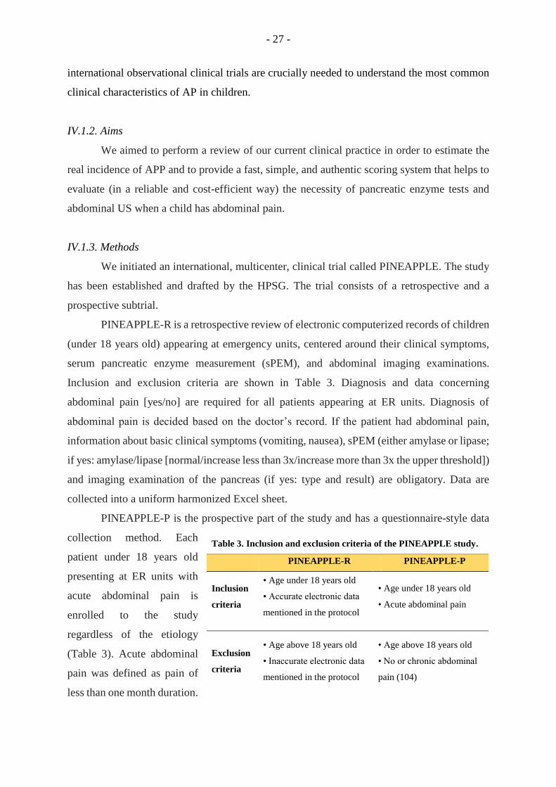
We initiated an international, multicenter, clinical trial called PINEAPPLE. The study
has been established and drafted by the HPSG. The trial consists of a retrospective and a
prospective subtrial.
PINEAPPLE-R is a retrospective review of electronic computerized records of children
(under 18 years old) appearing at emergency units, centered around their clinical symptoms,
serum pancreatic enzyme measurement (sPEM), and abdominal imaging examinations.
Inclusion and exclusion criteria are shown in Table 3. Diagnosis and data concerning
abdominal pain [yes/no] are required for all patients appearing at ER units. Diagnosis of
abdominal pain is decided based on the doctor’s record. If the patient had abdominal pain,
information about basic clinical symptoms (vomiting, nausea), sPEM (either amylase or lipase;
if yes: amylase/lipase [normal/increase less than 3x/increase more than 3x the upper threshold])
and imaging examination of the pancreas (if yes: type and result) are obligatory. Data are
collected into a uniform harmonized Excel sheet.
PINEAPPLE-P is the prospective part of the study and has a questionnaire-style data
collection method. Each
patient under 18 years old
presenting at ER units with
acute abdominal pain is
enrolled to the study
regardless of the etiology
(Table 3). Acute abdominal
pain was defined as pain of
less than one month duration.
Table 3. Inclusion and exclusion criteria of the PINEAPPLE study.
PINEAPPLE-R PINEAPPLE-P
Inclusion
criteria
• Age under 18 years old
• Accurate electronic data
mentioned in the protocol
• Age under 18 years old
• Acute abdominal pain
Exclusion
criteria
• Age above 18 years old
• Inaccurate electronic data
mentioned in the protocol
• Age above 18 years old
• No or chronic abdominal
pain (104)
- 28 -
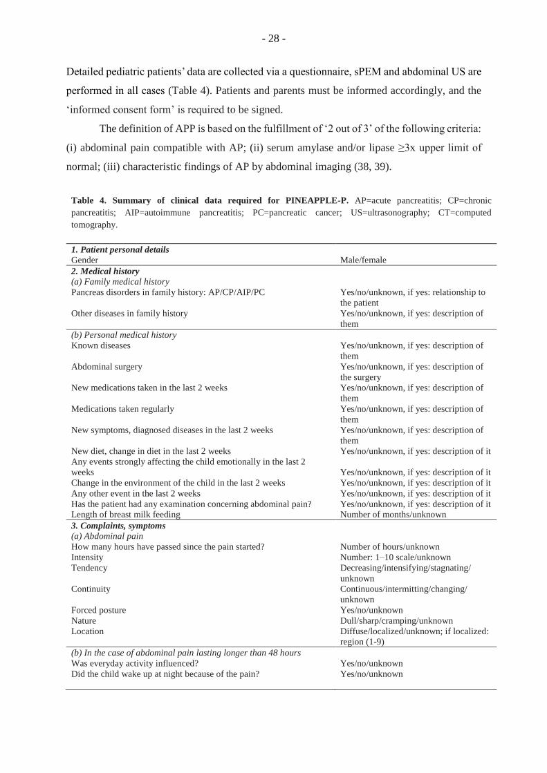
Detailed pediatric patients’ data are collected via a questionnaire, sPEM and abdominal US are
performed in all cases (Table 4). Patients and parents must be informed accordingly, and the
‘informed consent form’ is required to be signed.
The definition of APP is based on the fulfillment of ‘2 out of 3’ of the following criteria:
(i) abdominal pain compatible with AP; (ii) serum amylase and/or lipase ≥3x upper limit of
normal; (iii) characteristic findings of AP by abdominal imaging (38, 39).
Table 4. Summary of clinical data required for PINEAPPLE-P. AP=acute pancreatitis; CP=chronic
pancreatitis; AIP=autoimmune pancreatitis; PC=pancreatic cancer; US=ultrasonography; CT=computed
tomography.
1. Patient personal details
Gender
Male/female
2. Medical history
(a) Family medical history
Pancreas disorders in family history: AP/CP/AIP/PC
Other diseases in family history
Yes/no/unknown, if yes: relationship to
the patient
Yes/no/unknown, if yes: description of
them
(b) Personal medical history
Known diseases
Abdominal surgery
New medications taken in the last 2 weeks
Medications taken regularly
New symptoms, diagnosed diseases in the last 2 weeks
New diet, change in diet in the last 2 weeks
Any events strongly affecting the child emotionally in the last 2
weeks
Change in the environment of the child in the last 2 weeks
Any other event in the last 2 weeks
Has the patient had any examination concerning abdominal pain?
Length of breast milk feeding
Yes/no/unknown, if yes: description of
them
Yes/no/unknown, if yes: description of
the surgery
Yes/no/unknown, if yes: description of
them
Yes/no/unknown, if yes: description of
them
Yes/no/unknown, if yes: description of
them
Yes/no/unknown, if yes: description of it
Yes/no/unknown, if yes: description of it
Yes/no/unknown, if yes: description of it
Yes/no/unknown, if yes: description of it
Yes/no/unknown, if yes: description of it
Number of months/unknown
3. Complaints, symptoms
(a) Abdominal pain
How many hours have passed since the pain started?
Intensity
Tendency
Continuity
Forced posture
Nature
Location
Number of hours/unknown
Number: 1–10 scale/unknown
Decreasing/intensifying/stagnating/
unknown
Continuous/intermitting/changing/
unknown
Yes/no/unknown
Dull/sharp/cramping/unknown
Diffuse/localized/unknown; if localized:
region (1-9)
(b) In the case of abdominal pain lasting longer than 48 hours
Was everyday activity influenced?
Did the child wake up at night because of the pain?
Yes/no/unknown
Yes/no/unknown

- 29 -
Table 4. (continued)
In which part of the day did the pain appear mostly?
Was it related to eating?
Unrelated/after waking up/in the
morning/in the afternoon/in the
evening/at night/unknown
Yes/no/unknown; if yes: before/while/
after
(c) Other complaints
Nausea
Vomiting
Fever
Appetite
Weight loss
Jaundice
Stool
Yes/no/unknown
Yes/no/unknown; if yes: How many
times? Content of cast?
Yes/no/unknown; if yes: Since when?
Temperature? (C°)
Good/retained/bad/unknown
Yes/no/unknown, if yes: How much?
(kg), How long has it taken?
(weeks/months)
Yes/no/unknown
Normal/diarrhea/constipation/fatty/
putrid/undigested food/bloody/mucus/
unknown
4. Physical examination
Blood pressure
Heart rate
Body weight
Body height
Respiratory rate
Body temperature
Abdominal tenderness
Abdominal guarding
Jaundice
Bowel sounds
Number (mmHg)
Number (BPM)
Number (kg)
Number (m)
Number (/min)
Number (°C)
Yes/no, if yes: location of abdominal
tenderness
Yes/no
Yes/no
No/hypoactive/normal/hyperactive
5. Laboratory parameters
Amylase and/or lipase
Number (U/l)
6. Imaging examinations
Pancreas abnormalities suggesting AP
Pancreas abnormalities suggesting CP
Abdominal US
Abdominal CT scan
Yes/no
Yes/no
Yes/no, if yes: description
Yes/no, if yes: description
7. Diagnosis
8. Diagnosis – main group
9. Further step Admission/discharged/other
We aim to analyze patient data in different age groups. Association between each
collected parameter and AP will be determined. Statistical analysis will be carried out by data
mining methods. The applied methods will be determined based on the main characteristics of
the collected data, and the most suitable method – or method combination – will be chosen.
The following data mining methods are being contemplated: logistic regression, discriminant
analysis, random forest analysis, decision tree, and cluster analysis. ROC (receiver operating
characteristic) analysis will be performed to evaluate the predictive power of the classification
algorithm.

- 30 -
Four quality control points are established. First, the local clinical research assistant
must upload the data electronically and confirm that the data are the same as those in the hard
copy. Second, the local institutional principal investigator (who must have a medical doctoral
degree) must recheck the uploaded data and confirm their validity and accuracy. Third, the
central data administrator, who is based at HPSG headquarters, must control the accuracy, and
finally, the trial leader must go through the details. Patients with inadequate or insufficient data
will be excluded.
The protocols were introduced at our international meeting held in Szeged in
November 2014, which was attended by some of the best-established pediatric
pancreatologists. Around 100 clinicians – 60 Hungarians and 40 international investigators
from 9 different countries – attended. The trial was discussed and the suggested modifications
have been included. The study has been accepted by the scientific committee of the IAP, and
is therefore running under the auspices of HPSG and IAP. The PINEAPPLE trial has been
registered at the ISRCTN registry (ISRCTN35618458), a primary clinical trial registry
recognized by the World Health Organization (WHO) and the International Committee of
Medical Journal Editors (ICMJE) which accepts all clinical research studies, providing content
validation and curation as well as the unique identification number necessary for publication.
The study received the relevant ethical approval (No.: ad.52857-2/2014) issued by the
National Hungarian Ethical Authority (ETT TUKEB) in 2014. Completion of the ‘Letter of
intent’ form is mandatory for registering the participation of each institution. Study
management strictly follows the Ethical Guidelines for Observational Studies.
IV.1.4. Expected results
The PINEAPPLE trial is ongoing and expected to be finished by December 2020. The
PINEAPPLE-R study will aid understanding of our current clinical practice of APP in children
with abdominal pain in different countries and centers. The PINEAPPLE-P study will provide
the real incidence of APP and help to establish a fast, simple, and authentic scoring system to
evaluate the necessity of pancreatic enzyme tests and abdominal US when a child has
abdominal pain. As of the preparation of the thesis, 48,170 patient records have been enrolled
into PINEAPPLE-R, and 926 patients have been involved in PINEAPPLE-P.
- 31 -
IV.1.5. Discussion
The ‘2 out of 3’ criterion is used to diagnose AP both in adults and children (abdominal
pain, sPEM, and abdominal imaging) (39, 40, 42, 43, 49). Therefore, without measuring serum
pancreatic enzymes and/or performing transabdominal imaging, AP may remain undiagnosed.
According to previous pediatric studies in AP, abdominal pain is present in 66 to 95%
of the children with AP (57, 105-110); however, inconsistency and high variability exist
between the studies. Most of the trials investigating the characteristics of abdominal pain have
either low numbers or missing parameters causing inconsistencies between their data. Based
on the review of Bai et al. (41), abdominal pain was most commonly localized to the epigastric
region (62–89% of cases) (102, 105, 111) and was rarely associated with back pain (<10%) in
children with AP (57, 107). Radiation to the back was seen only in 1.6–5.6% (108, 110, 111)
of the cases. Diffuse abdominal pain was found in 12–20% of AP patients (105, 108, 110),
guarding in 29–37% (105, 107), whereas abdominal distension was reported in 21–46% (105-
107, 109, 110). Nausea or vomiting was noted in 40–80% of the AP cases (57, 102, 108-114).
Other symptoms might be fever, ascites, pleural effusion, and jaundice. Symptoms of infants
and toddlers are much more unspecific: abdominal pain was found in 43%, epigastric
tenderness in 57%, and nausea in 29% (102). In a study from Pittsburgh, 16% of the infants
and toddlers had abdominal distension and 40% had fever (115).
Based on a report published by Coffey et al., lipase, amylase, abdominal US, and
computed tomography (CT) were consistent with the diagnosis of AP only in 93%, 54%, 27%
and 67% of cases, respectively (116); however, the diagnostic yield for combinations of blood
and imaging tests was higher than any single test and blood tests alone (116).
In summary, diagnosing AP in children can be difficult and easily missed. Therefore, a
large, international prospective cohort is necessary to understand the complaints and symptoms
of AP in children. We have proposed an international observational clinical trial to collect a
critical mass of data from children with abdominal pain in order to develop an EBM guideline
concerning the necessity for obtaining serum pancreatic enzyme testing and abdominal US in
pediatric patients who present at the emergency room with abdominal pain.

- 32 -
IV.2. EARLY ENTERAL NUTRITION IN ACUTE PEDIATRIC PANCREATITIS
IV.2.1. Introduction
Common characteristics in both age groups are that no specific therapy is available to
treat AP, and the general supportive treatment at the early phase of the disease frequently
consists of volume resuscitation and NPO diet (38, 39, 42, 43, 117). Although there is clear
evidence in the literature that appropriate volume therapy is beneficial, the latter treatment is
questionable.
One of the main reasons
for the debate is that the
pathogenesis of the disease
clearly suggests the opposite.
Irrespective of the etiological
factors, mitochondrial damage
and energy depletion are the
leading intracellular responses
in the early phase of the disease
in the exocrine pancreas (11,
13, 91, 118). Bile acids (7-10),
fatty acids, and non-oxidative
ethanol metabolites (FAEEs)
(11-16) were shown to elevate the intracellular Ca2+ concentration, causing mitochondrial
damage and a resultant decrease of intracellular ATP concentration (Fig. 12). This leads to
inhibited fluid and bicarbonate secretion and dysfunction of the cystic fibrosis transmembrane
conductance regulator (CFTR) Cl¯ channel in the ductal cells with resultant secretory block
and intrapancreatic trypsinogen activation (11, 12, 119, 120). In addition, hypercatabolism
secondary to pancreatic and extrapancreatic inflammation further aggravates the energy deficit
(121). Consequently, restoration of ATP level both in acinar and ductal cells prevents (at least
in part) the toxic effect of the harmful causative factors noted above (91, 122, 123). These data
strongly suggest that early energy supply should be favorable for AP patients compared to nil
energy. Moreover, energy supply given by enteral nutrition in AP patients was shown to be
beneficial as a first-line treatment compared to TPN for several reasons (124, 125): (i) EEN
significantly decreases pathogenic bacteria in the stool and alteration of intestinal flora; (ii) gut
Fig. 12. Early events in acute pancreatitis. Bile acids, fatty acids or
non-oxidative ethanol metabolites (fatty acid ethyl esters, FAEEs)
induce calcium overload, causing mitochondrial damage and a
resultant decrease in intracellular adenosine triphosphate (ATP)
concentration both in acinar and ductal cells. This will lead to general
energy depletion in the pancreas.

- 33 -
plays an important role as a barrier in the immune system and EEN is able to optimize intestinal
permeability; (iii) this mucosal barrier integrity decreases the bacterial translocation from the
gut, therefore resultant bacteraemia and levels of serum endotoxins are reduced; (iv) EEN has
a favorable effect on immune dysregulation caused by SAP which can reduce the rate of
pancreatic infection, systemic inflammatory response syndrome (SIRS) and multiple organ
dysfunction syndrome as well as duration of ICU stay (37, 121, 126). Accordingly, enteral
nutrition has been shown to be beneficial regarding visual analogue pain score, LOH, local and
systemic infections, local complications, multi organ failure (MOF), need for surgical
interventions, and mortality (32-36, 124, 127-139). Additionally, enteral nutrition further
improves the outcome of AP if it is started within 48-72 hours (37, 39, 124, 132, 140, 141).
The type of enteral nutrition is administered based on disease severity (141, 142).
Furthermore, enteral nutrition has already been proven to be beneficial in other
inflammatory gastrointestinal diseases. The first-line recommendation to induce remission in
pediatric Crohn’s disease is exclusive enteral nutrition (143). Enteral nutrition could also be
effective in the maintenance of pediatric inflammatory bowel disease remission (144).
Regarding AP, three of the recent and most up-to-date guidelines for AP in adults have
shown the positive effect of early enteral tube feeding in moderately severe and SAP (39, 42,
43). Moreover, nasogastric tube feeding was shown to be as safe and as effective as nasojejunal
tube feeding in SAP (131, 132, 141, 145, 146). In the case of patients with predicted mild AP,
oral feeding is preferred as soon as possible (39, 42, 43, 147). However, no systematic review
is available concerning the role of EEN in children.
IV.2.2. Aims
We aimed to review the literature to analyze the effect of EEN versus NPO therapy on
the outcome of APP, and to aggregate the information in childhood onset AP, leading to a
higher statistical power and more robust point estimate than is possible from the individual
studies.
IV.2.3. Methods
The preferred reporting items for systematic review and meta-analysis protocol
(PRISMA-P) was followed (148). Our structured literature search was based on the
participants, intervention, comparison and outcomes (PICO) format [P: patients under the age
of twenty-one suffering from AP; I: EEN (per os/nasogastric- or nasojejunal tube started within

- 34 -
24-48 hours); C: NPO therapy (per os/nasogastric- or enteral tube started after 24-48 hours);
O: length of hospitalization, need for ICU, complications, necessity of antibiotics,
surgical/non-surgical interventions, and mortality].
In February 2016, a literature search was performed on the PubMed
(http://www.ncbi.nlm.nih.gov/pubmed) and EMBASE (https://www.embase.com) databases
using the following Medical Subject Headings and search terms: “pediatric” OR “paediatric”
AND “pancreatitis”. The search was limited to human studies, full-text publications with
abstracts in English with no time period, resulting in 632 articles altogether (PubMed: 131;
EMBASE: 501). The articles were checked separately. Meta-analyses, reviews, case reports
and articles on CP were excluded and duplicates were removed (Fig. 13). Potentially eligible
papers were selected, and finally five of them with relevant data on EEN or with NPO therapy
Fig. 13. Flow chart on the methods used in the literature search.
in APP in patients under 21 years old were included (Table 5). Details in the collected articles
were checked, and only articles where both EEN and NPO were presented separately were
used. Two articles met this criterion which contained three separate data pairs, where EEN was
- 35 -
compared to NPO (Fig. 14A). The following parameters were collected: LOH, need for
treatment at ICU, and development of SAP. Only one of the three investigated parameters
(LOH) contained a minimum of three items, which were analyzed statistically.
The meta-analytic
calculation was made with
Comprehensive
MetaAnalysis (V3)
software using the random
effects model (the
DerSimonian-Laird
method). We calculated a
weighted standard
difference in means and 95% CI. In the case of one study (Abu-El-Haija et al., 2016 (149)), we
converted the median and range values to means and standard deviation using the modified
Hozo’s formula by Wan et al., 2014 (153). For a visual inspection, we used a forest plot.
Fig. 14. Two articles containing three separate data pairs. A) Collected parameters. B) Forest plot analyses on
length of hospitalization (LOH). EEN=early enteral nutrition; NPO=nil per os; IVF=intravenous fluid; lo=low;
hi=high; ICU=intensive care unit; AP=acute pancreatitis; PO=per os.
Table 5. Studies included in the quantitative synthesis. EEN=early
enterals nutrition; NPO=nil per os; IVF=intravenous fluid; lo=low;
hi=high.
Ref. Data Groups n
Abu-El-Haija et al., 2016 (149) yes EEN 24
NPO 14
Flores-Calderón et al., 2009 (150) 18
Goh et al., 2003 (113) 12
Raizner et al., 2013 (151) 7
Szabo et al., 2015 (152)
yes EEN + IVF lo 55
NPO + IVF lo 20
yes EEN + IVF hi 96
NPO + IVF hi 30

- 36 -
IV.2.4. Results
Fig. 14A shows the parameters collected from the articles. It was only possible to
perform forest plot analyses on LOH (Fig. 14B). EEN significantly decreased LOH (SD=
0.806, p = 0.034) compared to the standard NPO diet in case of APP.
IV.2.5. Discussion
Several therapeutic recommendations are available in the literature on nutrition in AP.
The IAP/APA guideline suggests enteral tube feeding as the first-line therapy in patients with
predicted SAP, and oral feeding in the case of predicted mild AP (39). According to the
Japanese guideline, enteral nutrition can decrease the incidence of complications and elevate
the survival rate in the early phase of SAP (43). Recent meta-analysis of adult studies revealed
that EEN decreases mortality, rate of interventions, and the incidence of MOF in SAP.
Moreover, group analysis of 17 parameters including laboratory parameters (such as CRP and
WBC) and symptoms (such as pain or presence of SIRS) suggested that EEN also has merits
in mild AP (34).
Since the incidence of APP has risen in the past twenty years (3.6 and 13.2 per 100,000
children affected annually), we systematically reviewed the literature to understand whether
there is any beneficial effect of EEN versus NPO therapy in children (51, 52). We faced several
difficulties during our review: (i) APP is still underdiagnosed, thus decreasing the possibility
of performing clinical trials; (ii) the number of studies on the management of these patients is
very low, and there is still only a small number of studies focused on understanding the
characteristics of the childhood onset disease (154); (iii) studies have not focused on the early
management of the patients, therefore the groups were not separated; and finally, (iv) the
quality of the methods sections and data presentation in these articles is very low.
Consequently, in many cases it was impossible to obtain quality analyzable data from the
articles for proper broad-spectrum meta-analysis. By the end of the search, we identified five
articles containing relevant data on nutritional management during the early phase of APP.
Raizner et al. (151) published a retrospective analysis involving seven children with
necrotizing pancreatitis. All the children received a strict NPO diet for 1-11 days, five patients
received TPN for 8-18 days, and just one patient was treated with nasojejunal feeding for 7
days. All the children required intensive supportive treatment and a prolonged hospital stay
(with a mean of 20 days) because of the complications. Goh et al (113) included twelve patients

- 37 -
in their retrospective study. One patient needed a distal pancreatectomy, and eleven patients
recovered with conservative management. Three patients received TPN, and eight patients
were kept on bowel rest and nasogastric aspiration. Two patients had acute complications, and
two patients had recurrent AP. Flores-Calderon et al (150) studied eighteen patients with AP
caused by L-asparaginase due to acute lymphoblastic leukemia. All the patients were treated
with bowel resting for a mean of 22 days (range: from 3-66 days), fourteen of the patients
received TPN, and four had an elementary diet. Two of the patients required ICU admission,
and local complications developed in twelve patients. None of the patients died from
complications related to AP. On the contrary, data published by Wang et al. in 2020 confirmed
that enteral feeding is safe and tolerable in asparaginase-induced APP based on data from
twelve patients (155). Seven patients received nasojejunal tube feeding, and oral feeding was
started after 5-7 days in five patients. Pseudocyst and pancreatic duct stricture were found only
in two cases, and one patient required continuous renal replacement therapy among patients
with nasojejunal tube feeding without any death related to AP. Although the three included
studies mentioned above point out several disadvantages of the NPO diet, none of them could
be enrolled in our meta-analysis. Finally, it was possible to collect three sets of analyzable data
pairs where both NPO and EEN were present. Abu-El-Haija et al (149) conducted a prospective
study of 33 patients (38 admissions) suffering from mild AP, and retrospectively investigated
the relationship of nutrition with abdominal pain and LOH. EEN feeding meant per os feeding,
and NPO was identified as oral feeding not being allowed for 24 hours. Importantly, EEN, even
with high fat intake, did not cause elevation in pain in children, suggesting that EEN is a well
tolerable nutritional possibility in children. The fact that LOH was shorter in the EEN group
versus the NPO group points to EEN as a better way of treating APP. The most advanced study
was performed by Szabo et al (152), where several parameters were collected to understand
the effect of EEN on the course of APP. A total of 201 patients suffering from mild AP on
admission were enrolled retrospectively. They compared EEN versus NPO both with and
without aggressive fluid resuscitation. Fluid therapy was administered during the first 24 hours,
and the type of nutrition was determined during the first 48 hours. Besides the beneficial effects
of EEN on LOH, they also showed that EEN reduced the severity of the disease and the rate of
ICU transfer.
Although our aim was to perform a meta-analysis on several parameters to understand
the differences between EEN and NPO in childhood onset AP, we were only able to perform
the statistical analysis on LOH, which suggested that EEN is not only a safe method of nutrition

- 38 -
but also substantially decreases LOH, resulting in a better and less expensive treatment of APP
(152).
The first randomized control trial on early oral feeding in APP was published in 2020
by Ledder et al (156). A total of 33 patients were randomly assigned to initial fasting or an
immediate, unrestricted diet. There was no significant difference in the time elapsed until ready
for discharge, pain free status, serum amylase or lipase levels at discharge. Moreover, weight
was significantly higher in the early feeding group at follow-up. These data confirm our results,
that early oral feeding is not only a safe method of nutrition, but also has benefits.
IV.3. CONCLUSION
Based on our current knowledge, there are remarkable differences between childhood
and adult onset AP (41, 53, 54, 157). However, the majority of the current pediatric guidelines
are adopted from adult data (40, 45-47). Therefore, prospective observational and
interventional pediatric clinical trials would be necessary to understand the differences between
childhood and adult onset AP and to be able to provide appropriate patient care to children
suffering from AP. However, most of the pediatric cohorts are limited as a result of the low
incidence of AP and small sample size, which is particularly due to missing evidence-based
diagnostic guidelines and lower awareness of AP among pediatricians. Consequently, the
HPSG aimed to solve this unmet need and established the PINEAPPLE study to estimate the
real incidence of AP in children, and to create an evidence-based diagnostic guideline for APP.
Additionally, not only diagnostic, but also therapeutic guidelines for childhood onset AP are
based on adult data. EEN was proven to be beneficial for treating AP in adults compared to
NPO and TPN therapy. Therefore, we aimed to collect all the relevant data on EEN in APP
from the literature to achieve a higher level of evidence in childhood as well. Our meta-analysis
suggests that EEN should have priority in treating APP compared to NPO therapy, and
confirmed the necessity of further clinical trials in children.

- 39 -
V. SUMMARY AND NEW DISCOVERIES
Chapter III: Dose-dependent effect of hypertriglyceridemia on acute pancreatitis
1. Although we confirmed that biliary etiology is less common with HTG-AP, HTG-AP
was associated with biliary etiology in 5.4%, but with alcoholic etiology in 38.2%.
2. HTG was significantly and dose-dependently linked to younger age, male gender,
obesity and pre-existing DM in AP patients.
3. Amylase and lipase levels have shown a significant and dose-dependent reduction with
the elevation of TG in AP.
4. Our analysis has shown that local complications and organ failure were significantly
and dose-dependently increased by HTG in AP.
5. TG level above 11.3 mmol/l was associated with a significantly higher rate of
moderately severe AP and longer hospital stay, whereas TG level above 22.6 mmol/l
was significantly related to SAP as well.
6. Our data suggest that lipid-lowering therapy may be important clinically at a much
lower TG level in HTG-AP patients than we previously thought.
Chapter IV: Children are not small adults
The PINEAPPLE study will help to estimate the real incidence of AP in children and create
evidence-based diagnostic guidelines for APP.
Our meta-analysis: (i) proves that EEN shortens the LOH in the case of AP not only in adults,
but also in children; (ii) suggests that EEN is safe and should have priority in treating APP
compared to NPO therapy, and; (iii) confirms the necessity of further interventional clinical
trials in children.

- 40 -
VI. CONTRIBUTION
During my PhD work, we aimed to improve the clinical practice of AP in children and adults.
This has allowed me to learn the clinical methodologies of study design, observational clinical
trial, meta-analysis, and cohort analysis.
VI.1. Mosztbacher et al. Pancreatology, 2020.
I took part in the study design, data analysis and data interpretation, and I wrote the
article with the contributions of Péter Hegyi and Andrea Párniczky. Furthermore, figures were
created by myself and Lilla Hanák. I contributed to the patient involvement in the AP registry
operated by the HPSG.
VI.2. Mosztbacher et al. World J Gastroenterol, 2017.
I contributed to the study design and data interpretation, and I wrote the article with the
help of Péter Hegyi and Andrea Párniczky. Furthermore, figures were created by myself and
Nelli Farkas.
VI.3. Zsoldos et al. Digestion, 2016
As the principle investigator of the study, I am responsible for making presentations at
national and international conferences in order to involve as many centers as possible. In
addition, I organize and maintain relations with the enrolled centers and provide the
professional supervision of the study. I have enrolled 133 patients to the PINEAPPLE-P and 2
centers to the PINEAPPLE-R studies. I am responsible for data control and data analysis of
both subtrials.

- 41 -
VII. ACKNOWLEDGEMENT
First, I would like to thank my supervisor Péter Hegyi, for his support. He managed
my scientific studies and assisted my work with his advice and experience. I would also like to
express my thanks to Andrea Párniczky, who convinced me to join the HPSG and begin my
scientific work, and then supported me as a supervisor. Furthermore, I would like to thank
Miklós Sahin-Tóth and Jonas Rosendahl for the opportunity to widen my knowledge and do
basic research in the field of pancreas genetics.
I am also grateful to the interdisciplinary research unit led by Andrea Szentesi. My
PhD work would not have been possible without the work of administrators, patient
coordinators, local clinical investigators, and biobank leaders of the HPSG and the Institute
for Translational Medicine, University of Pécs. Furthermore, I would like to thank Nelli
Farkas and Lilla Hanák for their help in the statistical calculations.
My deepest gratefulness goes to my parents and my family who supported me during
my studies and research work. I would also like to thank my close friends who always assured
me that I would be able to manage and finish my PhD work.
- 42 -
VIII. REFERENCES
1. Chatila AT, Bilal M, Guturu P. Evaluation and management of acute pancreatitis.
World journal of clinical cases. 2019;7(9):1006.
2. Peery AF, Crockett SD, Murphy CC, Lund JL, Dellon ES, Williams JL, et al. Burden
and cost of gastrointestinal, liver, and pancreatic diseases in the United States: update 2018.
Gastroenterology. 2019;156(1):254-72. e11.
3. Yadav D, Lowenfels AB. The epidemiology of pancreatitis and pancreatic cancer.
Gastroenterology. 2013;144(6):1252-61.
4. Khatua B, El-Kurdi B, Singh VP. Obesity and pancreatitis. Current opinion in
gastroenterology. 2017;33(5):374-82.
5. Trikudanathan G, Wolbrink DR, van Santvoort HC, Mallery S, Freeman M, Besselink
MG. Current concepts in severe acute and necrotizing pancreatitis: an evidence-based
approach. Gastroenterology. 2019;156(7):1994-2007. e3.
6. Banks PA, Bollen TL, Dervenis C, Gooszen HG, Johnson CD, Sarr MG, et al.
Classification of acute pancreatitis—2012: revision of the Atlanta classification and definitions
by international consensus. Gut. 2013;62(1):102-11.
7. Hegyi P. Bile as a key aetiological factor of acute but not chronic pancreatitis: a possible
theory revealed. The Journal of physiology. 2016;594(21):6073.
8. Venglovecz V, Hegyi P, Rakonczay Z, Tiszlavicz L, Nardi A, Grunnet M, et al.
Pathophysiological relevance of apical large-conductance Ca2+-activated potassium channels
in pancreatic duct epithelial cells. Gut. 2011;60(3):361-9.
9. Venglovecz V, Rakonczay Z, Ozsvari B, Takacs T, Lonovics J, Varro A, et al. Effects
of bile acids on pancreatic ductal bicarbonate secretion in guinea pig. Gut. 2008;57(8):1102-
12.
10. Voronina SG, Gryshchenko OV, Gerasimenko OV, Green AK, Petersen OH, Tepikin
AV. Bile acids induce a cationic current, depolarizing pancreatic acinar cells and increasing
the intracellular Na+ concentration. Journal of Biological Chemistry. 2005;280(3):1764-70.
11. Hegyi P, Petersen OH. The exocrine pancreas: the acinar-ductal tango in physiology
and pathophysiology. Reviews of Physiology, Biochemistry and Pharmacology, Vol 165:
Springer; 2013. p. 1-30.
- 43 -
12. Maléth J, Balázs A, Pallagi P, Balla Z, Kui B, Katona M, et al. Alcohol disrupts levels
and function of the cystic fibrosis transmembrane conductance regulator to promote
development of pancreatitis. Gastroenterology. 2015;148(2):427-39. e16.
13. Maléth J, Hegyi P, Rakonczay Jr Z, Venglovecz V. Breakdown of bioenergetics evoked
by mitochondrial damage in acute pancreatitis: Mechanisms and consequences. Pancreatology.
2015;15(4):S18-S22.
14. Criddle DN. The role of fat and alcohol in acute pancreatitis: a dangerous liaison.
Pancreatology. 2015;15(4):S6-S12.
15. Hegyi P, Rakonczay Jr Z. The role of pancreatic ducts in the pathogenesis of acute
pancreatitis. Pancreatology. 2015;15(4):S13-S7.
16. Maléth J, Hegyi P. Calcium signaling in pancreatic ductal epithelial cells: an old friend
and a nasty enemy. Cell calcium. 2014;55(6):337-45.
17. Navina S, Acharya C, DeLany JP, Orlichenko LS, Baty CJ, Shiva SS, et al. Lipotoxicity
causes multisystem organ failure and exacerbates acute pancreatitis in obesity. Science
translational medicine. 2011;3(107):107ra10-ra10.
18. Yadav D, Pitchumoni C. Issues in hyperlipidemic pancreatitis. Journal of clinical
gastroenterology. 2003;36(1):54-62.
19. Baranyai T, Terzin V, Vajda Á, Wittmann T, Czakó L. Hypertriglyceridemia causes
more severe course of acute pancreatitis. Clinical Lipidology. 2012;7(6):731-6.
20. Kiss L, Fűr G, Mátrai P, Hegyi P, Ivány E, Cazacu IM, et al. The effect of serum
triglyceride concentration on the outcome of acute pancreatitis: systematic review and meta-
analysis. Scientific reports. 2018;8(1):14096.
21. Nawaz H, Koutroumpakis E, Easler J, Slivka A, Whitcomb DC, Singh VP, et al.
Elevated serum triglycerides are independently associated with persistent organ failure in acute
pancreatitis. The American journal of gastroenterology. 2015;110(10):1497.
22. Tai W-P, Lin X-C, Liu H, Wang C-H, Wu J, Zhang N-W, et al. A retrospective research
of the characteristic of hypertriglyceridemic pancreatitis in Beijing, China. Gastroenterology
research and practice. 2016;2016.
23. Wang S-H, Chou Y-C, Shangkuan W-C, Wei K-Y, Pan Y-H, Lin H-C. Relationship
between plasma triglyceride level and severity of hypertriglyceridemic pancreatitis. PloS one.
2016;11(10):e0163984.
24. Zhang R, Deng L, Jin T, Zhu P, Shi N, Jiang K, et al. Hypertriglyceridaemia-associated
acute pancreatitis: diagnosis and impact on severity. HPB. 2019.

- 44 -
25. Zheng Y, Zhou Z, Li H, Li J, Li A, Ma B, et al. A multicenter study on etiology of acute
pancreatitis in Beijing during 5 years. Pancreas. 2015;44(3):409-14.
26. Zhu Y, Pan X, Zeng H, He W, Xia L, Liu P, et al. A study on the etiology, severity, and
mortality of 3260 patients with acute pancreatitis according to the revised Atlanta classification
in Jiangxi, China over an 8-year period. Pancreas. 2017;46(4):504-9.
27. Yang AL, McNabb-Baltar J. Hypertriglyceridemia and acute pancreatitis.
Pancreatology. 2020.
28. Pan Y, Li Y, Gao L, Tong Z, Ye B, Liu S, et al. Development of a novel model of
hypertriglyceridemic acute pancreatitis in mice. Scientific reports. 2017;7:40799.
29. Goyal H, Smith B, Bayer C, Rutherford C, Shelnut D. Differences in severity and
outcomes between hypertriglyceridemia and alcohol-induced pancreatitis. North American
journal of medical sciences. 2016;8(2):82.
30. Navarro S, Cubiella J, Feu F, Zambon D, Fernandez-Cruz L, Ros E.
Hypertriglyceridemic acute pancreatitis. Is its clinical course different from lithiasic acute
pancreatitis? Medicina clinica. 2004;123(15):567-70.
31. Berger Z, Mancilla C, Tobar E, Morales MP, Baró M, Carrasco M, et al. Acute
pancreatitis in Chile: A multicenter study on epidemology, etiology and clinical outcome.
Retrospective analysis of clinical files. Pancreatology. 2020.
32. Al‐Omran M, AlBalawi ZH, Tashkandi MF, Al‐Ansary LA. Enteral versus parenteral
nutrition for acute pancreatitis. Cochrane database of systematic reviews. 2010(1).
33. Petrov MS, van Santvoort HC, Besselink MG, van der Heijden GJ, Windsor JA,
Gooszen HG. Enteral nutrition and the risk of mortality and infectious complications in patients
with severe acute pancreatitis: a meta-analysis of randomized trials. Archives of Surgery.
2008;143(11):1111-7.
34. Marta K, Farkas N, Hegyi P. Meta-analysis of early nutrition: the benefits of enteral
feeding compared to a nil per os diet not only in severe, but also in mild and moderate acute
pancreatitis. Pancreatology. 2017;17(4):S18.
35. Abou-Assi S, Craig K, O’Keefe SJ. Hypocaloric jejunal feeding is better than total
parenteral nutrition in acute pancreatitis: results of a randomized comparative study. The
American journal of gastroenterology. 2002;97(9):2255-62.
36. Petrov MS, McIlroy K, Grayson L, Phillips AR, Windsor JA. Early nasogastric tube
feeding versus nil per os in mild to moderate acute pancreatitis: a randomized controlled trial.
Clinical nutrition. 2013;32(5):697-703.

- 45 -
37. Sun J-K, Mu X-W, Li W-Q, Tong Z-H, Li J, Zheng S-Y. Effects of early enteral
nutrition on immune function of severe acute pancreatitis patients. World journal of
gastroenterology: WJG. 2013;19(6):917.
38. Hritz I, Czakó L, Dubravcsik Z, Farkas G, Kelemen D, Lásztity N, et al. Acute
pancreatitis. Evidence based management guidelines of the Hungarian Pancreatic Study Group.
Orvosi hetilap. 2015;156(7):244-61.
39. IAP WG, Guidelines AAP. IAP/APA evidence-based guidelines for the management
of acute pancreatitis. Pancreatology. 2013;13(4):e1-e15.
40. Párniczky A, Abu-El-Haija M, Husain S, Lowe M, Oracz G, Sahin-Tóth M, et al.
EPC/HPSG evidence-based guidelines for the management of pediatric pancreatitis.
Pancreatology. 2018;18(2):146-60.
41. Bai HX, Lowe ME, Husain SZ. What have we learned about acute pancreatitis in
children? Journal of pediatric gastroenterology and nutrition. 2011;52(3):262.
42. Tenner S, Baillie J, DeWitt J, Vege SS. American College of Gastroenterology
guideline: management of acute pancreatitis. American Journal of Gastroenterology.
2013;108(9):1400-15.
43. Yokoe M, Takada T, Mayumi T, Yoshida M, Isaji S, Wada K, et al. Japanese guidelines
for the management of acute pancreatitis: Japanese Guidelines 2015. Journal of Hepato‐
Biliary‐Pancreatic Sciences. 2015;22(6):405-32.
44. Abu-El-Haija M, Uc A, Werlin SL, Freeman AJ, Georgieva M, Jojkić–Pavkov D, et al.
Nutritional considerations in pediatric pancreatitis: A position paper from the NASPGHAN
pancreas committee and ESPGHAN cystic fibrosis/pancreas working group. Journal of
pediatric gastroenterology and nutrition. 2018;67(1):131.
45. Abu-El-Haija M, Kumar S, Quiros JA, Balakrishnan K, Barth B, Bitton S, et al. The
management of acute pancreatitis in the pediatric population: a clinical report from the
NASPGHAN Pancreas Committee. Journal of pediatric gastroenterology and nutrition.
2018;66(1):159.
46. Theodoridis X, Grammatikopoulou MG, Petalidou A, Stamouli E-M, Fotiadou I,
Gkiouras K, et al. Nutrition Interventions in Pediatric Pancreatitis: Guidelines We Can Trust.
Journal of Pediatric Gastroenterology and Nutrition. 2019;69(1):120-5.
47. Wall JM. Clinical Guideline Highlights for the Hospitalist: Management of Acute
Pancreatitis in the Pediatric Population. Journal of hospital medicine. 2019;14:E1-E2.

- 46 -
48. Sellers ZM, Dike C, Zhang K-Y, Giefer MJ, Uc A, Abu-El-Haija M. A Unified
Treatment Algorithm and Admission Order Set for Pediatric Acute Pancreatitis. Journal of
pediatric gastroenterology and nutrition. 2019;68(6):e109-e11.
49. Morinville VD, Husain SZ, Bai H, Barth B, Alhosh R, Durie PR, et al. Definitions of
pediatric pancreatitis and survey of current clinical practices: report from INSPPIRE
(International Study Group of Pediatric Pancreatitis: In Search for a Cure). Journal of pediatric
gastroenterology and nutrition. 2012;55(3):261.
50. Abu-El-Haija M, Kumar S, Szabo F, Werlin S, Conwell D, Banks P, et al. Classification
of acute pancreatitis in the pediatric population: clinical report from the NASPGHAN pancreas
committee. Journal of pediatric gastroenterology and nutrition. 2017;64(6):984-90.
51. Morinville VD, Barmada MM, Lowe ME. Increasing incidence of acute pancreatitis at
an American pediatric tertiary care center: is greater awareness among physicians responsible?
Pancreas. 2010;39(1):5-8.
52. Nydegger A, Heine RG, Ranuh R, Gegati‐Levy R, Crameri J, Oliver MR. Changing
incidence of acute pancreatitis: 10‐year experience at the Royal Children's Hospital,
Melbourne. Journal of gastroenterology and hepatology. 2007;22(8):1313-6.
53. Meyer A, Coffey MJ, Oliver MR, Ooi CY. Contrasts and comparisons between
childhood and adult onset acute pancreatitis. Pancreatology. 2013;13(4):429-35.
54. Uc A, Husain SZ. Pancreatitis in children. Gastroenterology. 2019;156(7):1969-78.
55. Majbar AA, Cusick E, Johnson P, Lynn RM, Hunt LP, Shield JP. Incidence and clinical
associations of childhood acute pancreatitis. Pediatrics. 2016;138(3).
56. Pohl JF, Uc A. Pediatric pancreatitis. Current opinion in gastroenterology.
2015;31(5):380.
57. Werlin SL, Kugathasan S, Frautschy BC. Pancreatitis in children. Journal of pediatric
gastroenterology and nutrition. 2003;37(5):591-5.
58. Keim V. Role of genetic disorders in acute recurrent pancreatitis. World journal of
gastroenterology: WJG. 2008;14(7):1011.
59. Whitcomb D. Value of genetic testing in the management of pancreatitis. Gut.
2004;53(11):1710-7.
60. Kumar S, Ooi CY, Werlin S, Abu-El-Haija M, Barth B, Bellin MD, et al. Risk factors
associated with pediatric acute recurrent and chronic pancreatitis: lessons from INSPPIRE.
JAMA pediatrics. 2016;170(6):562-9.

- 47 -
61. Liu QY, Abu-El-Haija M, Husain SZ, Barth B, Bellin M, Fishman DS, et al. Risk
Factors for Rapid Progression From Acute Recurrent to Chronic Pancreatitis in Children:
Report From INSPPIRE. Journal of pediatric gastroenterology and nutrition. 2019;69(2):206-
11.
62. Abu-El-Haija M, Lowe M, Barth B, Bellin MD, Fishman DS, Freedman S, et al.
Pediatric chronic pancreatitis without prior acute or acute recurrent pancreatitis: A report from
the INSPPIRE consortium. Pancreatology: official journal of the International Association of
Pancreatology (IAP)[et al]. 2020;20(4):781-4.
63. Singh VK, Yadav D, Garg PK. Diagnosis and Management of Chronic Pancreatitis: A
Review. Jama. 2019;322(24):2422-34.
64. Lucidi V, Alghisi F, Dall'Oglio L, D'Apice MR, Monti L, De Angelis P, et al. The
etiology of acute recurrent pancreatitis in children: a challenge for pediatricians. Pancreas.
2011;40(4):517-21.
65. Abu-El-Haija M, Valencia CA, Hornung L, Youssef N, Thompson T, Barasa NW, et
al. Genetic variants in acute, acute recurrent and chronic pancreatitis affect the progression of
disease in children. Pancreatology. 2019;19(4):535-40.
66. Zsoldos F, Párniczky A, Mosztbacher D, Tóth A, Lásztity N, Hegyi P. Pain in the early
phase of pediatric pancreatitis (PINEAPPLE Trial): pre-study protocol of a multinational
prospective clinical trial. Digestion. 2016;93(2):121-6.
67. Mosztbacher D, Farkas N, Solymár M, Pár G, Bajor J, Szűcs Á, et al. Restoration of
energy level in the early phase of acute pediatric pancreatitis. World journal of
gastroenterology. 2017;23(6):957.
68. Zádori N, Gede N, Antal J, Szentesi A, Alizadeh H, Vincze Á, et al. EarLy elimination
of fatty acids iN hypertriglyceridemia-induced acuTe pancreatitis (ELEFANT trial): Protocol
of an open-label, multicenter, adaptive randomized clinical trial. Pancreatology. 2019.
69. Mosztbacher D, Hanák L, Farkas N, Szentesi A, Mikó A, Bajor J, et al.
Hypertriglyceridemia-induced acute pancreatitis: A prospective, multicenter, international
cohort analysis of 716 acute pancreatitis cases. Pancreatology. 2020.
70. Ford ES, Li C, Zhao G, Pearson WS, Mokdad AH. Hypertriglyceridemia and its
pharmacologic treatment among US adults. Archives of Internal Medicine. 2009;169(6):572-
8.
71. Laufs U, Parhofer KG, Ginsberg HN, Hegele RA. Clinical review on triglycerides.
European heart journal. 2019.

- 48 -
72. Scherer J, Singh V, Pitchumoni C, Yadav D. Issues in hypertriglyceridemic
pancreatitis-an update. Journal of clinical gastroenterology. 2014;48(3):195.
73. Berglund L, Brunzell JD, Goldberg AC, Goldberg IJ, Sacks F, Murad MH, et al.
Evaluation and treatment of hypertriglyceridemia: an Endocrine Society clinical practice
guideline. The Journal of Clinical Endocrinology & Metabolism. 2012;97(9):2969-89.
74. Párniczky A, Kui B, Szentesi A, Balázs A, Szűcs Á, Mosztbacher D, et al. Prospective,
multicentre, nationwide clinical data from 600 cases of acute pancreatitis. PLoS One.
2016;11(10):e0165309.
75. Roberts S, Akbari A, Thorne K, Atkinson M, Evans P. The incidence of acute
pancreatitis: impact of social deprivation, alcohol consumption, seasonal and demographic
factors. Alimentary pharmacology & therapeutics. 2013;38(5):539-48.
76. Czakó L, Szabolcs A, Vajda Á, Csáti S, Venglovecz V, Rakonczay Jr Z, et al.
Hyperlipidemia induced by a cholesterol-rich diet aggravates necrotizing pancreatitis in rats.
European journal of pharmacology. 2007;572(1):74-81.
77. Wang Q, Wang G, Qiu Z, He X, Liu C. Elevated Serum Triglycerides in the Prognostic
Assessment of Acute Pancreatitis. Journal of clinical gastroenterology. 2017;51(7):586-93.
78. Yang N, Li B, Pan Y, Tu J, Liu G, Lu G, et al. Hypertriglyceridaemia delays pancreatic
regeneration after acute pancreatitis in mice and patients. Gut. 2019;68(2):378-80.
79. Padmanabhan A, Connelly‐Smith L, Aqui N, Balogun RA, Klingel R, Meyer E, et al.
Guidelines on the use of therapeutic apheresis in clinical practice–evidence‐based approach
from the writing committee of the American society for apheresis: the eighth special issue.
Journal of clinical apheresis. 2019;34(3):171-354.
80. Charlson ME, Pompei P, Ales KL, MacKenzie CR. A new method of classifying
prognostic comorbidity in longitudinal studies: development and validation. Journal of chronic
diseases. 1987;40(5):373-83.
81. Wilsnack RW, Wilsnack SC, Kristjanson AF, Vogeltanz‐Holm ND, Gmel G. Gender
and alcohol consumption: patterns from the multinational GENACIS project. Addiction.
2009;104(9):1487-500.
82. Van Geenen EJ, Van der Peet DL, Bhagirath P, Mulder CJ, Bruno MJ. Etiology and
diagnosis of acute biliary pancreatitis. Nature Reviews Gastroenterology & Hepatology.
2010;7(9):495.
83. Yourno J, Henry JB. Rapid amylase and lipase determinations by nephelometry.
American journal of clinical pathology. 1978;70(1):56-63.

- 49 -
84. Fallat RW, Vester JW, Glueck CJ. Suppression of amylase activity by
hypertriglyceridemia. Jama. 1973;225(11):1331-4.
85. Singh A, Shrestha M, Anand C. Acute pancreatitis with normal amylase and lipase—
an ED dilemma. The American journal of emergency medicine. 2016;34(5):940. e5-. e7.
86. Sotello D, Rivas AM, Nugent KM, editors. Newly diagnosed acromegaly presenting
with hypertriglyceridemic pancreatitis with normal amylase and lipase levels. Baylor
University Medical Center Proceedings; 2014: Taylor & Francis.
87. Pascual I, Sanahuja A, García N, Vázquez P, Moreno O, Tosca J, et al. Association of
elevated serum triglyceride levels with a more severe course of acute pancreatitis: Cohort
analysis of 1457 patients. Pancreatology. 2019;19(5):623-9.
88. Szentesi A, Párniczky A, Vincze A, Bajor J, Gódi S, Sarlos P, et al. Multiple hits in
acute pancreatitis: components of metabolic syndrome synergize each other’s deteriorating
effects. Frontiers in Physiology. 2019;10:1202.
89. Lu Z, Li M, Guo F, Zhang G, Song S, Liu N, et al. Timely Reduction of Triglyceride
Levels Is Associated With Decreased Persistent Organ Failure in Hypertriglyceridemic
Pancreatitis. Pancreas. 2020;49(1):105-10.
90. Yang F, Wang Y, Sternfeld L, Rodriguez J, Ross C, Hayden M, et al. The role of free
fatty acids, pancreatic lipase and Ca2+ signalling in injury of isolated acinar cells and
pancreatitis model in lipoprotein lipase‐deficient mice. Acta physiologica. 2009;195(1):13-28.
91. Maléth J, Hegyi P. Ca2+ toxicity and mitochondrial damage in acute pancreatitis:
translational overview. Philosophical Transactions of the Royal Society B: Biological
Sciences. 2016;371(1700):20150425.
92. Kimura W, Mössner J. Role of hypertriglyceridemia in the pathogenesis of
experimental acute pancreatitis in rats. International Journal of Gastrointestinal Cancer.
1996;20(3):177-84.
93. Pothoulakis I, Paragomi P, Archibugi L, Tuft M, Talukdar R, Kochhar R, et al. Clinical
features of hypertriglyceridemia-induced acute pancreatitis in an international, multicenter,
prospective cohort (APPRENTICE consortium). Pancreatology. 2020.
94. Balachandra S, Virlos I, King N, Siriwardana H, France M, Siriwardena A.
Hyperlipidaemia and outcome in acute pancreatitis. International journal of clinical practice.
2006;60(2):156-9.

- 50 -
95. Deng L-H, Xue P, Xia Q, Yang X-N, Wan M-H. Effect of admission
hypertriglyceridemia on the episodes of severe acute pancreatitis. World journal of
gastroenterology: WJG. 2008;14(28):4558.
96. Lowe ME, Greer JB. Pancreatitis in children and adolescents. Current gastroenterology
reports. 2008;10(2):128.
97. Joergensen M, Brusgaard K, Crüger DG, Gerdes A-M, de Muckadell OBS. Incidence,
prevalence, etiology, and prognosis of first-time chronic pancreatitis in young patients: a
nationwide cohort study. Digestive diseases and sciences. 2010;55(10):2988-98.
98. Satoh K, Shimosegawa T, Masamune A, Hirota M, Kikuta K, Kihara Y, et al.
Nationwide epidemiological survey of acute pancreatitis in Japan. Pancreas. 2011;40(4):503-
7.
99. Yadav D, Lowenfels AB. Trends in the epidemiology of the first attack of acute
pancreatitis: a systematic review. Pancreas. 2006;33(4):323-30.
100. Lopez MJ. The changing incidence of acute pancreatitis in children: a single-institution
perspective. The Journal of pediatrics. 2002;140(5):622-4.
101. Dhroove G, Chogle A, Saps M. A million-dollar work-up for abdominal pain: is it worth
it? Journal of pediatric gastroenterology and nutrition. 2010;51(5):579-83.
102. Park AJ, Latif SU, Ahmad MU, Bultron G, Orabi AI, Bhandari V, et al. A comparison
of presentation and management trends in acute pancreatitis between infants/toddlers and older
children. Journal of pediatric gastroenterology and nutrition. 2010;51(2):167.
103. Abu-El-Haija M, Palermo JJ, Fei L, Lin TK. Variability in pancreatitis care in
pediatrics: a single institution’s survey report. Pancreas. 2016;45(1):40-5.
104. Di Lorenzo C, Colletti RB, Lehmann HP, Boyle JT, Gerson WT, Hyams JS, et al.
Chronic abdominal pain in children: a clinical report of the American Academy of Pediatrics
and the North American Society for Pediatric Gastroenterology, Hepatology and Nutrition:
American Academy of Pediatrics Subcommittee on Chronic Abdominal Pain and NASPGHAN
Committee on Abdominal Pain. Journal of pediatric gastroenterology and nutrition.
2005;40(3):245-8.
105. Berney T, Belli D, Bugmann P, Beghetti M, Morel P, LeCoultre C. Influence of severe
underlying pathology and hypovolemic shock on the development of acute pancreatitis in
children. Journal of pediatric surgery. 1996;31(9):1256-61.
106. Yeung C-y, Lee H-c, Huang F-y, Ho M-y, Kao H-a, Liang D-c, et al. Pancreatitis in
children—experience with 43 cases. European journal of pediatrics. 1996;155(6):458-63.

- 51 -
107. Haddock G, Coupar G, Youngson GG, MacKinlay GA, Raine PA. Acute pancreatitis
in children: a 15-year review. Journal of pediatric surgery. 1994;29(6):719-22.
108. Jordan SC, Ament ME. Pancreatitis in children and adolescents. The Journal of
pediatrics. 1977;91(2):211-6.
109. Weizman Z, Durie P. Acute pancreatitis in childhood. The Journal of pediatrics.
1988;113(1):24-9.
110. Ziegler DW, Long JA, Philippart AI, Klein MD. Pancreatitis in childhood. Experience
with 49 patients. Annals of surgery. 1988;207(3):257.
111. Tiao M-M, Chuang J-H, Ko S-F, Kuo H-W, Liang C-D, Chen C-L. Pancreatitis in
children: clinical analysis of 61 cases in southern Taiwan. Chang Gung medical journal.
2002;25(3):162-8.
112. Chen C-F, Kong M-S, Lai M-W, Wang C-J. Acute pancreatitis in children: 10-year
experience in a medical center. Acta paediatrica Taiwanica= Taiwan er ke yi xue hui za zhi.
2006;47(4):192-6.
113. Goh S, Chui C, Jacobsen A. Childhood acute pancreatitis in a children's hospital.
Singapore medical journal. 2003;44(9):453-6.
114. Sánchez‐Ramírez CA, Larrosa‐Haro A, Flores‐Martínez S, Sánchez‐Corona J, Villa‐
Gómez A, Macías‐Rosales R. Acute and recurrent pancreatitis in children: etiological factors.
Acta Paediatrica. 2007;96(4):534-7.
115. Kandula L, Lowe ME. Etiology and outcome of acute pancreatitis in infants and
toddlers. The Journal of pediatrics. 2008;152(1):106-10. e1.
116. Coffey MJ, Nightingale S, Ooi CY. Diagnosing acute pancreatitis in children: what is
the diagnostic yield and concordance for serum pancreatic enzymes and imaging within 96 h
of presentation? Pancreatology. 2014;14(4):251-6.
117. Mirtallo JM, Forbes A, McClave SA, Jensen GL, Waitzberg DL, Davies AR, et al.
International consensus guidelines for nutrition therapy in pancreatitis. Journal of Parenteral
and Enteral Nutrition. 2012;36(3):284-91.
118. Hegyi P, Pandol S, Venglovecz V, Rakonczay Z. The acinar-ductal tango in the
pathogenesis of acute pancreatitis. Gut. 2011;60(4):544-52.
119. Hegyi P, Wilschanski M, Muallem S, Lukacs GL, Sahin-Tóth M, Uc A, et al. CFTR: a
new horizon in the pathomechanism and treatment of pancreatitis. Reviews of Physiology,
Biochemistry and Pharmacology Vol 170: Springer; 2016. p. 37-66.
- 52 -
120. Pallagi P, Venglovecz V, Rakonczay Jr Z, Borka K, Korompay A, Ózsvári B, et al.
Trypsin reduces pancreatic ductal bicarbonate secretion by inhibiting CFTR Cl− channels and
luminal anion exchangers. Gastroenterology. 2011;141(6):2228-39. e6.
121. Capurso G, Zerboni G, Signoretti M, Valente R, Stigliano S, Piciucchi M, et al. Role
of the gut barrier in acute pancreatitis. Journal of clinical gastroenterology. 2012;46:S46-S51.
122. Criddle DN, Murphy J, Fistetto G, Barrow S, Tepikin AV, Neoptolemos JP, et al. Fatty
acid ethyl esters cause pancreatic calcium toxicity via inositol trisphosphate receptors and loss
of ATP synthesis. Gastroenterology. 2006;130(3):781-93.
123. Judák L, Hegyi P, Rakonczay Z, Maléth J, Gray MA, Venglovecz V. Ethanol and its
non-oxidative metabolites profoundly inhibit CFTR function in pancreatic epithelial cells
which is prevented by ATP supplementation. Pflügers Archiv-European Journal of Physiology.
2014;466(3):549-62.
124. Grant JP. Nutritional support in acute and chronic pancreatitis. Surgical Clinics.
2011;91(4):805-20.
125. McClave SA, Heyland DK. The physiologic response and associated clinical benefits
from provision of early enteral nutrition. Nutrition in Clinical Practice. 2009;24(3):305-15.
126. Flint R, Windsor J. The role of the intestine in the pathophysiology and management
of severe acute pancreatitis. Hpb. 2003;5(2):69-85.
127. Eckerwall GE, Tingstedt BB, Bergenzaun PE, Andersson RG. Immediate oral feeding
in patients with mild acute pancreatitis is safe and may accelerate recovery—a randomized
clinical study. Clinical nutrition. 2007;26(6):758-63.
128. Kalfarentzos F, Kehagias J, Mead N, Kokkinis K, Gogos C. Enteral nutrition is superior
to parenteral nutrition in severe acute pancreatitis: results of a randomized prospective trial.
British Journal of Surgery. 1997;84(12):1665-9.
129. Li J, Xue G-J, Liu Y-L, Javed MA, Zhao X-L, Wan M-H, et al. Early oral refeeding
wisdom in patients with mild acute pancreatitis. Pancreas. 2013;42(1):88-91.
130. Petrov MS, Whelan K. Comparison of complications attributable to enteral and
parenteral nutrition in predicted severe acute pancreatitis: a systematic review and meta-
analysis. British journal of nutrition. 2010;103(9):1287-95.
131. Zhu Y, Yin H, Zhang R, Ye X, Wei J. Nasogastric nutrition versus nasojejunal nutrition
in patients with severe acute pancreatitis: a meta-analysis of randomized controlled trials.
Gastroenterology research and practice. 2016;2016.

- 53 -
132. Lodewijkx PJ, Besselink MG, Witteman BJ, Schepers NJ, Gooszen HG, van Santvoort
HC, et al. Nutrition in acute pancreatitis: a critical review. Expert review of gastroenterology
& hepatology. 2016;10(5):571-80.
133. Petrov M, Pylypchuk R, Emelyanov N. Systematic review: nutritional support in acute
pancreatitis. Alimentary pharmacology & therapeutics. 2008;28(6):704-12.
134. Oláah A, Pardavi G, Beláagyi T, Nagy A, Issekutz Á, Mohamed GE. Early nasojejunal
feeding in acute pancreatitis is associated with a lower complication rate. Nutrition.
2002;18(3):259-62.
135. Petrov MS, Kukosh MV, Emelyanov NV. A randomized controlled trial of enteral
versus parenteral feeding in patients with predicted severe acute pancreatitis shows a
significant reduction in mortality and in infected pancreatic complications with total enteral
nutrition. Digestive surgery. 2006;23(5-6):336-45.
136. Windsor A, Kanwar S, Li A, Barnes E, Guthrie J, Spark J, et al. Compared with
parenteral nutrition, enteral feeding attenuates the acute phase response and improves disease
severity in acute pancreatitis. Gut. 1998;42(3):431-5.
137. Yi F, Ge L, Zhao J, Lei Y, Zhou F, Chen Z, et al. Meta-analysis: total parenteral
nutrition versus total enteral nutrition in predicted severe acute pancreatitis. Internal medicine.
2012;51(6):523-30.
138. Louie BE, Noseworthy T, Hailey D, Gramlich LM, Jacobs P, Warnock GL. 2004
MacLean–Mueller Prize Enteral or parenteral nutrition for severe pancreatitis: a randomized
controlled trial and health technology assessment. Canadian journal of surgery.
2005;48(4):298.
139. Wu X-M, Ji K-Q, Wang H-Y, Li G-F, Zang B, Chen W-M. Total enteral nutrition in
prevention of pancreatic necrotic infection in severe acute pancreatitis. Pancreas.
2010;39(2):248-51.
140. Petrov MS, Pylypchuk RD, Uchugina AF. A systematic review on the timing of
artificial nutrition in acute pancreatitis. British Journal of Nutrition. 2008;101(6):787-93.
141. Marik PE. What is the best way to feed patients with pancreatitis? Current opinion in
critical care. 2009;15(2):131-8.
142. Murphy AE, Codner PA. Acute Pancreatitis: Exploring Nutrition Implications.
Nutrition in Clinical Practice. 2020.

- 54 -
143. Ruemmele F, Veres G, Kolho K-L, Griffiths A, Levine A, Escher J, et al. Consensus
guidelines of ECCO/ESPGHAN on the medical management of pediatric Crohn's disease.
Journal of Crohn's and Colitis. 2014;8(10):1179-207.
144. Penagini F, Dilillo D, Borsani B, Cococcioni L, Galli E, Bedogni G, et al. Nutrition in
pediatric inflammatory bowel disease: from etiology to treatment. A systematic review.
Nutrients. 2016;8(6):334.
145. Eatock F, Chong P, Menezes N, Murray L, McKay C, Carter C, et al. A randomized
study of early nasogastric versus nasojejunal feeding in severe acute pancreatitis. American
Journal of Gastroenterology. 2005;100(2):432-9.
146. Kumar A, Singh N, Prakash S, Saraya A, Joshi YK. Early enteral nutrition in severe
acute pancreatitis: a prospective randomized controlled trial comparing nasojejunal and
nasogastric routes. Journal of clinical gastroenterology. 2006;40(5):431-4.
147. Larino-Noia J, Lindkvist B, Iglesias-García J, Seijo-Ríos S, Iglesias-Canle J,
Domínguez-Muñoz J. Early and/or immediately full caloric diet versus standard refeeding in
mild acute pancreatitis: a randomized open-label trial. Pancreatology. 2014;14(3):167-73.
148. Shamseer L, Moher D, Clarke M, Ghersi D, Liberati A, Petticrew M, et al. Preferred
reporting items for systematic review and meta-analysis protocols (PRISMA-P) 2015:
elaboration and explanation. Bmj. 2015;349.
149. Abu-El-Haija M, Wilhelm R, Heinzman C, Siqueira BNF, Zou Y, Fei L, et al. Early
enteral nutrition in children with acute pancreatitis. Journal of pediatric gastroenterology and
nutrition. 2016;62(3):453-6.
150. Flores-Calderón J, Exiga-Gonzaléz E, Morán-Villota S, Martín-Trejo J, Yamamoto-
Nagano A. Acute pancreatitis in children with acute lymphoblastic leukemia treated with L-
asparaginase. Journal of pediatric hematology/oncology. 2009;31(10):790-3.
151. Raizner A, Phatak UP, Baker K, Patel MG, Husain SZ, Pashankar DS. Acute
necrotizing pancreatitis in children. The Journal of pediatrics. 2013;162(4):788-92.
152. Szabo FK, Fei L, Cruz LA, Abu-El-Haija M. Early enteral nutrition and aggressive fluid
resuscitation are associated with improved clinical outcomes in acute pancreatitis. The Journal
of pediatrics. 2015;167(2):397-402. e1.
153. Wan X, Wang W, Liu J, Tong T. Estimating the sample mean and standard deviation
from the sample size, median, range and/or interquartile range. BMC medical research
methodology. 2014;14(1):135.

- 55 -
154. Párniczky A, Mosztbacher D, Zsoldos F, Tóth A, Lásztity N, Hegyi P. Analysis of
pediatric pancreatitis (APPLE trial): pre-study protocol of a multinational prospective clinical
trial. Digestion. 2016;93(2):105-10.
155. Wang J, Cheng S, Hu L, Huang T, Huang Z, Hu S. Association of asparaginase-
associated pancreatitis and ULK2 gene polymorphism. International Journal of Clinical and
Experimental Pathology. 2020;13(3):347.
156. Ledder O, Duvoisin G, Lekar M, Lopez RN, Singh H, Dehlsen K, et al. Early feeding
in acute pancreatitis in children: a randomized controlled trial. Pediatrics. 2020;146(3).
157. Abu-El-Haija M, Hornung L, Lin TK, Nathan JD, Thompson T, Vitale DS, et al. Drug
induced pancreatitis is the leading known cause of first attack acute pancreatitis in children.
Pancreatology. 2020.

I.

Hypertriglyceridemia-induced acute pancreatitis: A prospective,multicenter, international cohort analysis of 716 acute pancreatitiscases
D�ora Mosztbacher a, b, c, Lilla Han�ak b, Nelli Farkas d, Andrea Szentesi b, e, Alexandra Mik�o f,Judit Bajor f, Patrícia Sarl�os f, J�ozsef Czimmer f, �Aron Vincze f, P�eter Jen}o Hegyi b,B�alint Er}oss b, Tam�as Tak�acs g, L�aszl�o Czak�o g, Bal�azs Csaba N�emeth g, Ferenc Izb�eki h,Adrienn Hal�asz h, L�aszl�o Gajd�an h, J�ozsef Hamvas i, M�aria Papp j, Ildik�o F€oldi j,Krisztina Eszter Feh�er j, M�arta Varga k, Kl�ara Csefk�o k, Imola T€or€ok l, Hunor P�al Farkas m,Artautas Mickevicius n, Elena Ramirez Maldonado o, Ville Sallinen p, q, J�anos Nov�ak r,Ali Tüzün Ince s, Shamil Galeev t, Barnab�as Bod u, J�anos Sümegi v, Petr Pencik w,Zsolt Dubravcsik x, D�ora Ill�es g, Szil�ard G�odi y, Bal�azs Kui g, Katalin M�arta b, D�aniel P�ecsi b,P�eter Varjú b, f, Zsolt Szak�acs b, Erika Darvasi e, Andrea P�arniczky b, c, z, 1, **,P�eter Hegyi b, e, y, aa, 1, *, on behalf of the Hungarian Pancreatic Study Groupa First Department of Paediatrics, Faculty of Medicine, Semmelweis University, Budapest, Hungaryb Institute for Translational Medicine, Szent�agothai Research Center, Medical School, University of P�ecs, P�ecs, Hungaryc Doctoral School of Theoretical Medicine, Faculty of Medicine, University of Szeged, Szeged, Hungaryd Institute of Bioanalysis, Medical School, University of P�ecs, P�ecs, Hungarye Centre for Translational Medicine, First Department of Medicine, Faculty of Medicine, University of Szeged, Szeged, Hungaryf First Department of Medicine, Medical School, University of P�ecs, P�ecs, Hungaryg First Department of Medicine, Faculty of Medicine, University of Szeged, Szeged, Hungaryh Szent Gy€orgy Teaching Hospital of County Fej�er, Sz�ekesfeh�erv�ar, Hungaryi Bajcsy-Zsilinszky Hospital, Budapest, Hungaryj Department of Internal Medicine, Division of Gastroenterology, Faculty of Medicine, University of Debrecen, Debrecen, Hungaryk Department of Gastroenterology, Dr. R�ethy P�al Hospital of County B�ek�es, B�ek�escsaba, Hungaryl County Emergency Clinical Hospital, George Emil Palade University of Medicine, Pharmacy, Sciences and Technology of Targu Mureș, Targu Mureș,Romaniam George Emil Palade University of Medicine, Pharmacy, Sciences and Technology of Targu Mureș, Targu Mureș, Romanian Vilnius University Hospital Santaros Clinics, Clinics of Abdominal Surgery, Nephrourology and Gastroenterology, Faculty of Medicine, Vilnius University,Vilnius, Lithuaniao Consorci Sanitari Del Garraf, Sant Pere de Ribes, Barcelona, Spainp Department of Transplantation and Liver Surgery, University of Helsinki and Helsinki University Hospital, Helsinki, Finlandq Department of Abdominal Surgery, University of Helsinki and Helsinki University Hospital, Helsinki, Finlandr P�andy K�alm�an Hospital of County B�ek�es, Gyula, Hungarys Hospital of Bezmialem Vakif University, School of Medicine, Istanbul, Turkeyt Saint Luke Clinical Hospital, St. Petersburg, Russiau Dr. Bugyi Istv�an Hospital, Szentes, Hungaryv Borsod-Abaúj-Zempl�en County Hospital and University Teaching Hospital, Miskolc, Hungaryw Centrum P�e�ce o Za�zívací Trakt, Vítkovick�a Nemocnice a.s., Ostrava, Czech Republicx Department of Gastroenterology, B�acs-Kiskun County Hospital, Kecskem�et, Hungaryy Division of Translational Medicine, First Department of Medicine, Medical School, University of P�ecs, P�ecs, Hungaryz Department of Gastroenterology, Heim P�al Children’s Hospital, Budapest, Hungaryaa Hungarian Academy of ScienceseUniversity of Szeged, Momentum Gastroenterology Multidisciplinary Research Group, Szeged, Hungary
* Corresponding author. 2nd floor, 12 Szigeti Road, P�ecs, 7624, Hungary. PO BOX 99, P�ecs, 7601, Hungary.** Corresponding author. 2nd floor, 12 Szigeti Road, P�ecs, 7624, Hungary. PO BOX 99, P�ecs, 7601, Hungary.
E-mail addresses: [email protected] (A. P�arniczky), [email protected] (P. Hegyi).1 P�eter Hegyi and Andrea P�arniczky (contributed equally).
Contents lists available at ScienceDirect
Pancreatology
journal homepage: www.elsevier .com/locate/pan
https://doi.org/10.1016/j.pan.2020.03.0181424-3903/© 2020 IAP and EPC. Published by Elsevier B.V. This is an open access article under the CC BY license (http://creativecommons.org/licenses/by/4.0/).
Pancreatology 20 (2020) 608e616

a r t i c l e i n f o
Article history:Received 28 March 2020Accepted 30 March 2020Available online 10 April 2020
Keywords:Acute pancreatitisHypertriglyceridemiaEtiologyCohortSeverity
a b s t r a c t
Background: Hypertriglyceridemia is the third most common cause of acute pancreatitis (AP). It has beenshown that hypertriglyceridemia aggravates the severity and related complications of AP; however,detailed analyses of large cohorts are contradictory. Our aim was to investigate the dose-dependenteffect of hypertriglyceridemia on AP.Methods: AP patients over 18 years old who underwent triglyceride measurement within the initialthree days were included into our cohort analysis from a prospective international, multicenter APregistry operated by the Hungarian Pancreatic Study Group. Data on 716 AP cases were analyzed. Sixgroups were created based on the highest triglyceride level (<1.7 mmol/l, 1.7e2.19 mmol/l, 2.2e5.59 mmol/l, 5.6e11.29 mmol/l, 11.3e22.59 mmol/l, �22.6 mmol/l).Results: Hypertriglyceridemia (�1.7 mmol/l) presented in 30.6% of the patients and was significantly anddose-dependently associated with younger age and male gender. In 7.7% of AP cases, hyper-triglyceridemia was considered as a causative etiological factor (�11.3 mmol/l); however, 43.6% of thesecases were associated with other etiologies (alcohol and biliary). Hypertriglyceridemia was significantlyand dose-dependently related to obesity and diabetes. The rates of local complications and organ failureand maximum CRP level were significantly and dose-dependently raised by hypertriglyceridemia. Tri-glyceride above 11.3 mmol/l was linked to a significantly higher incidence of moderately severe AP andlonger hospital stay, whereas triglyceride over 22.6 mmol/l was significantly associated with severe AP aswell.Conclusion: Hypertriglyceridemia dose-dependently aggravates the severity and related complications ofAP. Diagnostic workup for hypertriglyceridemia requires better awareness regardless of the etiology ofAP.© 2020 IAP and EPC. Published by Elsevier B.V. This is an open access article under the CC BY license(http://creativecommons.org/licenses/by/4.0/).
Introduction
Hypertriglyceridemia (HTG) affects 10e30% of the general adultpopulation [1,2]. Classifying HTG is complex; both genetic (pri-mary) and environmental (secondary) factors can lead to anelevated triglyceride (TG) level. In rare cases (2%), primary severeHTG (TG � 10 mmol/l) may arise as a result of autosomal recessive,monogenic familial chylomicronemia syndrome (FCS, former TypeI). However, a majority of severe HTG cases are multifactorial andhave polygenic (mixed HTG, former Type V) determinants withadditional secondary factors. Mild-to-moderate HTG cases(2e9.9 mmol/l TG) are similarly polygenic with complex geneticsusceptibility (former Type IV, Type IIB and Type III) [2e4]. Asregards environmental factors, alcohol, positive-energy balanceddiet, obesity, uncontrolled diabetes mellitus (DM), renal diseases,pregnancy, hypothyroidism and medications (e.g., estrogen, reti-noids, thiazides, and b-blockers) were shown to be responsible for araised TG level usually with the interaction of genetic susceptibility[5,6].
HTG is the third most common cause of acute pancreatitis (AP),and it is responsible for up to 15% of AP cases [7e9]. According tothe definition, the majority of experts agree that AP related to TGabove 5.6 mmol/l should be considered as suspectedhypertriglyceridemia-induced acute pancreatitis (HTG-AP) and APassociated with TG over 11.3 mmol/l is confirmed as HTG-AP [4,10].Importantly, the occurrence of AP increases with the increase in TGlevel. There is a 5% possibility of developing AP if TG exceeds11.3 mmol/l, and it rises to 10e20% if TG elevates over 22.6 mmol/l[4]. HTG-AP is of great importance for several reasons: 1) it hasshown a rising incidence worldwide as a result of increasingobesity-related dyslipidemia [11,12]; 2) it raises the risk of severeAP and related complications [8,10,12e16]; and 3) there is noevidence-based therapy for it [17e19].
We aimed to perform a cohort analysis for investigating thedose-dependent effect of HTG on AP and providing data for furtherprospective randomized clinical trials. In our current cohort study,we show clear evidence that TG level dose-dependently worsensthe outcome of AP. The rate of local complications is significantly
higher above 5.6 mmol/l, whereas significantly elevated organfailure presents above 11.3 mmol/l TG level, strongly suggestingthat TG-lowering therapy can achieve a better outcome of AP at amuch lower TG level than we previously thought.
Methods
AP patients (n ¼ 1435) over 18 years old were enrolled in theprospectively collected international, multicenter AP registryoperated by the Hungarian Pancreatic Study Group (HPSG) be-tween 2012 and 2017. Post-hoc cohort analysis was performed on716 AP cases who underwent TG measurement within 72 h fromadmission. AP was diagnosed based on the International Associa-tion of Pancreatology/American Pancreatic Association (IAP/APA)and HPSG evidence-based guidelines [17,18]. Participating coun-tries are shown in Sup. Fig. 1.
The threshold of the normal TG value was determined at1.7 mmol/l [6]. Six groups were established based on the EndocrineSociety Clinical Practice Guideline and previously published clinicaldata related to HTG-AP [4,6]: Group 1: <1.7 mmol/l; Group 2:1.7e2.19 mmol/l; Group 3: 2.2e5.59 mmol/l; Group 4:5.6e11.29 mmol/l; Group 5: 11.3e22.59 mmol/l; and Group 6:�22.6 mmol/l. To convert TG from mmol/l to mg/dl multiply by88.57. In case of each variable, elevated TG groups (Groups 2e6)were compared with the normal TG group (Group 1). TG categorieswere collapsed to three groups (<1.7 mmol/l; 1.7e11.29 mmol/l;�11.3 mmol/l) for the analysis of organ failure because of the lowevent number.
Seventy-three variables were collected from each AP case aslisted in Table S1. The analysis was performed on 42,655/52,268available data. Local complications, organ failure and severity weredefined based on the revised Atlanta classification [20]. The 716cases investigated have shown the same epidemiological andmajoroutcome distribution as the total cohort (1435 cases), demon-strating that our patients’ population represents a normal APcohort (Sup. Fig. 2).
The registry received ethical permission from the Scientific andResearch Ethics Committee of the Medical Research Council
D. Mosztbacher et al. / Pancreatology 20 (2020) 608e616 609

(22254e1/2012/EKU) in 2012, and all the patients provided writteninformed consent to participate. The study protocol conforms to theethical guidelines of the Declaration of Helsinki updated in 2013 asreflected in a priori approval by the institution’s human researchcommittee.
Statistical analysis
Prior to analysis of the dataset, descriptive statistical tools wereused to describe the basic characteristics. Mean and standard errorof the mean were calculated for continuous variables, whereas theincidence in each group was determined for categorical variables.Depending on the distribution of the data, the independent Stu-dent’s t-test or ManneWhitney U test was used to evaluate dif-ferences between continuous parameters. The chi-square test orFisher’s exact test was conducted to analyze the relations betweenthe variables. We compared the confidence intervals (CI) of theproportions to investigate differences in the incidence of moder-ately severe cases between groups. A p-value less than 0.05 (�0.05)was determined as statistically significance. All analyses wereperformed using IBM-SPSS Statistical Software Version 25 (IBMCorporation, Armonk, NY, USA).
Results
In our cohort, 30.6% (n ¼ 219) of the patients presented with
elevated TG level (�1.7 mmol/l). HTG was significantly and dose-dependently linked to younger age and male gender (Fig. 1AeC).In 7.7% of AP cases (n ¼ 55), TG level was above 11.3 mmol/l, whichis considered as a causative etiological factor [6,10]. In 56.4% ofthese cases, HTG-AP patients had no other etiology described;however, raised TG level was also accompanied by alcohol in 38.2%of these cases and by biliary etiology in 5.4%, showing that HTG-APis associated with other etiologies in a substantial number of cases(Fig. 1D).
Data from patients’ medical history revealed that HTG issignificantly and dose-dependently linked to obesity and DM(Fig. 2B and C); however, there is no relation to chronic pancreatitis(CP) and the Charlson comorbidity index (CCI) [21] (Fig. 2A and D).The amount of previous AP in the medical history was higher in theHTG group compared to the normal TG group (Fig. 2A). Generalsymptoms of AP and physical examination on admission (inci-dence, duration and intensity of pain, nausea, vomiting, abdominaltenderness and guarding, and blood pressure) have not shown asignificant link to elevated TG level (Fig. 3A and B). However, HTGwas significantly related to increased heart rate (Fig. 3C). As regardsthe laboratory parameters on admission showing significant dif-ferences with HTG, amylase, lipase, sodium, and calcium wereassociated inversely; however, glucose, C-reactive protein (CRP),cholesterol, red blood cell count (RBC), hemoglobin, and hematocritwere related parallel with TG level (Fig. 4). On admission laboratoryparameters consistent with cholestasis suggested that HTG is less
Fig. 1. Epidemiology and etiology. A) Age distribution of acute pancreatitis (AP) cases in males and females. B) Age distribution of triglyceride (TG) groups (a, c, d: p¼<0.001; b:p ¼ 0.010). C) Gender distribution of triglyceride groups (e: p ¼ 0.002; f, g: p < 0.001). D) Etiology. HTG ¼ hypertriglyceridemia; ERCP ¼ endoscopic retrogradecholangiopancreatography.
D. Mosztbacher et al. / Pancreatology 20 (2020) 608e616610
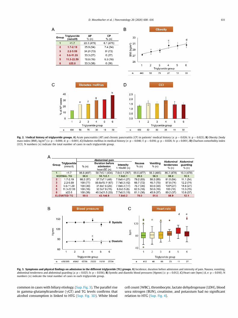
common in cases with biliary etiology. (Sup. Fig. 3). The parallel risein gamma-glutamyltransferase (gGT) and TG levels confirms thatalcohol consumption is linked to HTG (Sup. Fig. 3D). White blood
cell count (WBC), thrombocyte, lactate dehydrogenase (LDH), bloodurea nitrogen (BUN), creatinine, and potassium had no significantrelation to HTG (Sup. Fig. 4).
Fig. 2. Medical history of triglyceride groups. A) Acute pancreatitis (AP) and chronic pancreatitis (CP) in patients’ medical history (a: p ¼ 0.026; b: p ¼ 0.023). B) Obesity (bodymass index (BMI), kg/m2) (c: p ¼ 0.006; d: p ¼ 0.001). C) Diabetes mellitus in medical history (e: p ¼ 0.046; f: p ¼ 0.016; g: p ¼ 0.026; h: p < 0.001). D) Charlson comorbidity index(CCI). N numbers (n) indicate the total number of cases in each triglyceride group.
Fig. 3. Symptoms and physical findings on admission in the different triglyceride (TG) groups. A) Incidence, duration before admission and intensity of pain. Nausea, vomiting,abdominal tenderness and abdominal guarding (a: p ¼ 0.025; b: p ¼ 0.036). B) Systolic and diastolic blood pressures (Hgmm) (c: p ¼ 0.012). C) Heart rate (bpm) (d, e: p ¼ 0.010). Nnumbers (n) indicate the total number of cases in each triglyceride group.
D. Mosztbacher et al. / Pancreatology 20 (2020) 608e616 611

Fig. 4. Laboratory parameters on admission significantly associated with hypertriglyceridemia. A) Triglyceride groups [1e6]. B) Amylase (U/l) (a: p ¼ 0.036; b, c, e: p¼<0.001;d: p ¼ 0.009). C) Lipase (U/l) (f: p ¼ 0.017; g: p ¼ 0.001). D) Sodium (mmol/l) (h, i: p¼<0.001; j: p ¼ 0.005). E) Calcium (mmol/l) (k: p ¼ 0.012). F) Glucose (mmol/l) (l, m: p¼<0.001).G) C-reactive protein (CRP, mg/l) (n: p ¼ 0.021; o: p ¼ 0.014). H) Cholesterol (mmol/l) (p: p ¼ 0.008; q: p ¼ 0.006; r, s: p < 0.001). I) Red blood cell count (RBC, T/l) (t: p ¼ 0.017; u:p ¼ 0.004) J) Hemoglobin (G/l) (v: p ¼ 0.008; w: p < 0.001; x: p ¼ 0.002). K) Hematocrit (%) (y: p ¼ 0.009; z: p ¼ 0.014). N numbers (n) indicate the total number of cases in eachtriglyceride group.
D. Mosztbacher et al. / Pancreatology 20 (2020) 608e616612

The rate of local complications, including peripancreatic fluidcollection, pancreatic necrosis and DM, was significantly and dose-dependently increased with TG level (Fig. 5BeE); however,pancreatic pseudocysts did not show significant differences be-tween the investigated groups above 2.2 mmol/l (Fig. 5F). Organfailure, including heart and renal failure, and maximum CRP levelwere significantly and dose-dependently raised by TG level (Fig. 6B,D-G); however maximum WBC has not shown any significant dif-ferences by HTG (Fig. 6C).
As regards severity, TG level above 11.3 mmol/l was associatedwith a significantly higher rate of moderately severe AP and longerhospital stay, whereas TG level above 22.6 mmol/l was significantlyrelated to severe AP as well (Fig. 7A and B). Due to the low eventrate, the effect of HTG on mortality could not be determined(Fig. 7A). Detailed values of charts and statistical parameters areshown in Tables S2 and S3.
Plasmapheresis was carried out in 36.4% (20/55) of the HTG-APcases; 85% of these patients had an initial TG level higher than22.6 mmol/l and the average TG level was 70.1 ± 10.0 mmol/l.
Discussion
HTG-AP has grown in incidence and importance. According tothe previously published literature [7,9], HTG is the third mostcommon cause of AP (7.7%). However, it seemsmore than likely thatthe incidence of HTG-AP is higher than is usually recorded. Theprospective multicenter, international AP cohort run by the HPSGrevealed that TG measurement is performed in just 50% (716/1435)of AP cases within the first three days from admission and mostprobably this rate is even worse in centers which provide no data.Furthermore, our data also confirmed additional etiological factors(alcohol and biliary disease) besides HTG in 43.6% of HTG-AP casesand showed a dose-dependent relation between obesity (bodymass index), pre-existing DM and HTG. These data also suggest ahigher incidence rate since physicians finding an etiological factorbehind AP usually stop further investigation. Our data are inaccordance with Scherer et al. who recommend that HTG-APshould be suspected in the case of significant alcohol
consumption, poorly controlled DM and metabolic syndrome,including obesity [4]. Although our data clearly show that biliaryobstruction may be associated with HTG, serum TG was measuredin just 44.3% (266/601) of the biliary AP cases. Furthermore, in thecase of biliary AP, there is no recommendation for TGmeasurement.
Our data analysis confirmed results published by Zheng et al.[12] and Zhu et al. [8] which show that HTG is significantly linked toyounger age and male gender. This is not surprising, since under-lying genetic abnormalities behind HTG contribute to youngermanifestation and alcohol-related HTG affects male gender andyounger age more [3,4,22]. In contrast, biliary etiology is accom-panied by a higher rate for female gender and older population[3,23].
Diagnosing AP in the presence of HTG can be challenging due toin vitro interference between plasma TG level above 5.6 mmol/l(with grossly turbid plasma) and determination of amylase andlipase activities [24,25]. Our data confirmed a significant reductionof amylase and lipase levels with the elevation of TG. Furthermore,case reports have been published by Singh et al. [26] and Sotelloet al. [27], presenting HTG-AP patients with normal amylase andlipase levels.
Our analysis has shown that local complications and organfailure were significantly increased by HTG, just as published inprevious reports and a recent meta-analysis by Kiss et al.[10,13,15,28,29]. Nawaz et al. [30] confirmed that TG above2.3 mmol/l is independently associated with persistent organ fail-ure on a multivariate analysis controlling for age, gender, bodymass index, diabetes, and alcohol etiology, whereas Szentesi et al.[31] revealed that hyperlipidemia was an independent predictivefactor for local complications and new-onset DM. Although wecould not confirm a significantly higher risk of pancreatic pseu-docysts in the case of TG above 2.2 mmol/l, it is well known thatpseudocysts usually occur more than four weeks after the onset ofAP and the average hospital stay was 10.4 ± 0.3 days in our cohort[20].
Based on our data analysis, severity of AP and length of hospi-talizationwere significantly increased by HTG [8,10,12,13,29,30,32].Navarro et al. [33] and Goyal et al. [34] also confirmed that HTG
Fig. 5. Local pancreatic complications in the different triglyceride groups. A) Triglyceride groups [1e6]. B) Local pancreatic complications (a, b: p < 0.001). C) Peripancreatic fluidcollection (c: p ¼ 0.004; d: p < 0.001). D) Pancreatic necrosis (e: p ¼ 0.003; f: p < 0.001). E) Diabetes mellitus as complication (g: p ¼ 0.004; h: p < 0.001; i: p ¼ 0.011). F) Pancreaticpseudocyst (j: p ¼ 0.031). N numbers (n) indicate the total number of cases in each triglyceride group.
D. Mosztbacher et al. / Pancreatology 20 (2020) 608e616 613

aggravates the severity of AP compared to biliary and alcoholicetiology, respectively. The underlying mechanism is clearly com-plex. Unsaturated free fatty acids (UFAs) generated from TG are
responsible for cell injury by membrane lipid peroxidation, long-lasting cytosolic Ca2þ elevation and mitochondrial damage[35,36]. In the case of additional alcohol consumption, non-
Fig. 6. Systemic inflammatory effect and organ failure in the different triglyceride groups. A) Triglyceride groups [1e6]. B) Maximum C-reactive protein (Max CRP, mg/l) (a, c:p < 0.001; b: p ¼ 0.029). C)Maximumwhite blood cell count (Max WBC, G/l). D) Organ failure (d: p ¼ 0.001). E) Respiratory failure. F) Heart failure (e: p ¼ 0.007). G) Renal failure (f:p ¼ 0.002). N numbers (n) indicate the total number of cases in each triglyceride group.
D. Mosztbacher et al. / Pancreatology 20 (2020) 608e616614

oxidative ethanol metabolites (fatty acid ethyl esters, FAEEs)contribute to the persistent Ca2þ elevation and drop of ATP level[37,38]. Besides, the raised plasma viscosity caused by hyper-chylomicronemia leads to ischemia and acidosis in the pancreaticcapillaries [5]. This pathologic environment results in an earlytrypsinogen activation and pancreatic lipase leakage, leading tofurther free fatty acid (FFA) release and accumulation [5,35,39].Moreover, UFAs bring about a systemic pro-inflammation byincreased mRNA production of tumor necrosis factor-alpha (TNF-a)and neutrophil chemoattractants, thereby increasing the severity ofAP [36]. In our cohort, heart rate and maximum CRP were signifi-cantly raised by HTG, confirming the systemic inflammatory effectof relatively high TG level. In contrast, Pothoulakis et al. [40] andBalachandra et al. [41] reported that HTG does not worsen severity.Furthermore, Wang et al. showed that longer hospital stay wasassociated with higher TG level, but the difference was not signif-icant [29].
The overall mortality of AP is ~1% based on the literature [3,12]and 1.5% in our cohort, but we could not perform a further sub-group analysis because of the low event number. Zhu et al. [8] andDeng et al. [42] confirmed that HTG-AP is accompanied by asignificantly higher rate of mortality among severe AP casescompared to biliary AP and non-HTG etiology, respectively. How-ever, Tai et al. [32] showed that mortality was similar in HTG-APand biliary AP groups in a general AP cohort.
In our cohort, plasmapheresis was carried out in 36.4% of theHTG-AP cases. Although our data clearly suggest that the severity ofAP is significantly elevated above 11.3 mmol/l TG level, the averageTG level was 70.1 ± 10.0 mmol/l in patients with plasmapheresisand 85% of these cases had a TG level over 22.6 mmol/l. We couldnot state any further conclusion regarding the therapy because ofincomplete data and lack of randomization as a result of the cohortfeature of the dataset. Overall, in most cases, TG-lowering therapy,such as plasmapheresis and glucose-heparin-insulin (GLU-HEP-INS) administration, is performed above a TG level of 40 mmol/l. Inorder to solve this unmet need, the HPSG has initiated a prospectiverandomized clinical trial to investigate different lipid-loweringtherapies in AP [43].
Our study has several limitations. Although all data werecollected prospectively, all questions were raised retrospectively.Cases were included into the analysis with TGmeasurement withinthe first three days from admission, but unfortunately still just 50%of the entire cohort met the inclusion criteria. We attempted tominimize these limitations by comparing the epidemiological andmajor outcome distributions of the data analyzed and the wholecohort. We confirmed that the population under investigation
represents a normal AP cohort.
Conclusion
Our results confirm that HTG dose-dependently increases thecomplications and severity of AP and highlights the necessity ofbetter awareness of an accurate determination of causative andinfluencing risk factors in AP regardless of the etiology. Our datasuggest that lipid-lowering therapymay be important clinically at amuch lower TG level than we previously thought.
Declaration of competing interest
The authors declare that the research was conducted in theabsence of any conflict of interest.
Acknowledgements
The research was supported by Project Grants (K131996 to PH,FK131864 to AM, FK124632 to BCN and K120335 to TT) of the Na-tional Research Development and Innovation Office, an EconomicDevelopment and Innovation Operative Programme Grant (GINOP2.3.2-15-2016-00048 to PH), a Human Resources DevelopmentOperational Programme Grant (EFOP-3.6.2-16-2017-00006 to PH),J�anos Bolyai Research Scholarship of the Hungarian Academy ofSciences (to AP) and ÚNKP-19-4 New National Excellence Programof the Ministry of Human Capacities (to AP).
Appendix A. Supplementary data
Supplementary data to this article can be found online athttps://doi.org/10.1016/j.pan.2020.03.018.
References
[1] Ford ES, Li C, Zhao G, Pearson WS, Mokdad AH. Hypertriglyceridemia and itspharmacologic treatment among US adults. Arch Intern Med 2009;169(6):572e8.
[2] Laufs U, Parhofer KG, Ginsberg HN, Hegele RA. Clinical review on triglycerides.Eur Heart J 2019. https://doi.org/10.1093/eurheartj/ehz778. epub ahead ofprint.
[3] Yadav D, Lowenfels AB. The epidemiology of pancreatitis and pancreaticcancer. Gastroenterology 2013;144(6):1252e61.
[4] Scherer J, Singh V, Pitchumoni C, Yadav D. Issues in hypertriglyceridemicpancreatitis-an update. J Clin Gastroenterol 2014;48(3):195.
[5] Yadav D, Pitchumoni C. Issues in hyperlipidemic pancreatitis. J Clin Gastro-enterol 2003;36(1):54e62.
[6] Berglund L, Brunzell JD, Goldberg AC, Goldberg IJ, Sacks F, Murad MH, et al.Evaluation and treatment of hypertriglyceridemia: an Endocrine Society
Fig. 7. Outcomes in the different triglyceride (TG) groups. A) Length of hospitalization (LOH, day) and mortality (event n) (a: p ¼ 0.034; b: p ¼ 0.001). B) Severity (c: p ¼ 0.001; d:p < 0.001). Moderately severe acute pancreatitis (AP) cases (Group 1: 22.6% (95% CI: 19%e26.6%); Group 5: 66.7% (95% CI: 38.4%e88.2%); Group 6: 47.4% (95% CI: 31.0%e64.2%)).Severe AP cases (e: p ¼ 0.006). N numbers (n) indicate the total number of cases in each triglyceride group.
D. Mosztbacher et al. / Pancreatology 20 (2020) 608e616 615

clinical practice guideline. J Clin Endocrinol Metabol 2012;97(9):2969e89.[7] P�arniczky A, Kui B, Szentesi A, Bal�azs A, Sz}ucs �A, Mosztbacher D, et al. Pro-
spective, multicentre, nationwide clinical data from 600 cases of acutepancreatitis. PLoS One 2016;11(10):e0165309.
[8] Zhu Y, Pan X, Zeng H, He W, Xia L, Liu P, et al. A study on the etiology, severity,and mortality of 3260 patients with acute pancreatitis according to therevised Atlanta classification in Jiangxi, China over an 8-year period. Pancreas2017;46(4):504e9.
[9] Roberts S, Akbari A, Thorne K, Atkinson M, Evans P. The incidence of acutepancreatitis: impact of social deprivation, alcohol consumption, seasonal anddemographic factors. Aliment Pharmacol Therapeut 2013;38(5):539e48.
[10] Zhang R, Deng L, Jin T, Zhu P, Shi N, Jiang K, et al. Hypertriglyceridaemia-associated acute pancreatitis: diagnosis and impact on severity. HPB2019;21(9):1240e9.
[11] Khatua B, El-Kurdi B, Singh VP. Obesity and pancreatitis. Curr Opin Gastro-enterol 2017;33(5):374e82.
[12] Zheng Y, Zhou Z, Li H, Li J, Li A, Ma B, et al. A multicenter study on etiology ofacute pancreatitis in Beijing during 5 years. Pancreas 2015;44(3):409e14.
[13] Kiss L, F}ur G, M�atrai P, Hegyi P, Iv�any E, Cazacu IM, et al. The effect of serumtriglyceride concentration on the outcome of acute pancreatitis: systematicreview and meta-analysis. Sci Rep 2018;8(1):14096.
[14] Czak�o L, Szabolcs A, Vajda �A, Cs�ati S, Venglovecz V, Rakonczay Jr Z, et al.Hyperlipidemia induced by a cholesterol-rich diet aggravates necrotizingpancreatitis in rats. Eur J Pharmacol 2007;572(1):74e81.
[15] Wang Q, Wang G, Qiu Z, He X, Liu C. Elevated serum triglycerides in theprognostic assessment of acute pancreatitis. J Clin Gastroenterol 2017;51(7):586e93.
[16] Yang N, Li B, Pan Y, Tu J, Liu G, Lu G, et al. Hypertriglyceridaemia delayspancreatic regeneration after acute pancreatitis in mice and patients. Gut2019;68(2):378e80.
[17] IAP WG, Guidelines AAP. IAP/APA evidence-based guidelines for the man-agement of acute pancreatitis. Pancreatology 2013;13(4):e1e15.
[18] Hritz I, Czak�o L, Dubravcsik Z, Farkas G, Kelemen D, L�asztity N, et al. Acutepancreatitis. Evidence based management guidelines of the HungarianPancreatic Study Group. Orv Hetil 2015;156(7):244e61.
[19] Yokoe M, Takada T, Mayumi T, Yoshida M, Isaji S, Wada K, et al. Japaneseguidelines for the management of acute pancreatitis: Japanese Guidelines2015. J Hepato-Biliary-Pancreatic Sci 2015;22(6):405e32.
[20] Banks PA, Bollen TL, Dervenis C, Gooszen HG, Johnson CD, Sarr MG, et al.Classification of acute pancreatitisd2012: revision of the Atlanta classificationand definitions by international consensus. Gut 2013;62(1):102e11.
[21] Charlson ME, Pompei P, Ales KL, MacKenzie CR. A new method of classifyingprognostic comorbidity in longitudinal studies: development and validation.J Chron Dis 1987;40(5):373e83.
[22] Wilsnack RW, Wilsnack SC, Kristjanson AF, Vogeltanz-Holm ND, Gmel G.Gender and alcohol consumption: patterns from the multinational GENACISproject. Addiction 2009;104(9):1487e500.
[23] Van Geenen EJ, Van der Peet DL, Bhagirath P, Mulder CJ, Bruno MJ. Etiologyand diagnosis of acute biliary pancreatitis. Nat Rev Gastroenterol Hepatol2010;7(9):495.
[24] Yourno J, Henry JB. Rapid amylase and lipase determinations by nephelom-etry. Am J Clin Pathol 1978;70(1):56e63.
[25] Fallat RW, Vester JW, Glueck CJ. Suppression of amylase activity by hyper-triglyceridemia. Jama 1973;225(11):1331e4.
[26] Singh A, Shrestha M, Anand C. Acute pancreatitis with normal amylase and
lipasedan ED dilemma. Am J Emerg Med 2016;34(5):940. e5-. e7.[27] Sotello D, Rivas AM, Nugent KM, editors. Newly diagnosed acromegaly pre-
senting with hypertriglyceridemic pancreatitis with normal amylase andlipase levels. Baylor University Medical Center Proceedings. Taylor & Francis;2014.
[28] Baranyai T, Terzin V, Vajda �A, Wittmann T, Czak�o L. Hypertriglyceridemiacauses more severe course of acute pancreatitis. Clin Lipidol 2012;7(6):731e6.
[29] Wang S-H, Chou Y-C, ShangkuanW-C, Wei K-Y, Pan Y-H, Lin H-C. Relationshipbetween plasma triglyceride level and severity of hypertriglyceridemicpancreatitis. PloS One 2016;11(10):e0163984.
[30] Nawaz H, Koutroumpakis E, Easler J, Slivka A, Whitcomb DC, Singh VP, et al.Elevated serum triglycerides are independently associated with persistentorgan failure in acute pancreatitis. Am J Gastroenterol 2015;110(10):1497.
[31] Szentesi A, P�arniczky A, Vincze A, Bajor J, G�odi S, Sarlos P, et al. Multiple hits inacute pancreatitis: components of metabolic syndrome synergize each other’sdeteriorating effects. Front Physiol 2019;10:1202.
[32] Tai W-P, Lin X-C, Liu H, Wang C-H, Wu J, Zhang N-W, et al. A retrospectiveresearch of the characteristic of hypertriglyceridemic pancreatitis in Beijing,China. Gastroenterol Res Pract 2016;2016.
[33] Navarro S, Cubiella J, Feu F, Zambon D, Fernandez-Cruz L, Ros E. Hyper-triglyceridemic acute pancreatitis. Is its clinical course different from lithiasicacute pancreatitis? Med Clínica 2004;123(15):567e70.
[34] Goyal H, Smith B, Bayer C, Rutherford C, Shelnut D. Differences in severity andoutcomes between hypertriglyceridemia and alcohol-induced pancreatitis.N Am J Med Sci 2016;8(2):82.
[35] Yang F, Wang Y, Sternfeld L, Rodriguez J, Ross C, Hayden M, et al. The role offree fatty acids, pancreatic lipase and Ca2þ signalling in injury of isolatedacinar cells and pancreatitis model in lipoprotein lipase-deficient mice. ActaPhysiol 2009;195(1):13e28.
[36] Navina S, Acharya C, DeLany JP, Orlichenko LS, Baty CJ, Shiva SS, et al. Lip-otoxicity causes multisystem organ failure and exacerbates acute pancreatitisin obesity. Sci Transl Med 2011;3(107). 107ra10-ra10.
[37] Mal�eth J, Hegyi P. Ca2þ toxicity and mitochondrial damage in acute pancre-atitis: translational overview. Phil Trans Biol Sci 2016;371(1700):20150425.
[38] Hegyi P, Petersen OH. The exocrine pancreas: the acinar-ductal tango inphysiology and pathophysiology. In: Reviews of physiology, biochemistry andpharmacology, vol. 165. Springer; 2013. p. 1e30.
[39] Kimura W, M€ossner J. Role of hypertriglyceridemia in the pathogenesis ofexperimental acute pancreatitis in rats. Int J Gastrointest Canc 1996;20(3):177e84.
[40] Pothoulakis I, Paragomi P, Archibugi L, Tuft M, Talukdar R, Kochhar R, et al.Clinical features of hypertriglyceridemia-induced acute pancreatitis in an in-ternational, multicenter, prospective cohort (APPRENTICE consortium). Pan-creatology 2020;20(3):325e30.
[41] Balachandra S, Virlos I, King N, Siriwardana H, France M, Siriwardena A.Hyperlipidaemia and outcome in acute pancreatitis. Int J Clin Pract2006;60(2):156e9.
[42] Deng L-H, Xue P, Xia Q, Yang X-N, Wan M-H. Effect of admission hyper-triglyceridemia on the episodes of severe acute pancreatitis. World J Gastro-enterol: WJG 2008;14(28):4558.
[43] Z�adori N, Gede N, Antal J, Szentesi A, Alizadeh H, Vincze �A, et al. EarLyelimination of fatty acids iN hypertriglyceridemia-induced acuTe pancreatitis(ELEFANT trial): protocol of an open-label, multicenter, adaptive randomizedclinical trial. Pancreatology 2019;20(3):369e76.
D. Mosztbacher et al. / Pancreatology 20 (2020) 608e616616

II.

Dóra Mosztbacher, First Department of Pediatrics, Semmelweis University, 1083 Budapest, Hungary
Dóra Mosztbacher, Nelli Farkas, Margit Solymár, Katalin Márta, Alexandra Mikó, Zoltán Rumbus, Péter Varjú, Péter Hegyi, Andrea Párniczky, Institute for Translational Medicine, University of Pécs, 7624 Pécs, Hungary
Nelli Farkas, Institute for Bioanalyses, University of Pécs, 7624 Pécs, Hungary
Gabriella Pár, Judit Bajor, József Czimmer, Department of Gastroenterology, First Department of Medicine, University of Pécs, 7624 Pécs, Hungary
Judit Bajor, Péter Hegyi, Department of Translational Medicine, First Department of Medicine, University of Pécs, 7624 Pécs, Hungary
Ákos Szűcs, First Department of Surgery, Semmelweis University, 1082 Budapest, Hungary
Péter Hegyi, Hungarian Academy of Sciences - University of Szeged, Momentum Gastroenterology Multidisciplinary Research Group, 6720 Szeged, Hungary
Andrea Párniczky, Heim Pál Children’s Hospital, 1089 Budapest, Hungary
Author contributions: Solymár M, Pár G, Bajor J, Szűcs Á and Czimmer J searched for the articles in Pubmed and screened them using the titles and abstracts; Márta K, Mikó A, Rumbus Z and Varjú P searched for the articles in Embase and screened them using the titles and abstracts; data was collected from the eligible papers by Mosztbacher D and Párniczky A; Farkas N performed the statistical analysis; Mosztbacher D, Hegyi P and Párniczky A drafted the manuscript.
Supported by the Hungarian Scientific Research Fund, No. K116634 to Hegyi P; and the Momentum Grant of the Hungarian Academy of Sciences, No. LP2014-10/2014 to Hegyi P.
Conflict-of-interest statement: All the authors disclaim any form of conflict of interest.
Open-Access: This article is an open-access article which was selected by an in-house editor and fully peer-reviewed by external reviewers. It is distributed in accordance with the Creative Commons Attribution Non Commercial (CC BY-NC 4.0) license, which permits others to distribute, remix, adapt, build upon this work non-commercially, and license their derivative works on different terms, provided the original work is properly cited and the use is non-commercial. See: http://creativecommons.org/licenses/by-nc/4.0/
Manuscript source: Invited manuscript
Correspondence to: Andrea Párniczky, MD, Institute for Translational Medicine, University of Pécs, Szigeti út 12., II. em., 7624 Pécs, Hungary. [email protected]: +36-72-536246Fax: +36-72-536247
Received: October 3, 2016Peer-review started: October 7, 2016First decision: October 20, 2016Revised: November 21, 2016Accepted: January 11, 2017Article in press: January 11, 2017Published online: February 14, 2017
AbstractAcute pancreatitis (AP) is a serious inflammatory disease with rising incidence both in the adult and pediatric populations. It has been shown that mitoc-hondrial injury and energy depletion are the earliest intracellular events in the early phase of AP. Moreover, it has been revealed that restoration of intracellular ATP level restores cellular functions and defends the cells from death. We have recently shown in a systematic review and meta-analysis that early enteral feeding is beneficial in adults; however, no reviews are available concerning the effect of early enteral feeding in pediatric AP. In this minireview, our aim was to systematically analyse the literature on the treatment
Submit a Manuscript: http://www.wjgnet.com/esps/Help Desk: http://www.wjgnet.com/esps/helpdesk.aspxDOI: 10.3748/wjg.v23.i6.957
957 February 14, 2017|Volume 23|Issue 6|WJG|www.wjgnet.com
World J Gastroenterol 2017 February 14; 23(6): 957-963 ISSN 1007-9327 (print) ISSN 2219-2840 (online)
© 2017 Baishideng Publishing Group Inc. All rights reserved.
MINIREVIEWS
Restoration of energy level in the early phase of acute pediatric pancreatitis
Dóra Mosztbacher, Nelli Farkas, Margit Solymár, Gabriella Pár, Judit Bajor, Ákos Szűcs, József Czimmer, Katalin Márta, Alexandra Mikó, Zoltán Rumbus, Péter Varjú, Péter Hegyi, Andrea Párniczky
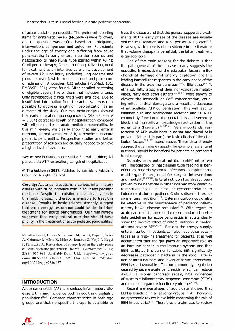
of acute pediatric pancreatitis. The preferred reporting items for systematic review (PRISMA-P) were followed, and the question was drafted based on participants, intervention, comparison and outcomes: P: patients under the age of twenty-one suffering from acute pancreatitis; I: early enteral nutrition (per os and nasogastric- or nasojejunal tube started within 48 h); C: nil per os therapy; O: length of hospitalization, need for treatment at an intensive care unit, development of severe AP, lung injury (including lung oedema and pleural effusion), white blood cell count and pain score on admission. Altogether, 632 articles (PubMed: 131; EMBASE: 501) were found. After detailed screening of eligible papers, five of them met inclusion criteria. Only retrospective clinical trials were available. Due to insufficient information from the authors, it was only possible to address length of hospitalization as an outcome of the study. Our mini-meta-analysis showed that early enteral nutrition significantly (SD = 0.806, P = 0.034) decreases length of hospitalization compared with nil per os diet in acute pediatric pancreatitis. In this minireview, we clearly show that early enteral nutrition, started within 24-48 h, is beneficial in acute pediatric pancreatitis. Prospective studies and better presentation of research are crucially needed to achieve a higher level of evidence.
Key words: Pediatric pancreatitis; Enteral nutrition; Nil per os diet; ATP restoration; length of hospitalization
© The Author(s) 2017. Published by Baishideng Publishing Group Inc. All rights reserved.
Core tip: Acute pancreatitis is a serious inflammatory disease with rising incidence both in adult and pediatric medicine. Despite the existing research activities in this field, no specific therapy is available to treat this disease. Results in basic science strongly suggest that early energy restoration could be the first-line treatment for acute pancreatitis. Our minireview suggests that early enteral nutrition should have priority in the treatment of acute pediatric pancreatitis.
Mosztbacher D, Farkas N, Solymár M, Pár G, Bajor J, Szűcs Á, Czimmer J, Márta K, Mikó A, Rumbus Z, Varjú P, Hegyi P, Párniczky A. Restoration of energy level in the early phase of acute pediatric pancreatitis. World J Gastroenterol 2017; 23(6): 957-963 Available from: URL: http://www.wjgnet.com/1007-9327/full/v23/i6/957.htm DOI: http://dx.doi.org/10.3748/wjg.v23.i6.957
INTRODUCTIONAcute pancreatitis (AP) is a serious inflammatory disease with rising incidence both in adult and pediatric populations[1,2]. Common characteristics in both age groups are that no specific therapy is available to
treat the disease and that the general supportive treatments at the early phase of the disease are usually volume resuscitation and a nil per os (NPO) diet[36]. However, while there is clear evidence in the literature that volume therapy is beneficial, the latter treatment is questionable.
One of the main reasons for the debate is that the pathogenesis of the disease clearly suggests the opposite. Irrespective of the etiological factors, mitochondrial damage and energy depletion are the leading intracellular responses in the early phase of the disease in the exocrine pancreas[710]. Bile acids[1114], ethanol, fatty acids and their nonoxidative metabolites, fatty acid ethyl esthers[8,9,1518] were shown to elevate the intracellular Ca2+ concentration, causing mitochondrial damage and a resultant decrease of intracellular ATP concentration. This will lead to inhibited fluid and bicarbonate secretion and CFTR Cl channel dysfunction in the ductal cells and secretory block and intracellular trypsinogen activation in the acinar cells (Figure 1)[9,16,19,20]. Very importantly, restoration of ATP levels both in acinar and ductal cells prevents (at least in part) the toxic effects of the etiological factors[7,21,22] noted above. These data strongly suggest that an energy supply, for example, via enteral nutrition, should be beneficial for patients as compared to nil energy.
Notably, early enteral nutrition (EEN) either via oral, nasogastric or nasojejunal tube feeding is beneficial as regards systemic infections, complications, multiorgan failure, need for surgical interventions and mortality[6,2330]. Enteral nutrition has already been proven to be beneficial in other inflammatory gastrointestinal diseases. The firstline recommendation to induce remission in pediatric Crohn’s disease is exclusive enteral nutrition[31]. Enteral nutrition could also be effective in the maintenance of pediatric inflammatory bowel disease remission[32]. With regard to acute pancreatitis, three of the recent and most uptodate guidelines for acute pancreatitis in adults clearly show the positive effect of enteral nutrition in moderate and severe AP[6,23,24]. Besides the energy supply, enteral nutrition in patients can also have other advantages as a firstline treatment for patients. It is well documented that the gut plays an important role as an immune barrier in the immune system and that EEN facilitates this barrier function. EEN significantly decreases pathogenic bacteria in the stool, alteration of intestinal flora and levels of serum endotoxins. EEN has a favourable effect on immune dysregulation caused by severe acute pancreatitis, which can reduce APACHE II scores, pancreatic sepsis, initial incidences of systemic inflammatory response syndrome (SIRS) and multiple organ dysfunction syndrome[33,34].
Recent metaanalyses of adult data showed that EEN is beneficial in all severity groups in AP; however, no systematic review is available concerning the role of EEN in pediatrics[35]. Therefore, the aim was to review
958 February 14, 2017|Volume 23|Issue 6|WJG|www.wjgnet.com
Mosztbacher D et al . Enteral feeding in acute pediatric pancreatitis

Table 1 Studies included in the quantitative synthesis
EEN: Early enteral nutrition; NPO: Nil per os.
the literature to analyse the effect of EEN vs NPO therapy on the outcome of acute pediatric pancreatitis (APP) and aggregate the information in APP leading to a higher statistical power and more robust point estimate than is possible from the individual studies.
The preferred reporting items for systematic review and metaanalysis protocol (PRISMAP) were followed[36]. Our structured literature search was based on the participants, intervention, comparison and outcomes format: P: patients under the age of twentyone suffering from acute pancreatitis; I: early enteral nutrition (per os and nasogastric or nasojejunal tube started within 48 h); C: NPO therapy [per os/nasogastric or enteral tube after 48 h and total parenteral nutrition (TPN) within or after 48 h]; O: length of hospitalization, need for intensive care unit (ICU), complications, necessity of antibiotics, surgical/nonsurgical interventions and mortality.
In February 2016, a literature search was performed on the PubMed (http://www.ncbi.nlm.nih.gov/pubmed) and EMBASE (https://www.embase.com) databases using the following Medical Subject Headings and search terms: “pediatric” OR “paediatric” AND “pancreatitis”. The search was limited to human studies, fulltext publications with abstracts in English with no time period, resulting in 632 articles altogether (PubMed: 131; EMBASE: 501).
The articles were checked separately. Metaanalyses, reviews, case reports and articles on chronic pancreatitis were excluded and duplicates were removed (Figure 2). Potentially eligible papers were selected, and, finally, five of them with relevant data on EEN or/with NPO therapy in acute pediatric pancreatitis in patients under twentyone years old were included (Table 1)[3741]. To reduce the risk of bias, the literature search was independently performed by three researchers following the inclusion criteria noted above.
The details in the collected articles were checked, and only articles where both EEN and NPO were presented separately were used. Two articles met this
criterion. The two articles contained three separate data pairs, where EEN was compared to NPO (Figure 3). The following parameters were collected: length of hospitalization (LOH), need for treatment at an ICU, development of severe AP, lung injury (including lung oedema and pleural effusion), white blood cell count and pain score on admission. Only one of the five investigated parameters (LOH) contained a minimum of three items, which were analysed statistically.
The metaanalytic calculation was made with Comprehensive MetaAnalysis (V3) software using the random effects model (the DerSimonianLaird method). We calculated a weighted standard difference in means and 95%CI. In the case of one study (AbuElHaija et al[37], 2016), we converted the median and range values to means and standard deviation using the modified Hozo’s formula by Wan et al[42]. For a visual inspection, we used a forest plot.
Figure 3 shows the parameters collected from the articles. It was only possible to perform forest plot analyses on LOH. EEN significantly decreased LOH (SD = 0.806, p = 0.034) compared to the standard NPO diet (Figure 3).
DISCUSSIONSeveral therapeutic recommendations are available in the literature on nutrition in acute pancreatitis. The IAP/APA guideline suggests enteral tube feeding as the firstline therapy in patients requiring nutritional support with predicted severe and severe acute pancreatitis[6]. According to the Japanese guideline, enteral nutrition in the early phase of severe acute pancreatitis can decrease the incidence of complications and elevate the survival rate[24]. Recent metaanalyses of adult studies revealed that EEN decreases mortality, rate of interventions and the incidence of multiorgan failure in severe acute pancreatitis. Moreover, group analyses of 17 parameters including laboratory parameters (such as CRP and white blood cells) and symptoms (such as pain or presence of SIRS) suggested that EEN also has merits in mild acute pancreatitis. Since the incidence of APP has risen in the past twenty years (with 3.6 and 13.2/100000 children affected annually), we
959 February 14, 2017|Volume 23|Issue 6|WJG|www.wjgnet.com
Ref. Data Groups NO. of patients
Abu-El-Haija et al[37], 2016 Yes EEN 24NPO 14
Flores-Calderón et al[41], 2009 Only NPO 18Goh et al[40], 2003 Only NPO 12Raizner et al[39], 2013 Only NPO 7Szabo et al[38], 2015 Yes EEN + IVF lo 55
NPO + IVF lo 20Yes EEN + IVF hi 96
NPO + IVF hi 30
Figure 1 Early events in acute pancreatitis. Bile acids, ethanol, fatty acids or their non-oxidative metabolites, fatty acid ethyl esthers, induce calcium overload, causing mitochondrial damage and a resultant decrease in intracellular ATP concentration both in acinar and ductal cells. This will lead to general energy depletion in the pancreas.
Energy depletion
Ductal cellsCa2+
Ca2+
Ca2+ overload
Mitochondrial injury
Acinar cells
Mosztbacher D et al . Enteral feeding in acute pediatric pancreatitis

systematically reviewed the literature to understand whether there is any beneficial effect of EEN vs NPO in children[43,44].
We faced several difficulties during our review: (1) APP is still underdiagnosed, thus decreasing the possibility of performing clinical trials[45]; (2) the number of studies on the management of these patients is very low, and there is still only a small number of studies focused on understanding the characteristics of the disease[46]; (3) the studies have not focused on the early management of the patients; the groups
960 February 14, 2017|Volume 23|Issue 6|WJG|www.wjgnet.com
were therefore not separated; and (4) finally, but very importantly, the methods sections and the quality of data presentation in these articles are very low. Consequently, in many cases, it was impossible to obtain quality analysable data from the manuscripts for a proper broadspectrum metaanalysis[3739].
By the end of the search, we identified five articles containing relevant data on nutritional management during the early phase of APP. Raizner et al[39] published a retrospective analysis involving seven children with necrotizing pancreatitis. All the children received a
Figure 2 Flow chart on the methods used in the literature search.
Records identified through database searchPubmed n = 131Embase n = 501
Records screened based on titles and abstracts n = 632
Full-text articles assessed for eligibility n = 61
Studies included in quantitative synthesisn = 5
Records excluded n = 571(meta-analyses, case reports, reviews,
chronic pancreatitis)
Records excluded n = 56(duplicates, adult data, no relevant data
on enteral nutrition or nil per os diet)
Iden
tifica
tion
Scre
enin
gEl
igib
ility
Inclu
ded
Figure 3 Two articles contained three separate data pairs where early enteral nutrition was compared to nil per os. LOH: Length of hospitalization; EEN: Early enteral nutrition; NPO: Nil per os. ICU: Intensive care unit; AP: Acute pancreatitis; WBC: White blood cell count.
Ref. Therapy n LOH Need for ICU Severe AP Lung injury
WBC Pain score
Abu-El-Haija et al [37], 2016
1 EEN (< 24 h) 24 81.7 hNPO (< 24 h) 14 94.7 h
Szabo et al [38], 2015
2 NPO (< 48 h) + IVF lo (< 24 h) 20 7.1 d (1.01 SE) 20.0% (8.94 SE) 35.0% (10.7 SE) 11 13.60 (6.44 SD) 4.95 (3.75 SD)EEN (< 48 h) + IVF lo (< 24 h) 55 2.8 d (0.24 SE) 1.8% (1.80 SE) 9.1% (3.88 SE) 21 9.89 (3.89 SD) 4.62 (3.50 SD)
3 NPO (< 48 h) + IVF hi (< 24 h) 30 5.0 d (0.58 SE) 13.0% (6.21 SE) 17.0% (6.80 SE) 14 13.30 (4.76 SD) 6.08 (3.19 SD)EEN (< 48 h) + IVF hi (< 24 h) 96 3.2 d (0.22 SE) 1.0% (1.04 SE) 4.2% (2.04 SE) 4 11.30 (5.25 SD) 5.47 (3.57 SD)
Study name Statistics for each studyStd diff
in meansStandard
errorLower limit
Upper limit
Z value P value
Abu-El-Haija et al [37], 2016 0.089 0.336 -0.570 0.749 0.266 0.790Szabo et al [38], 2015 A 1.554 0.290 0.985 2.122 5.352 0.000Szabo et al [38], 2015 B 0.739 0.214 0.319 1.159 3.447 0.001
0.806 0.380 0.062 1.551 2.124 0.034
Std diff in means and 95%CI
-2.50 1.25 0.00 1.25 2.50
PO NPO
Mosztbacher D et al . Enteral feeding in acute pediatric pancreatitis

strict NPO diet, five patients received TPN and just one patient was treated with nasojejunal feeding for seven days. All the children required a prolonged hospital stay (with a mean of 20 d) for acute complications, with three of them suffering from late complications[39]. Goh et al[40] included twelve patients in their retrospective study. One patient needed a distal pancreatectomy, and eleven patients recovered with conservative management, with none of them receiving EEN. Two patients had acute complications, and two patients had recurrent AP[40]. FloresCalderon et al[41] studied eighteen patients with acute pancreatitis caused by Lasparaginase due to acute lymphoblastic leukemia. All the patients were treated with bowel resting for a mean of 22 d, fourteen of the patients received TPN and four had an elementary diet. Two of the patients required intensive care unit admission, with local complications developing in twelve patients. None of the patients died from complications related to AP. Although these studies point out several disadvantages of that NPO diet, none of them could be enrolled in our metaanalysis.
Finally, it was possible to collect three sets of analysable data pairs where both NPO and EEN were present. AbuElHaija et al[37] conducted a prospective study of 38 children suffering from mild AP and retrospectively investigated the relationship of nutrition with pain and LOS. EEN feeding meant per os feeding and NPO was identified as oral feeding not being allowed for 24 h. Importantly, EEN, even with high fat intake, did not cause an elevation in pain in children, suggesting that EEN is a well tolerable nutritional possibility in children. The fact that LOS was much shorter in group EEN vs NPO points to EEN as a better way of treating APP[37]. The most advanced study was performed by Szabo et al[38], where several parameters were collected to understand the effect of EEN on the course of APP. Two hundred and one children suffering from mild AP were enrolled retrospectively. They compared EEN vs NPO both with and without aggressive fluid resuscitation. Fluid therapy was administered during the first 24 h, and the type of nutrition was determined during the first 48 h. Besides the beneficial effects of EEN on LOS, they also showed that EEN reduces the severity of the disease. Although our aim was to perform a metaanalysis on several parameters to understand the differences between EEN and NPO, we were only able to perform the statistical analyses on LOS, which clearly showed that EEN is not only a safe method of nutrition but also substantially decreases LOS, resulting in a better and less expensive treatment of mild APP[38].
CONCLUSIONThe information collected by basic scientists, retrospective clinical studies and metaanalyses suggests that EEN should have priority in treating APP. However, it is perhaps selfevident that randomized multicenter clinical intervention trials would be crucial to achieving
a higher level of evidence.
REFERENCES1 Peery AF, Dellon ES, Lund J, Crockett SD, McGowan CE,
Bulsiewicz WJ, Gangarosa LM, Thiny MT, Stizenberg K, Morgan DR, Ringel Y, Kim HP, Dibonaventura MD, Carroll CF, Allen JK, Cook SF, Sandler RS, Kappelman MD, Shaheen NJ. Burden of gastrointestinal disease in the United States: 2012 update. Gastroenterology 2012; 143: 1179-1187.e1-3 [PMID: 22885331 DOI: 10.1053/j.gastro.2012.08.002]
2 Pant C, Deshpande A, Olyaee M, Anderson MP, Bitar A, Steele MI, Bass PF, Sferra TJ. Epidemiology of acute pancreatitis in hospitalized children in the United States from 2000-2009. PLoS One 2014; 9: e95552 [PMID: 24805879 DOI: 10.1371/journal.pone.0095552]
3 Párniczky A, Czakó L, Dubravcsik Z, Farkas G, Hegyi P, Hritz I, Kelemen D, Morvay Z, Oláh A, Pap Á, Sahin-Tóth M, Szabó F, Szentkereszti Z, Szmola R, Takács T, Tiszlavicz L, Veres G, Szücs Á, Lásztity N. [Pediatric pancreatitis. Evidence based management guidelines of the Hungarian Pancreatic Study Group]. Orv Hetil 2015; 156: 308-325 [PMID: 25662148 DOI: 10.1556/OH.2015.30062]
4 Hritz I, Czakó L, Dubravcsik Z, Farkas G, Kelemen D, Lásztity N, Morvay Z, Oláh A, Pap Á, Párniczky A, Sahin-Tóth M, Szentkereszti Z, Szmola R, Szücs Á, Takács T, Tiszlavicz L, Hegyi P. [Acute pancreatitis. Evidence-based practice guidelines, prepared by the Hungarian Pancreatic Study Group]. Orv Hetil 2015; 156: 244-261 [PMID: 25661970 DOI: 10.1556/OH.2015.30059]
5 Morinville VD, Husain SZ, Bai H, Barth B, Alhosh R, Durie PR, Freedman SD, Himes R, Lowe ME, Pohl J, Werlin S, Wilschanski M, Uc A. Definitions of pediatric pancreatitis and survey of present clinical practices. J Pediatr Gastroenterol Nutr 2012; 55: 261-265 [PMID: 22357117 DOI: 10.1097/MPG.0b013e31824f1516]
6 Working Group IAP/APA Acute Pancreatitis Guidelines. IAP/APA evidence-based guidelines for the management of acute pancreatitis. Pancreatology 2013; 13: e1-15 [PMID: 24054878 DOI: 10.1016/j.pan.2013.07.063]
7 Maléth J, Hegyi P. Ca2+ toxicity and mitochondrial damage in acute pancreatitis: translational overview. Philos Trans R Soc Lond B Biol Sci 2016; 371: pii: 20150425 [PMID: 27377719 DOI: 10.1098/rstb.2015.0425]
8 Maléth J, Hegyi P, Rakonczay Z, Venglovecz V. Breakdown of bioenergetics evoked by mitochondrial damage in acute pancreatitis: Mechanisms and consequences. Pancreatology 2015; 15: S18-S22 [PMID: 26162756 DOI: 10.1016/j.pan.2015.06.002]
9 Hegyi P, Petersen OH. The exocrine pancreas: the acinar-ductal tango in physiology and pathophysiology. Rev Physiol Biochem Pharmacol 2013; 165: 1-30 [PMID: 23881310 DOI: 10.1007/112_ 2013_14]
10 Hegyi P, Pandol S, Venglovecz V, Rakonczay Z. The acinar-ductal tango in the pathogenesis of acute pancreatitis. Gut 2011; 60: 544-552 [PMID: 20876773 DOI: 10.1136/gut.2010.218461]
11 Venglovecz V, Rakonczay Z, Ozsvári B, Takács T, Lonovics J, Varró A, Gray MA, Argent BE, Hegyi P. Effects of bile acids on pancreatic ductal bicarbonate secretion in guinea pig. Gut 2008; 57: 1102-1112 [PMID: 18303091 DOI: 10.1136/gut.2007.134361]
12 Hegyi P. Bile as a key aetiological factor of acute but not chronic pancreatitis: a possible theory revealed. J Physiol 2016; 594: 6073-6074 [PMID: 27800624 DOI: 10.1113/JP273108]
13 Venglovecz V, Hegyi P, Rakonczay Z, Tiszlavicz L, Nardi A, Grunnet M, Gray MA. Pathophysiological relevance of apical large-conductance Ca²+-activated potassium channels in pancreatic duct epithelial cells. Gut 2011; 60: 361-369 [PMID: 20940280 DOI: 10.1136/gut.2010.214213]
14 Voronina SG, Gryshchenko OV, Gerasimenko OV, Green AK, Petersen OH, Tepikin AV. Bile acids induce a cationic current, depolarizing pancreatic acinar cells and increasing the intracellular Na+ concentration. J Biol Chem 2005; 280: 1764-1770 [PMID: 15536077 DOI: 10.1074/jbc.M410230200]
961 February 14, 2017|Volume 23|Issue 6|WJG|www.wjgnet.com
Mosztbacher D et al . Enteral feeding in acute pediatric pancreatitis

15 Hegyi P, Rakonczay Z. The role of pancreatic ducts in the patho-genesis of acute pancreatitis. Pancreatology 2015; 15: S13-S17 [PMID: 25921231 DOI: 10.1016/j.pan.2015.03.010]
16 Maléth J, Balázs A, Pallagi P, Balla Z, Kui B, Katona M, Judák L, Németh I, Kemény LV, Rakonczay Z, Venglovecz V, Földesi I, Pető Z, Somorácz Á, Borka K, Perdomo D, Lukacs GL, Gray MA, Monterisi S, Zaccolo M, Sendler M, Mayerle J, Kühn JP, Lerch MM, Sahin-Tóth M, Hegyi P. Alcohol disrupts levels and function of the cystic fibrosis transmembrane conductance regulator to promote development of pancreatitis. Gastroenterology 2015; 148: 427-439.e16 [PMID: 25447846 DOI: 10.1053/j.gastro.2014.11.002]
17 Maléth J, Hegyi P. Calcium signaling in pancreatic ductal epithe-lial cells: an old friend and a nasty enemy. Cell Calcium 2014; 55: 337-345 [PMID: 24602604 DOI: 10.1016/j.ceca.2014.02.004]
18 Criddle DN. The role of fat and alcohol in acute pancreatitis: A dangerous liaison. Pancreatology 2015; 15: S6-S12 [PMID: 25845855 DOI: 10.1016/j.pan.2015.02.009]
19 Hegyi P, Wilschanski M, Muallem S, Lukacs GL, Sahin-Tóth M, Uc A, Gray MA, Rakonczay Z, Maléth J. CFTR: A New Horizon in the Pathomechanism and Treatment of Pancreatitis. Rev Physiol Biochem Pharmacol 2016; 170: 37-66 [PMID: 26856995 DOI: 10.1007/112_2015_5002]
20 Pallagi P, Venglovecz V, Rakonczay Z, Borka K, Korompay A, Ozsvári B, Judák L, Sahin-Tóth M, Geisz A, Schnúr A, Maléth J, Takács T, Gray MA, Argent BE, Mayerle J, Lerch MM, Wittmann T, Hegyi P. Trypsin reduces pancreatic ductal bicarbonate secretion by inhibiting CFTR Cl⁻ channels and luminal anion exchangers. Gastroenterology 2011; 141: 2228-2239.e6 [PMID: 21893120 DOI: 10.1053/j.gastro.2011.08.039]
21 Judák L, Hegyi P, Rakonczay Z, Maléth J, Gray MA, Venglovecz V. Ethanol and its non-oxidative metabolites profoundly inhibit CFTR function in pancreatic epithelial cells which is prevented by ATP supplementation. Pflugers Arch 2014; 466: 549-562 [PMID: 23948742 DOI: 10.1007/s00424-013-1333-x]
22 Criddle DN, Murphy J, Fistetto G, Barrow S, Tepikin AV, Neoptolemos JP, Sutton R, Petersen OH. Fatty acid ethyl esters cause pancreatic calcium toxicity via inositol trisphosphate receptors and loss of ATP synthesis. Gastroenterology 2006; 130: 781-793 [PMID: 16530519 DOI: 10.1053/j.gastro.2005.12.031]
23 Tenner S, Baillie J, DeWitt J, Vege SS. American College of Gastroenterology guideline: management of acute pancreatitis. Am J Gastroenterol 2013; 108: 1400-1415; 1416 [PMID: 23896955 DOI: 10.1038/ajg.2013.218]
24 Yokoe M, Takada T, Mayumi T, Yoshida M, Isaji S, Wada K, Itoi T, Sata N, Gabata T, Igarashi H, Kataoka K, Hirota M, Kadoya M, Kitamura N, Kimura Y, Kiriyama S, Shirai K, Hattori T, Takeda K, Takeyama Y, Hirota M, Sekimoto M, Shikata S, Arata S, Hirata K. Japanese guidelines for the management of acute pancreatitis: Japanese Guidelines 2015. J Hepatobiliary Pancreat Sci 2015; 22: 405-432 [PMID: 25973947 DOI: 10.1002/jhbp.259]
25 Petrov MS, Whelan K. Comparison of complications attributable to enteral and parenteral nutrition in predicted severe acute pancre-atitis: a systematic review and meta-analysis. Br J Nutr 2010; 103: 1287-1295 [PMID: 20370944 DOI: 10.1017/S0007114510000887]
26 Kalfarentzos F, Kehagias J, Mead N, Kokkinis K, Gogos CA. Enteral nutrition is superior to parenteral nutrition in severe acute pancreatitis: results of a randomized prospective trial. Br J Surg 1997; 84: 1665-1669 [PMID: 9448611]
27 Abou-Assi S, Craig K, O’Keefe SJ. Hypocaloric jejunal feeding is better than total parenteral nutrition in acute pancreatitis: results of a randomized comparative study. Am J Gastroenterol 2002; 97: 2255-2262 [PMID: 12358242 DOI: 10.1111/j.1572-0241. 2002.05979.x]
28 Eckerwall GE, Tingstedt BB, Bergenzaun PE, Andersson RG. Immediate oral feeding in patients with mild acute pancreatitis is safe and may accelerate recovery--a randomized clinical study. Clin Nutr 2007; 26: 758-763 [PMID: 17719703 DOI: 10.1016/j.clnu. 2007.04.007]
29 Li J, Xue GJ, Liu YL, Javed MA, Zhao XL, Wan MH, Chen GY, Altaf K, Huang W, Tang WF. Early oral refeeding wisdom in
patients with mild acute pancreatitis. Pancreas 2013; 42: 88-91 [PMID: 22836861 DOI: 10.1097/MPA.0b013e3182575fb5]
30 Petrov MS, McIlroy K, Grayson L, Phillips AR, Windsor JA. Early nasogastric tube feeding versus nil per os in mild to moderate acute pancreatitis: a randomized controlled trial. Clin Nutr 2013; 32: 697-703 [PMID: 23340042 DOI: 10.1016/j.clnu.2012.12.011]
31 Ruemmele FM, Veres G, Kolho KL, Griffiths A, Levine A, Escher JC, Amil Dias J, Barabino A, Braegger CP, Bronsky J, Buderus S, Martín-de-Carpi J, De Ridder L, Fagerberg UL, Hugot JP, Kierkus J, Kolacek S, Koletzko S, Lionetti P, Miele E, Navas López VM, Paerregaard A, Russell RK, Serban DE, Shaoul R, Van Rheenen P, Veereman G, Weiss B, Wilson D, Dignass A, Eliakim A, Winter H, Turner D. Consensus guidelines of ECCO/ESPGHAN on the medical management of pediatric Crohn’s disease. J Crohns Colitis 2014; 8: 1179-1207 [PMID: 24909831 DOI: 10.1016/j.crohns.2014.04.005]
32 Penagini F, Dilillo D, Borsani B, Cococcioni L, Galli E, Bedogni G, Zuin G, Zuccotti GV. Nutrition in Pediatric Inflammatory Bowel Disease: From Etiology to Treatment. A Systematic Review. Nutrients 2016; 8: pii: E334 [PMID: 27258308 DOI: 10.3390/nu8060334]
33 Capurso G, Zerboni G, Signoretti M, Valente R, Stigliano S, Piciucchi M, Delle Fave G. Role of the gut barrier in acute pancreatitis. J Clin Gastroenterol 2012; 46 Suppl: S46-S51 [PMID: 22955357 DOI: 10.1097/MCG.0b013e3182652096]
34 Flint RS, Windsor JA. The role of the intestine in the patho-physiology and management of severe acute pancreatitis. HPB (Oxford) 2003; 5: 69-85 [PMID: 18332961 DOI: 10.1080/ 13651820310001108]
35 Márta K, Farkas N, Szabó I, Illés A, Vincze Á, Pár G, Sarlós P, Bajor J, Szűcs Á, Czimmer J, Mosztbacher D, Párniczky A, Szemes K, Pécsi D, Hegyi P. Meta-Analysis of Early Nutrition: The Benefits of Enteral Feeding Compared to a Nil Per Os Diet Not Only in Severe, but Also in Mild and Moderate Acute Pancreatitis. Int J Mol Sci 2016; 17: pii: E1691 [PMID: 27775609 DOI: 10.3390/ijms17101691]
36 Shamseer L, Moher D, Clarke M, Ghersi D, Liberati A, Petticrew M, Shekelle P, Stewart LA; PRISMA-P Group. Preferred report-ing items for systematic review and meta-analysis protocols (PRISMA-P) 2015: elaboration and explanation. BMJ 2015; 349: g7647 [PMID: 25555855 DOI: 10.1136/bmj.g7647]
37 Abu-El-Haija M, Wilhelm R, Heinzman C, Siqueira BN, Zou Y, Fei L, Cole CR. Early Enteral Nutrition in Children With Acute Pancreatitis. J Pediatr Gastroenterol Nutr 2016; 62: 453-456 [PMID: 26488122 DOI: 10.1097/MPG.0000000000001013]
38 Szabo FK, Fei L, Cruz LA, Abu-El-Haija M. Early Enteral Nutrition and Aggressive Fluid Resuscitation are Associated with Improved Clinical Outcomes in Acute Pancreatitis. J Pediatr 2015; 167: 397-402.e1 [PMID: 26210842 DOI: 10.1016/j.jpeds.2015.05.030]
39 Raizner A, Phatak UP, Baker K, Patel MG, Husain SZ, Pashankar DS. Acute necrotizing pancreatitis in children. J Pediatr 2013; 162: 788-792 [PMID: 23102790 DOI: 10.1016/j.jpeds.2012.09.037]
40 Goh SK, Chui CH, Jacobsen AS. Childhood acute pancreatitis in a children’s hospital. Singapore Med J 2003; 44: 453-456 [PMID: 14740774]
41 Flores-Calderón J, Exiga-Gonzaléz E, Morán-Villota S, Martín-Trejo J, Yamamoto-Nagano A. Acute pancreatitis in children with acute lymphoblastic leukemia treated with L-asparaginase. J Pediatr Hematol Oncol 2009; 31: 790-793 [PMID: 19770681 DOI: 10.1097/MPH.0b013e3181b794e8]
42 Wan X, Wang W, Liu J, Tong T. Estimating the sample mean and standard deviation from the sample size, median, range and/or interquartile range. BMC Med Res Methodol 2014; 14: 135 [PMID: 25524443 DOI: 10.1186/1471-2288-14-135]
43 Morinville VD, Barmada MM, Lowe ME. Increasing incidence of acute pancreatitis at an American pediatric tertiary care center: is greater awareness among physicians responsible? Pancreas 2010; 39: 5-8 [PMID: 19752770 DOI: 10.1097/MPA.0b013e3181baac47]
44 Lopez MJ. The changing incidence of acute pancreatitis in children: a single-institution perspective. J Pediatr 2002; 140:
962 February 14, 2017|Volume 23|Issue 6|WJG|www.wjgnet.com
Mosztbacher D et al . Enteral feeding in acute pediatric pancreatitis

622-624 [PMID: 12032533 DOI: 10.1067/mpd.2002.123880]45 Zsoldos F, Párniczky A, Mosztbacher D, Tóth A, Lásztity N, Hegyi
P. Pain in the Early Phase of Pediatric Pancreatitis (PINEAPPLE Trial): Pre-Study Protocol of a Multinational Prospective Clinical Trial. Digestion 2016; 93: 121-126 [PMID: 26641250 DOI:
10.1159/000441352]46 Párniczky A, Mosztbacher D, Zsoldos F, Tóth A, Lásztity N, Hegyi
P. Analysis of Pediatric Pancreatitis (APPLE Trial): Pre-Study Protocol of a Multinational Prospective Clinical Trial. Digestion 2016; 93: 105-110 [PMID: 26613586 DOI: 10.1159/000441353]
P- Reviewer: Cosen-Binker LI, Fujino Y, Luo HS, Peng SY, Sperti C S- Editor: Gong ZM L- Editor: A E- Editor: Liu WX
963 February 14, 2017|Volume 23|Issue 6|WJG|www.wjgnet.com
Mosztbacher D et al . Enteral feeding in acute pediatric pancreatitis

© 2016 Baishideng Publishing Group Inc. All rights reserved.
Published by Baishideng Publishing Group Inc8226 Regency Drive, Pleasanton, CA 94588, USA
Telephone: +1-925-223-8242Fax: +1-925-223-8243
E-mail: [email protected] Desk: http://www.wjgnet.com/esps/helpdesk.aspx
http://www.wjgnet.com
I S S N 1 0 0 7 - 9 3 2 7
9 7 7 1 0 07 9 3 2 0 45
0 6

III.

E-Mail [email protected]
Original Paper
Digestion 2016;93:121–126 DOI: 10.1159/000441352
Pain in the Early Phase of Pediatric Pancreatitis (PINEAPPLE Trial): Pre-Study Protocol of a Multinational Prospective Clinical Trial
Fanni Zsoldos c, e Andrea Párniczky a, c Dóra Mosztbacher d Anna Tóth b
Natália Lásztity c Peter Hegyi a on behalf of the Hungarian Pancreatic Study
Group and the International Association of Pancreatology
a 1st Department of Medicine, University of Szeged, MTA-SZTE Momentum Translational Gastroenterology Research Group, and b Department of Pediatrics, University of Szeged, Szeged, c Heim Pál Children’s Hospital, Budapest, Hungary, d Balassa János Hospital Country of Tolna, Szekszárd , Hungary, and e Department of Pediatrics, Paracelsus Medical University, Salzburg, Austria
reason behind this is the large differences that exist between the local managements of abdominal pain at emergency units. Unfortunately, no evidence-based medicine (EBM) guidelines are available to give proper instruction concern-ing the necessity of serum pancreatic enzyme measurement during abdominal pain. Summary: Pain in Early Phase of Pediatric Pancreatitis (PINEAPPLE) is an observational, multi-national observational clinical trial to explore the route from the first sign of abdominal pain to the diagnosis of pancre-
Key Words
Pediatric pancreatitis · Acute pancreatitis · Abdominal pain · Pancreatic enzyme · Evidence-based medicine
Abstract
Background: There are unexpectedly large differences be-tween the incidences of acute pancreatitis (AP) as indicated by different hospitals. Retrospective studies suggest that the
Received: July 3, 2015 Accepted after revision: September 29, 2015 Published online: December 4, 2015
Péter Hegyi, MD, PhD, DSc Professor of Medicine, 1st Department of Medicine University of Szeged Korányi fasor 8 6720 Szeged (Hungary) E-Mail hegyi.peter @ med.u-szeged.hu
© 2015 S. Karger AG, Basel0012–2823/15/0932–0121$39.50/0
www.karger.com/dig
What Is Known
• AP is an underdiagnosed disease in children. • No evidence-based guidelines are available to give proper instruction concerning the necessity of pan-
creatic enzyme measurement during abdominal pain. • No large worldwide prospective clinical trials exist for understanding the most common clinical char-
acteristics of AP.
What Is New
• A multinational prospective clinical trial is registered and open for all centers. • This is the first large worldwide study to explore the route from the first sign of abdominal pain to the
diagnosis of AP (PINEAPPLE-P). • This trial will help to diagnose AP in a reliable and cost-efficient way.
Dow
nloa
ded
by: S
. BA
SE
L
-
277
8119
8.14
3.58
.33
- 2/
23/2
016
4:33
:16
PM
jana1
copy hoch
Zsoldos et al. Digestion 2016;93:121–126DOI: 10.1159/000441352
122
atitis (PINEAPPLE trial). The PINEAPPLE-R subtrial is a retro-spective review on the records of children (patients under 18) appearing at emergency units – a review of their clinical symptoms, results of imaging examinations and laboratory parameters. The PINEAPPLE-P subtrial is a prospective trial designed to develop a fast and simple EBM guideline that helps to evaluate (in a reliable and cost-efficient way) the necessity of pancreatic enzyme test and abdominal ultraso-nography (or even computed tomography) when a child has abdominal pain. The trial has been registered at the ISRCTN registry and has received the relevant ethical approval. Key
Message: The PINEAPPLE trial will help to recognize AP in children in a highly efficient manner. © 2015 S. Karger AG, Basel
Introduction
Several publications describe an increasing incidence of acute pancreatitis (AP) in both children and adults [1–3] . Importantly, AP became the most common reason of hospitalization in the United States in 2012 with over 270,000 discharges [4] . The reason behind the impressive incidence of AP could be either improved awareness of the disease and/or the elevated number of the new inci-dence [5–7] .
Although two major studies have proved that the inci-dence of AP is not much less in children than in adults (3.6–13.2 per 100,000), the overall incidence is below 1 per 100,000 or even less worldwide [8, 9] . A retrospec-tive trial in Pittsburgh suggested a close relationship be-tween the number of serum amylase and lipase measure-ment and the rising incidence of the disease [7] . Their data showed that the increased pancreatic enzyme testing could account for 94% of the change in all AP admission, suggesting that pediatric pancreatitis is an underdiag-nosed disease.
There are factors that make the diagnosis of AP chal-lenging when it comes to ordering pancreatic enzymes: (i) abdominal pain is a common complaint in kids; 50% of the cases are in the category of pain-predominant func-tional gastrointestinal disorder with no significant mor-bidity [10] , (ii) most of the hospitals cannot afford mea-suring serum amylase/lipase in every children having ab-dominal pain [10] , however (iii) no evidence-based medicine (EBM) guidelines are available to give proper instruction concerning the necessity of pancreatic en-zyme measurement during abdominal pain. Therefore, most of the pancreatic enzyme test ordering are based on individual experience of the clinicians. It is almost need-
less to say that international clinical observational trials are crucially needed to understand the most common clinical characteristics of AP.
The Hungarian Pancreatic Study Group (HPSG) was established in 2011 in order to improve patients’ life suffering in pancreatic diseases. To achieve our aims, we (i) developed an electronic data registry and biobank for patients (www.pancreas.hu), (ii) published the cur-rently available EBM guidelines [11–15] , (iii) estab-lished specific study sessions including a pediatric ses-sion, and (iv) organized multicentre clinical trials [16, 17].
Here we propose a multicenter, clinical trial called PINEAPPLE (Pain in Early Phase of Pediatric Pancreati-tis), which is open to all institutions to join in. The trial protocol aims at exploring the route from the first sign of abdominal pain to the diagnosis of pancreatitis in a retro-spective (PINEAPPLE-R) and prospective (PINEAPPLE-P) way.
Methods
Preliminary Settings The study has been initiated and drafted by the HPSG. The
protocols have been introduced in our latest international meet-ing held in Szeged in November 2014 (http://pancreas.hu/sites/info/files/conferences/ALPD2014-Program.pdf), which was at-tended by some of the well-established pediatric pancreatologists. Around 100 clinicians – 60 Hungarians and 40 international (from 9 different countries) investigators attended. The trial has been discussed and the suggested modifications have been includ-ed. The study has been discussed and accepted by the scientific committee of the International Association of Pancreatology (IAP), and therefore, it is running under the auspices of HPSG and IAP.
Ethical Issues The studies have received the relevant ethical approval (No.:
ad.52857-2/2014) issued by the National Hungarian Ethical Au-thority (ETT TUKEB). Study management will strictly follow the Ethical Guidelines for Observational Studies.
Trial Registration The PINEAPPLE trial has been registered at the ISRCTN reg-
istry (ISRCTN35618458), which is a primary clinical trial registry recognized by WHO and ICMJE, which accepts all clinical re-search studies, providing content validation and curation and the unique identification number necessary for publication.
Centers throughout the world are welcome to participate in the PINEAPPLE trial. ‘Online Call for Centers’ will be available at http://www.pancreas.hu/en/studies/pineapple. Completion of the ‘LETTER OF INTENT’ form will be mandatory for registering participation of each institution. HPSG will acknowledge receipt of the ’LETTER OF INTENT’ form and will contact centers pro-viding them with additional study information.
Dow
nloa
ded
by: S
. BA
SE
L
-
277
8119
8.14
3.58
.33
- 2/
23/2
016
4:33
:16
PM

PINEAPPLE Study Digestion 2016;93:121–126DOI: 10.1159/000441352
123
Patients and Centers Involved in the Trial The PINEAPPLE trial is divided into 2 subtrials. The aim of PINEAPPLE-R is a retrospective review on records
of children (patients under 18) appearing at emergency units in one or two months depending on the size of the center; the review is centered around their clinical symptoms, results of imaging ex-aminations and laboratory parameters.
The aim of PINEAPPLE-P is to provide a fast, simple and au-thentic system that helps to evaluate (in a reliable and cost-efficient way) the necessity of pancreatic enzyme test and abdominal ultra-sonography (or even computed tomography) when a child has ab-dominal pain.
For the PINEAPPLE-R subtrial, we aim to collect around 1,000–2,000 cases (appearing in the ER unit with any kind of rea-sons/symptoms) from each center. Preliminary data suggested that around 5–10% of children admitted to the ER unit have ab-dominal pain. Therefore, per center, we expect around 100–200 cases with abdominal pain. Altogether, we wish to collect around 20,000 cases with abdominal pain within 3 years.
The PINEAPPLE-P is for patients under 18 years old appearing at ER unit with a leading symptom of abdominal pain. Our aim was to collect around 100 cases from each center. Altogether we wish to collect around 20,000 cases with abdominal pain within 3 years (http://www.pancreas.hu/en/studies/pineapple).
Protocol for Retrospective Data Collection for PINEAPPLE-R This is a pure retrospective review of electronic computerized
records of the relevant centers. Diagnosis and data concerning ba-sic clinical symptoms (abdominal pain [yes/no], vomiting [yes/no], nausea [yes/no]) are required for all patients appearing on the ER unit. A diagnosis of abdominal pain is decided based on the doctor’s opinion/record. If the patient has abdominal pain, infor-mation about the imaging examination of the pancreas (yes/no, if yes: positive/negative for pancreatitis) and laboratory parameters pancreatic enzyme measurements (either amylase or lipase, yes/no, if yes whether it is increased with 3×) are obligatory. Where data are available, information concerning the experience of the doctor in charge needs to be given (experienced doctor: at least 10 years experience with board certification, beginner: others). The information is collected into a uniform harmonized excel sheet that can be downloaded from the study website (http://pancreas.hu/en/studies/pineapple).
Inclusion Criteria for PINEAPPLE-R • Under 18 years old • Accurate electronic data mentioned in the protocol
Exclusion Criteria for PINEAPPLE-R • Above 18 years old • Inaccurate electronic data mentioned in the protocol
Protocol for Prospective Data Collection for PINEAPPLE-P The PINEAPPLE-P subtrial has a questionnaire style data-col-
lection method. The form is available on the web system http://www.pancreas.hu/en/studies/pineapple ( table 1 ). The patients and parents have to be informed accordingly. The ‘informed con-sent form’ needs to be signed and the ‘Questionnaire’ needs to be filled out. Four quality control points are established. First, the local clinical research assistant must upload the data electroni-cally and confirm that the data are the same as those in the hard
copy. Second, the local institutional principal investigator (who has to have a medical doctoral degree) must recheck the uploaded data and confirm their validity and accuracy. Third, the central data administrator, who is based at the headquarters of HPSG, must control the accuracy and finally, the trial leader must go through the details. Patients with inadequate or insufficient data will be excluded.
Inclusion Criteria for PINEAPPLE-P • Under 18 years old • The leading symptom is acute abdominal pain
Exclusion Criteria for PINEAPPLE-P • Above 18 years old • No or chronic abdominal pain
Statistical Analyses Patients data will be analyzed in 4 different age categories (0–
6, 6–10, 10–16, 16–18 years). Association between each collected parameters (medical history, symptoms, etc.) and AP will be de-termined. Statistical analysis will be carried out by data mining methods. The applied methods will be determined based on the main characteristics of the collected data, and the most suitable method – or method combination – will be chosen. The following data mining methods are being contemplated: logistic regression, discriminant analysis, random forest analysis, decision tree, clus-ter analysis. ROC analysis and/or confusion matrix will be per-formed to evaluate the predictive power of the classification al-gorithm.
Expected Results PINEAPPLE-R study will help to understand our current clin-
ical practice on patients with abdominal pain in different countries and centers. The PINEAPPLE-P study will help to establish an EBM guideline, which will help to provide a fast, simple and au-thentic system to evaluate (in a reliable and cost-efficient way) the necessity of the pancreatic enzyme test and abdominal ultrasonog-raphy when a child has abdominal pain.
Authorship Policy In order to give appropriate credit to each investigator/cen-
ter, we will use standardized authorship policy. Concerning the PINEAPPLE-R subtrial: under 1,500 patients will generate 1, whereas above 1,500 patients will generate maximum of 2 co-authors from the center PINEAPPLE-P: every 100 patients will generate 1 co-author. All other investigators/contributors who do not meet the criteria for authorship will be listed in an ‘Acknowl-edgements’ section. For example, those who provide purely techni-cal help or a department chair who provided only general support will appear in this section.
Discussion
The 2 out of 3 criteria rule is used to dianose AP both in adults and children [9, 11, 13] . Two of the following parameters are required: (1) abdominal pain, (2) serum amylase and/or lipase values ≥ 3 times upper limits of
Dow
nloa
ded
by: S
. BA
SE
L
-
277
8119
8.14
3.58
.33
- 2/
23/2
016
4:33
:16
PM

Zsoldos et al. Digestion 2016;93:121–126DOI: 10.1159/000441352
124
Table 1. Summary of clinical data required for PINEAPPLE-P
1. Patient personal detailsGender Male/femaleEthnicity/race White/Black/Asian-Indian/not known
2. Details from the medical history (a) Family medical history Pancreas disorders in family history: AP/CP/AIP/PC Yes/no, if yes: relationship to patient
(b) Medical history of the child Known diseases Yes/no, if yes: description of the diseaseAbdominal surgery Yes/no, if yes: description of the diseaseNew medications taken in the last 2 weeks Yes/no, if yes: description of themNew symptoms in the last 2 weeks Yes/no, if yes: description of themNew diet, change in diet in the last 2 weeks Yes/no, if yes: description of itAny event strongly affecting the child emotionally in the last 2 weeks Yes/no, if yes: description of itAny event strongly affecting the child emotionally in the last 2 weeks Yes/no, if yes: description of itChange in the environment of the child in the last 2 weeks Yes/no, if yes: description of itWas there any examination concerning abdominal pain? Yes/no, if yes: description of itLength of breast milk feeding Number of months
3. Complaints, symptoms (a) Abdominal pain How many hours have passed since the pain started? Number of hoursHow long did it last? Number of hoursIntensity on a 1–10 scale NumberIntensity Decreasing/intensifying/stagnatingForced posture Yes/noNature Dull/sharp/crampingLocation Diffuse/localized
(b) In case of abdominal pain longer than 48 hWas the everyday activity influenced? Yes/noDid the child wake up at night because of the pain? Yes/noWhich part of the day the pain appeared mostly? After waking up/in the morning/in the Afternoon/in the
evening/at nightWas it connected to eating? Yes/noSubfebrility, fever Before eating/while eating/after eating
(c) Other complains Nausea Yes/noVomiting Yes/no, if yes: how many times?, and content of castSubfebrility, fever Yes/no, if yes: since when? TemperatureAppetite Good/retained/badWeight loss Yes/no, if yes: how much? (kg), how long did it take? (weeks)Jaundice Yes/no, if yes: since when?Stool Normal/diarrhea/constipation/fatty/putrid/undigested food/
bloody/mucus
4. Admission details, status Blood pressure, mm Hg, heart rate, /min NumberBody weight, kg, body height, m NumberRespiratory rate, /min NumberBody temperature, °C NumberAbdominal tenderness Yes/no, location of abdominal tendernessAbdominal guarding Yes/noJaundice Yes/noBowel sounds No/hypoactive/normal/hyperactive
5. Laboratory parameters Amylase and/or lipase, U/l
Dow
nloa
ded
by: S
. BA
SE
L
-
277
8119
8.14
3.58
.33
- 2/
23/2
016
4:33
:16
PM

PINEAPPLE Study Digestion 2016;93:121–126DOI: 10.1159/000441352
125
normal, (3) characteristic imaging finding for AP. There-fore, without measuring serum pancreatic enzymes and/or performing either transabdominal ultrasonograpy or CT, AP cannot be diagnosed. According to previous pe-diatric studies in AP, abdominal pain is present in 66 to 95% of the children with AP [18–24] ; however, inconsis-tency and high variability exist between the studies. Most of the trials investigating the characteristics of abdominal pain have either low numbers or missing parameters causing inconsistencies between their data. Abdominal pain was most commonly localized to the epigastric re-gion (62–89% of cases) [18, 25, 26] and was rarely associ-ated with back pain (<10%) [20, 24] . Radiation to the back was only in 1.6–5.6% [22, 23, 26] of the cases. Dif-fuse abdominal pain was reported in 12–20% of patients [18, 22, 23] , guarding at 29–37% [18, 20] , whereas nausea or vomiting was found in 40–80% of patients [21–29] . A clinical study from Mexico described ileus at just under 50% of the children [27] . Abdominal distension was re-ported in 21–46% of the patients [18–22] . Other symp-toms might be fever, ascites, pleural effusion and jaun-dice. Palpable abdominal mass was reported in a quarter of the patients in a study from Taiwan [26] . Symptoms of infants and toddlers are much more unspecific: fever with abdominal pain is found in 43%, epigastric tender-ness in 57%, nausea in 29% [25] . In a study from Pitts-burgh, 16% of the infants and toddlers had abdominal distension and 40% had fever [30] . Therefore, it is almost needless to say that a large international prospective co-hort is necessary to understand the complaints and symptoms of AP in children.
In summary, here we propose an international obser-vational clinical trial (PINEAPPLE) to collect a critical mass of clinical data from children with abdominal pain in order to develop EBM guidelines concerning the ne-cessity for obtaining serum pancreatic enzyme testing and pancreatic imaging in pediatric patients who present to the emergency room with abdominal pain.
Acknowledgments
This study was supported by the MTA-SZTE Momentum Grant (LP2014-10/2014). The authors are grateful for the useful advice and suggestions of the international experts who attended the 3rd HPSG meeting in November 2014, especially to Maisam Abu-El-Haija (USA), Mark Lowe (USA), Grzegorz Oracz (Poland), Jonas, Rosendahl (Leipzig, Germany), Miklós Sahin-Tóth (Boston, USA), Aliye Uc (USA), Heiko Witt (Germany) and David Whit-comb (Pittsburgh, USA). We are also thankful for the administra-tive help of Andrea Szentesi and Péter Nagy.
Disclosure Statement
All authors disclose any sponsorship or funding arrangements relating to their research and disclose any possible conflicts of in-terest.
Authors’ Contributions
P.H. initiated, whereas F.Z. and P.H. drafted the study. All of the authors were involved in designing and conducting discus-sions on the study.
6. Imaging examinations at admission Pancreas deviation suggesting AP Yes/noPancreas deviation suggesting CP Yes/no
7. Diagnosis
8. Diagnosis – main group Unknown, autoimmun, cardiology, dermatology, endocrinology, gastroenterology, etc.
9. What happened with the patient? Admission to an inpatient department/went home/other
Table 1. (continued)
References 1 Satoh K, Shimosegawa T, Masamune A, Hi-rota M, Kikuta K, Kihara Y, Kuriyama S, Tsu-ji I, Satoh A, Hamada S; Research Committee of Intractable Diseases of the Pancreas: Na-tionwide epidemiological survey of acute pancreatitis in Japan. Pancreas 2011; 40: 503–507.
2 Yadav D, Lowenfels AB: Trends in the epide-miology of the first attack of acute pancreati-tis: a systematic review. Pancreas 2006; 33: 323–330.
3 Joergensen M, Brusgaard K, Crüger DG, Gerdes AM, de Muckadell OB: Incidence, prevalence, etiology, and prognosis of first-time chronic pancreatitis in young patients: a nationwide cohort study. Dig Dis Sci 2010; 55: 2988–2998.
Dow
nloa
ded
by: S
. BA
SE
L
-
277
8119
8.14
3.58
.33
- 2/
23/2
016
4:33
:16
PM

Zsoldos et al. Digestion 2016;93:121–126DOI: 10.1159/000441352
126
4 Peery AF, Dellon ES, Lund J, Crockett SD, McGowan CE, Bulsiewicz WJ, Gangarosa LM, Thiny MT, Stizenberg K, Morgan DR, Ringel Y, Kim HP, Dibonaventura MD, Car-roll CF, Allen JK, Cook SF, Sandler RS, Kap-pelman MD, Shaheen NJ: Burden of gastroin-testinal disease in the United States: 2012 up-date. Gastroenterology 2012; 143: 1179–1187.
5 Bai HX, Lowe ME, Husain SZ: What have we learned about acute pancreatitis in children? J Pediatr Gastroenterol Nutr 2011; 52: 262–270.
6 Park A, Latif SU, Shah AU, Tian J, Werlin S, Hsiao A, Pashankar D, Bhandari V, Nagar A, Husain SZ: Changing referral trends of acute pancreatitis in children: a 12-year single-cen-ter analysis. J Pediatr Gastroenterol Nutr 2009; 49: 316–322.
7 Morinville VD, Barmada MM, Lowe ME: In-creasing incidence of acute pancreatitis at an American pediatric tertiary care center: is greater awareness among physicians respon-sible? Pancreas 2010; 39: 5–8.
8 Nydegger A, Heine RG, Ranuh R, Gegati-Levy R, Crameri J, Oliver MR: Changing inci-dence of acute pancreatitis: 10-year experi-ence at the royal children’s hospital, Mel-bourne. J Gastroenterol Hepatol 2007; 22: 1313–1316.
9 Morinville VD, Husain SZ, Bai H, Barth B, Al-hosh R, Durie PR, Freedman SD, Himes R, Lowe ME, Pohl J, Werlin S, Wilschanski M, Uc A; INSPPIRE Group: Definitions of pedi-atric pancreatitis and survey of present clini-cal practices. J Pediatr Gastroenterol Nutr 2012; 55: 261–265.
10 Dhroove G, Chogle A, Saps M: A million-dol-lar work-up for abdominal pain: is it worth it? J Pediatr Gastroenterol Nutr 2010; 51: 579–583.
11 Hritz I, Czakó L, Dubravcsik Z, Farkas G, Kelemen D, Lásztity N, Morvay Z, Oláh A, Pap Á, Párniczky A, Sahin-Tóth M, Szent-kereszti Z, Szmola R, Szücs Á, Takács T, Tiszlavicz L, Hegyi P; Magyar Hasnyálmi-rigy Munkacsoport, Hungarian Pancreatic Study Group: [Acute pancreatitis. Evidence-based practice guidelines, prepared by the Hungarian Pancreatic Study Group]. Orv Hetil 2015; 156: 244–261.
12 Takács T, Czakó L, Dubravcsik Z, Farkas G, Hegyi P, Hritz I, Kelemen D, Lásztity N, Mor-vay Z, Oláh A, Pap Á, Párniczky A, Patai Á, Sahin-Tóth M, Szentkereszti Z, Szmola R, Tiszlavicz L, Szücs Á; Magyar Hasnyálmirigy Munkacsoport: [Chronic pancreatitis. Evi-dence based management guidelines of the Hungarian Pancreatic Study Group]. Orv Hetil 2015; 156: 262–288.
13 Párniczky A, Czakó L, Dubravcsik Z, Farkas G, Hegyi P, Hritz I, Kelemen D, Morvay Z, Oláh A, Pap Á, Sahin-Tóth M, Szabó F, Szent-kereszti Z, Szmola R, Takács T, Tiszlavicz L, Veres G, Szücs Á, Lásztity N; Magyar Has-nyálmirigy Munkacsoport Hungarian Pan-creatic Study Group: [Pediatric pancreatitis. Evidence based management guidelines of the Hungarian Pancreatic Study Group]. Orv Hetil 2015; 156: 308–325.
14 Dubravcsik Z, Farkas G, Hegyi P, Hritz I, Kele-men D, Lásztity N, Morvay Z, Oláh A, Pap Á, Párniczky A, Sahin-Tóth M, Szentkereszti Z, Szmola R, Takács T, Tiszlavicz L, Szücs Á, Cza-kó L; Magyar Hasnyálmirigy Munkacsoport Hungarian Pancreatic Study Group: [Autoim-mune pancreatitis. Evidence based manage-ment guidelines of the Hungarian Pancreatic Study Group]. Orv Hetil 2015; 156: 292–307.
15 Szmola R, Farkas G, Hegyi P, Czakó L, Du-bravcsik Z, Hritz I, Kelemen D, Lásztity N, Morvay Z, Oláh A, Párniczky A, Rubovszky G, Sahin-Tóth M, Szentkereszti Z, Szücs Á, Takács T, Tiszlavicz L, Pap Á; Magyar Has-nyálmirigy Munkacsoport Hungarian Pan-creatic Study Group: [Pancreatic cancer. Evi-dence based management guidelines of the Hungarian Pancreatic Study Group]. Orv Hetil 2015; 156: 326–339.
16 Hritz I, Hegyi P: Early achievable severity (EASY) index for simple and accurate expe-dite risk stratification in acute pancreatitis. J Gastrointestin Liver Dis 2015; 24: 177–182.
17 Dubravcsik Z, Madácsy L, Gyökeres T, Vinc-ze Á, Szepes Z, Hegyi P, Hritz I, Szepes A; Hungarian Pancreatic Study Group: Preven-tive pancreatic stents in the management of acute biliary pancreatitis (PREPAST trial): pre-study protocol for a multicenter, prospec-tive, randomized, interventional, controlled trial. Pancreatology 2015; 15: 115–123.
18 Berney T, Belli D, Bugmann P, Beghetti M, Morel P, LeCoultre C: Influence of severe un-derlying pathology and hypovolemic shock on the development of acute pancreatitis in children. J Pediatr Surg 1996; 31: 1256–1261.
19 Yeung CY, Lee HC, Huang FY, Ho MY, Kao HA, Liang DC, Hsu CH, Hung HY, Chang PY, Sheu JC: Pancreatitis in children – experi-ence with 43 cases. Eur J Pediatr 1996; 155: 458–463.
20 Haddock G, Coupar G, Youngson GG, MacKinlay GA, Raine PA: Acute pancreatitis in children: a 15-year review. J Pediatr Surg 1994; 29: 719–722.
21 Weizman Z, Durie PR: Acute pancreatitis in childhood. J Pediatr 1988; 113(1 pt 1):24–29.
22 Ziegler DW, Long JA, Philippart AI, Klein MD: Pancreatitis in childhood. Experience with 49 patients. Ann Surg 1988; 207: 257–261.
23 Jordan SC, Ament ME: Pancreatitis in chil-dren and adolescents. J Pediatr 1977; 91: 211–216.
24 Werlin SL, Kugathasan S, Frautschy BC: Pan-creatitis in children. J Pediatr Gastroenterol Nutr 2003; 37: 591–595.
25 Park AJ, Latif SU, Ahmad MU, Bultron G, Orabi AI, Bhandari V, Husain SZ: A compar-ison of presentation and management trends in acute pancreatitis between infants/toddlers and older children. J Pediatr Gastroenterol Nutr 2010; 51: 167–170.
26 Tiao MM, Chuang JH, Ko SF, Kuo HW, Liang CD, Chen CL: Pancreatitis in children: clini-cal analysis of 61 cases in southern Taiwan. Chang Gung Med J 2002; 25: 162–168.
27 Sánchez-Ramírez CA, Larrosa-Haro A, Flores-Martínez S, Sánchez-Corona J, Villa-Gómez A, Macías-Rosales R: Acute and re-current pancreatitis in children: etiological factors. Acta Paediatr 2007; 96: 534–537.
28 Chen CF, Kong MS, Lai MW, Wang CJ: Acute pancreatitis in children: 10-year experience in a medical center. Acta Paediatr Taiwan 2006; 47: 192–196.
29 Goh SK, Chui CH, Jacobsen AS: Childhood acute pancreatitis in a children’s hospital. Singapore Med J 2003; 44: 453–456.
30 Kandula L, Lowe ME: Etiology and outcome of acute pancreatitis in infants and toddlers. J Pediatr 2008; 152: 106–110.
Dow
nloa
ded
by: S
. BA
SE
L
-
277
8119
8.14
3.58
.33
- 2/
23/2
016
4:33
:16
PM
jana1
copy hoch