Challenges and Controversies in Fetal Diagnosis …. Desafios Controversias terapeuticas.pdf ·...
12
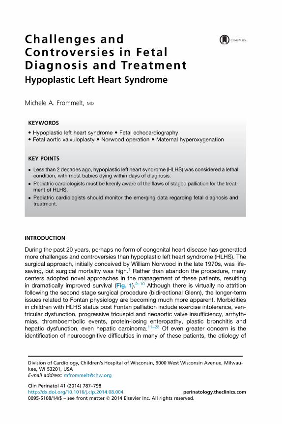
Challenges and Controversies in Fetal Diagnosis and Treatment Hypoplastic Left Heart Syndrome Michele A. Frommelt, MD INTRODUCTION During the past 20 years, perhaps no form of congenital heart disease has generated more challenges and controversies than hypoplastic left heart syndrome (HLHS). The surgical approach, initially conceived by William Norwood in the late 1970s, was life- saving, but surgical mortality was high. 1 Rather than abandon the procedure, many centers adopted novel approaches in the management of these patients, resulting in dramatically improved survival (Fig. 1). 2–10 Although there is virtually no attrition following the second stage surgical procedure (bidirectional Glenn), the longer-term issues related to Fontan physiology are becoming much more apparent. Morbidities in children with HLHS status post Fontan palliation include exercise intolerance, ven- tricular dysfunction, progressive tricuspid and neoaortic valve insufficiency, arrhyth- mias, thromboembolic events, protein-losing enteropathy, plastic bronchitis and hepatic dysfunction, even hepatic carcinoma. 11–23 Of even greater concern is the identification of neurocognitive difficulties in many of these patients, the etiology of Division of Cardiology, Children’s Hospital of Wisconsin, 9000 West Wisconsin Avenue, Milwau- kee, WI 53201, USA E-mail address: [email protected] KEYWORDS Hypoplastic left heart syndrome Fetal echocardiography Fetal aortic valvuloplasty Norwood operation Maternal hyperoxygenation KEY POINTS Less than 2 decades ago, hypoplastic left heart syndrome (HLHS) was considered a lethal condition, with most babies dying within days of diagnosis. Pediatric cardiologists must be keenly aware of the flaws of staged palliation for the treat- ment of HLHS. Pediatric cardiologists should monitor the emerging data regarding fetal diagnosis and treatment. Clin Perinatol 41 (2014) 787–798 http://dx.doi.org/10.1016/j.clp.2014.08.004 perinatology.theclinics.com 0095-5108/14/$ – see front matter Ó 2014 Elsevier Inc. All rights reserved.
Transcript of Challenges and Controversies in Fetal Diagnosis …. Desafios Controversias terapeuticas.pdf ·...
Challenges andControversies in Fetal
Diagnosis and TreatmentHypoplastic Left Heart SyndromeMichele A. Frommelt, MD
KEYWORDS
� Hypoplastic left heart syndrome � Fetal echocardiography� Fetal aortic valvuloplasty � Norwood operation � Maternal hyperoxygenation
KEY POINTS
� Less than 2 decades ago, hypoplastic left heart syndrome (HLHS) was considered a lethalcondition, with most babies dying within days of diagnosis.
� Pediatric cardiologists must be keenly aware of the flaws of staged palliation for the treat-ment of HLHS.
� Pediatric cardiologists should monitor the emerging data regarding fetal diagnosis andtreatment.
INTRODUCTION
During the past 20 years, perhaps no form of congenital heart disease has generatedmore challenges and controversies than hypoplastic left heart syndrome (HLHS). Thesurgical approach, initially conceived by William Norwood in the late 1970s, was life-saving, but surgical mortality was high.1 Rather than abandon the procedure, manycenters adopted novel approaches in the management of these patients, resultingin dramatically improved survival (Fig. 1).2–10 Although there is virtually no attritionfollowing the second stage surgical procedure (bidirectional Glenn), the longer-termissues related to Fontan physiology are becoming much more apparent. Morbiditiesin children with HLHS status post Fontan palliation include exercise intolerance, ven-tricular dysfunction, progressive tricuspid and neoaortic valve insufficiency, arrhyth-mias, thromboembolic events, protein-losing enteropathy, plastic bronchitis andhepatic dysfunction, even hepatic carcinoma.11–23 Of even greater concern is theidentification of neurocognitive difficulties in many of these patients, the etiology of
Division of Cardiology, Children’s Hospital of Wisconsin, 9000 West Wisconsin Avenue, Milwau-kee, WI 53201, USAE-mail address: [email protected]
Clin Perinatol 41 (2014) 787–798http://dx.doi.org/10.1016/j.clp.2014.08.004 perinatology.theclinics.com0095-5108/14/$ – see front matter � 2014 Elsevier Inc. All rights reserved.

Fig. 1. Actuarial survival, Norwood operation. Children’s Hospital of Wisconsin, 2003 to2013. BT, Blalock-Taussig; RV-PA, right ventricle to pulmonary artery.
Frommelt788
which is likely multifactorial.24–26 Although medical therapy and ventricular assist de-vices can temper the situation, eventual cardiac replacement therapy for many ofthese patients seems inevitable. Unfortunately, the risk associated with cardiac trans-plantation in the Fontan population is high, as patients are frequently listed with signif-icant chronic heart failure symptoms and end organ dysfunction, and there is noconsensus on optimal timing of listing.27–29 Pediatric cardiologists should be keenlyaware of the flaws of staged palliation for treatment of HLHS, and need to monitorthe emerging data regarding fetal diagnosis and treatment.
ETIOLOGY OF HYPOPLASTIC LEFT HEART SYNDROME
Before the advent of fetal echocardiography, the embryologic cause of HLHS was notentirely clear. However, in 1989 Allan and colleagues30 observed the in utero evolutionof HLHS in a fetus initially diagnosed with critical aortic stenosis. It is now postulatedthat many cases of HLHS are dynamic and progressive throughout gestation, resultingfrom altered left ventricular outflow as already described or, less commonly, alteredleft ventricular inflow (ie, mitral valve stenosis/foramen ovale restriction).31–33
In normal fetal circulation, the fetal left ventricle is predominantly filled with oxygen-ated blood that returns from the placenta and traverses the foramen ovale. If bloodflow across the foramen ovale is diminished or reversed, the combined cardiac outputis redistributed to the right ventricle and pulmonary artery, resulting in enlargement ofthe right heart structures and creating less impetus for normal growth of left heartstructures. Perhaps the most well-recognized mechanism for decreased flow orreversal of flow through the foramen ovale in utero is the presence of severe aorticvalve disease.34 With significant aortic valve stenosis, alterations in left ventricularcompliance occur, either secondary to the development of left ventricular hypertrophy
Hypoplastic Left Heart Syndrome 789
or to the development of left ventricular dilation and dysfunction. Endocardial fibroe-lastosis, a poorly understood phenomenon whereby the endocardial lining of the leftventricle becomes fibrotic, may also be present. As the disease state progresses,with subsequent elevation in left atrial pressure, flow across the foramen ovale be-comes bidirectional and eventually left to right, the result of which may be the cessa-tion of left ventricular growth.In a classic study by Hornberger and colleagues,35 the prenatal and postnatal echo-
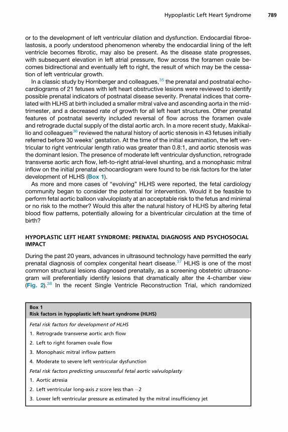
cardiograms of 21 fetuses with left heart obstructive lesions were reviewed to identifypossible prenatal indicators of postnatal disease severity. Prenatal indices that corre-lated with HLHS at birth included a smaller mitral valve and ascending aorta in themid-trimester, and a decreased rate of growth for all left heart structures. Other prenatalfeatures of postnatal severity included reversal of flow across the foramen ovaleand retrograde ductal supply of the distal aortic arch. In a more recent study, Makikal-lio and colleagues36 reviewed the natural history of aortic stenosis in 43 fetuses initiallyreferred before 30 weeks’ gestation. At the time of the initial examination, the left ven-tricular to right ventricular length ratio was greater than 0.8:1, and aortic stenosis wasthe dominant lesion. The presence of moderate left ventricular dysfunction, retrogradetransverse aortic arch flow, left-to-right atrial-level shunting, and a monophasic mitralinflow on the initial prenatal echocardiogram were found to be risk factors for the laterdevelopment of HLHS (Box 1).As more and more cases of “evolving” HLHS were reported, the fetal cardiology
community began to consider the potential for intervention. Would it be feasible toperform fetal aortic balloon valvuloplasty at an acceptable risk to the fetus andminimalor no risk to the mother? Would this alter the natural history of HLHS by altering fetalblood flow patterns, potentially allowing for a biventricular circulation at the time ofbirth?
HYPOPLASTIC LEFT HEART SYNDROME: PRENATAL DIAGNOSIS AND PSYCHOSOCIALIMPACT
During the past 20 years, advances in ultrasound technology have permitted the earlyprenatal diagnosis of complex congenital heart disease.37 HLHS is one of the mostcommon structural lesions diagnosed prenatally, as a screening obstetric ultrasono-gram will preferentially identify lesions that dramatically alter the 4-chamber view(Fig. 2).38 In the recent Single Ventricle Reconstruction Trial, which randomized
Box 1
Risk factors in hypoplastic left heart syndrome (HLHS)
Fetal risk factors for development of HLHS
1. Retrograde transverse aortic arch flow
2. Left to right foramen ovale flow
3. Monophasic mitral inflow pattern
4. Moderate to severe left ventricular dysfunction
Fetal risk factors predicting unsuccessful fetal aortic valvuloplasty
1. Aortic atresia
2. Left ventricular long-axis z score less than �2
3. Lower left ventricular pressure as estimated by the mitral insufficiency jet

Fig. 2. Screening obstetric ultrasound identifying left heart hypoplasia. Arrow identifiesbowing of atrial septum from left to right. RA, right atrium; RV, right ventricle.
Frommelt790
patients with HLHS to a modified Blalock-Taussig shunt versus right ventricular to pul-monary artery conduit during stage I palliation (Norwood) procedure, 75% of the pa-tients were diagnosed prenatally.39 Prenatal diagnosis has resulted in complexdecision making before the birth of the baby, ranging from termination of pregnancyto alteration in care and delivery plans, and, in a select few, referral for fetal cardiacintervention.There is no doubt that the presence of any significant abnormality identified prena-
tally can result in a variety of emotions including grief, anxiety, and stress.40,41 In arecent study by Larsson and colleagues,42 16 parents (mothers and fathers) wereinterviewed after their fetus had been diagnosed with an abnormality. Although theinitial identification of the anomaly resulted in broken expectations (“You are verysad and shocked in the beginning.because the dream of a perfect child is broken)and anxiety (“I was laying there and becoming more anxious about everything”), par-ents quickly became involved in change and adaptation. The parents wanted accurateinformation about the anomaly, without a prolonged delay, and in a suitable environ-ment. In the author’s experience, the best way to approach this is the development ofa multidisciplinary fetal heart program that includes fetal cardiologists, maternal fetalmedicine experts, cardiac surgeons, and, importantly, a dedicated fetal cardiac nursecoordinator (Box 2). Having a team immediately available to consult with these fam-ilies seems to help them cope with what will be a difficult period of time in their life.The dedicated nurse coordinator guides the family through the prenatal experience,fielding any questions or concerns that arise, and coordinating care, which oftenincludes a change in delivery plan. Families are also referred to other parents whohave gone through a similar experience, who are therefore able to give them

Box 2
Key elements of multidisciplinary fetal cardiac program
Nurse coordinator
24/7 intake; gathering of data; greet and welcome at visit; observation of counseling with allproviders; coordination of care and follow-up appointments; tours of cardiac intensive careunit; dedicated follow-up as needed for continued support of family.
Fetal cardiologist
24/7 availability for performance and interpretation of complete fetal echocardiogram;same-day counseling and education in a quiet, suitable environment with the availability ofdescriptive images and data; knowledge of institutional surgical volume and outcomes; serialechocardiographic follow-up as indicated by heart diagnosis.
Maternal fetal medicine expert
24/7 availability for performance/interpretation of high-risk scan identifying any associatedextracardiac anomalies; discussion of genetic screening; assessment of fetal growth and fetalwell-being; plans for delivery with discussion of delivery options.
Cardiothoracic surgery
Available for consultation with family during pregnancy to answer any specific surgical ques-tions that the family may have; part of planning for IMPACT (Immediate Postnatal Access toCardiac Therapy) if necessary.
Additional support
Genetics, neonatology, social work, lactation, child life, interstage home monitoring program,research coordinators, and, if indicated, other pediatric specialties including pediatric surgery.
Hypoplastic Left Heart Syndrome 791
first-hand knowledge of raising a child with HLHS. All of these services provide the re-ality that their baby does have a chance of a good outcome.
HYPOPLASTIC LEFT HEART SYNDROME: RISK FACTORS FOR ADVERSE OUTCOME
Extracardiac anomalies, including genetic disorders, occur in 15% to 30% of patientswith HLHS.43,44 A few of the identifiable heritable syndromes and genetic disordersassociated with HLHS include Kabuki syndrome, Noonan syndrome, trisomy 13, tri-somy 18, Turner syndrome, and Jacobsen syndrome. Prenatal detection may affectparental decision making, as it is well known that any additional anomaly will decreasesurvival after Norwood palliation.45 To better identify these associated problems, theauthor recommends a complete fetal ultrasonography examination by maternal fetalmedicine colleagues, in addition to a genetics evaluation, again supporting the multi-disciplinary team approach (see Box 2).Parents may also have questions regarding surgical volumes and outcomes, as they
do have the ability to access this information through many avenues, especially theInternet. Several recent publications suggest that smaller center and surgeon volumesare associated with adverse outcomes in children undergoing a variety of surgical pro-cedures, including the Norwood operation.46,47 Based on these findings, the investi-gators postulate that overall outcomes in the United States may be improved
Frommelt792
through regional collaboration and the development of quality improvement initiativeswithin and across centers. This notion also raises the controversial question, “Shouldwe have centers of excellence?”, an approach taken in some European countries.Although the operative survival for infants born with HLHS has improved signifi-
cantly over time, the subgroup of patients with a highly restrictive or intact atrialseptum continues to experience a higher mortality.48,49 These infants can be pro-foundly cyanotic at the time of delivery and are often unresponsive to medical inter-vention. Even with prompt resuscitation and adequate decompression of the atrialseptum, there is ongoing morbidity and mortality, likely related to secondary anatomicchanges in the lung. Some investigators have reported “arterialization” of the pulmo-nary veins and lymphatic dilation in this setting; others have postulated that there isassociated pulmonary artery hypoplasia.50
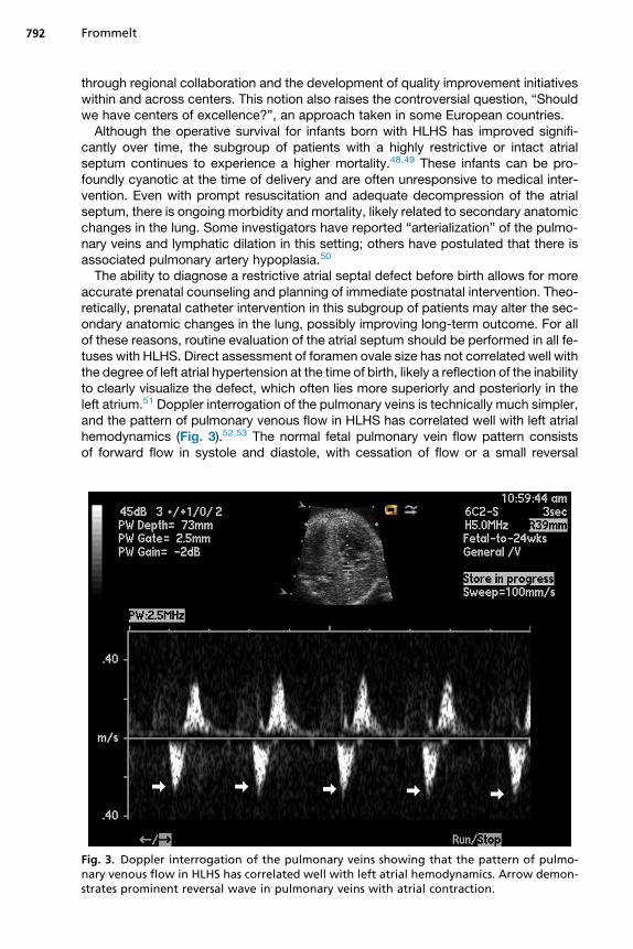
The ability to diagnose a restrictive atrial septal defect before birth allows for moreaccurate prenatal counseling and planning of immediate postnatal intervention. Theo-retically, prenatal catheter intervention in this subgroup of patients may alter the sec-ondary anatomic changes in the lung, possibly improving long-term outcome. For allof these reasons, routine evaluation of the atrial septum should be performed in all fe-tuses with HLHS. Direct assessment of foramen ovale size has not correlated well withthe degree of left atrial hypertension at the time of birth, likely a reflection of the inabilityto clearly visualize the defect, which often lies more superiorly and posteriorly in theleft atrium.51 Doppler interrogation of the pulmonary veins is technically much simpler,and the pattern of pulmonary venous flow in HLHS has correlated well with left atrialhemodynamics (Fig. 3).52,53 The normal fetal pulmonary vein flow pattern consistsof forward flow in systole and diastole, with cessation of flow or a small reversal
Fig. 3. Doppler interrogation of the pulmonary veins showing that the pattern of pulmo-nary venous flow in HLHS has correlated well with left atrial hemodynamics. Arrow demon-strates prominent reversal wave in pulmonary veins with atrial contraction.

Hypoplastic Left Heart Syndrome 793
wave with atrial systole. In a study by Taketazu and colleagues,51 a pattern of a largereversal wave back into the pulmonary veins was associated with the need for imme-diate respiratory support and emergent atrial decompression. More specifically,calculation of the forward flow versus reverse flow Doppler derived velocity time inte-gral has also been predictive of the need for immediate neonatal intervention, andshould be routinely performed in all fetuses with HLHS.54
HYPOPLASTIC LEFT HEART SYNDROME: MANAGEMENT STRATEGIESDelivery
When babies with HLHS are diagnosed de novo postnatally, they can present inextremis related to ductal closure and low cardiac output. If the atrial septum is restric-tive, they can present with severe hypoxemia as well. When there is a prenatal diag-nosis of HLHS, the author recommends delivery at the birthing center, so there can beimmediate stabilization and initiation of prostaglandin to maintain ductal patency. Thisapproach also allows the family to be in close contact with the physicians taking careof the baby, and allows for maternal bonding. Onemight expect that the improved pre-operative condition of the baby with prenatally diagnosed HLHS would result inimproved surgical outcome; however, recent studies suggest otherwise.54 A retro-spective study at the University of California in San Francisco evaluated 81 patientswith HLHS who presented to their hospital between 1999 and 2010; more than halfof the patients were diagnosed prenatally. Although the postnatally diagnosedpatients had increased preoperative acidosis, multiorgan failure, right ventriculardysfunction, and tricuspid insufficiency on presentation, there was no difference inNorwood survival between groups. Long-term follow-up will be important, however,as there is a possibility that neurocognitive outcomes may be worse in those who pre-sent with cardiovascular collapse.
Maternal Hyperoxygenation
In the normal fetus, only a small proportion of the fetal cardiac output is directed to thelungs, with most flow directed across the ductus arteriosus to descending aorta.Studies have demonstrated that maternal hyperoxygenation later in pregnancy can in-crease pulmonary blood flow, and this has been used to assess pulmonary reactivity infetuses with suspected pulmonary hypoplasia related to diaphragmatic hernia andsevere renal disease.55 In a recent study by Szwast and colleagues,56 maternal hyper-oxygenation was used to assess pulmonary reactivity in fetuses with HLHS and eitheran open atrial septum or a restrictive/intact atrial septum. The mother was adminis-tered 100% oxygen via a nonrebreather face mask for 10 minutes, and fetal Dopplerassessment of pulmonary artery flow was measured at baseline, with maternaloxygen, and after recovery. The pulsatility index, a surrogate measure of vascularimpedance, was used for assessment of pulmonary blood flow. Maternal hyperoxyge-nation led to a significant increase in pulmonary blood flow in fetuses with an openatrial septum; however, this was not the case in fetuses with atrial septal restrictionthat required immediate intervention on the atrial septum at birth. It seems that thefetal response to maternal hyperoxygenation is predictive of the need for urgent inter-vention at the time of birth, and the use of this diagnostic technique can be very helpfulwhen planning the delivery. In the setting of a fetus with HLHS and restrictive or intactatrial septum and lack of pulmonary reactivity, the author recommends cesarean sec-tion in either the delivery room or operating room suite so that there can be ImmediatePostnatal Access to Cardiac Therapy (IMPACT procedure), either surgical or interven-tional. The IMPACT procedure was designed to manage high-risk patients and

Frommelt794
assemble the multidisciplinary resources required to care for these critically ill neo-nates in the immediate postpartum period.49
Therapeutic use of maternal hyperoxygenation has also been proposed, as therehas also been evidence that chronic intermittent maternal hyperoxygenation in lategestation may cause growth of hypoplastic cardiac structures. Thomas Kohl per-formed repetitive daily maternal hyperoxygenation in 15 pregnant women between33 and 38 weeks’ gestation.57 The fetal cardiac disease was variable, but 13 of the15 fetuses had hypoplasia of at least one left heart structure. Kohl demonstrated in-creases in cardiovascular dimensions (improvements in z scores for gestational age)in most fetuses with small ventricles and no inflow/outflow obstruction. The presenceof inflow/outflow tract obstruction or a large ventricular septal defect seemed toameliorate the effect of hyperoxygenation. Maternal hyperoxygenation is a new andexciting potential therapy in select fetal patients, especially considering its simplicityand universal availability. However, the long-term effects of hyperoxygenation onthe fetus remain unknown.
Fetal Catheter Intervention
In 2000, Kohl and colleagues58 reported the world experience of fetal aortic balloonvalvuloplasty. The small early clinical experience (n 5 12) was poor, with only 1“long-term” survivor. However, more encouraging data were recently reported byMcElhinney and colleagues59 from the Children’s Hospital of Boston and the Brighamand Women’s Hospital. These data included 70 fetuses who underwent attemptedaortic valvuloplasty for critical aortic stenosis with evolving HLHS between March2000 and October 2008. There was a significant improvement in technical success(74%), and most procedures were performed with only percutaneous access (73%).Eight fetuses died in relation to the procedure (11% mortality). Although fetuseswith a technically successful valvuloplasty had improved growth of the aortic valveand mitral valve, intervention did not effectively promote left ventricular growth. There-fore, fetuses with a larger left ventricular dimensions initially were more likely to sustaina biventricular circulation at the time of birth (n 5 15). Based on these results, theinvestigators were able to create a multivariable scoring system to improve patientselection. Predictors of an unsuccessful fetal aortic valvuloplasty include the presenceof aortic atresia, a left ventricular long axis z score of less than �2, and lower left ven-tricular pressure as estimated by the mitral insufficiency jet (see Box 1).It is important to bear in mind that most fetuses with critical aortic stenosis will sur-
vive gestation. Therefore, fetal cardiac intervention in this setting does not serve as alife-saving procedure, but rather a procedure that may improve postnatal surgicaloptions and outcomes. More specifically, the goal is that successful intervention willlead to a biventricular circulation at the time of birth. There is also an assumptionthat a biventricular circulation is better than a univentricular circulation. That beingsaid, the possible benefits must be weighed against the risks of the procedure, whichinclude fetal demise or extreme prematurity. One must also consider maternal risk,although no maternal deaths associated with fetal intervention have been reportedin the United States. Because the risk/benefit ratio of fetal cardiac intervention inthe setting of critical aortic stenosis is still largely unknown, it is not surprising thatthese procedures are not universally accepted. Some centers have advocated fetalcardiac intervention only when it is considered to be a life-saving procedure, suchas in the setting of critical aortic stenosis with hydrops fetalis.In utero therapy for HLHS with a severely restrictive or intact atrial septum has also
been described. Successful decompression of the left atrium in utero may avoidsevere hypoxemia at birth, and theoretically may also reduce prenatal lung damage

Hypoplastic Left Heart Syndrome 795
and improve otherwise dismal outcomes. In a recent publication by Marshall and col-leagues,60 technically successful atrial septoplasty was performed in 19 of 21 fetusesbetween October 2001 and November 2007, with 2 episodes of fetal demise.59 Theinvestigators determined that creation of a larger defect was associated with betterpostnatal oxygenation; however, whether this confers a benefit to later survival ispresently unknown.
SUMMARY
Less than 2 decades ago HLHS was considered a lethal condition, with most babiesdying within days of diagnosis. Today it is estimated that almost 70% of patients withHLHS will survive into adulthood.61 Despite this advance, there is significant long-term morbidity and mortality in this patient group, and clinicians are continually work-ing to achieve better outcomes. At the same time there has been exciting progress infetal diagnosis and intervention. Most babies with HLHS can be expected to be iden-tified prenatally, with features likely to lead to a poor outcome able to be recognized,leading to improved counseling. It is now known that in utero dilatation of the aorticvalve or atrial septum is technically feasible; however, these procedures require com-plex choreography, and there needs to be a dedicated team involved. One must bearin mind that 2 lives are at risk, and the safety of the mother is paramount. A salientpoint is that a biventricular circulation with multiple left-sided lesions has associatedchronic morbidity and mortality, and there are essentially no long-term data on pa-tients with a biventricular circulation after fetal intervention.
REFERENCES
1. Norwood WI, Kirkland JK, Sanders SP. Hypoplastic left heart syndrome: experi-ence with palliative surgery. Am J Cardiol 1980;45:87–91.
2. Azakie A, Merklinger SL, McCrindle BW, et al. Evolving strategies and improvingoutcomes of the modified Norwood procedure: a 10-year single-institution expe-rience. Ann Thorac Surg 2001;72:1349–53.
3. Sano S, Ishino K, Kawada M, et al. Right ventricle-pulmonary artery shunt in first-stage palliation of hypoplastic left heart syndrome. Semin Thorac CardiovascSurg Pediatr Card Surg Annu 2004;7:22–31.
4. Pizarro C, Malec E, Maher KO, et al. Right ventricle to pulmonary artery conduitimproves outcome after stage I Norwood for hypoplastic left heart syndrome.Circulation 2003;108:II155–60.
5. Tweddell JS, Hoffman GM, Fedderly RT, et al. Phenoxybenzamine improvessystemic oxygen delivery after the Norwood procedure. Ann Thorac Surg1999;67:161–7.
6. Hoffman GM, Ghanayem NS, Kampine JM, et al. Venous saturation and theanaerobic threshold in neonates after the Norwood procedure for hypoplasticleft heart syndrome. Ann Thorac Surg 2000;70:1515–20.
7. Tweddell JS, Hoffman GM, Mussatto KA, et al. Improved survival of patientsundergoing palliation of hypoplastic left heart syndrome: lessons learned from115 consecutive patients. Circulation 2002;106:I82–9.
8. Tweddell JS, Ghanayem NS, Mussatto KA, et al. Mixed venous oxygen satura-tion monitoring after stage 1 palliation for hypoplastic left heart syndrome. AnnThorac Surg 2007;84:1301–11.
9. Hoffman GM, Mussatto KA, Brosig CL, et al. Systemic venous oxygen saturationafter the Norwood procedure and childhood neurodevelopmental outcome.J Thorac Cardiovasc Surg 2005;130:1094–100.

Frommelt796
10. Tortoriello TA, Stayer SA, Mott AR, et al. A noninvasive estimation of mixedvenous oxygen saturation using near-infrared spectroscopy by cerebral ox-imetry in pediatric cardiac surgery patients. Paediatr Anaesth 2005;15:495–503.
11. Ranucci M, Isgro G, De La Torre T, et al. Near-infrared spectroscopy correlateswith continuous superior vena cava oxygen saturation in pediatric cardiac sur-gery patients. Paediatr Anaesth 2008;18:1163–9.
12. Ghanayem NS, Hoffman GM, Mussatto KA, et al. Home surveillance programprevents interstage mortality after the Norwood procedure. J Thorac CardiovascSurg 2003;126:1367–75.
13. Kugler JD, Beekman Iii RH, Rosenthal GL, et al. Development of a pediatric car-diology quality improvement collaborative: from inception to implementation.From the Joint Council on Congenital Heart Disease Quality ImprovementTask Force. Congenit Heart Dis 2009;4:318–28.
14. Anderson PA, Sleeper LA, Mahony L, et al. Contemporary outcomes after theFontan procedure: a Pediatric Heart Network multicenter study. J Am Coll Car-diol 2008;52:85–98.
15. Shachar G, Fuhrman B, Wang Y, et al. Rest and exercise hemodynamics afterthe Fontan procedure. Circulation 1982;65:1043–8.
16. Cohen MS, Marino BS, McElhinney DB, et al. Neo-aortic root dilation and valveregurgitation up to 21 years after staged reconstruction for hypoplastic left heartsyndrome. J Am Coll Cardiol 2003;42:533–40.
17. Ghaferi AA, Hutchins GM. Progression of liver pathology in patients undergoingthe Fontan procedure: chronic passive congestion, cardiac cirrhosis, hepaticadenoma, and hepatocellular carcinoma. J Thorac Cardiovasc Surg 2005;129:1348–52.
18. Kiesewetter CH, Sheron N, Vettukattill JJ, et al. Hepatic changes in the failingFontan circulation. Heart 2007;93:579–84.
19. Crupi G, Locatelli G, Tiraboschi R, et al. Protein-losing enteropathy after Fontanoperation for tricuspid atresia (imperforate tricuspid valve). Thorac CardiovascSurg 1980;28:359–63.
20. Feldt RH, Driscoll DJ, Offord KP, et al. Protein-losing enteropathy after theFontan operation. J Thorac Cardiovasc Surg 1996;112:672–80.
21. Goo HW, Jhang WK, Kim YH, et al. CT findings of plastic bronchitis in childrenafter a Fontan operation. Pediatr Radiol 2008;38:989–93.
22. Ghai A, Harris L, Harrison DA, et al. Outcomes of late atrial tachyarrhythmias inadults after the Fontan operation. J Am Coll Cardiol 2001;37:585–92.
23. Stephenson EA, Lu M, Berul CI, et al. Arrhythmias in a contemporary Fontancohort: prevalence and clinical associations in a multicenter cross-sectionalstudy. J Am Coll Cardiol 2010;56:890–6.
24. Mahle WT, Clancy RR, Moss EM, et al. Neurodevelopmental outcome and life-style assessment in school-aged and adolescent children with hypoplastic leftheart syndrome. Pediatrics 2000;105:1082–9.
25. Goldberg CS, Schwartz EM, Brunberg JA, et al. Neurodevelopmental outcomeof patients after the Fontan operation: a comparison between children withhypoplastic left heart syndrome and other functional single ventricle lesions.J Pediatr 2000;137:646–52.
26. Limperopoulos C. Disorders of the fetal circulation and the fetal brain. ClinPerinatol 2009;36:561–77.
27. Gamba A, Merlo M, Fiocchi R, et al. Heart transplantation in patients with previ-ous Fontan operations. J Thorac Cardiovasc Surg 2004;127:555–62.

Hypoplastic Left Heart Syndrome 797
28. Bernstein D, Naftel D, Chin C, et al. Outcome of listing for cardiac transplanta-tion for failed Fontan: a multi-institutional study. Circulation 2006;114:273–80.
29. Michielon G, Parisi F, Di Carlo D, et al. Orthotopic heart transplantation for failingsingle ventricle physiology. Eur J Cardiothorac Surg 2003;24:502–10.
30. Allan LD, Sharland GK, Tynan MJ. The natural history of hypoplastic left heartsyndrome. Int J Cardiol 1989;25:341–3.
31. Feit LR, Copel JA, Kleinman CS. Foramen ovale size in the normal and abnormalhuman fetal heart: an indicator of transatrial flow physiology. Ultrasound ObstetGynecol 1991;1:313–9.
32. Chin AJ, Weinberg PM, Barber G. Subcostal two-dimensional echocardiographicidentification of anomalous attachment of septum primum in patients with leftatrioventricular valve underdevelopment. J Am Coll Cardiol 1990;15:678–81.
33. Lurie PR. Changing concepts of endocardial fibroelastosis. Cardiol Young 2010;20:115–23.
34. Danford DA, Cronican P. Hypoplastic left heart syndrome: progression of leftventricular dilation and dysfunction to left ventricular hypoplasia in utero. AmHeart J 1992;123:1712–3.
35. Hornberger LK, Sanders SP, Rein AJ, et al. Left heart obstructive lesions and leftventricular growth in the midtrimester fetus: a longitudinal study. Circulation1995;92:1531–8.
36. Makikallio K, Levine JC, Marx GR, et al. Fetal aortic valve stenosis and the evo-lution of hypoplastic left heart syndrome: patient selection for fetal intervention[abstract]. Circulation 2004;110:III690.
37. Allan LD, Crawford DC, Chita SK, et al. Prenatal screening for congenital heartdisease. BMJ 1986;292:1717–9.
38. Copel JA, Pilu G, Green J, et al. Fetal echocardiographic screening for congen-ital heart disease: the importance of the four chamber view. Am J Obstet Gyne-col 1987;157(3):648–55.
39. Atz AM, Travison TG, Williams IA, et al. Prenatal diagnosis and risk factors forpreoperative death in neonates with single right ventricle and systemic outflowobstruction: screening data from the Pediatric Heart Network Single VentricleReconstruction Trial. J Thorac Cardiovasc Surg 2010;140(6):1245–50.
40. Brosig CL, Whitstne BN, Frommelt MA, et al. Psychological distress in parents ofchildren with severe congenital heart disease: the impact of prenatal versuspostnatal diagnosis. J Perinatol 2007;27:687–92.
41. Sklansky M, Tang A, Levy D, et al. Maternal psychological impact of fetal echo-cardiography. J Am Soc Echocardiogr 2002;15:159–66.
42. Larrson AK, Svalenius EC, Lundqvist A, et al. Parents’ experiences of anabnormal ultrasound examination – vacillating between emotional confusionand sense of reality. Reprod Health 2010;7:1–10.
43. Ye M, Coldren C, Liang X, et al. Deletion of ETS-1, a gene in the Jacobsen syn-drome critical region, causes ventricular septal defects and abnormal ventricu-lar morphology in mice. Hum Mol Genet 2010;19:648–56.
44. Natowicz M, Chatten J, Clancy R, et al. Genetic disorders and major extracar-diac anomalies associated with the hypoplastic left heart syndrome. Pediatrics1988;82:698–706.
45. Stasik CN, Goldberg CS, Bove EL, et al. Current outcomes and risk factors forthe Norwood procedure. J Thorac Cardiovasc Surg 2006;131:412–7.
46. Hornik CP, He X, Jacobs JP, et al. Relative impact of surgeon and center volumeon early mortality after the Norwood procedure. Ann Thorac Surg 2012;93(6):1992–7.

Frommelt798
47. Checchia PA, McCollegan J, Daher N, et al. The effect of surgical case volumeon outcome after the Norwood procedure. J Thorac Cardiovasc Surg 2005;129(4):754–9.
48. Vlahos AP, Lock JE, McElhinney DB, et al. Hypoplastic left heart syndrome withintact or highly restrictive atrial septum: outcome after neonatal transcatheteratrial septostomy. Circulation 2004;109:2326–30.
49. Glatz JA, Gaynor JW, Rome JJ, et al. Hypoplastic left heart syndrome with atriallevel restriction in the era of prenatal diagnosis. Ann Thorac Surg 2007;84(5):1633–8.
50. Rychik J, Rome JJ, Collins MH, et al. The hypoplastic left heart syndrome withintact atrial septum: atrial morphology, pulmonary vascular histopathology andoutcome. J Am Coll Cardiol 1999;34:554–60.
51. Taketazu M, Barrea C, Smallhorn JF, et al. Intrauterine pulmonary venous flowand restrictive foramen ovale in fetal hypoplastic left heart syndrome. J AmColl Cardiol 2004;43:1902–7.
52. Better DJ, Apfel HD, Zidere V, et al. Pattern of pulmonary venous blood flow inthe hypoplastic left heart syndrome in the fetus. Heart 1999;81:646–9.
53. Michelfelder E, Gomez C, Border W, et al. Predictive value of fetal pulmonaryvenous flow patterns in identifying the need for atrial septoplasty in the newbornwith hypoplastic left ventricle. Circulation 2005;112(19):2974–9.
54. Kipps AK, Feuille C, Azakie A, et al. Prenatal diagnosis of hypoplastic left heartsyndrome in current era. Am J Cardiol 2011;108(3):421–7.
55. Broth RE, Wood DC, Rasanen J, et al. Prenatal prediction of lethal pulmonaryhypoplasia: the hyperoxygenation test for pulmonary artery reactivity. Am JObstet Gynecol 2002;187:940–5.
56. Szwast A, Tian Z, McCann M, et al. Vasoreactive response to maternal hyper-oxygenation in the fetus with hypoplastic left heart syndrome. Circ CardiovascImaging 2010;3(2):172–8.
57. Kohl T. Chronic intermittent materno-fetal hyperoxygenation in late gestationmay improve on hypoplastic cardiovascular structures associated with cardiacmalformations in human fetuses. Pediatr Cardiol 2010;31:250–63.
58. Kohl T, Sharland G, Allan LD, et al. World experience of percutaneousultrasound-guided balloon valvuloplasty in human fetuses with severe aorticvalve obstruction. Am J Cardiol 2000;85:1230–3.
59. McElhinney DB, Marshall AC, Wilkins-Haug LE, et al. Predictors of technicalsuccess and postnatal biventricular outcome after in utero aortic valvuloplastyfor aortic stenosis with evolving hypoplastic left heart syndrome. Circulation2009;120:1482–90.
60. Marshall A, Levine J, Morash D, et al. Results of in utero atrial septoplasty infetuses with hypoplastic left heart syndrome. Prenat Diagn 2008;28:1023–8.
61. Feinstein JA, Benson DW, Dubin AM, et al. Hypoplastic left heart syndrome: cur-rent considerations and expectations. J Am Coll Cardiol 2012;59(Suppl 1):S1–42.