Challenges and advances in clinical applications of ...
24
Zhou et al. J Hematol Oncol (2021) 14:24 https://doi.org/10.1186/s13045-021-01037-x REVIEW Challenges and advances in clinical applications of mesenchymal stromal cells Tian Zhou 1,2 , Zenan Yuan 3 , Jianyu Weng 1 , Duanqing Pei 4 , Xin Du 1* , Chang He 2* and Peilong Lai 1* Abstract Mesenchymal stromal cells (MSCs), also known as mesenchymal stem cells, have been intensely investigated for clinical applications within the last decades. However, the majority of registered clinical trials applying MSC therapy for diverse human diseases have fallen short of expectations, despite the encouraging pre-clinical outcomes in varied animal disease models. This can be attributable to inconsistent criteria for MSCs identity across studies and their inher- ited heterogeneity. Nowadays, with the emergence of advanced biological techniques and substantial improvements in bio-engineered materials, strategies have been developed to overcome clinical challenges in MSC application. Here in this review, we will discuss the major challenges of MSC therapies in clinical application, the factors impacting the diversity of MSCs, the potential approaches that modify MSC products with the highest therapeutic potential, and finally the usage of MSCs for COVID-19 pandemic disease. Keywords: Mesenchymal stromal cells, Clinical applications, Heterogeneity, Artificial intelligence (AI), Extracellular vesicles, COVID-19 © The Author(s) 2021. Open Access This article is licensed under a Creative Commons Attribution 4.0 International License, which permits use, sharing, adaptation, distribution and reproduction in any medium or format, as long as you give appropriate credit to the original author(s) and the source, provide a link to the Creative Commons licence, and indicate if changes were made. The images or other third party material in this article are included in the article’s Creative Commons licence, unless indicated otherwise in a credit line to the material. If material is not included in the article’s Creative Commons licence and your intended use is not permitted by statutory regulation or exceeds the permitted use, you will need to obtain permission directly from the copyright holder. To view a copy of this licence, visit http://creativecommons.org/licenses/by/4.0/. The Creative Commons Public Domain Dedication waiver (http://creativeco mmons.org/publicdomain/zero/1.0/) applies to the data made available in this article, unless otherwise stated in a credit line to the data. Background Mesenchymal stromal cells (MSCs) are pluripotent non- hematopoietic stem cells with self-renewal capability [1] and being intensively investigated in clinical trials. Since the discovery of MSCs from bone marrow by Frieden- stein in 1970s, MSCs have been isolated from various sources including muscle, umbilical cord, liver, placenta, skin, amniotic fluid, synovial membrane, and tooth root [2, 3], and tested in amounts of preclinical and clinical studies (Fig. 1). It is now understood that MSCs have wide-ranging physiological effects including the main- tenance of tissue homeostasis and regeneration [4, 5], as well as the immunomodulatory activities suitable for therapeutic application [6]. So their indications have been expanded to graft-versus-host disease (GVHD), multiple sclerosis (MS), Crohn’s disease (CD), amyotrophic lateral sclerosis (ALS), myocardial infarction (MI), and acute respiratory distress syndrome (ARDS) [7–9]. Over 300 clinical trials of MSC therapies have been completed in patients including but not limited to degen- erative or autoimmune diseases (Table 1 lists some of the representative completed studies). Overall, MSCs have exhibited tolerable safety profile and demonstrated prom- ising therapeutic benefits in some clinical settings, which led to regulatory approvals of MSCs in a few countries. In 2011, the Ministry of Food and Drug Safety (Korea FDA) approved Cartistem ® , a MSC product derived from umbilical cord blood and developed by Medipost for the treatment of traumatic or degenerative osteoar- thritis [10]. ereafter, more MSC products including HeartiCellgram ® , Mesoblast, TiGenix, and Stempeutics, were approved by regulatory authorities worldwide for the treatment of a variety of diseases. In the USA, Ryon- cil (remestemcel-L) is promising to be the first FDA- approved GVHD treatment for children younger than 12, but is still in the stage of safety verification. e amount Open Access *Correspondence: [email protected]; [email protected]; [email protected] 1 Department of Hematology, Guangdong Provincial People’s Hospital, Guangdong Academy of Medical Sciences, Guangzhou 510080, People’s Republic of China 2 State Key Laboratory of Ophthalmology, Zhongshan Ophthalmic Center, Sun Yat-Sen University, Guangzhou 510060, People’s Republic of China Full list of author information is available at the end of the article
Transcript of Challenges and advances in clinical applications of ...

Zhou et al. J Hematol Oncol (2021) 14:24 https://doi.org/10.1186/s13045-021-01037-x
REVIEW
Challenges and advances in clinical applications of mesenchymal stromal cellsTian Zhou1,2, Zenan Yuan3, Jianyu Weng1, Duanqing Pei4, Xin Du1*, Chang He2* and Peilong Lai1*
Abstract
Mesenchymal stromal cells (MSCs), also known as mesenchymal stem cells, have been intensely investigated for clinical applications within the last decades. However, the majority of registered clinical trials applying MSC therapy for diverse human diseases have fallen short of expectations, despite the encouraging pre-clinical outcomes in varied animal disease models. This can be attributable to inconsistent criteria for MSCs identity across studies and their inher-ited heterogeneity. Nowadays, with the emergence of advanced biological techniques and substantial improvements in bio-engineered materials, strategies have been developed to overcome clinical challenges in MSC application. Here in this review, we will discuss the major challenges of MSC therapies in clinical application, the factors impacting the diversity of MSCs, the potential approaches that modify MSC products with the highest therapeutic potential, and finally the usage of MSCs for COVID-19 pandemic disease.
Keywords: Mesenchymal stromal cells, Clinical applications, Heterogeneity, Artificial intelligence (AI), Extracellular vesicles, COVID-19
© The Author(s) 2021. Open Access This article is licensed under a Creative Commons Attribution 4.0 International License, which permits use, sharing, adaptation, distribution and reproduction in any medium or format, as long as you give appropriate credit to the original author(s) and the source, provide a link to the Creative Commons licence, and indicate if changes were made. The images or other third party material in this article are included in the article’s Creative Commons licence, unless indicated otherwise in a credit line to the material. If material is not included in the article’s Creative Commons licence and your intended use is not permitted by statutory regulation or exceeds the permitted use, you will need to obtain permission directly from the copyright holder. To view a copy of this licence, visit http://creat iveco mmons .org/licen ses/by/4.0/. The Creative Commons Public Domain Dedication waiver (http://creat iveco mmons .org/publi cdoma in/zero/1.0/) applies to the data made available in this article, unless otherwise stated in a credit line to the data.
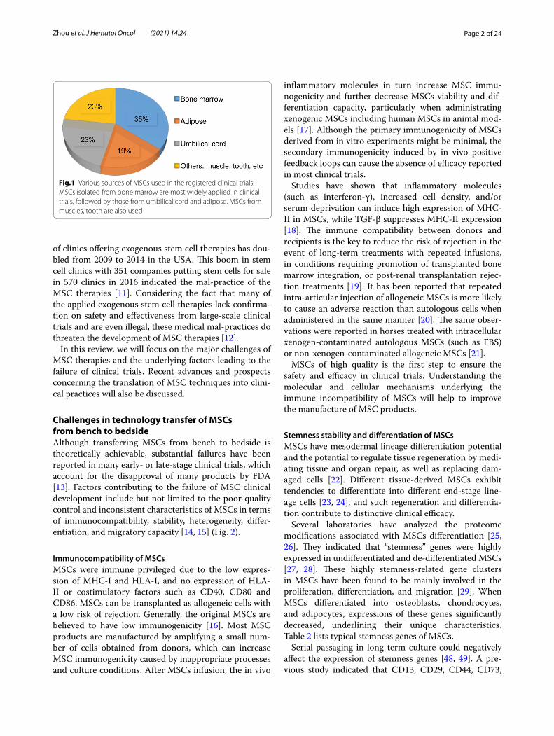
BackgroundMesenchymal stromal cells (MSCs) are pluripotent non-hematopoietic stem cells with self-renewal capability [1] and being intensively investigated in clinical trials. Since the discovery of MSCs from bone marrow by Frieden-stein in 1970s, MSCs have been isolated from various sources including muscle, umbilical cord, liver, placenta, skin, amniotic fluid, synovial membrane, and tooth root [2, 3], and tested in amounts of preclinical and clinical studies (Fig. 1). It is now understood that MSCs have wide-ranging physiological effects including the main-tenance of tissue homeostasis and regeneration [4, 5], as well as the immunomodulatory activities suitable for therapeutic application [6]. So their indications have been
expanded to graft-versus-host disease (GVHD), multiple sclerosis (MS), Crohn’s disease (CD), amyotrophic lateral sclerosis (ALS), myocardial infarction (MI), and acute respiratory distress syndrome (ARDS) [7–9].
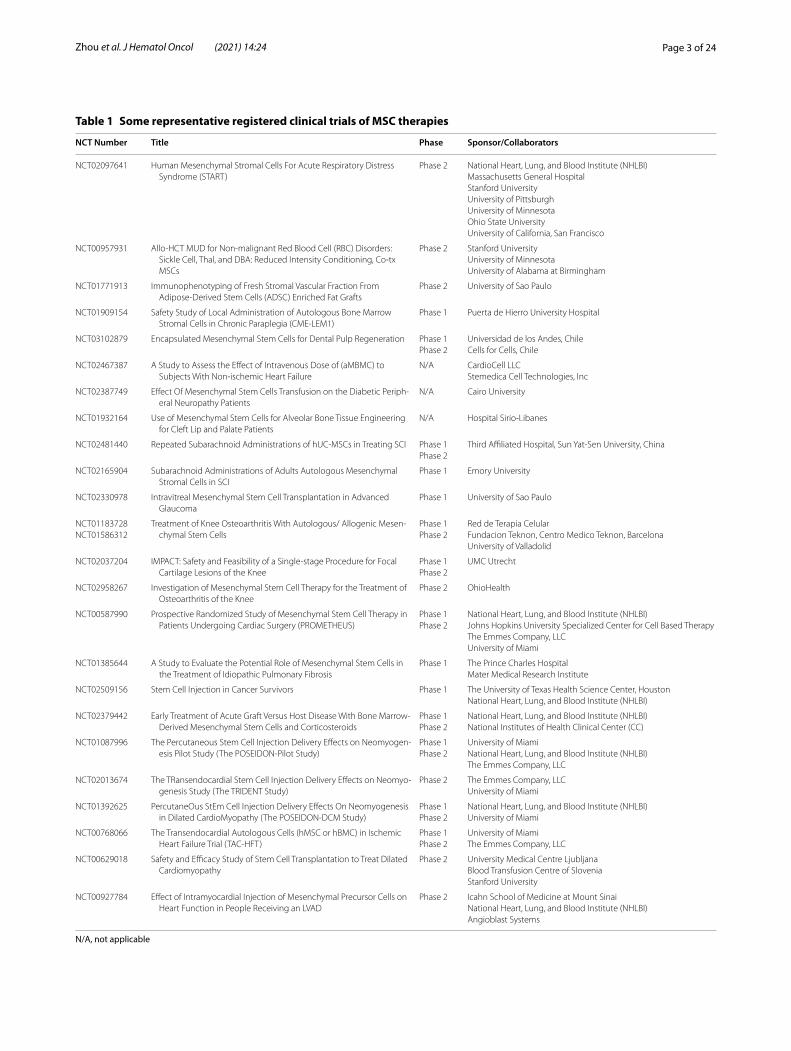
Over 300 clinical trials of MSC therapies have been completed in patients including but not limited to degen-erative or autoimmune diseases (Table 1 lists some of the representative completed studies). Overall, MSCs have exhibited tolerable safety profile and demonstrated prom-ising therapeutic benefits in some clinical settings, which led to regulatory approvals of MSCs in a few countries. In 2011, the Ministry of Food and Drug Safety (Korea FDA) approved Cartistem®, a MSC product derived from umbilical cord blood and developed by Medipost for the treatment of traumatic or degenerative osteoar-thritis [10]. Thereafter, more MSC products including HeartiCellgram®, Mesoblast, TiGenix, and Stempeutics, were approved by regulatory authorities worldwide for the treatment of a variety of diseases. In the USA, Ryon-cil (remestemcel-L) is promising to be the first FDA-approved GVHD treatment for children younger than 12, but is still in the stage of safety verification. The amount
Open Access
*Correspondence: [email protected]; [email protected]; [email protected] Department of Hematology, Guangdong Provincial People’s Hospital, Guangdong Academy of Medical Sciences, Guangzhou 510080, People’s Republic of China2 State Key Laboratory of Ophthalmology, Zhongshan Ophthalmic Center, Sun Yat-Sen University, Guangzhou 510060, People’s Republic of ChinaFull list of author information is available at the end of the article
Page 2 of 24Zhou et al. J Hematol Oncol (2021) 14:24
of clinics offering exogenous stem cell therapies has dou-bled from 2009 to 2014 in the USA. This boom in stem cell clinics with 351 companies putting stem cells for sale in 570 clinics in 2016 indicated the mal-practice of the MSC therapies [11]. Considering the fact that many of the applied exogenous stem cell therapies lack confirma-tion on safety and effectiveness from large-scale clinical trials and are even illegal, these medical mal-practices do threaten the development of MSC therapies [12].
In this review, we will focus on the major challenges of MSC therapies and the underlying factors leading to the failure of clinical trials. Recent advances and prospects concerning the translation of MSC techniques into clini-cal practices will also be discussed.
Challenges in technology transfer of MSCs from bench to bedsideAlthough transferring MSCs from bench to bedside is theoretically achievable, substantial failures have been reported in many early- or late-stage clinical trials, which account for the disapproval of many products by FDA [13]. Factors contributing to the failure of MSC clinical development include but not limited to the poor-quality control and inconsistent characteristics of MSCs in terms of immunocompatibility, stability, heterogeneity, differ-entiation, and migratory capacity [14, 15] (Fig. 2).
Immunocompatibility of MSCsMSCs were immune privileged due to the low expres-sion of MHC-I and HLA-I, and no expression of HLA-II or costimulatory factors such as CD40, CD80 and CD86. MSCs can be transplanted as allogeneic cells with a low risk of rejection. Generally, the original MSCs are believed to have low immunogenicity [16]. Most MSC products are manufactured by amplifying a small num-ber of cells obtained from donors, which can increase MSC immunogenicity caused by inappropriate processes and culture conditions. After MSCs infusion, the in vivo
inflammatory molecules in turn increase MSC immu-nogenicity and further decrease MSCs viability and dif-ferentiation capacity, particularly when administrating xenogenic MSCs including human MSCs in animal mod-els [17]. Although the primary immunogenicity of MSCs derived from in vitro experiments might be minimal, the secondary immunogenicity induced by in vivo positive feedback loops can cause the absence of efficacy reported in most clinical trials.
Studies have shown that inflammatory molecules (such as interferon-γ), increased cell density, and/or serum deprivation can induce high expression of MHC-II in MSCs, while TGF-β suppresses MHC-II expression [18]. The immune compatibility between donors and recipients is the key to reduce the risk of rejection in the event of long-term treatments with repeated infusions, in conditions requiring promotion of transplanted bone marrow integration, or post-renal transplantation rejec-tion treatments [19]. It has been reported that repeated intra-articular injection of allogeneic MSCs is more likely to cause an adverse reaction than autologous cells when administered in the same manner [20]. The same obser-vations were reported in horses treated with intracellular xenogen-contaminated autologous MSCs (such as FBS) or non-xenogen-contaminated allogeneic MSCs [21].
MSCs of high quality is the first step to ensure the safety and efficacy in clinical trials. Understanding the molecular and cellular mechanisms underlying the immune incompatibility of MSCs will help to improve the manufacture of MSC products.
Stemness stability and differentiation of MSCsMSCs have mesodermal lineage differentiation potential and the potential to regulate tissue regeneration by medi-ating tissue and organ repair, as well as replacing dam-aged cells [22]. Different tissue-derived MSCs exhibit tendencies to differentiate into different end-stage line-age cells [23, 24], and such regeneration and differentia-tion contribute to distinctive clinical efficacy.
Several laboratories have analyzed the proteome modifications associated with MSCs differentiation [25, 26]. They indicated that ‘‘stemness’’ genes were highly expressed in undifferentiated and de-differentiated MSCs [27, 28]. These highly stemness-related gene clusters in MSCs have been found to be mainly involved in the proliferation, differentiation, and migration [29]. When MSCs differentiated into osteoblasts, chondrocytes, and adipocytes, expressions of these genes significantly decreased, underlining their unique characteristics. Table 2 lists typical stemness genes of MSCs.
Serial passaging in long-term culture could negatively affect the expression of stemness genes [48, 49]. A pre-vious study indicated that CD13, CD29, CD44, CD73,
Fig.1 Various sources of MSCs used in the registered clinical trials. MSCs isolated from bone marrow are most widely applied in clinical trials, followed by those from umbilical cord and adipose. MSCs from muscles, tooth are also used
Page 3 of 24Zhou et al. J Hematol Oncol (2021) 14:24
Table 1 Some representative registered clinical trials of MSC therapies
N/A, not applicable
NCT Number Title Phase Sponsor/Collaborators
NCT02097641 Human Mesenchymal Stromal Cells For Acute Respiratory Distress Syndrome (START)
Phase 2 National Heart, Lung, and Blood Institute (NHLBI)Massachusetts General HospitalStanford UniversityUniversity of PittsburghUniversity of MinnesotaOhio State UniversityUniversity of California, San Francisco
NCT00957931 Allo-HCT MUD for Non-malignant Red Blood Cell (RBC) Disorders: Sickle Cell, Thal, and DBA: Reduced Intensity Conditioning, Co-tx MSCs
Phase 2 Stanford UniversityUniversity of MinnesotaUniversity of Alabama at Birmingham
NCT01771913 Immunophenotyping of Fresh Stromal Vascular Fraction From Adipose-Derived Stem Cells (ADSC) Enriched Fat Grafts
Phase 2 University of Sao Paulo
NCT01909154 Safety Study of Local Administration of Autologous Bone Marrow Stromal Cells in Chronic Paraplegia (CME-LEM1)
Phase 1 Puerta de Hierro University Hospital
NCT03102879 Encapsulated Mesenchymal Stem Cells for Dental Pulp Regeneration Phase 1Phase 2
Universidad de los Andes, ChileCells for Cells, Chile
NCT02467387 A Study to Assess the Effect of Intravenous Dose of (aMBMC) to Subjects With Non-ischemic Heart Failure
N/A CardioCell LLCStemedica Cell Technologies, Inc
NCT02387749 Effect Of Mesenchymal Stem Cells Transfusion on the Diabetic Periph-eral Neuropathy Patients
N/A Cairo University
NCT01932164 Use of Mesenchymal Stem Cells for Alveolar Bone Tissue Engineering for Cleft Lip and Palate Patients
N/A Hospital Sirio-Libanes
NCT02481440 Repeated Subarachnoid Administrations of hUC-MSCs in Treating SCI Phase 1Phase 2
Third Affiliated Hospital, Sun Yat-Sen University, China
NCT02165904 Subarachnoid Administrations of Adults Autologous Mesenchymal Stromal Cells in SCI
Phase 1 Emory University
NCT02330978 Intravitreal Mesenchymal Stem Cell Transplantation in Advanced Glaucoma
Phase 1 University of Sao Paulo
NCT01183728NCT01586312
Treatment of Knee Osteoarthritis With Autologous/ Allogenic Mesen-chymal Stem Cells
Phase 1Phase 2
Red de Terapia CelularFundacion Teknon, Centro Medico Teknon, BarcelonaUniversity of Valladolid
NCT02037204 IMPACT: Safety and Feasibility of a Single-stage Procedure for Focal Cartilage Lesions of the Knee
Phase 1Phase 2
UMC Utrecht
NCT02958267 Investigation of Mesenchymal Stem Cell Therapy for the Treatment of Osteoarthritis of the Knee
Phase 2 OhioHealth
NCT00587990 Prospective Randomized Study of Mesenchymal Stem Cell Therapy in Patients Undergoing Cardiac Surgery (PROMETHEUS)
Phase 1Phase 2
National Heart, Lung, and Blood Institute (NHLBI)Johns Hopkins University Specialized Center for Cell Based TherapyThe Emmes Company, LLCUniversity of Miami
NCT01385644 A Study to Evaluate the Potential Role of Mesenchymal Stem Cells in the Treatment of Idiopathic Pulmonary Fibrosis
Phase 1 The Prince Charles HospitalMater Medical Research Institute
NCT02509156 Stem Cell Injection in Cancer Survivors Phase 1 The University of Texas Health Science Center, HoustonNational Heart, Lung, and Blood Institute (NHLBI)
NCT02379442 Early Treatment of Acute Graft Versus Host Disease With Bone Marrow-Derived Mesenchymal Stem Cells and Corticosteroids
Phase 1Phase 2
National Heart, Lung, and Blood Institute (NHLBI)National Institutes of Health Clinical Center (CC)
NCT01087996 The Percutaneous Stem Cell Injection Delivery Effects on Neomyogen-esis Pilot Study (The POSEIDON-Pilot Study)
Phase 1Phase 2
University of MiamiNational Heart, Lung, and Blood Institute (NHLBI)The Emmes Company, LLC
NCT02013674 The TRansendocardial Stem Cell Injection Delivery Effects on Neomyo-genesis Study (The TRIDENT Study)
Phase 2 The Emmes Company, LLCUniversity of Miami
NCT01392625 PercutaneOus StEm Cell Injection Delivery Effects On Neomyogenesis in Dilated CardioMyopathy (The POSEIDON-DCM Study)
Phase 1Phase 2
National Heart, Lung, and Blood Institute (NHLBI)University of Miami
NCT00768066 The Transendocardial Autologous Cells (hMSC or hBMC) in Ischemic Heart Failure Trial (TAC-HFT)
Phase 1Phase 2
University of MiamiThe Emmes Company, LLC
NCT00629018 Safety and Efficacy Study of Stem Cell Transplantation to Treat Dilated Cardiomyopathy
Phase 2 University Medical Centre LjubljanaBlood Transfusion Centre of SloveniaStanford University
NCT00927784 Effect of Intramyocardial Injection of Mesenchymal Precursor Cells on Heart Function in People Receiving an LVAD
Phase 2 Icahn School of Medicine at Mount SinaiNational Heart, Lung, and Blood Institute (NHLBI)Angioblast Systems

Page 4 of 24Zhou et al. J Hematol Oncol (2021) 14:24
CD90, CD105, and CD106 in MSCs are down-regulated during culture expansion compared to MSCs in the stro-mal fraction [50]. The senescence-related proteins p53, p21, and p16 expressed under different conditions [51]. Rene et al. reported that after short-term in vitro cul-ture, wild-type MSCs became senescent, and p21(−/−)p53(+/+) MSCs showed an elevated spontaneous apop-tosis rate but no sign of tumoral transformation [52]. On the other hand, Mclean et al. discovered cancer-associated MSCs (CA-MSCs), which are determined by the expression of CD44, CD73, and CD90, exhibited the upregulation of the TGF-β superfamily/bone morphoge-netic protein (BMP) family [53], and MSCs harbored the potential to differentiate into cancer-associated fibro-blasts (CAFs) at latter passages [54–57]. The malignant phenotypes of MSCs associated with CAFs could express Meflin, which is also a marker of MSCs maintaining their undifferentiated state [57–59].
To provide sufficient MSCs for clinical trials, MSCs need to be amplified in a large scale, which will inevita-bly face the issue of MSCs senescence and subsequent modifications of gene expressions [60]. Therefore, the long-term culture of MSCs often results in decreased proliferation and differentiation capacities and shortened
life expectancy [61]. A standardized manufacturing pro-cess is essential for the success of clinical trials. Though the above molecules have been found to mediate the stemness of MSCs and regulate their differentiation, it remains challenging to control the fate of MSCs in a complex in vivo environment.
Heterogeneity of MSCsHeterogeneity of MSCs is determined by multiple factors including but not limited to donors and tissue sources, cell populations, culture conditions, cell isolation tech-niques, cryoprotective and thawing protocols [62–64] (Fig. 3).
MSCs were defined as adherent cells with a spindle-shaped morphology in standard culture conditions according to the minimal criteria developed by the Inter-national Society of Cell Therapy in 2006 [65]. They were characterized by the following features: (1) expression of CD105, CD73, and CD90, but no expression of CD45, CD34, CD14 or CD11b, CD79a, CD19, or HLA-DR; (2) capacity to differentiate into osteoblasts, adipocytes, and chondroblasts in vitro. However, these criteria were insufficient to define MSCs as variations exist at multi-ple levels. First, MSCs from different donors have distinct
Fig. 2 The main challenges in clinical applications of MSCs. During preparation of the MSC products, the main challenges include: (1) heterogeneity of MSCs resulted from donor variations such as the health status, genetics, gender, and age. (2) The varying degree of stability of stemness and differentiation capacities between MSCs isolated from different sources, such as bone marrow, adipose tissue, umbilical cord, or muscles. (3) The varying level of expansion capacities under different culture conditions, including confluence, culture surface, oxygen levels, flasks/bioreactors, passage number, and cell surface modifications. At the state of application, challenges remain due to the influence of (1) the homing or migratory capacity of MSCs under different administration route (local/systemic), injection site, infusion time, and cell carrier materials. (2) The immune compatibility between donors and recipients is the key to reduce the risk of rejection, but is affected by environmental inflammatory molecules which could induce distinct expression of MHC-II in MSCs. (3) The complex effective components released by MSCs depending on the host microenvironment (inflammation status, hypoxia, and ECM), which can result in highly variable factors shaping distinct functions of MSCs

Page 5 of 24Zhou et al. J Hematol Oncol (2021) 14:24
functions due to differences in age, health condition, and other individual characteristics. Second, MSCs from dif-ferent tissues ranging from adipose tissue to bone mar-row could be distinct in terms of surface markers and differentiation capacities. This variation probably results from different biological, chemical, and mechanical stresses in stem cell niches, though the culture condi-tions are similar in vitro. Moreover, MSCs form clones, and cell heterogeneity exists both inter-clonally and intra-clonally. Extracellular matrix genes and osteo-genic transcription factor-related genes show increased expression in highly osteogenic clones compared to poor osteogenic clones. Cell morphology and differentiation ability within one clone can also be remarkably different. For instance, cells located at the outer periphery express higher levels of genes related to cell proliferation (MKI67 and PODXL), while extracellular matrix genes (VCAM1) tend to be expressed in interior MSCs [66].
To identify specific cell subsets in heterogeneous MSCs, researchers have been continuously exploring character-istic cell surface markers and molecular signatures. Sin-gle cell-derived colony with rapidly dividing cells shows high colony-forming efficiency. STRO-1, CD146, and CD271 have been identified as cell surface markers for this subset [67]. However, cell subsets sharing similar surface markers would exhibit different chondrogenic
Table 2 Some typical stemness genes of MSCs
Abbreviation Names Functional description References
HMGB1 High Mobility Group Box 1 Interacts with SDF-1 and CXCR4; required for tissue repairment [30]
KLF2 Krüppel-like Factor 2 Enhances MSC proliferation; required for the maintenance of stemness
[31]
MCM2 Minichromosome maintenance marker 2 Required for cell division and DNA replication [32]
CCNA2 Cyclin A2 Regulates cell cycle [33]
PCNA Proliferating cell nuclear antigen Recruits and retains many enzymes required for DNA replication and repairment
[34]
POLA1 DNA Polymerase Alpha 1 Required for DNA replication [35]
POLD1 DNA Polymerase Delta 1 Required for DNA replication [36]
RFC4 replication factor C subunit 4 Required for DNA replication [37]
MAD2L1 mitotic arrest-deficient 2 like 1 Executes mitotic checkpoint [38]
CDK1 Cyclin-Dependent Kinase 1 A catalytic subunit of a protein kinase complex that induces cell entry into mitosis
[39]
CCNB1 Cyclin B1 Predominantly expressed in the G2/M phase of cell division [40]
CDC45 Cell Division Cycle 45 An important component of the replication fork, in DNA unwind-ing
[41]
TUBA1B Tubulin Alpha 1b Mitosis, cell movement, intracellular movement, and other biologi-cal processes
[42]
E2F1 E2F Transcription Factor 1 Promotes proliferation or apoptosis in response to DNA damage [43]
BIRC5 Baculoviral IAP Repeat Containing 5 Regulates apoptosis [44]
BLM Bloom syndrome, RecQ helicase-like Maintains genome integrity [45]
ITGAV Integrin Subunit Alpha V Belongs to α-V integrin family, required for cell surface adhesion [46]
MAD2L1 Mitotic spindle assembly checkpoint protein MAD2A Required for chromosomes alignment at metaphase plate [47]
Fig. 3 MSCs exhibit heterogeneity at multiple levels. Heterogeneity of MSCs is determined by factors at multiple levels. (1) Donors at different health status, genetics, gender, and age may result in variations. (2) Tissue from different sources exhibits distinct characteristics, therefore leading to heterogeneity. (3) Cell isolation techniques may lead to distinct purity and sub-populations. (4) Cell culture environment and preservation conditions could affect the expansion and states of MSCs, therefore also affecting the heterogeneity

Page 6 of 24Zhou et al. J Hematol Oncol (2021) 14:24
differentiation capacities even under the same culture conditions [68]. RNA sequencing and microarray analy-sis have showed transcriptional signals predicting differ-entiation potential. Osterix and distal-less homeobox5 are the main transcription factors involved in osteoblast differentiation, while peroxisome proliferator-activated receptor gamma (PPAR-γ) and CCAAT/enhancer-bind-ing protein alpha are associated with adipogenic poten-tial [69]. In addition, MSCs with specific surface markers of differentiation potential may present various physi-ological functions [70]. For example, CD105 + MSCs exhibited myogenic potential assisting the repairment of the infarcted myocardium [71], while CD106 + MSCs showed enhanced multipotency and immunosuppres-sive ability [72]. Increasing evidence shows that MSCs comprise multiple subsets with specific surface mark-ers. More work is needed to define these subpopulations based on biomarkers and biological functions.
Directed migratory capacity of MSCsThe therapeutic efficacy of MSCs is highly dependent on their in vivo migration and homing capacities. The migrating direction is determined by chemokine recep-tors expressed on MSCs and chemokines in tissues [73]. Freshly isolated MSCs have a good homing effect, which is decreased after somatic expansion. For exam-ple, the chemokine receptor CXCR4 is highly expressed on primary bone marrow MSCs, but gradually lost with passages, resulting in the less recognition of its ligand CXCL12 (also known as SDF-1α) [74, 75]. Together, the primary MSCs are expected to have a better therapeutic efficacy due to more potent migration capacity.
However, the expression profile of chemokines in dam-aged tissues is often not compatible with that of recep-tors on MSCs. For instance, CXCL1, CXCL2 and CCL7 increased in infarcted myocardium, while expression of corresponding receptors (CCR1 and CXCR2) on MSCs was very low, resulting in low efficiency in the migration of MSCs to infarct sites [76]. To improve the migration rate, MSCs are genetically modified to express specific chemokine receptors [73]. For example, CCR7-modified MSCs efficiently migrated to secondary lymphoid organs and demonstrated significant clinical efficacy in the GVHD mouse model [77, 78]. CXCR5-modified MSCs migrated to the damaged sites by binding to CXCL13, which was highly expressed in damaged tissues [79]. Taken together, genetically modified MSCs are an inde-pendent treatment entity and could be used as targeted therapy.
The delivery of MSCs emerges as a prerequisite to the unfoldment of their full therapeutic potential. Differ-ent delivery routes could affect cell homing, survival, and paracrine function. Systemic delivery is considered
a reasonable approach. However, the reported effect in terms of homing rate, survival rate, and maintenance of cellular function was modest and transient [80] for rea-sons including poor migration rate from vessels to tissues and high retention rate in the liver, lungs, and spleen [81]. In contrast to intravenous delivery, intra-tissue or intra-organ delivery showed higher delivery retention and efficiency, as evidenced by a large body of studies [82]. However, clustering of MSCs and occlusions in microvas-culature has been reported in some disease models such as myocardial infarction [83]. Walczak et al. reported that only cells with a diameter between 20 and 50 μm could avoid intracerebral entrapment [84]. Therefore, to maxi-mize therapeutic efficacy, both the migratory capacity of MSCs and appropriate delivery methods should be considered.
Limited expansion of MSCsTheoretically, MSCs can be expanded in vitro in tradi-tional culture plates and flasks to any amount that meets experimental purpose. However, with prolonged culture duration and increased passage numbers, MSCs reach the Hayflick limit, exhibiting a marked decrease in prolif-eration with a transformation in morphology from a thin spindle shape to a flattened square shape. The cell density seeded in the culture containers also plays a role in the senescence of MSCs. Neuhuber et al. found the optimal cell growth of rat MSCs at 200 cells per cm2 compared with 20 cells or 2000 cells per cm2 [85]. In other studies, a relatively low density (~ 1.5–200 cells per cm2) was sug-gested to support better proliferation [86]. Alterations in autocrine secretion and contact inhibition may contrib-ute to the slow growth at high density.
Large-scale expansion in 2D plates over long term also impacts stem cell characteristics of MSCs. According to Zhao et al., hUC-MSCs at various passages have multiple mutation spectra on signatures and functions, and cells at high passage showed declined therapeutic effect in aGVHD mouse model [87]. It has been shown that chon-drogenic differentiation of MSCs in 2D culture is less effi-cient than that of MSCs in 3D culture [88]. Therefore, 3D expansion of MSCs was developed to prevent phenotypic changes caused by monolayers, where a broad and flat-tened morphology upon passaging was well preserved.
Moreover, MSCs have shown the capacity to differenti-ate into numerous cell types such as neural cells, hepat-ocyte-like cells, and pancreatic islet-like cells [89, 90]. The transient differentiation of MSCs into neural pre-cursor-like cells may experience de-differentiation dur-ing extended culture [91]. Therefore, in vitro induction is often insufficient to yield pure functionally competent cells.
Page 7 of 24Zhou et al. J Hematol Oncol (2021) 14:24
Taken together, developing the technique that can pro-duce a huge number of cells rapidly and cost-effectively with guaranteed cell quality is paramount for the clinical progress of MSCs.
Effective components of MSC treatmentsThe secretion of cytoprotective factors by MSCs was first reported by Gnecchi and colleagues. They observed that Akt-MSCs (MSCs overexpressing Akt) prevented ventricular remodeling and improved the heart func-tion following surgical myocardial infarction (MI). Since cell transplantation and myogenic pathways would be ineffective over such a brief interval, a new mechanism was proposed that the injected MSCs might act through releasing trophic factors that contribute to myocardial protection following an ischemic insult. This hypothesis was then confirmed by evident improvements in cardiac performance following injection of conditioned medium (CM) collected from hypoxic Akt-MSCs into an induced MI model, which protected ventricular cardiomyocytes with less apoptosis when subjected to a hypoxic condi-tion [92].
In 2007, Dai et al. observed that MSCs-CM had a similar, albeit less intense, effect of MSCs in myocar-dial infarction, indicating that at least part of the effect observed following MSCs injection could be attributed to soluble factors [93]. In the context of neuronal damage, it has been established that the presence of BDNF, GDNF, NGF, and IGF in the MSCs secretome is necessary for the neuronal survival in vitro and in vivo [94, 95]. MSCs-CM has demonstrated therapeutic efficacy in some other disease models including chronic kidney disease, certain lung, and liver diseases [96, 97].
The paracrine effects of MSCs as an initial mechanism of action inspired further biological analysis of MSCs secretome [98]. Subsequent studies found more parac-rine effectors, including soluble cytokines, growth fac-tors, hormones, miRNAs, or lncRNAs that targeting a variety of cells such as immune cells and injured tissue cells [99]. In addition, the paracrine effectors could be loaded in extracellular vesicles (EVs) and exerted long-term effects [100]. In accordance, many studies have shown that MSC-derived EVs retain the biological activ-ity of parental MSCs. It has been demonstrated that EVs showed a similar therapeutic effect as MSCs in selected animal models [101]. However, different studies found various effective components of MSCs in specific animal models and human diseases, and the interactions and functional differences between effectors remain elusive. Therefore, novel in-depth analytical techniques and plat-forms are warranted to investigate the MSCs secretome in the future.
Attempts to improve the therapeutic outcomes of MSCsAlthough there were no attributable serious adverse events after MSC therapy, fever within 24 h and tempo-rary pain at the injection sites are commonly occurred. Here we summarize four strategies to limit adverse events related to MSC treatments and improve the therapeutic outcomes, including genetic modifications or priming strategies to change the inherent characteristics of MSCs, and biomaterial strategies to modify the outside circum-stances, and the usage of MSCs secretome (Fig. 4).
Biomaterial strategies to maintain more homogeneous MSCsBiomaterials for delivering MSCs have been extensively investigated. These materials showed advantages in offer-ing a scaffold for the adherence and survival of MSCs, as well as preserving the functional components MSCs secreted, thus elongating the effective durations in clini-cal treatment. However, the implantation of biomateri-als could induce the foreign-body responses (FBR) in the host immune system, which can potentially result in fibrosis and failure of the implantation. Therefore, bioma-terials suitable for MSCs were constructed to ameliorate the FBR and subsequent fibrotic encapsulation [102]. For example, loading MSCs with small-molecule encapsulat-ing microparticles (MPs) can boost the duration of the products. MPs are composed of biocompatible materials that can be therapeutically tuned according to their com-position, polymer molecular weight, drug loading, and release capacities [103]. MSCs loaded with degradable budesonide-containing MPs exhibited fourfold increase in IDO activity in vitro compared to MSCs without being pre-treated with budesonide [104]. This led to a twofold improvement in the suppression of peripheral blood mononuclear cells (PBMCs) activation following IFN-γ stimulation [105].
MSCs are typically delivered to a graft site using a decellularized extracellular matrix (ECM) scaffold. The advent of synthetic polymers has revolutionized tissue engineering. These polymers are highly tunable, homog-enous, and cell-free materials and have a high batch-to-batch consistency taking the form of porous hydrogels, sponges, plates, or membranes [106, 107]. However, their unique properties could exert different influences on MSCs function. Table 3 summarizes the influence of biomaterials properties on the function of MSCs, includ-ing dimensionality, stiffness, topographical cues, surface chemistry, and microstructure of biomaterials.

Page 8 of 24Zhou et al. J Hematol Oncol (2021) 14:24
Genetic modification to produce MSCs with desired biologic functionViral DNA transduction and mRNA/DNA transfectionTo further optimize the therapeutic efficacy of MSCs, MSCs have been genetically engineered to produce trophic cytokines or other beneficial gene products in numerous preclinical models by transfecting MSCs with viral or non-viral vectors. Over the last few dec-ades, these MSCs have successfully been engineered to express therapeutic peptides and proteins in animal models [119]. For instance, MSCs expressing thiore-doxin-1 (Trx1, a powerful antioxidant, transcription factor and growth factor regulator) improved cardiac function in post-myocardial infarction rat models [120]. MSCs expressing IL-12 showed potent anticancer activity against melanoma, breast cancer, and hepatoma [121, 122]. And MSCs expressing interferon-γ inhibited tumor growth in mouse neuroblastoma and lung car-cinoma models [123, 124]. In line with these advances achieved in animal models, several MSCs-based thera-pies are under clinical development (Table 4).
However, both viral and non-viral vectors have some limitations. Non-viral vectors present transient gene expression and low-transfection efficiency, while viral
transduction is associated with a higher risk of chro-mosomal instability, insertional mutagenesis, and proto-oncogene activation despite the inherent high transfection efficiency [125]. The adverse immune reac-tions induced by viral transduction were reported to impair the stability of transgenes [126, 127]. Therefore, the limitations and adverse responses should be valued when modifying MSCs by transfection.
Some studies made attempts on human-induced pluripotent stem cell (iPSC)-derived MSCs to obtain improved expandability. Actually, therapeutic transgenes could be inserted into iPSC-derived MSCs before MSCs derivation. This strategy could eliminate insertional mutation as well as guarantee stable expres-sion of transgenes during prolonged expansion [128]. So iPSC-derived MSCs may be a candidate of MSCs for usage.
Fig. 4 Current attempts to improve MSC treatment. To improve the therapeutic efficiency of MSCs treatment, modification was made mainly in the following aspects: (1) genetic modification of MSCs by viral transduction or CRISPR/Cas9 techniques to engineer MSCs with enhanced homing, potency, or expansion capacities; (2) priming MSCs with small molecules, hypoxia, or structural stimulations by biomaterials to improve MSC function, survival, and therapeutic efficacy, thus boosting their therapeutic efficacy; (3) biomaterial strategies to improve the survival and function of MSCs by offering a scaffold for MSCs adherence, including modifications on dimensionality, stiffness, topographical cues, surface chemistry, and microstructure of biomaterials. (4) Utilize the MSCs secretome as a drug delivery platform for treatment

Page 9 of 24Zhou et al. J Hematol Oncol (2021) 14:24
Tabl
e 3
The
prop
erti
es o
f bio
mat
eria
ls a
ffec
ting
the
func
tion
of M
SCs
Biom
ater
ial p
rope
rtie
sBi
omat
eria
lM
SCs
Sour
ceEx
peri
men
t mod
elM
SCs
func
tion
Refe
renc
es
Dim
ensi
onal
ity3D
alg
inat
e m
icro
-enc
apsu
latio
n ve
rsus
2D
TC
PH
uman
bon
e m
arro
wIn
vitr
o co
cultu
re w
ith ra
t hip
poca
m-
pal s
lice
Redu
ced
TNF-
α an
d en
hanc
ed P
GE-
2 in
the
cocu
lture
slic
e[1
08]
3D a
lgin
ate
hydr
ogel
ver
sus
2D T
CP
Hum
an a
dipo
se ti
ssue
In v
itro
Enha
nced
pot
entia
l of s
uppr
essi
ng
the
prol
ifera
tion
of P
BMC
s[1
09]
HA
hyd
roge
l enc
apsu
latio
n ve
rsus
fre
e ce
llsRa
t bon
e m
arro
wIn
viv
o im
plan
tatio
n in
to ra
t SC
I m
odel
Enca
psul
ated
MSC
s re
duce
d M
1 m
acro
phag
es[1
10]
Stiff
ness
Fibr
in h
ydro
gel
Hum
an b
one
mar
row
In v
itro
Cha
nged
ela
stic
mod
ulus
of h
ydro
-ge
l and
pro
tein
sec
retio
n le
vels
of
VEG
F an
d PG
E-2
by M
SCs
[111
]
Elec
tros
pun
PCL
fibro
us s
caffo
lds
with
rand
om, a
ligne
d, a
nd m
esh-
like
fiber
alig
nmen
t
Rat a
dipo
se ti
ssue
In v
itro
MSC
s on
mes
h-lik
e fib
ers
had
the
grea
test
pot
entia
l of i
mm
unom
od-
ulat
ion
[112
]
Topo
grap
hica
l cue
s: fib
er a
lignm
ent
Elec
tros
pun
PLLA
fibr
ous
scaff
olds
w
ith ra
ndom
or a
ligne
d fib
er
alig
nmen
t
Hum
an a
dipo
se ti
ssue
In v
itro
MSC
s on
alig
ned
fiber
s ha
d en
hanc
ed e
xpre
ssio
n an
d se
cre-
tion
leve
l of T
SG-6
and
CO
X-2
[113
]
Ti-a
lloy
disk
s w
ith m
acro
–mic
ro-
nano
scal
e-ro
ughe
ned
surf
ace
or
smoo
th s
urfa
ce
Hum
anIn
vitr
oM
SCs
on ro
ugh
surf
ace
had
redu
ced
secr
etio
n le
vels
of p
roin
flam
ma-
tory
gen
e ex
pres
sion
[114
]
Topo
grap
hica
l cue
s: su
rfac
e ro
ugh-
ness
Biph
asic
cal
cium
pho
spha
te
bioc
eram
ics
with
mic
ro-n
anos
cale
-ro
ughe
ned
surf
ace
or s
moo
th
surf
ace
Mou
se b
one
mar
row
In v
itro
MSC
s on
roug
h su
rfac
e ha
d re
duce
d ex
pres
sion
leve
ls o
f pro
infla
mm
a-to
ry c
ytok
ines
[115
]
Topo
grap
hica
l cue
s: su
rfac
e st
ruc-
ture
Ther
mop
last
ic p
olyu
reth
ane
plat
es
with
grid
-like
cav
ities
or n
o-st
ruct
ure
Hum
an b
one
mar
row
In v
itro
MSC
s on
grid
-like
str
uctu
re h
ad
enha
nced
sec
retio
n le
vels
of P
GE-
2 an
d IL
-1RA
[116
]
Bioc
hem
istr
yH
A w
ith d
iffer
ent m
olec
ular
wei
ghts
: 1.
6 M
Da,
150
kD
a, o
r 7.5
kD
aH
uman
bon
e m
arro
wIn
vitr
oM
olec
ular
wei
ght o
f HA
had
neg
ligi-
ble
effec
t on
MSC
exp
ress
ion
leve
ls
of im
mun
e m
odul
ator
s
[117
]
Mic
ro-s
truc
ture
Type
-I co
llage
n hy
drog
el, s
pong
e an
d m
embr
ane
Neo
nata
l rab
bit b
one
mar
row
In v
itro
MSC
s in
a h
ydro
gel t
hat h
as th
e sm
alle
st p
ore
size
sho
wed
the
grea
test
sup
pres
sive
effe
ct o
n th
e pr
olife
ratio
n of
PBM
Cs
[118
]

Page 10 of 24Zhou et al. J Hematol Oncol (2021) 14:24
CRISPR‑Cas9 technology to obtain highly homogeneous MSCsWith CRISPR/Cas9 technology, genetic modification of MSCs can be done with higher efficiency and specificity [129]. Compared to transcription activator like effector nuclease (TALEN) and the zinc-finger nucleases (ZFNs), CRISPR/Cas9 technology is faster, more economically efficient, and user-friendly [130]. CRISPR/Cas9-based gene manipulation has been widely employed in stem cell field particularly MSCs research, including gene knock-in, knock-out, activation or silence, etc.
CRISPR/Cas9-mediated gene knockdown in MSCs has been proved effective in treating diseases such as myocar-dial infarction [131]. Targeted gene knock-in promoted the differentiation capacity of MSCs and, in turn, ame-liorated the insufficiency of functional cells in local sites [132]. Genetically modified MSCs have been evaluated in clinical trials. The TREAT-ME-1 study, an open-label, multicenter, and first-in-human Phase 1/2 trial, evaluated the safety, tolerability, and efficacy of genetically modified autologous MSC-apceth-101 treatment in patients with advanced gastrointestinal adenocarcinoma [133]. Further investigations are still needed to obtain unequivocal evi-dence on the differentiation and regeneration potentials of MSCs in vivo. Moreover, next-generation sequencing and genotypic techniques might serve as a new paradigm to improve the efficacy on targeting specific cell types for personalized medicine. CRISPR gene-engineered MSCs studies are illustrated in Table 5.
Despite the specificity of CRISPR/Cas technol-ogy in gene delivery [143], only one clinical trial of
MSCs modified with CRISPR/Cas9 has been registered (NCT03855631).
“Priming” MSCs with small molecules to exogenously boost their therapeutic functionGiven current manufacture of MSCs cannot meet the requirement for clinical trials in terms of production scale, the alternative is to boost the function of limited cells through priming MSCs. Priming has also been referred to as licensing or preconditioning, which is a concept commonly used in the field of immunology, and it has been adapted to the scope of stem cells [144, 145]. One of the commonly used strategies is priming MSCs with pro-inflammatory mediators, including IFN-γ, TNF-α, IL-1α, and IL-1β, and more priming approaches are being proposed to improve the function, survival, and therapeutic efficacy of MSCs [146, 147]. The priming approaches could be divided into three categories based on the stimulations: (a) MSCs priming with small mole-cules, (b) MSCs priming with hypoxia, (c) MSCs priming with biomaterials. Table 6 summarizes some representa-tive priming MSCs.
“Priming” MSCs resulted in exogenously boosted therapeutic function in comparison with original state. Several “primed” MSC products have been applied clin-ically, with the most notable being NurOwn from Brain-storm Cell Therapeutics Company. NurOwn boosted the expression of multiple neurotrophic factors (NTFs) including GDNF, BDNF, VEGF, and HGF [173]. When administered to patients with neurodegenerative dis-eases, NurOwn delivered multiple NTFs as well as the
Table 4 Engineered MSCs for treatment reaching the clinical stage
Delivery system Administration route
Sponsor Indication Development phase
Status NCT number
MSCs secreting IFN-β
Intraperitoneal M.D. Anderson Cancer Center, Dallas TX
Ovarian cancer Phase 1 Active, not recruit-ing
NCT02530047
MV-NIS infected adipose tissue–derived MSCs
Intraperitoneal Mayo Clinic, Roches-ter MN
Recurrent ovarian cancer
Phase 1/2 Recruiting NCT02068794
Bone marrow-derived autolo-gous MSCs infected with ICOVIR5, an onco-lytic adenovirus (CELYVIR)
Intravenous Hospital Infantil Universitario Niño Jesús, Madrid, Spain
Metastatic and refractory solid tumors
Phase 1/2 Completed NCT01844661
MSCs genetically modified to express TRAIL
Intravenous University College, London
Lung adenocarci-noma
Phase 1/2 Recruiting NCT03298763
Autologous human MSCs geneti-cally modified to express HSV-TK
Intravenous Apceth GmbH & Co. KG, Germany
Advanced gastroin-testinal cancer
Phase 1/2 Completed 2012–003,741-15 (EudraCT number)

Page 11 of 24Zhou et al. J Hematol Oncol (2021) 14:24
immunomodulatory components secreted by MSCs. This combination demonstrated impressive therapeu-tic efficacy in a phase 2 clinical trial (NCT02017912), in which ALS patients got reduced ALS progression 24 months after NurOwn infusion compared to the controls [174]. So the indication of NurOwn has been expanded to include multiple sclerosis.
However, priming approaches of MSCs still have many limitations in clinical translation, such as induc-tion of immunogenicity, high costs, variable effects, and lack of good manufacturing practices (GMP) suitable for clinical application [175]. Moreover, the long-term effect of priming MSCs has not been evaluated yet. Further studies are needed to evaluate (1) the effects
of different priming approaches in clinic; (2) the best sources for MSCs isolation; (3) the epigenetic modifi-cations, immunogenicity, and tumorigenicity of primed and non-primed MSCs; and (4) the appropriate GMP standards for quality control of MSC products, includ-ing quality of cryopreserved primed-MSCs at different passages.
Utilize the MSCs secretome as a drug delivery platform for treatmentThe “secretome” of MSCs, including secretory proteins such as growth factors, cytokines, and chemokines and EVs such as microvesicles (MVs; 100–1000 nm diameter) and exosomes (40–150 nm diameter), has been shown to
Table 5 The tests of modified MSCs using CRISPR-Cas9 technology
Source of MSCs Gene Outcome References
Human umbilical cord-derived MSCs MCP-1/CCL2 CCL2-overexpressing hUC-MSCs showed better functional recovery relative to naïve hUC-MSCs, promoting subsequent endogenous brain repair
[134]
Human pancreatic ductal tissue MSCs PTEN gene PTEN mRNA synthesized in vitro is capable of being applied to a MSC-mediated anticancer strategy for the treatment of glioblastoma patients
[135]
Mouse bone marrow MSCs SV40T into a safe harboring site at Rosa26 locus CRISPR/Cas9 HDR-mediated immortalization of BMSCs can be more effectively reversed than that of retrovirus-mediated random integra-tions
[136]
Human bone marrow MSCs Promotor of ectodysplasin (EDA) After transfection with sgRNA-guided dCas9-E, the BM-MSCs acquired significantly higher transcription and expression of EDA by doxy-cycline (Dox) induction
[137]
Mouse bone marrow-derived MSCs IL-10 Transplantation of CRISPR system engineered IL10-overexpressing bone marrow-derived MSCs for the treatment of myocardial infarc-tion in diabetic mice
[138]
Rat bone marrow MSCs Smad7 Smad7-MSCs is effective in treating liver fibrosis in the CCl4-induced liver cirrhosis model via inhibition of TGF-β1 signaling pathway
[139]
Human mesenchymal stem cells First intron of the PPP1R12C gene exogenous gene hFIX was effectively expressed following site-specific targeting into the AAVS1 locus in MSCs; MSCs may be used as potential cell carriers for gene therapy of hemophilia B
[140]
Immortalized human bone marrow MSC cell line (ATCC PCS-500–041)
PUMILIO2 (PUM2) Depletion of PUM2 blocks MSC adipogenesis and enhances osteogenesis. PUM2 works as a negative regulator on the 3′ UTRs of JAK2 and RUNX2 via direct binding. CRISPR/CAS9-medi-ated gene silencing of Pum2 inhibited lipid accumulation and excessive bone formation
[141]
Human bone marrow-MSCs Platelet-derived growth factor B (PDGF-B) PDGFB-MSCs increased anti-apoptotic signal-ing and exhibited enhanced survival and expansion after transplantation, resulting in an enlarged humanized niche cell pool that pro-vide a better humanized microenvironment to facilitate superior engraftment and prolifera-tion of human hematopoietic cells
[142]

Page 12 of 24Zhou et al. J Hematol Oncol (2021) 14:24
Tabl
e 6
Repr
esen
tati
ve p
rim
ing
MSC
s in
pre
clin
ical
and
clin
ical
stu
dies
Stim
uli
Sour
ce M
SCs
Mod
el/d
isea
seIn
viv
o/in
vitr
oRe
sults
Refe
renc
es
IFN
-γBo
ne m
arro
wgr
aft-
vers
us-h
ost d
isea
se (G
VHD
)In
viv
oIF
N-γ
prim
ed M
SCs
sign
ifica
ntly
redu
ced
the
sym
ptom
s of
GVH
D in
NO
D-S
CID
m
ice,
ther
eby
incr
easi
ng s
urvi
val r
ate
whe
n co
mpa
red
with
naï
ve M
SC-
infu
sed
mic
e
[146
]
IFN
-γBo
ne m
arro
w–
In v
itro
Inhi
bite
d T
cell
effec
tor f
unct
ion
thro
ugh
the
ligan
ds fo
r PD
1 an
d Th
1 cy
toki
nes
prod
uctio
n
[148
]
IFN
-γBo
ne m
arro
wID
O1,
whi
ch d
eple
tes
tryp
toph
an
nece
ssar
y to
sup
port
pro
lifer
atio
n of
ac
tivat
ed T
cel
ls
In v
itro
MSC
s pr
imin
g ca
uses
chr
omat
in
rem
odel
ing
at th
e ID
O1
prom
oter
, tha
t th
is a
ltera
tion
is m
aint
aine
d du
ring
proc
essi
ng c
omm
only
use
d to
pre
pare
M
SCs
for c
linic
al u
se a
nd th
at, o
nce
prim
ed, M
SCs
are
pois
ed fo
r ID
O1
expr
essi
on e
ven
in th
e ab
senc
e of
cy
toki
nes
[149
]
IFN
-γBo
ne m
arro
w–
In v
itro
Xeno
tran
spla
ntat
ion
of IF
N-γ
-pre
trea
ted
hum
an M
SCs
indu
ces
mou
se c
alva
rial
bone
rege
nera
tion
[150
]
IFN
-γBo
ne m
arro
wD
SS-in
duce
d co
litis
mod
elIn
vitr
o/ in
viv
o (m
ice)
Att
enua
ted
deve
lopm
ent o
f col
itis,
redu
ced
pro-
infla
mm
ator
y cy
toki
ne
leve
ls in
col
on a
nd in
crea
sed
mig
ratio
n po
tent
ial
[151
]
IFN
-γU
mbi
lical
cor
d–
In v
itro
Incr
ease
d su
ppre
ssio
n of
NK
cells
and
re
duce
d N
K-m
edia
ted
cyto
toxi
city
[152
]
IL-1
βU
mbi
lical
cor
dD
SS-in
duce
d co
litis
mod
elIn
vitr
o/ in
viv
o (m
ice)
Att
enua
ted
the
deve
lopm
ent o
f mur
ine
colit
is, i
ncre
ased
mig
ratio
n po
tent
ial t
o in
flam
mat
ory
site
s by
CXC
R4 u
preg
ula-
tion
[153
]
TNF-
α an
d LP
SBo
ne m
arro
w–
In v
itro
Incr
ease
d al
kalin
e ph
osph
ate
activ
ity a
nd
bone
min
eral
izat
ion
[154
]
IL-1
7ABo
ne m
arro
w–
In v
itro
Incr
ease
d su
ppre
ssiv
e po
tent
ial o
f T c
ell
prol
ifera
tion
corr
elat
ed w
ith in
crea
sed
IL-6
, inh
ibite
d su
rfac
e C
D25
and
Th1
cy
toki
nes
expr
essi
on, a
nd in
duce
d iT
regs
[155
]
5% O
2W
hart
on’s
jelly
–In
vitr
oCo
nditi
oned
-med
ium
incr
ease
d m
igra
-tio
n an
d tu
be fo
rmat
ion
in v
itro,
pa
rtia
lly re
duce
d by
prio
r inh
ibiti
on
auto
phag
y
[156
]
2.5%
O2
Bone
mar
row
Radi
atio
n-in
duce
d lu
ng in
jury
mod
elIn
vitr
o/ in
viv
o (m
ice)
Upr
egul
ated
HIF
-1α,
incr
ease
d su
rviv
al
and
the
antio
xida
nt a
bilit
y, in
crea
sed
effici
ency
in th
e tr
eatm
ent o
f rad
iatio
n-in
duce
d lu
ng in
jury
[157
]

Page 13 of 24Zhou et al. J Hematol Oncol (2021) 14:24
Tabl
e 6
(con
tinu
ed)
Stim
uli
Sour
ce M
SCs
Mod
el/d
isea
seIn
viv
o/in
vitr
oRe
sults
Refe
renc
es
2–2.
5% O
2Pl
acen
ta–
In v
itro
Upr
egul
ated
glu
cose
tran
spor
ters
, ad
hesi
on m
olec
ules
and
incr
ease
d an
giog
enic
pot
entia
l
[156
]
2% O
2A
dipo
se ti
ssue
Mur
ine
hind
limb
isch
emia
mod
elIn
vitr
o/ in
viv
o (m
ice)
Enha
nced
pro
lifer
atio
n, s
urvi
val,
and
angi
ogen
ic c
ytok
ine
secr
etio
n in
viv
o[1
58]
1.5%
O2
Bone
mar
row
Bleo
myc
in-in
duce
d pu
lmon
ary
fibro
sis
mod
elIn
vitr
o/ in
viv
o (m
ice)
Impr
oved
pul
mon
ary
func
tions
and
re
duce
d in
flam
mat
ory
and
fibro
tic
med
iato
rs in
viv
o
[159
]
1% O
2H
uman
cor
d bl
ood
–In
vitr
oIn
crea
sed
the
surv
ival
and
pro
-an
giog
enic
cap
acity
in is
chem
ia-li
ke
envi
ronm
ent,
indu
ced
anti-
apop
totic
m
echa
nism
s, an
d in
crea
sed
VEG
F se
cret
ion
[160
]
1% O
2Bo
ne m
arro
wIn
tram
uscu
lar i
njec
tion
into
imm
une-
defic
ient
mic
eIn
vitr
o/ in
viv
o (m
ice)
Redu
ced
cell
deat
h un
der s
erum
-de
priv
atio
n co
nditi
ons,
decr
ease
d cy
toch
rom
e c
and
HO
-1 le
vels
, en
hanc
ed s
urvi
val i
n vi
vo
[161
]
3D c
ell c
ultu
re in
col
lage
n-hy
drog
el
scaff
old
Um
bilic
al C
ord
–In
vitr
oIn
duce
d ch
ondr
ogen
esis
diff
eren
tiatio
n by
incr
easi
ng e
xpre
ssio
ns o
f col
lage
n II,
ag
grec
an, C
OM
PS
[162
]
3D c
ell c
ultu
re in
chi
tosa
n sc
affol
dBo
ne m
arro
w (r
at)
–In
vitr
oIn
duce
d ch
ondr
ogen
esis
diff
eren
tiatio
n by
incr
ease
d pr
oduc
tion
of c
olla
gen
type
II
[163
]
3D c
ell c
ultu
re o
f com
posi
te c
ombi
ning
an
affi
nity
pep
tide
sequ
ence
(E7)
and
hy
drog
el
Bone
mar
row
(rat
)–
In v
itro
Incr
ease
d ce
ll su
rviv
al, m
atrix
pro
duct
ion,
an
d im
prov
ed c
hond
roge
nic
diffe
ren-
tiatio
n ab
ility
[164
]
3D c
ell c
ultu
re in
hyd
roge
lbo
ne m
arro
w (H
uman
)Ra
t myo
card
ial i
nfar
ctio
n m
odel
In v
itro/
in v
ivo
The
epic
ardi
al p
lace
men
t of M
SC-lo
aded
PO
x hy
drog
els
prom
oted
the
reco
very
of
car
diac
func
tion
and
stru
ctur
e w
ith re
duce
d in
ters
titia
l fibr
osis
and
im
prov
ed n
eova
scul
ar fo
rmat
ion
[165
]
Enca
psul
atio
n in
hyd
roge
lBo
ne m
arro
w (r
at)
Dia
betic
ulc
ers
mod
elIn
vitr
o/ in
viv
o (ra
ts)
Prom
oted
gra
nula
tion
tissu
e fo
rmat
ion,
an
giog
enes
is, e
xtra
cellu
lar m
atrix
se
cret
ion,
wou
nd c
ontr
actio
n, a
nd re
-ep
ithel
ializ
atio
n
[166
]
Hig
h gl
ucos
e co
ncen
trat
ion
in th
e cu
lture
med
ium
Bone
mar
row
In v
itro
Dec
reas
ed c
hond
roge
nic
capa
city
[167
]
Med
ium
from
car
diom
yocy
tes
expo
sed
to o
xida
tive
stre
ss a
nd h
igh
gluc
ose
Bone
mar
row
(dia
betic
mou
se)
Dia
bete
s in
duce
d w
ith s
trep
tozo
toci
n m
odel
In v
itro/
in v
ivo
(mic
e)En
hanc
ed s
urvi
val,
prol
ifera
tion
and
angi
ogen
ic a
bilit
y, in
crea
sed
the
abili
ty
to im
prov
e fu
nctio
n in
a d
iabe
tic h
eart
[168
]

Page 14 of 24Zhou et al. J Hematol Oncol (2021) 14:24
Tabl
e 6
(con
tinu
ed)
Stim
uli
Sour
ce M
SCs
Mod
el/d
isea
seIn
viv
o/in
vitr
oRe
sults
Refe
renc
es
Sphe
roid
form
atio
n (d
iffer
ent t
ech-
niqu
es)
Bone
mar
row
In v
itro
Enha
nced
hom
ogen
ous
cellu
lar
aggr
egat
es fo
rmat
ion
and
impr
oved
os
teog
enic
diff
eren
tiatio
n (lo
w a
ttac
h-m
ent p
late
s)
[169
]
Sphe
roid
s fo
rmat
ion
(han
ging
-dro
p)Bo
ne m
arro
wZy
mos
an-in
duce
d pe
riton
itis
mod
elIn
vitr
o/ in
viv
o (m
ice)
Expr
esse
d hi
gh le
vels
of a
nti-i
nflam
ma-
tory
(TSG
-6 a
nd S
TC-1
) and
ant
i-tu
mor
igen
ic m
olec
ules
com
pare
d to
2D
cul
ture
, sup
pres
sed
infla
mm
atio
n in
viv
o
[170
]
mat
rilin
-3-p
rimed
sph
eroi
d ge
nera
tion
Adi
pose
tiss
uein
terv
erte
bral
dis
c (IV
D) d
egen
erat
ion
In v
itro/
in v
ivo
(rabb
it)Pr
imin
g M
SCs
with
mat
rilin
-3 a
nd s
phe-
roid
form
atio
n co
uld
be a
n eff
ectiv
e st
rate
gy to
ove
rcom
e th
e ch
alle
nges
as
soci
ated
with
the
use
of M
SCs
for t
he
trea
tmen
t of I
VD d
egen
erat
ion
[171
]
Sphe
roid
s fo
rmat
ion
(han
ging
dro
p)Co
rd b
lood
Hin
dlim
b is
chem
ia m
odel
In v
itro/
in v
ivo
(mic
e)Im
prov
ed e
ngra
ftm
ent;
incr
ease
d th
e nu
mbe
r of m
icro
vess
els
and
smoo
th
mus
cle
α-ac
tin-p
ositi
ve v
esse
ls
[172
]

Page 15 of 24Zhou et al. J Hematol Oncol (2021) 14:24
exhibit many of the therapeutic properties of MSCs. For example, MSC-derived EVs have demonstrated similar or even superior therapeutic capacity for autoimmune dis-eases and neurodegenerative disorders compared with their parental MSCs [176, 177]. They also have better safety profiles due to their better immunocompatibility. In addition, they can bypass the endothelial layers in the blood–brain barrier or blood-retinal barrier, providing an ideal cargo to deliver biomolecules to the central nerv-ous system [178].
Several studies have demonstrated the clinical effec-tiveness of MSC-EVs. For example, hBMMSC-EVs revealed significant improvements in patients suffer-ing from refractory graft-versus-host disease [179]. In another study, administration of hUCMSC-EVs resulted in overall improvement in patients with grade III-IV chronic kidney disease [180]. Nassar et al. conducted a clinical trial to assess the effects of hUCMSC-EVs on pancreatic islet beta cell mass in Type-1 diabetic patients (NCT02138331). And there are other ongoing trials con-ducted to determine the safety and efficacy of human MSC-EVs in ocular diseases such as promoting the heal-ing of large and refractory macular holes (NCT03437759) and relieving dry eye symptoms in oGVHD patients (NCT04213248). Moreover, MSC-EVs have been modi-fied to load small molecules. For example, miR-124 was loaded in exosomes to treat patients with acute ischemic stroke (NCT03384433).
Advances and perspectives to overcome challenges in MSC clinical applicationArtificial intelligence (AI) in MSC treatmentDigital technology and AI are driving the revolution of healthcare industry [181]. The drug research and devel-opment became an important application field of AI technology [182]. AI in de novo design has successfully produced biologically active molecules with desired properties [183]. The discovery of drug molecules by AI has been selected as one of the "top ten global break-through technologies" by MIT Technology Review in 2020. The advances of AI are likewise expected to boost the understanding of MSCs therapies and help identify-ing the essential elements of MSCs.
AI can find new molecular compounds and emerging drug targets much faster than traditional methods, thus speeding up the progress of drug development [184, 185]. At the same time, AI can more accurately predict the follow-up experimental results of new drugs, so as to improve the accuracy at each stage of drug development [186]. Computer-aided drug design techniques are thus revolutionizing MSCs therapies.
To understand the essential elements in MSCs treat-ment, AI may recognize the dynamic molecular char-acteristics of essential elements, which include different protein sequences, molecular structures, as well as the binding forces and stabilities between targeted molecules and cell receptors. These data could be used to train a predictive model to the utmost accuracy [187]. Predicted elements may also be produced under AI guidance. Pow-ered by a robotic platform, a system developed by MIT researchers partially automates the production of small molecules that could be used in medicine, solar energy, and polymer chemistry. Reportedly, the new system com-bines three main steps. First, software guided by AI pro-poses a route for synthesizing a molecule, then chemical experts review this route and refine it into a chemical "recipe," and lastly, the recipe is sent to a robotic platform that automatically assembles the hardware and performs the reactions that build the molecule [188].
At present, the pharmaceutical world is increasingly engaged in technologies to shorten the time required to identify new drugs and repurpose current drugs. Since MSC therapies showed beneficial effects with complex undetermined components, AI may be well-suited to analyzing and revealing essential elements. Companies such as Merck, GSK, and Roche have developed partner-ships with AI companies to construct suitable platforms [189, 190]. However, the drug discovery process with AI is a long shot, which need to be verified in clinical trials.
Engineered MSC‑EVs for treatmentParacrine effect was discovered to mediate MSCs thera-peutic efficacy in previous studies [191–193]. EVs are one of the major paracrine effectors, which are bilayer membrane structures transferring bioactive compo-nents [194]. The best-studied EVs can be classified into exosomes and microvesicles according to their sizes, shapes, biogenesis, origins, and compositions [195, 196]. Due to their liposome-like structures reflecting biophysi-cal characteristics of the parental cells, EVs are stable in vivo compared to other foreign particles [197]. Moreo-ver, it is relatively easy to modify and/or improve the con-tents of EVs and their surface properties to enhance the therapeutic potential or to act as a drug delivery system [198]. These advantages make EVs promising for clini-cal treatment. Currently, there are 15 clinical trials reg-istered in ClinicalTrial.gov (Table 7). However, none has been completed and challenges remained for the practi-cal application of EVs.
First of all, the manufacture of large scales of MSC-EVs with high purity is difficult. MSC-EVs are isolated from MSC culture media, of which conditions includ-ing the seeding cell number, media volume, and isolation method and time of EVs can influence both the quantity

Page 16 of 24Zhou et al. J Hematol Oncol (2021) 14:24
and quality of EVs [199]. Therefore, optimization of cul-ture methods (e.g., hypoxia, sheer stress, and bioreactor) combining with intensive evaluation of the pros and cons of the different EVs isolation methods is prerequisites for MSC-EVs to yield improvements. These procedures should be regulated and controlled to ensure the clini-cal-grade EVs production [200]. Recently, Mendt et al. reported using a bioreactor system in the GMP facility to obtain sterile, clinical-grade EVs from BM-MSCs. In that instance, the therapeutic effects of BM-MSCs on pancre-atic cancer xenograft mouse models were evaluated, and
feasible directions for clinical application of MSC-EVs were provided [201].
Safety and efficacy of MSC-EVs in various disease conditions need to be ensured in further preclinical and clinical evaluation. In vivo distribution analysis of flu-orescence-labeled EVs has shown that MSC-EVs might have homing capacity for injured or tumor-bearing sites comparable as MSCs [202]. Long-term toxicity and immunogenicity of repetitive EVs administration using hematological examination, histopathological analysis, and immunotyping test should also be performed to find
Table 7 The registered clinical trials of treatment using EVs or exosomes derived from MSCs
NCT number Title Status Condition Phase Start date
NCT04173650 MSC EVs in Dystrophic Epidermolysis Bullosa
Not yet recruiting Dystrophic Epidermolysis Bullosa Phase 1Phase 2
Sep-2020
NCT04276987 A Pilot Clinical Study on Inhalation of Mesenchymal Stem Cells Exosomes Treating Severe Novel Coronavirus Pneumonia
Completed Coronavirus Phase 1 Feb-2020
NCT02138331 Effect of Microvesicles and Exosomes Therapy on cell Mass in Type I Diabe-tes Mellitus (T1DM)
Unknown status Diabetes Mellitus Type 1 Phase 2Phase 3
Apr-2014
NCT04313647 A Tolerance Clinical Study on Aerosol Inhalation of Mesenchymal Stem Cells Exosomes In Healthy Volunteers
Recruiting Healthy Phase 1 Mar-2020
NCT03384433 Allogenic Mesenchymal Stem Cell-Derived Exosome in Patients With Acute Ischemic Stroke
Recruiting Cerebrovascular Disorders Phase 1Phase 2
Apr-2019
NCT04223622 Effects of ASC Secretome on Human Osteochondral Explants
Not yet recruiting Osteoarthritis - Feb-2020
NCT04213248 Effect of UMSCs-Derived Exosomes on Dry Eye in Patients With cGVHD
Recruiting Dry Eye Phase 1Phase 2
Feb-2020
NCT03437759 MSC-Exos Promote Healing of MHs Recruiting Macular Holes Early Phase 1 Mar-2017
NCT04356300 Exosome of Mesenchymal Stem Cells for Multiple Organ Dysfunction Syn-drome After Surgical Repair of Acute Type A Aortic Dissection
Not yet recruiting Multiple Organ Failure Not Applicable Sep-2020
NCT04388982 The Safety and the Efficacy Evaluation of Allogenic Adipose MSC-Exos in Patients With Alzheimer’s Disease
Recruiting Alzheimer Disease Phase 1Phase 2
Jul-2020
NCT03608631 Exosomes in Treating Participants with Metastatic Pancreas Cancer with KrasG12D Mutation
Not yet recruiting Metastatic Pancreatic Adenocarcinoma|Pancreatic Ductal Adenocarcinoma|Stage IV Pancreatic Cancer
Phase 1 Mar-2020
NCT04602442 Safety and Efficiency of Method of Exosome Inhalation in COVID-19 Associated Pneumonia
Enrolling by invitation Covid19 Phase 2 Oct-2020
NCT04491240 Evaluation of Safety and Efficiency of Method of Exosome Inhalation in SARS-CoV-2 Associated Pneumonia
Completed Covid19 Phase 1Phase 2
July 2020
NCT04602104 A Clinical Study of Mesenchymal Stem Cell Exosomes Nebulizer for the Treat-ment of ARDS
Not yet recruiting Acute Respiratory Distress Syndrome Phase 1Phase 2
Oct-2020
NCT03857841 A Safety Study of IV Stem Cell-derived Extracellular Vesicles (UNEX-42) in Pre-term Neonates at High Risk for BPD
Recruiting Bronchopulmonary Dysplasia Phase 1 June-2019
Page 17 of 24Zhou et al. J Hematol Oncol (2021) 14:24
whether MSC-EVs might trigger immune responses or toxic reactions [203].
After the disclosure of precise mechanisms of action or key therapeutic factors in MSC-EVs therapy, targeted-EVs could be expanded in uniform proliferative cells such as fibroblasts via gene modification technology. There-fore, with big data-based analysis of transcriptome and proteome, engineered EVs may be manufactured with desired elements. For instance, Thomas C. Roberts et al. engineered EVs to express IL6 signal transducer (IL6ST) decoy receptors to selectively inhibit the IL6 trans-sign-aling pathway. Treatment in the Duchenne muscular dystrophy mouse model with these IL6ST decoy recep-tor EVs resulted in a reduced phosphorylation of STAT3 in muscles; further functional studies verified the in vivo activity of the decoy receptor EVs as a potential therapy [204]. Similarly, CXCR4/TRAIL-enriched exosomes were successfully obtained from MSCs overexpressing both CXCR4 and TRAIL. These exosomes exerted activity as a cooperative agent with carboplatin against brain metas-tasis of breast cancer in vivo, improving the efficacy of chemotherapy and highlighting a novel synergistic pro-tocol with anticancer agents to treat brain diseases [205, 206]. Moreover, in a Phase 1 clinical trial, IL-12 was engi-neered to express on the exosome surface using Codiak’s proprietary engEx Platform. This product could enhance the dose control of IL-12 and limit systemic exposure and associated toxicity. EVs can overcome the reported limitations of parental cells on various aspects, including safety, reproducibility, and cost-effectiveness related to storage and maintenance. Engineered EVs might be novel promising therapeutics for clinical application. Further-more, to resolve current hurdles in EVs-based therapeu-tics, the production of EVs should be standardized and optimized, and its underlying mechanisms need further investigation.
MSC usage for pandemic diseases such as COVID‑19Pandemic diseases like 2019 novel coronavirus disease (COVID-19) have dramatically increased the number of sickness and death worldwide. Though vaccines have been developed recently, the viruses are still rapidly mutating and expanding, and the available specific and effective treatment options are currently very limited [207]. For severe or critical COVID-19 patients requiring hospitalization, acute lung injures (ALI)/acute respira-tory distress syndrome (ARDS) was the main pathologic features, characterized by immunopathological com-plications with cellular fibromyxoid exudates, extensive pulmonary inflammation, pulmonary edema, and hyaline membrane formation [208]. Besides, inflammation and sepsis are also the leading causes of mortality in COVID-19 patients [209]. In all these cases, any treatment that
could hasten recovery would be in substantial demand. MSC therapy may be one such treatment.
MSC therapeutics may be the ideal candidates for han-dling the broad spectrum of COVID-19 symptoms due to their multifactorial mode-of-action [210]. They can release various factors including keratinocyte growth factor, prostaglandin E2, granulocyte–macrophage col-ony-stimulating factor (GM-CSF), IL-6, and IL-13 to facilitate the phagocytosis and alternative activation of alveolar macrophages, alter the cytokine secretion pro-file of dendritic cell subsets, and decrease the release of interferon γ from natural killer cells [211]. For exam-ple, IL-10, TGF-β, and tryptophan catabolizing enzyme indoleamine 2,3-dioxygenase secreted from them were reported to suppress the proliferation of T cells and change the cytokine secretion profile of T cell subsets [212]. Moreover, the proliferation, differentiation, and chemotactic properties of B cells were impaired by MSCs as well. Except for the immune regulatory effects, MSCs can enhance the restoration of capillary barriers, inhibit bacterial growth, and restore alveolar ATP. All these functions mentioned above might also be effective in COVID-19 infection.
COVID-19 has been the top priority of global health-care systems since its emergence. There have been more than 160 vaccines in development and more than 60 clinical trials are ongoing, and now, only a few vaccines have been approved [213]. The representative clinical trials of MSC therapy in COVID-19 disease were listed in Table 8. But the rapid mutation of SARS-CoV-2 virus leads to challenges on the effect of the available vaccine. It is an urgent need to develop more universal and stable therapy to reverse or combat. Though no evidence has showed that coronavirus was eliminated completely after stem cell treatments, preliminary results were promising. Diseased patients were more likely to survive the infec-tion after the treatment. The specific primed MSCs were also investigated for COVID-19 treatment [212, 214]. The results will provide a strong foundation for future sci-entific research and clinical applications for a variety of diseases including pandemic crisis and pulmonary com-plications. Hopefully, the approaches utilizing MSCs par-ticularly the primed MSCs could be vital for the success of cell therapy in treating COVID-19.
ConclusionsAlthough MSCs therapies have achieved tremendous advancements over the past decades, substantial chal-lenges remain to be overcome. The main challenges include the immunocompatibility, stability, heterogene-ity, differentiation, and migratory capacity. More and more studies are focusing on the attempts to overcome these shortcomings. Although the detailed mechanism of

Page 18 of 24Zhou et al. J Hematol Oncol (2021) 14:24
Table 8 MSCs therapies for COVID-19 in clinical trials
Study name NCT number Starting date Phase Key findings/study status
Mesenchymal Stem Cell Therapy for SARS-CoV-2-related Acute Respira-tory Distress Syndrome
NCT04366063 April 2020 2–3 Recruiting
UC-MSCs in the treatment of novel coronavirus severe pneumonia NCT04273646 February 2020 Not applicable Not yet recruiting
A pilot clinical study on inhalation of MSCs exosomes treating severe novel coronavirus pneumonia
NCT04276987 February 2020 1 Not yet recruiting
UC-MSCs treatment for the 2019-novel coronavirus pneumonia NCT04269525 February 2020 2 Recruiting
Treatment with MSCs for severe corona virus disease 2019 NCT04288102 February 2020 1–2 Not yet recruiting
MSCs treatment for pneumonia patients infected with 2019 novel coronavirus
NCT04252118 January 2020 1 Recruiting
Nest Cell ®Mesenchymal Stem Cell to Treat Patients with Severe COVID19 Pneumonia
NCT04315987 April 2020 1 Not yet recruiting
Treatment of COVID19 Patients Using Wharton’s Jelly Mesenchymal Stem Cells
NCT04313322 March 2020 1 Recruiting
Novel Coronavirus Induced Severe Pneumonia Treated by Dental Pulp Mesenchymal Stem Cells
NCT04302519 March 2020 Early phase 1 Not yet recruiting
Safety and Efficacy Study of Allogeneic Human Dental Pulp Mesenchy-mal Stem Cells to Treat Severe COVID19 Patients
NCT04336254 April 2020 1 and 2 Recruiting
Clinical Research of Human Mesenchymal Stem Cells in the Treatment of COVID19 Pneumonia
NCT04339660 February 2020 1 and 2 Recruiting
Bone Marrow-Derived Mesenchymal Stem Cell Treatment for Severe Patients With Coronavirus Disease 2019 (COVID19)
NCT04346368 April 2020 1 and 2 Not yet recruiting
Adipose Mesenchymal Cells for Abatement of SARS CoV-2 Respiratory Compromise in COVID-19 Disease
NCT04352803 April 2020 1 Not yet recruiting
A Clinical Trial to Determine the Safety and Efficacy of Hope Biosciences Autologous Mesenchymal Stem Cell Therapy (HBadMSCs) to Provide Protection Against COVID19
NCT04349631 May 2020 2 Enrolling by invitation
Repair of Acute Respiratory Distress Syndrome by Stromal Cell Adminis-tration (REALIST) (COVID19) (REALIST)
NCT03042143 January 2019 1 and 2 Recruiting
Safety and Efficacy of Intravenous Wharton’s Jelly-Derived Mesenchymal Stem Cells in Acute Respiratory Distress Syndrome due to COVID19
NCT04390152 June 2020 1 and 2 Not yet recruiting
Treatment of COVID19 Associated Pneumonia with Allogenic Pooled Olfactory Mucosa-derived Mesenchymal Stem Cells
NCT04382547 May 2020 1 and 2 Not yet recruiting
Clinical Trial to Assess the Safety and Efficacy of Intravenous Administra-tion of Allogeneic Adult Mesenchymal Stem Cells of Expanded Adipose Tissue in Patients with Severe Pneumonia due to COVID19
NCT04366323 April 2020 1 and 2 Not yet recruiting
Study of the Safety of Therapeutic Tx with Immunomodulatory MSC in Adults with COVID19 Infection Requiring Mechanical Ventilation
NCT04397796 June 2020 1 Not yet recruiting
Efficacy and Safety Evaluation of Mesenchymal Stem Cells for the Treat-ment of Patients with Respiratory Distress to COVID19
NCT04390139 May 2020 1 and 2 Recruiting
Mesenchymal Stem Cells (MSCs) in Inflammation-Resolution Programs of Coronavirus Disease 2019 (COVID19) Induced Acute Respiratory Distress Syndrome
NCT04377334 May 2020 2 Not yet Recruiting
Efficacy and Safety Study of Allogeneic HB-adMSCs for the Treatment of COVID19
NCT04362189 May 2020 3 Not yet Recruiting
Clinical Trial of Allogeneic Mesenchymal Cells from Umbilical Cord Tissue in Patients with COVID19
NCT04366271 May 2020 2 Recruiting
A Randomized, Double-Blind, Placebo-Controlled Clinical Trial to Determine the Safety and Efficacy of Hope Biosciences Allogeneic Mesenchymal Stem Cell Therapy (HBadMSCs) to Provide Protection Against COVID19
NCT04348435 April 2020 2 Enrolling by invitation
Safety and Effectiveness of Mesenchymal Stem Cells in the Treatment of Pneumonia of Coronavirus Disease 2019
NCT04371601 March 2020 2 Active not Recruiting
Use of UC-MSCs for COVID19 Patients NCT04355728 April 2020 Early Phase 1 Recruiting
Clinical Use of Stem Cells for the Treatment of COVID19 NCT04392778 April 2020 1 and 2 Recruiting
Study of the Safety of Therapeutic Tx with Immunomodulatory MSC in Adults with COVID19 Infection Requiring Mechanical Ventilation
NCT04397796 June 2020 1 and 2 Not yet Recruiting

Page 19 of 24Zhou et al. J Hematol Oncol (2021) 14:24
MSCs immunomodulatory effects is still elusive and any attempts to improve MSCs efficacy are still lack of evi-dence, the preclinical studies are developing rapidly and more standardized clinical trials are wildly carried out. It might be expected that the conversion to canonically registered MSC therapies will flourish with time. The lessons from the current MSCs investigations may pro-vide critical guidance for investigators pursuing further translational processes. With the clarification of MSCs effectors and the emergences of new technologies assist-ing in-depth studies, MSCs are promising to be proved as effective treatment options for a variety of devastating conditions.
AbbreviationsAI: Artificial intelligence; ALI: Acute lung injures; ALS: Amyotrophic lateral scle-rosis; ARDS: Acute respiratory distress syndrome; BMP: Bone morphogenetic protein; BDNF: Brain-derived neurotrophic factor; CD: Crohn’s disease; CM: Conditioned medium; ECM: Extracellular matrix; EDA: Ectodysplasin; EVs: Extra-cellular vesicles; FBR: Foreign-body response; FDA: Food and Drug Administra-tion; GDNF: Glial cell line-derived neurotrophic factor; GM-CSF: Granulocyte–macrophage colony-stimulating factor; GMP: Good manufacturing practices; GVHD: Graft-versus-host disease; hBMMSC-EVs: Extracellular vesicles from human bone marrow-derived mesenchymal stromal cells; HGF: Hepatocyte growth factor; HLA: Human leukocyte antigen; hUCMSC-EVs: Extracellular vesicles from human umbilical cord-derived mesenchymal stromal cells; IDO: Indoleamine 2,3-dioxygenase; IGF: Insulin-like growth factor; IL6ST: IL6 signal transducer; MHC: Major histocompatibility complex; MI: Myocardial infarction; MIT: Massachusetts Institute of Technology; MPs: Microparticles; MS: Multiple sclerosis; MSCs: Mesenchymal stromal cells; MVs: Microvesicles; NGF: Nerve growth factor; NTFs: Neurotrophic factors; oGVHD: Ocular graft-versus-host disease; PBMCs: Peripheral blood mononuclear cells; TALEN: Transcription activator like effector nuclease; Trx1: Thioredoxin-1; VCAM1: Vascular cell adhe-sion molecule-1; VEGF: Vascular endothelial growth factor; ZFNs: Zinc-finger nucleases.
AcknowledgementsThe authors regret that it was not possible to include many interesting studies in the field due to limited space.
Authors’ contributionsPLL, CH, and XD designed and wrote the review. TZ, ZNY, and JYW drafted the manuscript and prepared the figures. DQP helped to modify the manuscript. All authors read and approved the final manuscript.
FundingThis work was supported by the National Key R&D Program of China (No. 2017YFE0131600), National Natural Science Foundation of China (Nos. 81870121, 81700825, 81671585, 82070176), Natural Science Foundation of Guangdong Province, China (Nos. 2019B020236004, 2019B151502006), Sci-ence and Technology Planning Project of Guangdong Province, China (No. 2017B020230004), and Science and Technology Program of Guangzhou, China (Nos. 201906010076, 201803040005).
Availability of data and materialsThe material supporting the conclusions of this review is included within the article.
Ethics approval and consent to participateNot applicable.
Consent for publicationNot applicable.
Competing interestsThe authors declare that they have no competing interests.
Author details1 Department of Hematology, Guangdong Provincial People’s Hospital, Guang-dong Academy of Medical Sciences, Guangzhou 510080, People’s Republic of China. 2 State Key Laboratory of Ophthalmology, Zhongshan Ophthal-mic Center, Sun Yat-Sen University, Guangzhou 510060, People’s Republic of China. 3 Department of Hepatic Surgery and Liver Transplantation Center, The Third Affiliated Hospital of Sun Yat-Sen University, Guangzhou 510630, People’s Republic of China. 4 Guangdong Provincial Key Laboratory of Stem Cell and Regenerative Medicine, South China Institute for Stem Cell Biology and Regenerative Medicine, Guangzhou Institutes of Biomedicine and Health, University of Chinese Academy of Sciences, Chinese Academy of Sciences, Guangzhou 510530, People’s Republic of China.
Received: 14 December 2020 Accepted: 26 January 2021
References 1. Pittenger MF, Mackay AM, Beck SC, Jaiswal RK, Douglas R, Mosca JD,
et al. Multilineage potential of adult human mesenchymal stem cells. Science. 1999;284(5411):143–7.
2. Midha S, Jain KG, Bhaskar N, Kaur A, Rawat S, Giri S, et al. Tissue-specific mesenchymal stem cell-dependent osteogenesis in highly porous chitosan-based bone analogs. Stem Cells Transl Med. 2020. https ://doi.org/10.1002/sctm.19-0385.
3. Vaananen HK. Mesenchymal stem cells. Ann Med. 2005;37(7):469–79. 4. Ullah I, Subbarao RB, Rho GJ. Human mesenchymal stem cells—current
trends and future prospective. Biosci Rep. 2015;35(2):e00191. 5. Reagan MR, Kaplan DL. Concise review: Mesenchymal stem cell tumor-
homing: detection methods in disease model systems. Stem Cells. 2011;29(6):920–7.
6. Levy O, Kuai R, Siren EMJ, Bhere D, Milton Y, Nissar N, et al. Shatter-ing barriers toward clinically meaningful MSC therapies. Sci Adv. 2020;6(30):6884.
7. Martínez-Carrasco R, Sánchez-Abarca LI, Nieto-Gómez C, Martín García E, Sánchez-Guijo F, Argüeso P, et al. Subconjunctival injection of mesen-chymal stromal cells protects the cornea in an experimental model of GVHD. Ocul Surf. 2019;17(2):285–94.
8. Petrou P, Gothelf Y, Argov Z, Gotkine M, Levy YS, Kassis I, et al. Safety and clinical effects of mesenchymal stem cells secreting neurotrophic factor transplantation in patients with amyotrophic lateral sclerosis: results of phase 1/2 and 2a clinical trials. JAMA Neurol. 2016;73(3):337–44.
9. Zhao K, Liu Q. The clinical application of mesenchymal stromal cells in hematopoietic stem cell transplantation. J Hematol Oncol. 2016;9(1):46.
10. Park YB, Ha CW, Lee CH, Yoon YC, Park YG. Cartilage regeneration in osteoarthritic patients by a composite of allogeneic umbilical cord blood-derived mesenchymal stem cells and hyaluronate hydrogel: results from a clinical trial for safety and proof-of-concept with 7 years of extended follow-up. Stem Cells Transl Med. 2017;6(2):613–21.
11. Rubin R. Unproven but profitable: the boom in US stem cell clinics. JAMA. 2018;320(14):1421–3.
Table 8 (continued)
Study name NCT number Starting date Phase Key findings/study status
Efficacy and Safety Evaluation of Mesenchymal Stem Cells for the Treat-ment of Patients with Respiratory Distress to COVID19
NCT04390139 May 2020 1 Recruiting
Page 20 of 24Zhou et al. J Hematol Oncol (2021) 14:24
12. Dimmeler S, Ding S, Rando TA, Trounson A. Translational strategies and challenges in regenerative medicine. Nat Med. 2014;20(8):814–21.
13. Wang S, Qu X, Zhao RC. Clinical applications of mesenchymal stem cells. J Hematol Oncol. 2012;5:19.
14. Conrad C, Niess H, Huss R, Huber S, von Luettichau I, Nelson PJ, et al. Multipotent mesenchymal stem cells acquire a lymphendothelial phenotype and enhance lymphatic regeneration in vivo. Circulation. 2009;119(2):281–9.
15. Haga H, Yan IK, Takahashi K, Wood J, Zubair A, Patel T. Tumour cell-derived extracellular vesicles interact with mesenchymal stem cells to modulate the microenvironment and enhance cholangiocarcinoma growth. J Extracell Vesicles. 2015;4:24900.
16. Ankrum JA, Ong JF, Karp JM. Mesenchymal stem cells: immune evasive, not immune privileged. Nat Biotechnol. 2014;32(3):252–60.
17. Barrachina L, Remacha AR, Romero A, Vázquez FJ, Albareda J, Prades M, et al. Priming equine bone marrow-derived mesenchymal stem cells with proinflammatory cytokines: implications in immunomodulation-immunogenicity balance, cell viability, and differentiation potential. Stem Cells Dev. 2017;26(1):15–24.
18. Chan JL, Tang KC, Patel AP, Bonilla LM, Pierobon N, Ponzio NM, et al. Antigen-presenting property of mesenchymal stem cells occurs during a narrow window at low levels of interferon-gamma. Blood. 2006;107(12):4817–24.
19. Skrahin A, Ahmed RK, Ferrara G, Rane L, Poiret T, Isaikina Y, et al. Autologous mesenchymal stromal cell infusion as adjunct treatment in patients with multidrug and extensively drug-resistant tuberculosis: an open-label phase 1 safety trial. Lancet Respir Med. 2014;2(2):108–22.
20. Joswig AJ, Mitchell A, Cummings KJ, Levine GJ, Gregory CA, Smith R 3rd, et al. Repeated intra-articular injection of allogeneic mesenchymal stem cells causes an adverse response compared to autologous cells in the equine model. Stem Cell Res Ther. 2017;8(1):42.
21. Rowland AL, Xu JJ, Joswig AJ, Gregory CA, Antczak DF, Cummings KJ, et al. In vitro MSC function is related to clinical reaction in vivo. Stem Cell Res Ther. 2018;9(1):295.
22. Xia X, Chan KF, Wong GTY, Wang P, Liu L, Yeung BPM, et al. Mesenchy-mal stem cells promote healing of nonsteroidal anti-inflammatory drug-related peptic ulcer through paracrine actions in pigs. Sci Transl Med. 2019;11(516):eaat7455.
23. Ciuffreda MC, Malpasso G, Musarò P, Turco V, Gnecchi M. Protocols for in vitro differentiation of human mesenchymal stem cells into osteogenic, chondrogenic and adipogenic lineages. Methods Mol Biol. 2016;1416:149–58.
24. Čamernik K, Zupan J. Complete assessment of multilineage differentia-tion potential of human skeletal muscle-derived mesenchymal stem/stromal cells. Methods Mol Biol. 2019;2045:131–44.
25. Haraszti RA, Didiot MC, Sapp E, Leszyk J, Shaffer SA, Rockwell HE, et al. High-resolution proteomic and lipidomic analysis of exosomes and microvesicles from different cell sources. J Extracell Vesicles. 2016;5:32570.
26. Wang X, Shah FA, Vazirisani F, Johansson A, Palmquist A, Omar O, et al. Exosomes influence the behavior of human mesenchymal stem cells on titanium surfaces. Biomaterials. 2020;230:119571.
27. Jozkowiak M, Hutchings G, Jankowski M, Kulcenty K, Mozdziak P, Kemp-isty B, et al. The stemness of human ovarian granulosa cells and the role of resveratrol in the differentiation of MSCs-A review based on cellular and molecular knowledge. Cells. 2020;9(6):1418.
28. Lin GL, Hankenson KD. Integration of BMP, Wnt, and notch sign-aling pathways in osteoblast differentiation. J Cell Biochem. 2011;112(12):3491–501.
29. Lu GM, Rong YX, Liang ZJ, Hunag DL, Ma YF, Luo ZZ, et al. Multiomics global landscape of stemness-related gene clusters in adipose-derived mesenchymal stem cells. Stem Cell Res Ther. 2020;11(1):310.
30. Tao X, Sun MY, Chen M, Ying RC, Su WJ, Zhang J, et al. HMGB1-modified mesenchymal stem cells attenuate radiation-induced vascular injury possibly via their high motility and facilitation of endothelial differentia-tion. Stem Cell Res Ther. 2019;10(1):92.
31. Wang HM, Zhou Y, Yu D, Zhu HY. Klf2 contributes to the stemness and self-renewal of human bone marrow stromal cells. Cytotechnology. 2016;68(4):839–48.
32. Choi MR, In YH, Park J, Park T, Jung KH, Chai JC, et al. Genome-scale DNA methylation pattern profiling of human bone marrow mesenchymal stem cells in long-term culture. Exp Mol Med. 2012;44(8):503–12.
33. Murphy M. Delayed early embryonic lethality following disruption of the murine cyclin A2 gene. Nat Genet. 1997;15(1):83–6.
34. Baple EL, Chambers H, Cross HE, Fawcett H, Nakazawa Y, Chioza BA, et al. Hypomorphic PCNA mutation underlies a human DNA repair disorder. J Clin Invest. 2014;124(7):3137–46.
35. Toukoki C, Gryllos I. PolA1, a Putative DNA Polymerase I, Is coexpressed with PerR and contributes to peroxide stress defenses of group A strep-tococcus. J Bacteriol. 2013;195(4):717–25.
36. Palles C, Cazier JB, Howarth KM, Domingo E, Jones AM, Broderick P, et al. Germline mutations affecting the proofreading domains of POLE and POLD1 predispose to colorectal adenomas and carcinomas. Nat Genet. 2013;45(2):136–44.
37. Xiang J, Fang LK, Luo YX, Yang ZL, Liao Y, Cui J, et al. Levels of human replication factor C4, a clamp loader, correlate with tumor progres-sion and predict the prognosis for colorectal cancer. J Transl Med. 2014;12:320.
38. Li Y, Benezra R. Identification of a human mitotic checkpoint gene: hsMAD2. Science. 1996;274(5285):246–8.
39. Al Jord A, Shihavuddin A, Servignat d’Aout R, Faucourt M, Genovesio A, Karaiskou A, et al. Calibrated mitotic oscillator drives motile ciliogenesis. Science. 2017;358(6364):803–6.
40. Gong D, Ferrell JE. The roles of cyclin A2, B1, and B2 in early and late mitotic events. Mol Biol Cell. 2010;21(18):3149–61.
41. Fenwick AL, Kliszczak M, Cooper F, Murray J, Sanchez-Pulido L, Twigg SRF, et al. Mutations in CDC45, encoding an essential component of the pre-initiation complex, cause meier-gorlin syndrome and craniosynos-tosis. Am J Hum Genet. 2016;99(1):125–38.
42. Aiken J, Moore JK, Bates EA. TUBA1A mutations identified in lissenceph-aly patients dominantly disrupt neuronal migration and impair dynein activity. Hum Mol Genet. 2019;28(8):1227–43.
43. Morris EJ, Ji JY, Yang FJ, Di Stefano L, Herr A, Moon NS, et al. E2F1 represses beta-catenin transcription and is antagonized by both pRB and CDK8. Nature. 2008;455(7212):552–6.
44. Caldas H, Holloway MP, Hall BM, Qualman SJ, Altura RA. Survivin-directed RNA interference cocktail is a potent suppressor of tumour growth in vivo. J Med Genet. 2006;43(2):119–28.
45. Wang YB, Li S, Smith K, Waldman BC, Waldman AS. Intrachromosomal recombination between highly diverged DNA sequences is ena-bled in human cells deficient in Bloom helicase. DNA Repair (Amst). 2016;41:73–84.
46. Kim H, Wrann CD, Jedrychowski M, Vidoni S, Kitase Y, Nagano K, et al. Irisin mediates effects on bone and fat via αV integrin receptors. Cell. 2018;175(7):1756–68.
47. Pan SH, Tai CC, Lin CS, Hsu WB, Chou SF, Lai CC, et al. Epstein-Barr virus nuclear antigen 2 disrupts mitotic checkpoint and causes chromo-somal instability. Carcinogenesis. 2009;30(2):366–75.
48. Ng CP, Sharif AR, Heath DE, Chow JW, Zhang CB, Chan-Park MB, et al. Enhanced ex vivo expansion of adult mesenchymal stem cells by fetal mesenchymal stem cell ECM. Biomaterials. 2014;35(13):4046–57.
49. Zhang L, Mack R, Breslin P, Zhang J. Molecular and cellular mechanisms of aging in hematopoietic stem cells and their niches. J Hematol Oncol. 2020;13(1):157.
50. Cao H, Xiao J, Reeves ME, Payne K, Chen CS, Baylink DJ, et al. Discov-ery of proangiogenic CD44+mesenchymal cancer stem cells in an acute myeloid leukemia patient’s bone marrow. J Hematol Oncol. 2020;13(1):63.
51. Kim JH, Shin SH, Li TZ, Suh H. Influence of in vitro biomimicked stem cell “niche” for regulation of proliferation and differentiation of human bone marrow-derived mesenchymal stem cells to myocardial pheno-types: serum starvation without aid of chemical agents and prevention of spontaneous stem cell transformation enhanced by the matrix environment. J Tissue Eng Regen Med. 2016;10(1):E1-13.
52. Rodriguez R, Rubio R, Masip M, Catalina P, Nieto A, de la Cueva T, et al. Loss of p53 induces tumorigenesis in p21-deficient mesenchymal stem cells. Neoplasia. 2009;11(4):397–407.
53. McLean K, Gong Y, Choi Y, Deng N, Yang K, Bai S, et al. Human ovarian carcinoma-associated mesenchymal stem cells regulate cancer stem

Page 21 of 24Zhou et al. J Hematol Oncol (2021) 14:24
cells and tumorigenesis via altered BMP production. J Clin Invest. 2011;121(8):3206–19.
54. Direkze NC, Hodivala-Dilke K, Jeffery R, Hunt T, Poulsom R, Oukrif D, et al. Bone marrow contribution to tumor-associated myofibroblasts and fibroblasts. Cancer Res. 2004;64(23):8492–5.
55. Yoshida GJ, Azuma A, Miura Y, Orimo A. Activated fibroblast program orchestrates tumor initiation and progression; molecular mechanisms and the associated therapeutic strategies. Int J Mol Sci. 2019;20(9):2256.
56. Jeon ES, Moon HJ, Lee MJ, Song HY, Kim YM, Cho M, et al. Cancer-derived lysophosphatidic acid stimulates differentiation of human mesenchymal stem cells to myofibroblast-like cells. Stem Cells. 2008;26(3):789–97.
57. Yoshida GJ. Regulation of heterogeneous cancer-associated fibroblasts: the molecular pathology of activated signaling pathways. J Exp Clin Cancer Res. 2020;39(1):112.
58. Maeda K, Enomoto A, Hara A, Asai N, Kobayashi T, Horinouchi A, et al. Identification of meflin as a potential marker for mesenchymal stromal cells. Sci Rep. 2016;6:22288.
59. Mizutani Y, Kobayashi H, Iida T, Asai N, Masamune A, Hara A, et al. Meflin-positive cancer-associated fibroblasts inhibit pancreatic carcino-genesis. Cancer Res. 2019;79(20):5367–81.
60. Li X, Zeng X, Xu Y, Wang B, Zhao Y, Lai X, et al. Mechanisms and rejuve-nation strategies for aged hematopoietic stem cells. J Hematol Oncol. 2020;13(1):31.
61. Gallina C, Capelôa T, Saviozzi S, Accomasso L, Catalano F, Tullio F, et al. Human mesenchymal stem cells labelled with dye-loaded amorphous silica nanoparticles: long-term biosafety, stemness preservation and traceability in the beating heart. J Nanobiotechnol. 2015;13:77.
62. Nicolay NH, Lopez Perez R, Debus J, Huber PE. Mesenchymal stem cells—a new hope for radiotherapy-induced tissue damage? Cancer Lett. 2015;366(2):133–40.
63. Fong CY, Subramanian A, Biswas A, Bongso A. Freezing of fresh Wharton’s jelly from human umbilical cords yields high post-thaw mes-enchymal stem cell numbers for cell-based therapies. J Cell Biochem. 2016;117(4):815–27.
64. Yin Z, Dong C, Jiang K, Xu Z, Li R, Guo K, et al. Heterogeneity of cancer-associated fibroblasts and roles in the progression, prognosis, and therapy of hepatocellular carcinoma. J Hematol Oncol. 2019;12(1):101.
65. Tallone T, Realini C, Böhmler A, Kornfeld C, Vassalli G, Moccetti T, et al. Adult human adipose tissue contains several types of multipotent cells. J Cardiovasc Transl Res. 2011;4(2):200–10.
66. Andrzejewska A, Dabrowska S, Nowak B, Walczak P, Lukomska B, Janowski M. Mesenchymal stem cells injected into carotid artery to target focal brain injury home to perivascular space. Theranostics. 2020;10(15):6615–28.
67. Ducret M, Farges JC, Pasdeloup M, Perrier-Groult E, Mueller A, Mallein-Gerin F, et al. Phenotypic identification of dental pulp mesenchymal stem/stromal cells subpopulations with multiparametric flow cytom-etry. Methods Mol Biol. 2019;1922:77–90.
68. Yang H, Gao LN, An Y, Hu CH, Jin F, Zhou J, et al. Comparison of mesenchymal stem cells derived from gingival tissue and peri-odontal ligament in different incubation conditions. Biomaterials. 2013;34(29):7033–47.
69. Choi YS, Park SN, Suh H. Adipose tissue engineering using mesen-chymal stem cells attached to injectable PLGA spheres. Biomaterials. 2005;26(29):5855–63.
70. Holan V, Trosan P, Cejka C, Javorkova E, Zajicova A, Hermankova B, et al. A comparative study of the therapeutic potential of mesenchymal stem cells and limbal epithelial stem cells for ocular surface reconstruction. Stem Cells Transl Med. 2015;4(9):1052–63.
71. Roura S, Farré J, Soler-Botija C, Llach A, Hove-Madsen L, Cairó JJ, et al. Effect of aging on the pluripotential capacity of human CD105+ mes-enchymal stem cells. Eur J Heart Fail. 2006;8(6):555–63.
72. Pogozhykh O, Pogozhykh D, Neehus AL, Hoffmann A, Blasczyk R, Müller T. Molecular and cellular characteristics of human and non-human primate multipotent stromal cells from the amnion and bone marrow during long term culture. Stem Cell Res Ther. 2015;6(1):150.
73. Sarkar D, Spencer JA, Phillips JA, Zhao W, Schafer S, Spelke DP, et al. Engineered cell homing. Blood. 2011;118(25):e184-191.
74. Won YW, Patel AN, Bull DA. Cell surface engineering to enhance mes-enchymal stem cell migration toward an SDF-1 gradient. Biomaterials. 2014;35(21):5627–35.
75. Pallares V, Unzueta U, Falgas A, Sanchez-Garcia L, Serna N, Gallardo A, et al. An Auristatin nanoconjugate targeting CXCR4+ leukemic cells blocks acute myeloid leukemia dissemination. J Hematol Oncol. 2020;13(1):36.
76. Huang J, Zhang Z, Guo J, Ni A, Deb A, Zhang L, et al. Genetic modifica-tion of mesenchymal stem cells overexpressing CCR1 increases cell viability, migration, engraftment, and capillary density in the injured myocardium. Circ Res. 2010;106(11):1753–62.
77. Li H, Jiang Y, Jiang X, Guo X, Ning H, Li Y, et al. CCR7 guides migra-tion of mesenchymal stem cell to secondary lymphoid organs: a novel approach to separate GvHD from GvL effect. Stem Cells. 2014;32(7):1890–903.
78. Robles JD, Liu YP, Cao J, Xiang Z, Cai Y, Manio M, et al. Immunosup-pressive mechanisms of human bone marrow derived mesenchymal stromal cells in BALB/c host graft versus host disease murine models. Exp Hematol Oncol. 2015;4:13.
79. Zhang X, Huang W, Chen X, Lian Y, Wang J, Cai C, et al. CXCR5-overex-pressing mesenchymal stromal cells exhibit enhanced homing and can decrease contact hypersensitivity. Mol Ther. 2017;25(6):1434–47.
80. Liu Z, Mikrani R, Zubair HM, Taleb A, Naveed M, Baig M, et al. Systemic and local delivery of mesenchymal stem cells for heart renovation: chal-lenges and innovations. Eur J Pharmacol. 2020;876:173049.
81. Li L, Dong L, Zhang J, Gao F, Hui J, Yan J. Mesenchymal stem cells with downregulated Hippo signaling attenuate lung injury in mice with lipopolysaccharide-induced acute respiratory distress syndrome. Int J Mol Med. 2019;43(3):1241–52.
82. Dick AJ, Guttman MA, Raman VK, Peters DC, Pessanha BS, Hill JM, et al. Magnetic resonance fluoroscopy allows targeted delivery of mesenchymal stem cells to infarct borders in Swine. Circulation. 2003;108(23):2899–904.
83. Monsanto MM, Wang BJ, Ehrenberg ZR, Echeagaray O, White KS, Alvarez R Jr, et al. Enhancing myocardial repair with CardioClusters. Nat Com-mun. 2020;11(1):3955.
84. Srivastava AK, Bulte CA, Shats I, Walczak P, Bulte JW. Co-transplantation of syngeneic mesenchymal stem cells improves survival of allogeneic glial-restricted precursors in mouse brain. Exp Neurol. 2016;275 Pt 1((0-1)):154–61.
85. Neuhuber B, Swanger SA, Howard L, Mackay A, Fischer I. Effects of plat-ing density and culture time on bone marrow stromal cell characteris-tics. Exp Hematol. 2008;36(9):1176–85.
86. Colter DC, Class R, DiGirolamo CM, Prockop DJ. Rapid expansion of recy-cling stem cells in cultures of plastic-adherent cells from human bone marrow. Proc Natl Acad Sci U S A. 2000;97(7):3213–8.
87. Zhao Q, Zhang L, Wei Y, Yu H, Zou L, Huo J, et al. Systematic comparison of hUC-MSCs at various passages reveals the variations of signatures and therapeutic effect on acute graft-versus-host disease. Stem Cell Res Ther. 2019;10(1):354.
88. Ryan AE, Lohan P, O’Flynn L, Treacy O, Chen X, Coleman C, et al. Chondrogenic differentiation increases antidonor immune response to allogeneic mesenchymal stem cell transplantation. Mol Ther. 2014;22(3):655–67.
89. Nekouian S, Sojoodi M, Nadri S. Fabrication of conductive fibrous scaf-fold for photoreceptor differentiation of mesenchymal stem cell. J Cell Physiol. 2019. https ://doi.org/10.1002/jcp.28238 .
90. Khan AA, Huat TJ, Al Mutery A, El-Serafi AT, Kacem HH, Abdallah SH, et al. Significant transcriptomic changes are associated with differentia-tion of bone marrow-derived mesenchymal stem cells into neural progenitor-like cells in the presence of bFGF and EGF. Cell Biosci. 2020;10:126.
91. Venkatesh K, Sen D. Mesenchymal stem cells as a source of dopaminer-gic neurons: a potential cell based therapy for Parkinson’s disease. Curr Stem Cell Res Ther. 2017;12(4):326–47.
92. Gnecchi M, He H, Noiseux N, Liang OD, Zhang L, Morello F, et al. Evidence supporting paracrine hypothesis for Akt-modified mes-enchymal stem cell-mediated cardiac protection and functional improvement. FASEB J. 2006;20(6):661–9.

Page 22 of 24Zhou et al. J Hematol Oncol (2021) 14:24
93. Dai W, Hale SL, Kloner RA. Role of a paracrine action of mesenchy-mal stem cells in the improvement of left ventricular function after coronary artery occlusion in rats. Regen Med. 2007;2(1):63–8.
94. Ko HR, Ahn SY, Chang YS, Hwang I, Yun T, Sung DK, et al. Human UCB-MSCs treatment upon intraventricular hemorrhage contributes to attenuate hippocampal neuron loss and circuit damage through BDNF-CREB signaling. Stem Cell Res Ther. 2018;9(1):326.
95. Whone AL, Kemp K, Sun M, Wilkins A, Scolding NJ. Human bone marrow mesenchymal stem cells protect catecholaminergic and serotonergic neuronal perikarya and transporter function from oxida-tive stress by the secretion of glial-derived neurotrophic factor. Brain Res. 2012;1431:86–96.
96. Silva M, Monteiro GA, Fialho AM, Bernardes N, da Silva CL. Con-ditioned medium from azurin-expressing human mesenchymal stromal cells demonstrates antitumor activity against breast and lung cancer cell lines. Front Cell Dev Biol. 2020;8:471.
97. Guo L, Lai P, Wang Y, Huang T, Chen X, Geng S, et al. Extracellular vesicles derived from mesenchymal stem cells prevent skin fibrosis in the cGVHD mouse model by suppressing the activation of macrophages and B cells immune response. Int Immunopharmacol. 2020;84:106541.
98. Lai P, Weng J, Guo L, Chen X, Du X. Novel insights into MSC-EVs therapy for immune diseases. Biomark Res. 2019;7:6.
99. Guo L, Lai P, Wang Y, Huang T, Chen X, Luo C, et al. Extracellular vesicles from mesenchymal stem cells prevent contact hypersensitiv-ity through the suppression of Tc1 and Th1 cells and expansion of regulatory T cells. Int Immunopharmacol. 2019;74:105663.
100. Wang W, Han Y, Jo HA, Lee J, Song YS. Non-coding RNAs shuttled via exosomes reshape the hypoxic tumor microenvironment. J Hematol Oncol. 2020;13(1):67.
101. Rostom DM, Attia N, Khalifa HM, Abou Nazel MW, El Sabaawy EA. The therapeutic potential of extracellular vesicles versus mesenchymal stem cells in liver damage. Tissue Eng Regen Med. 2020;17(4):537–52.
102. Swartzlander MD, Blakney AK, Amer LD, Hankenson KD, Kyriakides TR, Bryant SJ. Immunomodulation by mesenchymal stem cells combats the foreign body response to cell-laden synthetic hydrogels. Bioma-terials. 2015;41:79–88.
103. Tzouanas SN, Ekenseair AK, Kasper FK, Mikos AG. Mesenchymal stem cell and gelatin microparticle encapsulation in thermally and chemi-cally gelling injectable hydrogels for tissue engineering. J Biomed Mater Res A. 2014;102(5):1222–30.
104. Zhang X, Yang Y, Zhang L, Lu Y, Zhang Q, Fan D, et al. Mesenchymal stromal cells as vehicles of tetravalent bispecific Tandab (CD3/CD19) for the treatment of B cell lymphoma combined with IDO pathway inhibitor D-1-methyl-tryptophan. J Hematol Oncol. 2017;10(1):56.
105. Ankrum JA, Dastidar RG, Ong JF, Levy O, Karp JM. Performance-enhanced mesenchymal stem cells via intracellular delivery of steroids. Sci Rep. 2014;4:4645.
106. Luo L, Tang J, Nishi K, Yan C, Dinh PU, Cores J, et al. Fabrication of syn-thetic mesenchymal stem cells for the treatment of acute myocardial infarction in mice. Circ Res. 2017;120(11):1768–75.
107. Valles G, Bensiamar F, Crespo L, Arruebo M, Vilaboa N, Saldana L. Topographical cues regulate the crosstalk between MSCs and mac-rophages. Biomaterials. 2015;37:124–33.
108. Stucky EC, Schloss RS, Yarmush ML, Shreiber DI. Alginate micro-encapsulation of mesenchymal stromal cells enhances modulation of the neuro-inflammatory response. Cytotherapy. 2015;17(10):1353–64.
109. Follin B, Juhl M, Cohen S, Pedersen AE, Gad M, Kastrup J, et al. Human adipose-derived stromal cells in a clinically applicable injectable alginate hydrogel: phenotypic and immunomodulatory evaluation. Cytotherapy. 2015;17(8):1104–18.
110. Li LM, Han M, Jiang XC, Yin XZ, Chen F, Zhang TY, et al. Peptide-tethered hydrogel scaffold promotes recovery from spinal cord transection via synergism with mesenchymal stem cells. ACS Appl Mater Interfaces. 2017;9(4):3330–42.
111. Murphy KC, Whitehead J, Zhou D, Ho SS, Leach JK. Engineering fibrin hydrogels to promote the wound healing potential of mesenchymal stem cell spheroids. Acta Biomater. 2017;64:176–86.
112. Su N, Gao PL, Wang K, Wang JY, Zhong Y, Luo Y. Fibrous scaffolds potentiate the paracrine function of mesenchymal stem cells: a new dimension in cell-material interaction. Biomaterials. 2017;141:74–85.
113. Wan S, Fu X, Ji Y, Li M, Shi X, Wang Y. FAK- and YAP/TAZ dependent mechanotransduction pathways are required for enhanced immu-nomodulatory properties of adipose-derived mesenchymal stem cells induced by aligned fibrous scaffolds. Biomaterials. 2018;171:107–17.
114. Olivares-Navarrete R, Hyzy SL, Slosar PJ, Schneider JM, Schwartz Z, Boyan BD. Implant materials generate different peri-implant inflamma-tory factors: poly-ether-ether-ketone promotes fibrosis and micro-textured titanium promotes osteogenic factors. Spine (Phila Pa 1976). 2015;40(6):399–404.
115. Zhu Y, Zhang K, Zhao R, Ye X, Chen X, Xiao Z, et al. Bone regeneration with micro/nano hybrid-structured biphasic calcium phosphate biocer-amics at segmental bone defect and the induced immunoregulation of MSCs. Biomaterials. 2017;147:133–44.
116. Roger Y, Schack LM, Koroleva A, Noack S, Kurselis K, Krettek C, et al. Grid-like surface structures in thermoplastic polyurethane induce anti-inflammatory and anti-fibrotic processes in bone marrow-derived mesenchymal stem cells. Colloids Surf B Biointerfaces. 2016;148:104–15.
117. Gomez-Aristizabal A, Kim KP, Viswanathan S. A systematic study of the effect of different molecular weights of hyaluronic acid on mesenchymal stromal cell-mediated immunomodulation. PLoS ONE. 2016;11(1):e0147868.
118. Yuan T, Li K, Guo L, Fan H, Zhang X. Modulation of immunological prop-erties of allogeneic mesenchymal stem cells by collagen scaffolds in cartilage tissue engineering. J Biomed Mater Res A. 2011;98(3):332–41.
119. Yang Y, Zhang X, Lin F, Xiong M, Fan D, Yuan X, et al. Bispecific CD3-HAC carried by E1A-engineered mesenchymal stromal cells against meta-static breast cancer by blocking PD-L1 and activating T cells. J Hematol Oncol. 2019;12(1):46.
120. Suresh SC, Selvaraju V, Thirunavukkarasu M, Goldman JW, Husain A, Alexander Palesty J, et al. Thioredoxin-1 (Trx1) engineered mesen-chymal stem cell therapy increased pro-angiogenic factors, reduced fibrosis and improved heart function in the infarcted rat myocardium. Int J Cardiol. 2015;201:517–28.
121. Gao P, Ding Q, Wu Z, Jiang H, Fang Z. Therapeutic potential of human mesenchymal stem cells producing IL-12 in a mouse xenograft model of renal cell carcinoma. Cancer Lett. 2010;290(2):157–66.
122. Han J, Zhao J, Xu J, Wen Y. Mesenchymal stem cells genetically modified by lentivirus-mediated interleukin-12 inhibit malignant ascites in mice. Exp Ther Med. 2014;8(4):1330–4.
123. Relation T, Yi T, Guess AJ, La Perle K, Otsuru S, Hasgur S, et al. Intratu-moral delivery of interferongamma-secreting mesenchymal stromal cells repolarizes tumor-associated macrophages and suppresses neuroblastoma proliferation in vivo. Stem Cells. 2018;36(6):915–24.
124. Seo SH, Kim KS, Park SH, Suh YS, Kim SJ, Jeun SS, et al. The effects of mesenchymal stem cells injected via different routes on modified IL-12-mediated antitumor activity. Gene Ther. 2011;18(5):488–95.
125. Cheng S, Nethi SK, Rathi S, Layek B, Prabha S. Engineered mesenchymal stem cells for targeting solid tumors: therapeutic potential beyond regenerative therapy. J Pharmacol Exp Ther. 2019;370(2):231–41.
126. Mingozzi F, High KA. Immune responses to AAV vectors: overcoming barriers to successful gene therapy. Blood. 2013;122(1):23–36.
127. Wang J, Liu X, Qiu Y, Shi Y, Cai J, Wang B, et al. Cell adhesion-mediated mitochondria transfer contributes to mesenchymal stem cell-induced chemoresistance on T cell acute lymphoblastic leukemia cells. J Hema-tol Oncol. 2018;11(1):11.
128. Zhao QG, Gregory CA, Lee RH, Reger RL, Qin LZ, Hai B, et al. MSCs derived from iPSCs with a modified protocol are tumor-tropic but have much less potential to promote tumors than bone marrow MSCs. Proc Natl Acad Sci U S A. 2015;112(2):530–5.
129. Gerace D, Martiniello-Wilks R, Nassif NT, Lal S, Steptoe R, Simpson AM. CRISPR-targeted genome editing of mesenchymal stem cell-derived therapies for type 1 diabetes: a path to clinical success? Stem Cell Res Ther. 2017;8(1):62.
130. Faulkner J, Jiang P, Farris D, Walker R, Dai Z. CRISPR/CAS9-mediated knockout of Abi1 inhibits p185(Bcr-Abl)-induced leukemogenesis and signal transduction to ERK and PI3K/Akt pathways. J Hematol Oncol. 2020;13(1):34.
131. Golchin A, Shams F, Karami F. Advancing mesenchymal stem cell therapy with CRISPR/Cas9 for clinical trial studies. Adv Exp Med Biol. 2020;1247:89–100.

Page 23 of 24Zhou et al. J Hematol Oncol (2021) 14:24
132. Miwa H, Era T. Tracing the destiny of mesenchymal stem cells from embryo to adult bone marrow and white adipose tissue via Pdgfral-pha expression. Development. 2018;145(2):dev155879.
133. von Einem JC, Guenther C, Volk HD, Grutz G, Hirsch D, Salat C, et al. Treatment of advanced gastrointestinal cancer with genetically modified autologous mesenchymal stem cells: results from the phase 1/2 TREAT-ME-1 trial. Int J Cancer. 2019;145(6):1538–46.
134. Lee S, Kim OJ, Lee KO, Jung H, Oh SH, Kim NK. Enhancing the thera-peutic potential of CCL2-overexpressing mesenchymal stem cells in acute stroke. Int J Mol Sci. 2020;21(20):7795.
135. Guo XR, Hu QY, Yuan YH, Tang XJ, Yang ZS, Zou DD, et al. PTEN-mRNA engineered mesenchymal stem cell-mediated cytotoxic effects on U251 glioma cells. Oncol Lett. 2016;11(4):2733–40.
136. Hu X, Li L, Yu X, Zhang R, Yan S, Zeng Z, et al. CRISPR/Cas9-mediated reversibly immortalized mouse bone marrow stromal stem cells (BMSCs) retain multipotent features of mesenchymal stem cells (MSCs). Oncotarget. 2017;8(67):111847–65.
137. Sun S, Xiao J, Huo J, Geng Z, Ma K, Sun X, et al. Targeting ectodys-plasin promotor by CRISPR/dCas9-effector effectively induces the reprogramming of human bone marrow-derived mesenchymal stem cells into sweat gland-like cells. Stem Cell Res Ther. 2018;9(1):8.
138. Meng X, Zheng M, Yu M, Bai W, Zuo L, Bu X, et al. Transplantation of CRISPRa system engineered IL10-overexpressing bone marrow-derived mesenchymal stem cells for the treatment of myocardial infarction in diabetic mice. J Biol Eng. 2019;13:49.
139. Su DN, Wu SP, Xu SZ. Mesenchymal stem cell-based Smad7 gene therapy for experimental liver cirrhosis. Stem Cell Res Ther. 2020;11(1):395.
140. Li SJ, Luo Y, Zhang LM, Yang W, Zhang GG. Targeted introduction and effective expression of hFIX at the AAVS1 locus in mesenchymal stem cells. Mol Med Rep. 2017;15(3):1313–8.
141. Lee MH, Wu X, Zhu Y. RNA-binding protein PUM2 regulates mesen-chymal stem cell fate via repression of JAK2 and RUNX2 mRNAs. J Cell Physiol. 2020;235(4):3874–85.
142. Yin X, Hu L, Zhang Y, Zhu C, Cheng H, Xie X, et al. PDGFB-expressing mesenchymal stem cells improve human hematopoietic stem cell engraftment in immunodeficient mice. Bone Marrow Transplant. 2020;55(6):1029–40.
143. Marina RJ, Brannan KW, Dong KD, Yee BA, Yeo GW. Evaluation of engi-neered CRISPR-Cas-mediated systems for site-specific RNA editing. Cell Rep. 2020;33(5):108350.
144. Carvalho JL, Braga VB, Melo MB, Campos AC, Oliveira MS, Gomes DA, et al. Priming mesenchymal stem cells boosts stem cell therapy to treat myocardial infarction. J Cell Mol Med. 2013;17(5):617–25.
145. Noronha NC, Mizukami A, Caliari-Oliveira C, Cominal JG, Rocha JLM, Covas DT, et al. Priming approaches to improve the efficacy of mesenchymal stromal cell-based therapies. Stem Cell Res Ther. 2019;10(1):131.
146. Kim DS, Jang IK, Lee MW, Ko YJ, Lee DH, Lee JW, et al. Enhanced immu-nosuppressive properties of human mesenchymal stem cells primed by interferon-gamma. EBioMedicine. 2018;28:261–73.
147. Mead B, Chamling X, Zack DJ, Ahmed Z, Tomarev S. TNFalpha-mediated priming of mesenchymal stem cells enhances their neuroprotective effect on retinal ganglion cells. Invest Ophthalmol Vis Sci. 2020;61(2):6.
148. Chinnadurai R, Copland IB, Patel SR, Galipeau J. IDO-independent suppression of T cell effector function by IFN-gamma-licensed human mesenchymal stromal cells. J Immunol. 2014;192(4):1491–501.
149. Rovira Gonzalez YI, Lynch PJ, Thompson EE, Stultz BG, Hursh DA. In vitro cytokine licensing induces persistent permissive chroma-tin at the Indoleamine 2,3-dioxygenase promoter. Cytotherapy. 2016;18(9):1114–28.
150. Takeshita K, Motoike S, Kajiya M, Komatsu N, Takewaki M, Ouhara K, et al. Xenotransplantation of interferon-gamma-pretreated clumps of a human mesenchymal stem cell/extracellular matrix complex induces mouse calvarial bone regeneration. Stem Cell Res Ther. 2017;8(1):101.
151. Jo H, Eom YW, Kim HS, Park HJ, Kim HM, Cho MY. Regulatory dendritic cells induced by mesenchymal stem cells ameliorate dextran sodium sulfate-induced chronic colitis in mice. Gut Liver. 2018;12(6):664–73.
152. Hu CD, Kosaka Y, Marcus P, Rashedi I, Keating A. Differential immu-nomodulatory effects of human bone marrow-derived mesenchymal stromal cells on natural killer cells. Stem Cells Dev. 2019;28(14):933–43.
153. Ma ZJ, Wang YH, Li ZG, Wang Y, Li BY, Kang HY, et al. Immunosuppres-sive effect of exosomes from mesenchymal stromal cells in defined medium on experimental colitis. Int J Stem Cells. 2019;12(3):440–8.
154. Lin T, Pajarinen J, Nabeshima A, Lu L, Nathan K, Jamsen E, et al. Preconditioning of murine mesenchymal stem cells synergistically enhanced immunomodulation and osteogenesis. Stem Cell Res Ther. 2017;8(1):277.
155. Sivanathan KN, Rojas-Canales DM, Hope CM, Krishnan R, Carroll RP, Gronthos S, et al. Interleukin-17A-induced human mesenchymal stem cells are superior modulators of immunological function. Stem Cells. 2015;33(9):2850–63.
156. Mathew SA, Chandravanshi B, Bhonde R. Hypoxia primed placental mesenchymal stem cells for wound healing. Life Sci. 2017;182:85–92.
157. Li B, Li C, Zhu M, Zhang Y, Du J, Xu Y, et al. Hypoxia-induced mes-enchymal stromal cells exhibit an enhanced therapeutic effect on radiation-induced lung injury in mice due to an increased prolifera-tion potential and enhanced antioxidant ability. Cell Physiol Biochem. 2017;44(4):1295–310.
158. Lee JH, Yoon YM, Lee SH. Hypoxic preconditioning promotes the bio-activities of mesenchymal stem cells via the HIF-1alpha-GRP78-Akt axis. Int J Mol Sci. 2017;18(6):1320.
159. Lan YW, Choo KB, Chen CM, Hung TH, Chen YB, Hsieh CH, et al. Hypoxia-preconditioned mesenchymal stem cells attenuate bleomycin-induced pulmonary fibrosis. Stem Cell Res Ther. 2015;6(1):97.
160. Bader AM, Klose K, Bieback K, Korinth D, Schneider M, Seifert M, et al. Hypoxic preconditioning increases survival and pro-angiogenic capac-ity of human cord blood mesenchymal stromal cells in vitro. PLoS ONE. 2015;10(9):e0138477.
161. Beegle J, Lakatos K, Kalomoiris S, Stewart H, Isseroff RR, Nolta JA, et al. Hypoxic preconditioning of mesenchymal stromal cells induces meta-bolic changes, enhances survival, and promotes cell retention in vivo. Stem Cells. 2015;33(6):1818–28.
162. Chen X, Zhang F, He X, Xu Y, Yang Z, Chen L, et al. Chondrogenic dif-ferentiation of umbilical cord-derived mesenchymal stem cells in type I collagen-hydrogel for cartilage engineering. Injury. 2013;44(4):540–9.
163. Breyner NM, Hell RC, Carvalho LR, Machado CB, Peixoto Filho IN, Valerio P, et al. Effect of a three-dimensional chitosan porous scaffold on the differentiation of mesenchymal stem cells into chondrocytes. Cells Tis-sues Organs. 2010;191(2):119–28.
164. Meng Q, Man Z, Dai L, Huang H, Zhang X, Hu X, et al. A composite scaffold of MSC affinity peptide-modified demineralized bone matrix particles and chitosan hydrogel for cartilage regeneration. Sci Rep. 2015;5:17802.
165. You Y, Kobayashi K, Colak B, Luo P, Cozens E, Fields L, et al. Engineered cell-degradable poly(2-alkyl-2-oxazoline) hydrogel for epicardial place-ment of mesenchymal stem cells for myocardial repair. Biomaterials. 2020. https ://doi.org/10.1016/j.bioma teria ls.2020.12035 6.
166. Chen S, Shi J, Zhang M, Chen Y, Wang X, Zhang L, et al. Mesenchymal stem cell-laden anti-inflammatory hydrogel enhances diabetic wound healing. Sci Rep. 2015;5:18104.
167. Tsai TL, Manner PA, Li WJ. Regulation of mesenchymal stem cell chon-drogenesis by glucose through protein kinase C/transforming growth factor signaling. Osteoarthritis Cartilage. 2013;21(2):368–76.
168. Khan M, Ali F, Mohsin S, Akhtar S, Mehmood A, Choudhery MS, et al. Preconditioning diabetic mesenchymal stem cells with myogenic medium increases their ability to repair diabetic heart. Stem Cell Res Ther. 2013;4(3):58.
169. Hildebrandt C, Buth H, Thielecke H. A scaffold-free in vitro model for osteogenesis of human mesenchymal stem cells. Tissue Cell. 2011;43(2):91–100.
170. Bartosh TJ, Ylostalo JH, Mohammadipoor A, Bazhanov N, Coble K, Clay-pool K, et al. Aggregation of human mesenchymal stromal cells (MSCs) into 3D spheroids enhances their antiinflammatory properties. Proc Natl Acad Sci U S A. 2010;107(31):13724–9.
171. Muttigi MS, Kim BJ, Kumar H, Park S, Choi UY, Han I, et al. Efficacy of matrilin-3-primed adipose-derived mesenchymal stem cell spheroids in a rabbit model of disc degeneration. Stem Cell Res Ther. 2020;11(1):363.
172. Bhang SH, Lee S, Shin JY, Lee TJ, Kim BS. Transplantation of cord blood mesenchymal stem cells as spheroids enhances vascularization. Tissue Eng Part A. 2012;18(19–20):2138–47.

Page 24 of 24Zhou et al. J Hematol Oncol (2021) 14:24
173. Gothelf Y, Abramov N, Harel A, Offen D. Safety of repeated transplanta-tions of neurotrophic factors-secreting human mesenchymal stromal stem cells. Clin Transl Med. 2014;3:21.
174. Berry JD, Cudkowicz ME, Windebank AJ, Staff NP, Owegi M, Nicholson K, et al. NurOwn, phase 2, randomized, clinical trial in patients with ALS: safety, clinical, and biomarker results. Neurology. 2019;93(24):e2294–305.
175. Guess AJ, Daneault B, Wang R, Bradbury H, La Perle KMD, Fitch J, et al. Safety profile of good manufacturing practice manufactured interferon gamma-primed mesenchymal stem/stromal cells for clinical trials. Stem Cells Transl Med. 2017;6(10):1868–79.
176. de Godoy MA, Saraiva LM, de Carvalho LRP, Vasconcelos-Dos-Santos A, Beiral HJV, Ramos AB, et al. Mesenchymal stem cells and cell-derived extracellular vesicles protect hippocampal neurons from oxidative stress and synapse damage induced by amyloid-beta oligomers. J Biol Chem. 2018;293(6):1957–75.
177. Wang JH, Liu XL, Sun JM, Yang JH, Xu DH, Yan SS. Role of mesenchymal stem cell derived extracellular vesicles in autoimmunity: a systematic review. World J Stem Cells. 2020;12(8):879–96.
178. Wang C, Borger V, Sardari M, Murke F, Skuljec J, Pul R, et al. Mesenchy-mal stromal cell-derived small extracellular vesicles induce ischemic neuroprotection by modulating leukocytes and specifically neutrophils. Stroke. 2020;51(6):1825–34.
179. Lai P, Chen X, Guo L, Wang Y, Liu X, Liu Y, et al. A potent immunomodu-latory role of exosomes derived from mesenchymal stromal cells in preventing cGVHD. J Hematol Oncol. 2018;11(1):135.
180. Dreyer GJ, Groeneweg KE, Heidt S, Roelen DL, van Pel M, Roelofs H, et al. Human leukocyte antigen selected allogeneic mesenchymal stromal cell therapy in renal transplantation: the Neptune study, a phase I single-center study. Am J Transplant. 2020;20(10):2905–15.
181. McCradden MD, Stephenson EA, Anderson JA. Clinical research under-lies ethical integration of healthcare artificial intelligence. Nat Med. 2020;26(9):1325–6.
182. Mak KK, Pichika MR. Artificial intelligence in drug development: present status and future prospects. Drug Discov Today. 2019;24(3):773–80.
183. Schneider P, Walters WP, Plowright AT, Sieroka N, Listgarten J, Goodnow RA Jr, et al. Rethinking drug design in the artificial intelligence era. Nat Rev Drug Discov. 2020;19(5):353–64.
184. Alberto AVP, da Silva Ferreira NC, Soares RF, Alves LA. Molecular mod-eling applied to the discovery of new lead compounds for P2 receptors based on natural sources. Front Pharmacol. 2020;11:01221.
185. Piazza I, Beaton N, Bruderer R, Knobloch T, Barbisan C, Chandat L, et al. A machine learning-based chemoproteomic approach to identify drug targets and binding sites in complex proteomes. Nat Commun. 2020;11(1):4200.
186. Adeshina YO, Deeds EJ, Karanicolas J. Machine learning classification can reduce false positives in structure-based virtual screening. Proc Natl Acad Sci U S A. 2020;117(31):18477–88.
187. Paul D, Sanap G, Shenoy S, Kalyane D, Kalia K, Tekade RK. Artificial intelligence in drug discovery and development. Drug Discov Today. 2020;26(1):80–93.
188. Bedard AC, Adamo A, Aroh KC, Russell MG, Bedermann AA, Torosian J, et al. Reconfigurable system for automated optimization of diverse chemical reactions. Science. 2018;361(6408):1220–5.
189. Norgeot B, Quer G, Beaulieu-Jones BK, Torkamani A, Dias R, Gianfranc-esco M, et al. Minimum information about clinical artificial intelligence modeling: the MI-CLAIM checklist. Nat Med. 2020;26(9):1320–4.
190. Gonem S, Janssens W, Das N, Topalovic M. Applications of artificial intelligence and machine learning in respiratory medicine. Thorax. 2020;75(8):695–701.
191. Ranganath SH, Levy O, Inamdar MS, Karp JM. Harnessing the mesen-chymal stem cell secretome for the treatment of cardiovascular disease. Cell Stem Cell. 2012;10(3):244–58.
192. Santamaria G, Brandi E, Vitola P, Grandi F, Ferrara G, Pischiutta F, et al. Intranasal delivery of mesenchymal stem cell secretome repairs the brain of Alzheimer’s mice. Cell Death Differ. 2021;28(1):203–18.
193. Sajeesh S, Broekelman T, Mecham RP, Ramamurthi A. Stem cell derived extracellular vesicles for vascular elastic matrix regenerative repair. Acta Biomater. 2020;113:267–78.
194. Qiu G, Zheng G, Ge M, Wang J, Huang R, Shu Q, et al. Mesenchymal stem cell-derived extracellular vesicles affect disease outcomes via transfer of microRNAs. Stem Cell Res Ther. 2018;9(1):320.
195. Jeppesen DK, Fenix AM, Franklin JL, Higginbotham JN, Zhang Q, Zimmerman LJ, et al. Reassessment of exosome composition. Cell. 2019;177(2):428–45.
196. Kalluri R, LeBleu VS. The biology, function, and biomedical applications of exosomes. Science. 2020;367(6478):eaau6977.
197. Pluchino S, Smith JA. Explicating exosomes: reclassifying the rising stars of intercellular communication. Cell. 2019;177(2):225–7.
198. Poggio M, Hu T, Pai CC, Chu B, Belair CD, Chang A, et al. Suppression of Exosomal PD-L1 induces systemic anti-tumor immunity and memory. Cell. 2019;177(2):414–27.
199. Doyle LM, Wang MZ. Overview of extracellular vesicles, their origin, composition, purpose, and methods for exosome isolation and analysis. Cells. 2019;8(7):727.
200. Yin K, Wang S, Zhao RC. Exosomes from mesenchymal stem/stromal cells: a new therapeutic paradigm. Biomark Res. 2019;7:8.
201. Mendt M, Kamerkar S, Sugimoto H, McAndrews KM, Wu CC, Gagea M, et al. Generation and testing of clinical-grade exosomes for pancreatic cancer. JCI Insight. 2018;3(8):e99263.
202. Roberts-Dalton HD, Cocks A, Falcon-Perez JM, Sayers EJ, Webber JP, Watson P, et al. Fluorescence labelling of extracellular vesicles using a novel thiol-based strategy for quantitative analysis of cellular delivery and intracellular traffic. Nanoscale. 2017;9(36):13693–706.
203. Saleh AF, Lazaro-Ibanez E, Forsgard MA, Shatnyeva O, Osteikoetxea X, Karlsson F, et al. Extracellular vesicles induce minimal hepatotoxicity and immunogenicity. Nanoscale. 2019;11(14):6990–7001.
204. Conceicao M, Forcina L, Wiklander OPB, Gupta D, Nordin JZ, Vrellaku B, et al. Engineered extracellular vesicle decoy receptor-mediated modulation of the IL6 trans-signalling pathway in muscle. Biomaterials. 2021;266:120435.
205. Liu M, Hu Y, Chen G. The antitumor effect of gene-engineered exosomes in the treatment of brain metastasis of breast cancer. Front Oncol. 2020;10:1453.
206. Tian X, Shen H, Li Z, Wang T, Wang S. Tumor-derived exosomes, mye-loid-derived suppressor cells, and tumor microenvironment. J Hematol Oncol. 2019;12(1):84.
207. Stebbing J, Phelan A, Griffin I, Tucker C, Oechsle O, Smith D, et al. COVID-19: combining antiviral and anti-inflammatory treatments. Lancet Infect Dis. 2020;20(4):400–2.
208. Carlet J, Payen D, Opal SM. Steroids for sepsis and ARDS: this eternal controversy remains with COVID-19. Lancet. 2020;396(10259):e61-62.
209. Jiang L, Tang K, Levin M, Irfan O, Morris SK, Wilson K, et al. COVID-19 and multisystem inflammatory syndrome in children and adolescents. Lancet Infect Dis. 2020;20(11):e276–88.
210. Moll G, Drzeniek N, Kamhieh-Milz J, Geissler S, Volk HD, Reinke P. MSC therapies for COVID-19: importance of patient coagulopathy, throm-boprophylaxis, cell product quality and mode of delivery for treatment safety and efficacy. Front Immunol. 2020;11:1091.
211. Jayaramayya K, Mahalaxmi I, Subramaniam MD, Raj N, Dayem AA, Lim KM, et al. Immunomodulatory effect of mesenchymal stem cells and mesenchymal stem-cell-derived exosomes for COVID-19 treatment. BMB Rep. 2020;53(8):400–12.
212. Leng Z, Zhu R, Hou W, Feng Y, Yang Y, Han Q, et al. Transplantation of ACE2(-) mesenchymal stem cells improves the outcome of patients with COVID-19 pneumonia. Aging Dis. 2020;11(2):216–28.
213. Haynes BF, Corey L, Fernandes P, Gilbert PB, Hotez PJ, Rao S, et al. Prospects for a safe COVID-19 vaccine. Sci Transl Med. 2020;12(568):eabe0948.
214. Shetty AK. Mesenchymal stem cell infusion shows promise for combating coronavirus (COVID-19)-induced pneumonia. Aging Dis. 2020;11(2):462–4.
Publisher’s NoteSpringer Nature remains neutral with regard to jurisdictional claims in pub-lished maps and institutional affiliations.