Chairside Protocol for Posterior Single-Unit Implant ...
3
Citation: Joda T, Ferrari M and Zitzmann NU. Chairside Protocol for Posterior Single-Unit Implant Restorations in a Complete Digital Workflow. Austin J Dent. 2018; 5(2): 1104. Austin J Dent - Volume 5 Issue 2 - 2018 ISSN : 2381-9189 | www.austinpublishinggroup.com Joda et al. © All rights are reserved Austin Journal of Dentistry Open Access Abstract Aim: Introduction of a step-wise sequence for chairside treatment with a posteriorsingle-unit implant restoration in a complete digital workflow. Materials and Methods: The clinical case report describes the surgical andprosthodontic workflows for the rehabilitation with an implant crown using Computer-Assisted Implant Dentistry (CAID): starting with digital data acquisition and virtual treatment planning, following by guided implant placement including digital impression-taking, and finally CAD/CAM-processing and -production of the implant crown. Results: The entire treatment protocol plus technical work steps are integrated in avalidated seamless process using original-to-original implant components for in-house production. Conclusion: Surgical and prosthodontic protocols for the treatment with monolithicimplant crowns using CAID in a complete digital workflow without any physical models have to be considered in place of conventional manufacturing. Keywords: Implant dentistry; Digital workflow; Chairside dentistry; Computer-Assisted Implant Dentistry (CAID); Guided implant surgery; Monolithic restoration Introduction Restoration driven implant placement is a key factor for successful implant therapy. In this context, Computer-Assisted Implant Dentistry (CAID) offers a powerful instrument for treatment planning, surgical placement and prosthodontic rehabilitation in an interdisciplinary approach. A prerequisite for a complete digital implant rehabilitation concept is the use of monolithic CAD/CAM-processed restorations combined with pre-fabricated abutments based on a restorative driven backward planning by 3D radiographic imaging and optical surface scanning to digitalize the individual patient situation for virtual implant simulation in order to guarantee a predictable outcome [1]. is simplified prosthodontic protocol avoids demanding laboratory work steps and ensures a streamlined production with material- specific advantages as a result of standardized fabrication quality [2]. e clinical case shows the step-wise sequence for chairside treatment with a posterior single-unit implant restoration in a complete digital workflow. Surgical and prosthodontic work steps as well as technical fabrication are integrated in a validated seamless protocol using original-to-original implant components for in-house production. Clinical Case A 32-yrs old male patient was referred to our department for single-tooth replacement with an implant-supported crown. e patient was diagnosed with tooth agenesis in position FDI 45 aſter extraction of the resisting milk tooth (second primary molar) by the general practitioner. Case Report Chairside Protocol for Posterior Single-Unit Implant Restorations in a Complete Digital Workflow Joda T 1 *, Ferrari M 2 and Zitzmann NU 3 1 Department of Reconstructive Dentistry & Gerodontology, School of Dental Medicine, University of Bern, Switzerland 2 Department of Prosthodontics & Dental Materials, School of Dental Medicine, University of Siena, Italy 3 Department of Reconstructive Dentistry,University Center for Dental Medicine Basel, University of Basel, Switzerland *Corresponding author: Joda T, Head, Section for Digital Reconstructive Technology and Implant Dentistry, Department of Reconstructive Dentistry School of Dental Medicine, University of Bern, Switzerland Received: December 22, 2017; Accepted: January 19, 2018; Published: January 31, 2018 e entire implant treatment for surgical and prosthodontic steps was planned and performed in a complete digital approach applying CAID: (6) Starting with digital data acquisition; following by (2) virtual treatment planning; and consecutively (3) guided surgical implant placement; (4) digital impression; CAD/CAM-processing using a chairside milling approach for (5) designing and (6) production of the implant crown including (7) post-processing as well as prosthodontic rehabilitation with the (8) final restoration. Step 1: Data acquisition Aſter clinical assessment, Cone Beam Computed Tomography (CBCT) and Intraoral Optical Scanning (IOS) were performed to digitalize the individual patient situation, and secondary processing of the 3D anatomical DICOM- and STL-files. Neither physical models nor a radiographic template was necessary during the phase of digital data acquisition. Step 2: Planning e DICOM-file was transferred to the virtual implant planning soſtware (coDiagnostiX, Dental Wings, Montreal, Canada). Initially, the individual patient coordinates, the occlusal plane and the 3D visualization of the N. alveolaris inf. were prepared prior to the implant planning. en, the STL-file of the pre-operative intraoral situation was implemented in the computer system. Based on a point-to-point superimposition technique, both datasets were matched to each other using an automatic algorithm of the soſtware. A virtual set-up of the prospective implant reconstruction as well as a surgical guide
Transcript of Chairside Protocol for Posterior Single-Unit Implant ...
Citation: Joda T, Ferrari M and Zitzmann NU. Chairside Protocol for Posterior Single-Unit Implant Restorations in a Complete Digital Workflow. Austin J Dent. 2018; 5(2): 1104.
Austin J Dent - Volume 5 Issue 2 - 2018ISSN : 2381-9189 | www.austinpublishinggroup.com Joda et al. © All rights are reserved
Austin Journal of DentistryOpen Access
Abstract
Aim: Introduction of a step-wise sequence for chairside treatment with a posteriorsingle-unit implant restoration in a complete digital workflow.
Materials and Methods: The clinical case report describes the surgical andprosthodontic workflows for the rehabilitation with an implant crown using Computer-Assisted Implant Dentistry (CAID): starting with digital data acquisition and virtual treatment planning, following by guided implant placement including digital impression-taking, and finally CAD/CAM-processing and -production of the implant crown.
Results: The entire treatment protocol plus technical work steps are integrated in avalidated seamless process using original-to-original implant components for in-house production.
Conclusion: Surgical and prosthodontic protocols for the treatment with monolithicimplant crowns using CAID in a complete digital workflow without any physical models have to be considered in place of conventional manufacturing.
Keywords: Implant dentistry; Digital workflow; Chairside dentistry; Computer-Assisted Implant Dentistry (CAID); Guided implant surgery; Monolithic restoration
IntroductionRestoration driven implant placement is a key factor for
successful implant therapy. In this context, Computer-Assisted Implant Dentistry (CAID) offers a powerful instrument for treatment planning, surgical placement and prosthodontic rehabilitation in an interdisciplinary approach.
A prerequisite for a complete digital implant rehabilitation concept is the use of monolithic CAD/CAM-processed restorations combined with pre-fabricated abutments based on a restorative driven backward planning by 3D radiographic imaging and optical surface scanning to digitalize the individual patient situation for virtual implant simulation in order to guarantee a predictable outcome [1]. This simplified prosthodontic protocol avoids demanding laboratory work steps and ensures a streamlined production with material-specific advantages as a result of standardized fabrication quality [2].
The clinical case shows the step-wise sequence for chairside treatment with a posterior single-unit implant restoration in a complete digital workflow. Surgical and prosthodontic work steps as well as technical fabrication are integrated in a validated seamless protocol using original-to-original implant components for in-house production.
Clinical CaseA 32-yrs old male patient was referred to our department for
single-tooth replacement with an implant-supported crown. The patient was diagnosed with tooth agenesis in position FDI 45 after extraction of the resisting milk tooth (second primary molar) by the general practitioner.
Case Report
Chairside Protocol for Posterior Single-Unit Implant Restorations in a Complete Digital WorkflowJoda T1*, Ferrari M2 and Zitzmann NU3
1Department of Reconstructive Dentistry & Gerodontology, School of Dental Medicine, University of Bern, Switzerland2Department of Prosthodontics & Dental Materials, School of Dental Medicine, University of Siena, Italy3Department of Reconstructive Dentistry,University Center for Dental Medicine Basel, University of Basel, Switzerland
*Corresponding author: Joda T, Head, Section for Digital Reconstructive Technology and Implant Dentistry, Department of Reconstructive Dentistry School of Dental Medicine, University of Bern, Switzerland
Received: December 22, 2017; Accepted: January 19, 2018; Published: January 31, 2018
The entire implant treatment for surgical and prosthodontic steps was planned and performed in a complete digital approach applying CAID:
(6) Starting with digital data acquisition; following by (2) virtual treatment planning; and consecutively (3) guided surgical implant placement; (4) digital impression; CAD/CAM-processing using a chairside milling approach for (5) designing and (6) production of the implant crown including (7) post-processing as well as prosthodontic rehabilitation with the (8) final restoration.
Step 1: Data acquisitionAfter clinical assessment, Cone Beam Computed Tomography
(CBCT) and Intraoral Optical Scanning (IOS) were performed to digitalize the individual patient situation, and secondary processing of the 3D anatomical DICOM- and STL-files. Neither physical models nor a radiographic template was necessary during the phase of digital data acquisition.
Step 2: PlanningThe DICOM-file was transferred to the virtual implant planning
software (coDiagnostiX, Dental Wings, Montreal, Canada). Initially, the individual patient coordinates, the occlusal plane and the 3D visualization of the N. alveolaris inf. were prepared prior to the implant planning.
Then, the STL-file of the pre-operative intraoral situation was implemented in the computer system. Based on a point-to-point superimposition technique, both datasets were matched to each other using an automatic algorithm of the software. A virtual set-up of the prospective implant reconstruction as well as a surgical guide
Austin J Dent 5(2): id1104 (2018) - Page - 02
Joda T Austin Publishing Group
Submit your Manuscript | www.austinpublishinggroup.com
with optimal 3D implant positioning was realized using a restoration driven backward planning concept, whilst considering the individual anatomical situation. Once the planning phase was finished in the planning software, a 3D printer plotted the virtual construction of the surgical guide with the rapid prototyping technique based on the STL-file gathered from the IOS without the need of any physical models. Finally, the implant planning software delivers a case-specific drilling protocol with sequenced instruments for a safe and predictable 3D implant placement (Figure 1).
Step 3: Guided surgeryThe surgical treatment protocol involved of a fully guided
approach for the drilling sequence as well as implant placement of a soft tissue level type dental implant (Straumann TL RN 4.1 x 12 mm). According to the virtual planning, guided implant surgery ensured a safe, predictable and streamlined workflow.
Prior to implant placement, the plotted surgical guide was checked for a gap-free fit in the patient’s mouth. Built-in viewing windows adjacent to the implant site and in contralateral position improved
the level of control that could be clinically achieved. After anesthesia and raising a full-thickness muco-periosteal flap, the preparation of the implant bed was made successively using specialized guiding tools and corresponding spiral drills that could clinically be inserted into the slots of the sleeve for fully guided implant surgery.
An implant depth gauge was placed in order to proof accurate positioning of the osteotomy. Possible error detection could be noticed at this stage and a deviation of the proposed implant position would have been corrected manually. The placement of a soft tissue
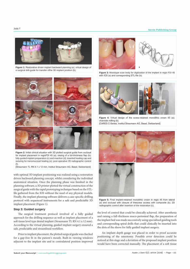
Figure 1: Restorative driven implant backward planning (a); virtual design of a surgical drill-guide for transfer ofthe 3D implant position (b).
Figure 2: Initial clinical situation with 3D plotted surgical guide from occlusal for implant placement in regioFDI 45 (a); raising of a full-thickness flap (b); fully guided implant preparation (c) and insertion (d); inserted healing cap and suturing for transmucosal healing (e); post-operative 2D radiographic control (f).[Straumann TL RN 4.1 x 12 mm, Institut Straumann AG, Basel, Switzerland]
Figure 3: Monotype scan body for digitization of the implant in regio FDI 45 with IOS (a) and corresponding STL-file (b).
Figure 4: Virtual design of the screw-retained monolithic crown 45 (a); chairside milling (b).[CARES C-Series, Institut Straumann AG, Basel, Switzerland]
Figure 5: Final implant-retained monolithic crown in regio 45 from lateral (a) and occlusal with closure of thescrew access with composite (b); 2D radiographic control after insertion of the restoration (c).

Austin J Dent 5(2): id1104 (2018) - Page - 03
Joda T Austin Publishing Group
Submit your Manuscript | www.austinpublishinggroup.com
level type implant was made directly fully guided via the integrated 5mm drill sleeve (Straumann TL RN 4.1 x 12 mm, Institut Straumann AG, Basel, Switzerland).
The post-operative clinical situation and related radiograph showed the correct prosthodontic 3D position of the implant with sufficient safety distance from the N. alveolaris inf. and the adjacent dentition (Figure 2).
Step 4: Digital impressionProsthodontic and technical work steps followed a digitalized
approach including IOS (TRIOS Pod, 3Shape, Copenhagen, Denmark) and CAD/CAM-processing using a pre-fabricated titanium abutment (Variobase RN, Institut Straumann AG, Basel, Switzerland).
After guided implant placement, a second IOS captured the peri-implant mucosal architecture including the neighboring teeth in a quadrant-like approach. Then, a monotype scanbody was screwed and the 3D implant position was scanned. The corresponding opposite arch was scanned in the same way; and finally, the bite registration was also digitally transferred (Figure 3).
Step 5: DesignThe final implant crown was planned as screw-retained monolithic
restoration out of a Lithium-di-silicate (LS2) CAD/CAM-blank (n!ce CAD, Institut Straumann AG, Basel, Switzerland) and bonded pre-fabricated titanium abutment (Variobase RN, Institut Straumann AG, Basel, Switzerland).
Based on the STL-file from the IOS, the anatomically full-contoured shape and contour of the crown was designed and produced completely digital without any physical models and casting. Interproximal and occlusal contacts were defined virtually according to the threshold settings of the dental design software for chairside solutions (CARES C-Series, Institut Straumann AG, Basel, Switzerland) (Figure 4).
Step 6: ProductionThe virtual crown design was processed and produced with a
4-axis wet milling and grinding equipment (CARES C-Series, Institut Straumann AG, Basel, Switzerland) for in-house manufacturing out of a monolithic LS2 CAD/CAM-blank (n!ce CAD, Institut Straumann AG, Basel, Switzerland) (Figure 4).
Step 7: Post-processingAfter milling of the monolithic LS2 crown (Straumann CARES
C-Series, Institut Straumann AG, Basel, Switzerland), the restoration was cleaned with 95 % ethanol, and for further post-processing finally polished and individually characterized. Then, the prepared LS2 crown was directly bonded to the pre-fabricated titanium abutment (Variobase RN, Institut Straumann AG, Basel, Switzerland) under extraoral conditions (Multilink Implant, IvoclarVivadent, Schaan, Liechtenstein).
Step 8: Final restorationFirst, the interproximal fit, and secondary, the marginal integrity
of the restoration was clinically assessed. Identical continuity with dental floss was separately controlled for mesial and distal contact
surfaces. Next, the occlusal scheme was checked statically and dynamically with shimstock foil achieving light occlusal contacts without dynamic interactions. The monolithic LS2 restoration was screwed with a controlled torque of 35 Ncm according to the implant provider’s recommendations. The screw access hole was sealed with teflon and composite application (Figures 5).
DiscussionThe major advantages of digital dental technologies are related to
standardized quality with high accuracy and streamlined production combined with reduced manpower resources [1]. Initially clinical RCTs showed the superiority of complete digital processing compared to conventional pathways analyzing socio-economic considerations, such as clinical and technical time-efficiency [3]. Regarding treatment costs, cost-minimization estimations showed decreased overall treatment costs for first line therapy, including laboratory fees, for implant crowns fabricated using quadrant-like IOS plus CAD/CAM-technology [4].
In addition, the need for chairside corrections, such as secondary grinding and polishing, can be minimized, or may not even be necessary, within a complete digital protocol with monolithic restorations. This reduces work time but may also decrease the risk for cracks and chipping as a result of the absence of veneered ceramics [5]. Digital applications may facilitate not only forward-thinking opportunities of functionality by means of implant restorations but also allow clinical treatment protocols in a more convenient approach for the patients when using IOS instead of conventional impression-taking procedures [1].
Besides technical processing, the type of material suitable for full-contoured crowns is debatable. These implant restorative materials must be able to tolerate and absorb excessive forces, but should also protect the antagonist for abrasion, especially in the case of naturally teeth.
Treatment protocols for single-unit monolithic implant restorations in posterior sites applying CAID with virtual implant planning, guided surgery, IOS and CAD/CAM-technology without any physical master model situations have to be considered in place of conventional manufacturing.
References1. Joda T, Ferrari M, Gallucci GO, Wittneben JG, Bragger U. Digital technology
in fixed implant prosthodontics. Periodontol 2000. 2017; 73: 178-192.
2. Joda T, Bragger U. Complete digital workflow for the production of implant-supported single-unit monolithic crowns. Clin Oral Impl Res. 2014; 25: 1304-1306.
3. Joda T, Bragger U. Time-efficiency analysis of the treatment with monolithic implant crowns in a digital workflow: A randomized controlled trial. Clin Oral Impl Res. 2016; 27: 1401-1406.
4. Joda T, Bragger U. Digital vs. conventional implant prosthetic workflows: A time/cost analysis. Clin Oral Impl Res. 2015; 26: 1430-1435.
5. Joda T, Ferrari M, Bragger U. Monolithic implant-supported lithium disilicate (LS2) crowns in a complete digital workflow: A prospective clinical trial with a 2-year follow-up. ClinImpl Dent Relat Res. 2017; 19: 505-511.