Ch07 part 1
39
1 Chapter 7 Skeletal System
-
Upload
ben-earl-alverson-jr -
Category
Documents
-
view
222 -
download
0
Transcript of Ch07 part 1

8/3/2019 Ch07 part 1
http://slidepdf.com/reader/full/ch07-part-1 1/39
1
Chapter 7
Skeletal System

8/3/2019 Ch07 part 1
http://slidepdf.com/reader/full/ch07-part-1 2/39
2
A. Components: bones, cartilage andligaments (connect bones to bones)
B. Tissues: include cartilage, fibrous
connective tissue, blood, and nervous
tissue
C. Function: support, protection,
attachment sites for muscles, formation
of blood cells, and mineral storage
CopyrightThe McGraw-Hill Companies, Inc. Permission required for reproduction or display.

8/3/2019 Ch07 part 1
http://slidepdf.com/reader/full/ch07-part-1 3/39
Two Types of Bone
A. Compact bone: has a continuous
matrix without any spaces
B. Spongy bone: has many branchingbony plates with spaces, spongy bone has
osteocytes (mature bone cells) but they
are not arranged around osteonic canals
3

8/3/2019 Ch07 part 1
http://slidepdf.com/reader/full/ch07-part-1 4/39
4
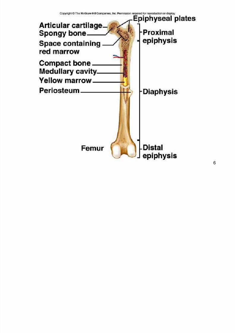
Bone Structure
Parts of a Long Bone
1. Expanded ends of bones that form jointswith adjacent bones are called epiphyses.
2. Articular cartilages (hyaline cartilage) cover the epiphyses.
3. The shaft of the bone is the diaphysis.4. A tough layer of vascular connective tissue,called the periosteum, covers the bone and iscontinuous with ligaments and tendons.
CopyrightThe McGraw-Hill Companies, Inc. Permission required for reproduction or display.

8/3/2019 Ch07 part 1
http://slidepdf.com/reader/full/ch07-part-1 5/39
5
5. A bone's shape makes possible its
function; bony processes or grooves indicateplaces of attachment for muscles.
6. Compact bone makes up the wall of the
diaphysis; the epiphyses are filled withspongy bone to reduce the weight of theskeleton.
7. The diaphysis contains a hollow
medullary cavity that is lined with endosteumand filled with marrow.
CopyrightThe McGraw-Hill Companies, Inc. Permission required for reproduction or display.
8/3/2019 Ch07 part 1
http://slidepdf.com/reader/full/ch07-part-1 6/39
6

8/3/2019 Ch07 part 1
http://slidepdf.com/reader/full/ch07-part-1 7/39
7
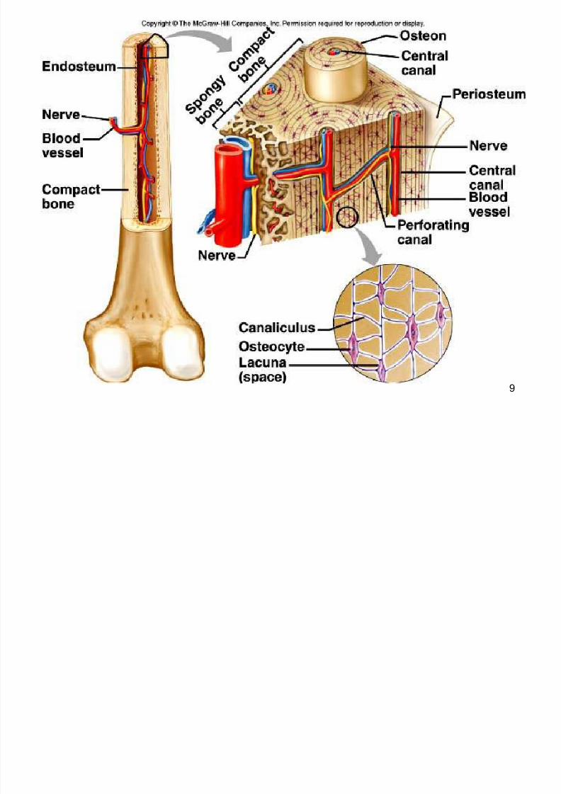
D. Microscopic Structure
1. Osteocytes- mature bone cells located in thelacunae (cavity)
2. Osteoblast ± bone forming cells which will
form a matrix around themselves, form
spongy bone3. Osteoclasts ± cells which break down a
calcified matrix during bone formation
leading to the formation of a medullary cavity
CopyrightThe McGraw-Hill Companies, Inc. Permission required for reproduction or display.
8/3/2019 Ch07 part 1
http://slidepdf.com/reader/full/ch07-part-1 8/39
8
4. Osteon: (Haversian system) consists of
osteonic canal lamella and an osteocytes.Many osteons cemented together to formthe substance of bone. Contains one or two blood vessels and a nerve
5. Lamellae: layers of bone matrix whichform concentric patterns around theocteonic canals.
CopyrightThe McGraw-Hill Companies, Inc. Permission required for reproduction or display.
8/3/2019 Ch07 part 1
http://slidepdf.com/reader/full/ch07-part-1 9/39
9

8/3/2019 Ch07 part 1
http://slidepdf.com/reader/full/ch07-part-1 10/39
10
Bone Development and Gr owth
A. Bones form by replacing connective
tissues in the fetus.
B. Some form within sheetlike layers of connective tissue (intramembranous bones),
while others replace masses of cartilage(endochondral bones).
C. Intramembranous Bones
1. The flat bones of the skull form as
intramembranous bones that develop fromlayers of connective tissue.
2. Osteoblasts deposit bony tissue aroundthemselves.
CopyrightThe McGraw-Hill Companies, Inc. Permission required for reproduction or display.

8/3/2019 Ch07 part 1
http://slidepdf.com/reader/full/ch07-part-1 11/39
11
3. Once osteoblasts deposit bone are
located in lacunae, they are calledosteocytes.
4. Cells of the membranous connective
tissue that lie outside the developing bone
give rise to the periosteum.
CopyrightThe McGraw-Hill Companies, Inc. Permission required for reproduction or display.
8/3/2019 Ch07 part 1
http://slidepdf.com/reader/full/ch07-part-1 12/39
12
D. Endochondral Bones
1. Most of the bones of the skeletonfall into this category.
2. They first develop as hyaline cartilage
models and are then replaced with bone.
3. Cartilage is broken down in thediaphysis and progressively replaced with
bone while the periosteum develops onthe outside.
4. Cartilage tissue is invaded by blood
vessels and osteoblasts that first form
spongy bone at the primary ossificationcenter in the diaphysis.
CopyrightThe McGraw-Hill Companies, Inc. Permission required for reproduction or display.

8/3/2019 Ch07 part 1
http://slidepdf.com/reader/full/ch07-part-1 13/39
13
5. Osteoblasts beneath the periosteum lay
down compact bone outside the spongybone.
6. S econdary ossification centers appear later in the epiphyses.
7. A band of hyaline cartilage, theepiphyseal plate, forms between the twoossification centers.
8. Layers of cartilage cells undergoing
mitosis make up the epiphyseal plate.9. Osteoclasts break down the calcified
matrix and are replaced with bone-building osteoblasts that deposit bone inplace of calcified cartilage.
CopyrightThe McGraw-Hill Companies, Inc. Permission required for reproduction or display.
8/3/2019 Ch07 part 1
http://slidepdf.com/reader/full/ch07-part-1 14/39

8/3/2019 Ch07 part 1
http://slidepdf.com/reader/full/ch07-part-1 15/39
15

8/3/2019 Ch07 part 1
http://slidepdf.com/reader/full/ch07-part-1 16/39
16
E. Homeostasis of Bone Tissue
1. Osteoclasts tear down and
osteoblasts build bone throughout
the lifespan with the processes of
resorption and deposition, with an
average of 3% to 5% of bone
calcium exchanged annually.
CopyrightThe McGraw-Hill Companies, Inc. Permission required for reproduction or display.
8/3/2019 Ch07 part 1
http://slidepdf.com/reader/full/ch07-part-1 17/39
17
Bone Function
A. Support and Protection
1. Bones give shape to thehead, thorax, and limbs.
2. Bones such as the pelvis andlower limbs provide support for the body.
3. Bones of the skull protect the
brain, ears, and eyes.
CopyrightThe McGraw-Hill Companies, Inc. Permission required for reproduction or display.

8/3/2019 Ch07 part 1
http://slidepdf.com/reader/full/ch07-part-1 18/39
18
B. Body Movement
1. Bones can act as levers.
a. A lever has four components:
a rigid bar, a pivot or fulcrum,
an object that is moved against resistance, and a force that
supplies energy.
CopyrightThe McGraw-Hill Companies, Inc. Permission required for reproduction or display.

8/3/2019 Ch07 part 1
http://slidepdf.com/reader/full/ch07-part-1 19/39
19

8/3/2019 Ch07 part 1
http://slidepdf.com/reader/full/ch07-part-1 20/39
20
C. Blood Cell Formation
1. Blood cells begin to form through
hematopoieses in the yolk sac; they arelater manufactured in bone marrow.
2. Two kinds of marrow occupy themedullary cavities of bone.
a. Red marrow functions in theformation of red blood cells,white blood cells, and platelets,and is found in the spongy bone
of the skull, ribs, sternum,clavicles, vertebrae, and pelvis.
b. Yellow marrow, occupying thecavities of most bones, stores
fat.
CopyrightThe McGraw-Hill Companies, Inc. Permission required for reproduction or display.

8/3/2019 Ch07 part 1
http://slidepdf.com/reader/full/ch07-part-1 21/39
21
D. Storage of Inorganic Salts
1. The inorganic matrix of bone storesinorganic mineral salts in the form of
calcium phosphate that is important inmany metabolic processes.
2. Calcium in bone is a reservoir for
body calcium; when blood levels are
low, osteoclasts release calcium from
bone.
CopyrightThe McGraw-Hill Companies, Inc. Permission required for reproduction or display.

8/3/2019 Ch07 part 1
http://slidepdf.com/reader/full/ch07-part-1 22/39
22
3. Calcium is stored in bone under the
influence of calcitonin when bloodlevels of calcium are high.
4. Bone also stores magnesium, sodium, potassium, and carbonate ions.
5. Bones can also accumulate harmful
elements, such as lead, radium, and
strontium.
CopyrightThe McGraw-Hill Companies, Inc. Permission required for reproduction or display.

8/3/2019 Ch07 part 1
http://slidepdf.com/reader/full/ch07-part-1 23/39
23

8/3/2019 Ch07 part 1
http://slidepdf.com/reader/full/ch07-part-1 24/39
24
Skeletal Organization
A. The axial skeleton consists of theskull, hyoid bone, vertebral column(vertebrae and intervertebral disks), andthorax (ribs and sternum).
B. The appendicular skeleton consists of the pectoral girdle (scapulae andclavicles), upper limbs (humerus, radius,ulna, carpals, metacarpals, andphalanges), pelvic girdle (coxal bones
articulating with the sacrum), and lower limbs (femur, tibia, fibula, patella,tarsals, metatarsals, phalanges).
CopyrightThe McGraw-Hill Companies, Inc. Permission required for reproduction or display.

8/3/2019 Ch07 part 1
http://slidepdf.com/reader/full/ch07-part-1 25/39
25

8/3/2019 Ch07 part 1
http://slidepdf.com/reader/full/ch07-part-1 26/39
26
CopyrightThe McGraw-Hill Companies, Inc. Permission required for reproduction or display.
Joints
A. Joints (articulations) are the functional junctions between bones.
B. Joints enable a wide variety of body
movements.C. Joints can be classified according to the
degree of movement possible and can
be immovable, slightly movable, or freely
movable.

8/3/2019 Ch07 part 1
http://slidepdf.com/reader/full/ch07-part-1 27/39
27
CopyrightThe McGraw-Hill Companies, Inc. Permission required for reproduction or display.
D. Joints can also classified according to the
type of tissue that binds them together.
E. Fibrous Joints
1. Fibrous joints are held close together
by dense connective tissue andare immovable (sutures of skull) or only
slightly movable (joint between the
distal tibia and fibula).

8/3/2019 Ch07 part 1
http://slidepdf.com/reader/full/ch07-part-1 28/39
28

8/3/2019 Ch07 part 1
http://slidepdf.com/reader/full/ch07-part-1 29/39
29
CopyrightThe McGraw-Hill Companies, Inc. Permission required for reproduction or display.
F. Cartilaginous Joints
1. Hyaline cartilage or disks of
fibrocartilage unite the bones incartilaginous joints.
2. Intervertebral disks betweenvertebrae help absorb shock andare slightly movable.
3. Other examples of cartilaginous
joints include the symphysis pubisand the first rib with the sternum.

8/3/2019 Ch07 part 1
http://slidepdf.com/reader/full/ch07-part-1 30/39
30
CopyrightThe McGraw-Hill Companies, Inc. Permission required for reproduction or display.
G. Synovial Joints
1. Most joints of the skeleton are
synovial joints, which are more complexthan fibrous or cartilaginous joints.
2. The articular ends of bone in asynovial joint are covered with hyaline
cartilage.

8/3/2019 Ch07 part 1
http://slidepdf.com/reader/full/ch07-part-1 31/39
31

8/3/2019 Ch07 part 1
http://slidepdf.com/reader/full/ch07-part-1 32/39
32
CopyrightThe McGraw-Hill Companies, Inc. Permission required for reproduction or display.
3. A joint capsule consists of an outer layer of
dense connective tissue that joins the
periosteum, and an inner layer made up of synovial membrane.
S ynovial fluid has the consistency of
egg whites and lubricates articulatingsurfaces within the joint.
4. Some synovial joints contain shock-
absorbing pads of fibrocartilage called
menisci.5. Some synovial joints have fluid-filled sacs called bursae.

8/3/2019 Ch07 part 1
http://slidepdf.com/reader/full/ch07-part-1 33/39
33

8/3/2019 Ch07 part 1
http://slidepdf.com/reader/full/ch07-part-1 34/39
34
CopyrightThe McGraw-Hill Companies, Inc. Permission required for reproduction or display.
6. Based on the shapes of their parts and the
movements they permit, synovial joints canbe classified as follows:
a. A ball-and-socket joint consists
of a bone with a globular or egg-shaped head articulating with the
cup-shaped cavity of another bone; a
very wide range of motion is
possible; examples include the hipand shoulder joint.

8/3/2019 Ch07 part 1
http://slidepdf.com/reader/full/ch07-part-1 35/39
35
CopyrightThe McGraw-Hill Companies, Inc. Permission required for reproduction or display.
b. A condyloid joint consists of an
ovoid condyle fitting into an ellipticalcavity, permitting a variety of motions;
an example is the joint between a
metacarpal and a phalange.
c. Gliding joints occur where articulating
surfaces are nearly flat or slightly
curved, allowing a back-and-forth
motion; the joints of the wrist and ankle,as well as those between vertebrae,
are gliding joints.

8/3/2019 Ch07 part 1
http://slidepdf.com/reader/full/ch07-part-1 36/39
36
CopyrightThe McGraw-Hill Companies, Inc. Permission required for reproduction or display.
d. In a hinge joint, a convex surface
fits into a concave surface, as isfound in the elbow and phalange
joints; movement is in one plane
only.
e. In a pivot joint, a cylindrical
surface rotates within a ring of
bone and fibrous tissue; examples
include the joint between theproximal ends of the radius and
ulna.

8/3/2019 Ch07 part 1
http://slidepdf.com/reader/full/ch07-part-1 37/39
37
CopyrightThe McGraw-Hill Companies, Inc. Permission required for reproduction or display.
f. A saddle joint forms where
articulating surfaces have bothconcave and convex areas,
permitting a wide range of
movements; the joint between the
trapezium and the metacarpal of
the thumb is of this type.

8/3/2019 Ch07 part 1
http://slidepdf.com/reader/full/ch07-part-1 38/39
38
CopyrightThe McGraw-Hill Companies, Inc. Permission required for reproduction or display.
H. Types of Joint Movements
1. When a muscle contracts, its fibers pull
its movable end (insertion) toward its
stationary end (origin), causing
movement at a joint.
2. These terms describe movements thatoccur at joints: flexion, extension,
dorsiflexion, plantar flexion,
hyperextension, abduction, adduction,
rotation, circumduction, pronation,supination, eversion, inversion,
retraction, protraction, elevation, anddepression.

8/3/2019 Ch07 part 1
http://slidepdf.com/reader/full/ch07-part-1 39/39
39