CH 6 Skeletal System Part I. Fun Bone Facts When you were born you had over 300 bones. Now, you have...
99
CH 6 Skeletal System Part I
-
Upload
amelia-morgan -
Category
Documents
-
view
214 -
download
1
Transcript of CH 6 Skeletal System Part I. Fun Bone Facts When you were born you had over 300 bones. Now, you have...
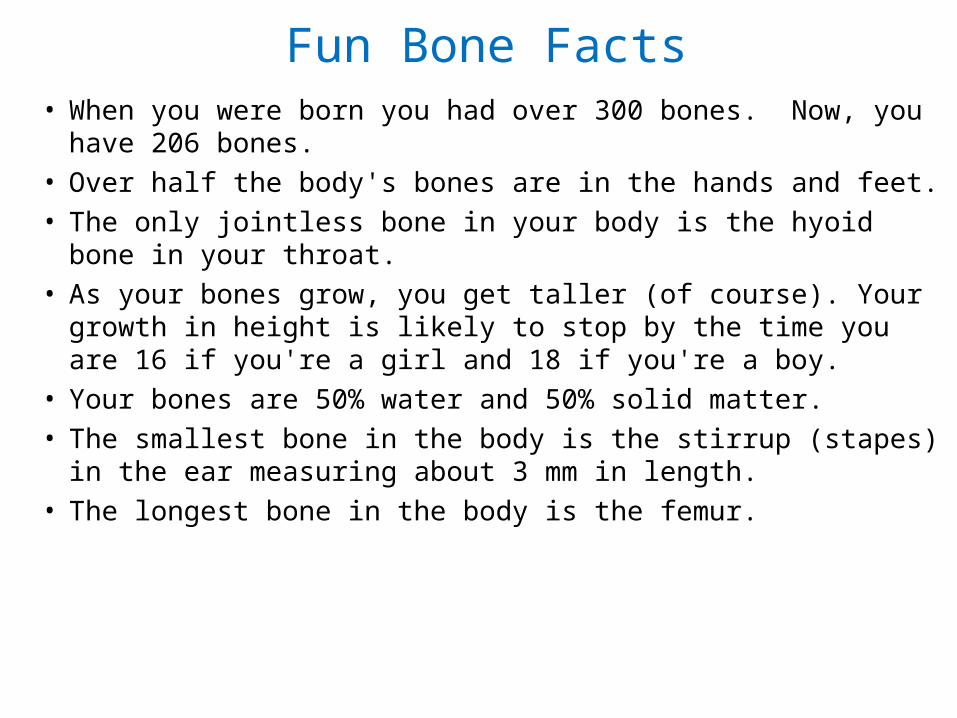
Fun Bone Facts• When you were born you had over 300 bones. Now, you have
206 bones.
• Over half the body's bones are in the hands and feet.
• The only jointless bone in your body is the hyoid bone in your throat.
• As your bones grow, you get taller (of course). Your growth in height is likely to stop by the time you are 16 if you're a girl and 18 if you're a boy.
• Your bones are 50% water and 50% solid matter.
• The smallest bone in the body is the stirrup (stapes) in the ear measuring about 3 mm in length.
• The longest bone in the body is the femur.

Bone• Osteology- The study of bone structure and the
treatment of bone disordersBone Functions:• Support- Provides points of attachment for muscle and other
soft tissues• Protection- Protects organs from injury• Body Movement- As muscles contract, they pull and move
bones• Stores Minerals- Especially calcium and phosphorous- When
minerals are in demand by the body, they are released from bones to the bloodstream for distribution
• Stores Chemical Energy- Yellow bone marrow is found in certain bones and contains adipocytes. This stored fat can be used for energy– Newborns do not have yellow bone marrow- just red bone
marrow

• Produces blood cells- Red bone marrow in certain bones produces all red blood cells, white blood cells and platelets. This process is called hematopoiesis– Red bone marrow is found in adults at the pelvis, ribs,
sternum, vertebrae, skull and the ends of arm and thigh bones
Though once used in various preparations, bone marrow has fallen out of favor as a food in the United States; However many other countries still feast on it. Diners in the US in the 18th century used a marrow scoop (or marrow spoon), often of silver and with a long thin bowl, as a table implement for removing marrow from a bone.

“Bone” Appétit!!

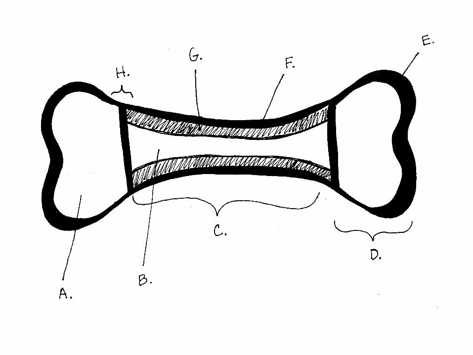
Parts of a Long Bone• Bones are either long, short, flat or irregular. Their
shape is relative to their function. • Examples of long bones include the femur, humerus,
tibia, fibula, radius and ulna.• The parts of a long bone are:
• Diaphysis – The shaft of the bone (longest section)
• Epiphyses – The distal and proximal ends of bones
• Metaphyses- The 2 regions in a bone where the diaphysis meets the epiphyses
• Each includes an epiphyseal plate (growth plate) made up of hyaline cartilage. This is where bones elongate.
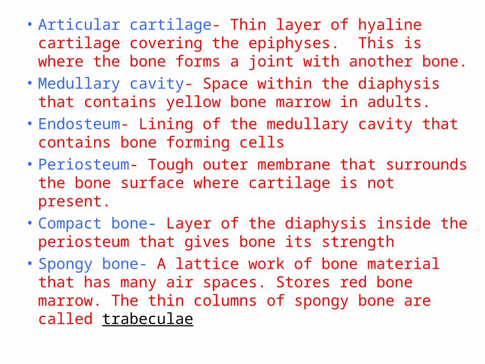
• Articular cartilage- Thin layer of hyaline cartilage covering the epiphyses. This is where the bone forms a joint with another bone.
• Medullary cavity- Space within the diaphysis that contains yellow bone marrow in adults.
• Endosteum- Lining of the medullary cavity that contains bone forming cells
• Periosteum- Tough outer membrane that surrounds the bone surface where cartilage is not present.
• Compact bone- Layer of the diaphysis inside the periosteum that gives bone its strength
• Spongy bone- A lattice work of bone material that has many air spaces. Stores red bone marrow. The thin columns of spongy bone are called trabeculae


Pop Quiz1. What is the tough outer layer of a long
bone?2. What are the ends of long bone covered
with?3. What is the shaft of the bone called?4. What material is found in the medullary
cavity?5. What does hematopoesis mean?

Bone Cells• The formation of mature bone cells is as follows:
Osteogenic cells Osteoblasts Osteocytes
• Osteogenic cells undergo cell division in the inner portion of the periosteum and endosteum
• Osteoblasts are developed osteogenic cells that do not have the ability to divide. They secrete collagen- gives bone some flexibility.
• Osteocytes are mature bone cells that are trapped in collagen (matrix) and perform the daily metabolic activities of the bone.

Microscopic Structure of Compact Bone• Perforating Canals (Volkmann’s Canals)- Tunnels
that extend transversely across the width of bones. These canals carry blood vessels and nerves.
• Haversian Canals- Tunnels that run lengthwise through bone and connect with Perforating Canals
• Lamellae- Concentric circles of hard, mineralized matrix (collagen strengthened with calcium and phosphorus)
• Lacunnae- Spaces between lamellae which contain osteocytes

Structure of compact bone continued…
• Canaliculi- Channels projecting outward from lacunnae. These channels connect lacunnae with each other. This allows many routes for nutrients and oxygen to reach bone cells.
• Osteon- Each Haversian canal with its surrounding lamellae, lacunnae, osteocytes and canaliculi


ANOTHER POP QUIZ….1. What are the repeating circular units found
in bone tissue called?
2. What is in the center of an osteon?
3. What two things are found that run through #2?
4. What are bone cells called?
5. Where are they found (the space)?
6. What is bone matrix composed of?

ANOTHER POP QUIZ….1. What are the repeating circular units found in
bone tissue called? Osteon/Haversian system
2. What is in the center of an osteon? Haversian/central canal
3. What two things are found that run through #2? Blood vessel/nerve fibers
4. What are bone cells called? osteocytes
5. Where are they found (the space)? lacunae
6. What is bone matrix composed of? Collagen and minerals like calcium and phosphorus


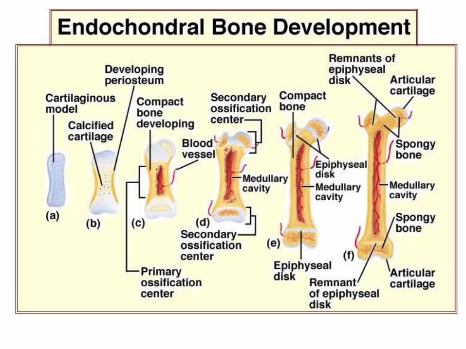
Bone Formation• The process by which bone forms is called
ossification. This begins during the 6th or 7th month of fetal development and continues throughout adulthood.
• During ossification, hyaline cartilage becomes bone. This happens by the following steps:
1.A membrane develops around the cartilage called the perichondrium (This eventually become the periosteum).
2.Chondrocytes (cartilage cells) trigger the uptake of calcium.

Ossification continued…
3. Chondrocytes die because they can’t receive nutrients through the calcified matrix.
4. Osteogenic cells in the perichondrium develop into osteoblasts and ultimately osteocytes.
5. This continues until the hyaline cartilage of the epiphyseal plate is completely replaced with bone. The new bone in this area is called the epiphyseal line.


Bone Surface Markings
• The markings on bones serve as sites of attachment for muscles and/or ligaments.
• Openings in bones serve as passageways for blood vessels and/or nerves.
• Projections on bones take part in forming joints.

Trochanter
Femur
A trochanter is a smooth,rounded projection

Spine
A spine is a sharp,slender, often pointedprojection

Fossa - a surface depression or concavity

Facet
A facet is a small, flatsurface

Condyle
Condyle – a large, convex projection thatusually contacts anotherbone

Fissure
A fissure is a deepcleft between adjacent parts of bone

Meatus
A meatus is a canal within a bone

Foramen
A foramen is anatural openingon a bone surface
Tubercle – rough, rounded projection

Suture
A suture is a line at the junction of bones in theskull
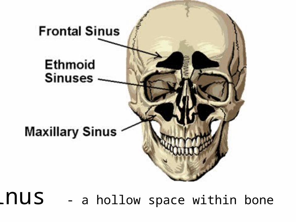
Sinus - a hollow space within bone

Fractures• What is a fracture?
A break in a bone•What are the main types of fractures?
• Displaced- The bone snaps into two or more parts and moves so that the two ends are not lined up straight
• Non-displaced- The bone cracks either part or all of the way through, but maintains its proper alignment.
• Open(Also called compound) - Breaks through the skin • Closed (Also called simple)- Does not break through the
skin
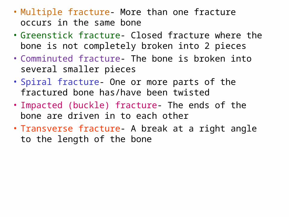
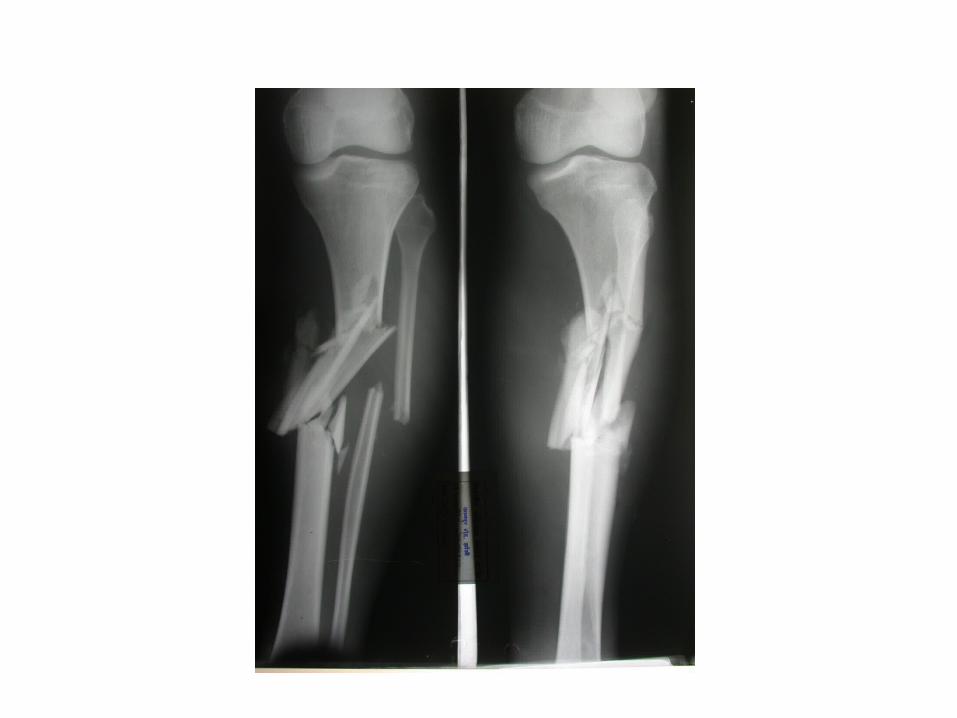
• Multiple fracture- More than one fracture occurs in the same bone
• Greenstick fracture- Closed fracture where the bone is not completely broken into 2 pieces
• Comminuted fracture- The bone is broken into several smaller pieces
• Spiral fracture- One or more parts of the fractured bone has/have been twisted
• Impacted (buckle) fracture- The ends of the bone are driven in to each other
• Transverse fracture- A break at a right angle to the length of the bone


Organization of the Skeleton• The adult human skeleton has 206 bones.• The skeleton can be divided into…
• Axial skeleton – Includes the bones of the skull, ear, hyoid, vertebral column, ribs and sternum
• Appendicular skeleton- Includes the bones of the upper and lower limbs, pectoral girdle and pelvic girdle– What is a girdle? Appendicular bone that connects to the
axial skeleton


AppendicularSkeleton


Types of Joints• Joints are either immovable, semi-movable or
movable.
• Immovable joints- The joints of the skull are an example of this. It is important that these joints don’t move so that they protect the brain.
• Semi-movable joints- The joints of the backbone are an example of this. They have limited movement, but can allow us to touch our toes.

Moveable Joints• Ball and Socket- The shoulder- This joint allows for a
full range of motion.• Hinge- The elbow and knee- This joint allows for
extension, but no rotation.• Pivot- Where the top vertebra joins with the skull.
This allows us to move our heads as to say “no”.• Saddle- Base of the thumb- This allows for a wide
range of movement.• Gliding- Bones of the hands and feet- Bones can
slide or twist against each other.
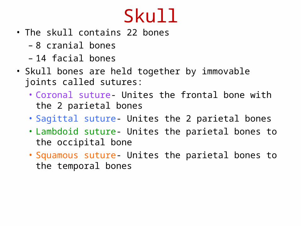
Skull• The skull contains 22 bones
– 8 cranial bones– 14 facial bones
• Skull bones are held together by immovable joints called sutures:• Coronal suture- Unites the frontal bone with the 2 parietal
bones• Sagittal suture- Unites the 2 parietal bones• Lambdoid suture- Unites the parietal bones to the occipital
bone• Squamous suture- Unites the parietal bones to the temporal
bones

Cranial Bones
• The 8 cranial bones are as follows:
Frontal bone, 2 Parietal bones, 2 Temporal bones, Occipital bone, Sphenoid bone, Ethmoid bone
* Color these on the picture and label the sutures


Facial bones
• You need to be able to identify the following facial bones:
Maxillae, Mandible, Nasal, Palatine, Zygomatic, Lacrimal, Vomer
* Color these on the picture


Sinuses• A hollow in a bone• Function in producing mucus (trap infection) and
acting as resonating chambers for our speaking and singing.
• Sinuses are continuous with the nasal cavity; therefore, infection in the nasal cavity can travel to the sinuses and cause membranes of the sinuses to become inflammed.
• The 4 paranasal sinuses are: Frontal sinus, Sphenoid sinus, Ethmoid sinus, and Maxillary sinus


Hyoid Bone• The hyoid bone does not attach to any other bone.
It is suspended by ligaments and muscles.
• The hyoid bone is in the neck between the mandible and larynx (voice box).
• It is often fractured during strangulation.
• It supports the tongue and provides attachment sites for tongue and neck muscles.

Vertebral Column• The vertebral column extends from the skull to
the pelvis • It protects the spinal cord and provides
attachment sites for ribs and back muscles• It is composed of 26 vertebrae separated by
intervertebral disks of cartilage• There are 5 vertebral regions: Cervical (7
vertebrae), Thoracic (12 vertebrae), Lumbar (5 vertebrae), Sacrum (5 fused vertebrae = 1 bone), and Coccyx (1 bone)


Typical vertebra• A typical vertebra consists of a body and a vertebral arch
that surrounds the spinal cord
• The open part is the vertebral foramen through which the spinal cord runs
• Vertebral processes (projections) are where muscles attach. When you feel your backbone, you are feeling the spines of the vertebra.
Vertebral Arch Vertebral Foramen

Cervical Vertebrae (C1-C7)• The top 2 cervical vertebrae are considerably different
than the other 5.• Atlas (first vertebra – C1) supports and balances the head• Axis- second vertebra- C2• Odontoid process – projection of the axis that provides a
pivot for the atlas
Odontoid Process


THORAX• The skeletal part of the thorax is called the
thoracic cage which includes the sternum, costal cartilages, and ribs.
• The sternum is divided into the upper part (manubrium), middle part (body) and lower part (xiphoid process)• The manubrium attaches the 1st and 2nd ribs• The body attaches the 3rd through 10th ribs• The xiphoid process does not attach any ribs
only abdominal muscles.

THORAX continued…• There are 12 sets of ribs.• Each rib attaches posteriorly with its
corresponding thoracic vertebra (Rib pair #1 attaches to T1).
• The first 7 ribs attach anteriorly to the sternum by costal cartilage. These are true ribs.
• The costal cartilage of ribs 8-10 does not attach to the sternum but to the cartilage of rib 7. They are called false ribs.
• The costal cartilage of the 11th and 12th pairs does not attach to anything; therefore, they are called floating ribs.


PECTORAL GIRDLE• The pectoral girdle attaches the upper limbs to the
axial skeleton.• It includes the clavicle and scapula.• The clavicle (collarbone) attaches to the
manubrium of the sternum and the acromion of the scapula
• The scapula (shoulder blade) has 5 parts:• Acromion- High point of the shoulder• Body- Flattened triangular part• Spine- Runs diagonally across the body• Glenoid cavity- Depression where the humerus fits• Coracoid process- Where muscles and ligaments attach


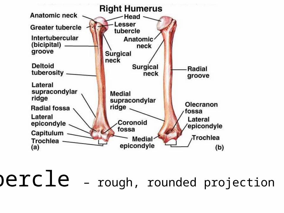
UPPER LIMB• Each upper limb consists of 30 bones: 1
humerus, 1 ulna, 1 radius, 8 carpals, 5 metacarpals and 14 phalanges
– Humerus:• Proximal end articulates (is in contact) with
the Glenoid cavity of the scapula• The body contains a “v” shaped area called
the deltoid tuberosity which is where the deltoid muscle attaches
• The distal end has a rounded knob called the capitulum that articulates with the radius. Also is the trochlea that articulates with the ulna.

UPPER LIMB continued…– Ulna-
• On the medial side (pinky side) of the forearm (in anatomical position).
• At the proximal end, the olecranon forms the elbow.
• The trochlear notch is a concavity in the bone where the trochlea rests.
– Radius- • On the lateral side (thumb side) of the forearm• Has a raised, roughened area called the
radial tuberosity- where the biceps muscle attaches.

UPPER LIMB continued……• Carpals-
– There are 8 carpal bones on each hand. They are organized into 2 rows of 4 bones.
– Carpal bones include: scaphoid, lunate, triquetrum, pisiform, trapezium, trapezoid, capitate, & hamate.
– These bones and the connective tissue that holds them together create a tunnel called the carpal tunnel. The median nerve runs through this tunnel. As the joints of the wrist are overworked, they become inflamed. This puts pressure on the median nerve and is painful…..called carpal tunnel syndrome.

UPPER LIMB continued……• Metacarpals-
• Located between the fingers and wrist.• There are 5 bones and are named 1st-5th
metacarpal. The 1st is the thumb’s metacarpal.
• Phalanges-• There are 14 on each hand (each finger has 3
and the thumb has 2)• They are named 1st-5th proximal, intermediate
and distal phalanges.

Label & color the hand bones

Lower Limb• The 2 lower lower limbs are each composed of
30 bones:– Femur, patella, tibia, fibula, 7 tarsals, 5 metatarsals,
and 14 phalanges.– Femur is the longest, strongest and heaviest bone in
the body.– The distal end expands into the medial and lateral
condyle which articulate with the tibia.– The patella (kneecap) is a triangular bone in front of
the joint between the femur and tibia. It functions in protecting that joint.

Lower Limb continued….• The tibia (shin bone) is the larger, medial
bone of the lower leg. – Its proximal end expands into a lateral and
medial condyle which articulate with the femur. – On the anterior surface is the tibial tuberosity
where the patellar tendon attaches.– On the distal, medial surface is the medial
malleolus which articulates with the talus of the ankle and can be felt as a projection of the medial surface of the ankle.

• The fibula is parallel and lateral to the tibia.– The head of the fibula articulates with the lateral
condyle of the tibia– The distal end of the fibula is the lateral malleolus
which articulates with the talus.– The distal end also articulates with the tibia at the
fibular notch• The ankle bones are called tarsals.
• These bones include the calcaneus, talus, cuboid, navicular and the 1st, 2nd, and 3rd cuneiforms.
• Each foot contains 5 metatarsals and 14 phalanges. The big toe has 2 while the other toes have 3.
Lower Limb continued….


Anatomy of the Knee• A joint is a place where 2 bones meet.• The knee is the largest and most frequently injured joint
of the body. It is an example of a hinge joint. • The bones that form the knee joint are the femur, tibia,
fibula and patella. They are held together by ligaments.• The ends of the femur and tibia and the back of the
patella are covered with articular cartilage.• In between the femur and tibia there are also 2 pieces of
cartilage that cushion and absorb shock during motion. These are called the medial meniscus and lateral meniscus.

• Medial Collateral Ligament (MCL)- This runs along the inside of the femur and tibia. It limits any joint movement to the inside.
• Lateral Collateral Ligament (LCL)- This runs along the outside of the femur and fibula. This limits any joint movement to the outside.
• Anterior Cruciate Ligament (ACL)- This is called cruciate because it crosses with the PCL. The ACL attaches the front of the tibia with the back of the femur. This limits the tibia from moving forward.
• Posterior Cruciate Ligament (PCL)- This attaches the back of the tibia with the front of the femur. The PCL limits the tibia from moving backward.


Types of Joints• Joints are either immovable, semi-movable or
movable.
• Immovable joints- The joints of the skull are an example of this. It is important that these joints don’t move so that they protect the brain.
• Semi-movable joints- The joints of the backbone are an example of this. They have limited movement, but can allow us to touch our toes.

Moveable Joints• Ball and Socket- The shoulder- This joint allows for a
full range of motion.• Hinge- The elbow and knee- This joint allows for
extension, but no rotation.• Pivot- Where the atlas and axis join together. This
allows us to move our heads as to say “no”.• Saddle- Base of the thumb- This allows for a wide
range of movement.• Gliding- Bones of the hands and feet- Bones can
slide or twist against each other.


Fractures• What is a fracture?
A break in a bone•What are the main types of fractures?
• Displaced- The bone snaps into two or more parts and moves so that the two ends are not lined up straight
• Non-displaced- The bone cracks either part or all of the way through, but maintains its proper alignment.
• Open(Also called compound) - Breaks through the skin • Closed (Also called simple)- Does not break through the
skin

Common Types of Fractures
• Multiple fracture- More than one fracture occurs in the same bone.
• Greenstick fracture- Closed fracture where the bone is not completely broken into 2 or more pieces.
• Comminuted fracture- The bone is broken into several smaller pieces
• Spiral fracture- One or more parts of the fractured bone has/have been twisted


Let’s see what you remember 1. What is the long shaft of a bone?
2. What is hematopoiesis?
3. Where does hematopoiesis occur?
4. What is the space within the diaphysis?
5. What is the tough outer membrane of the diaphysis?
6. Where is the epiphyseal plate located?
7. What are the cells of bone that divide to produce new bone cells?
8. What are mature bone cells?

9. What is the membrane that surrounds the epiphyseal plate during ossification?
10. Where are osteocytes found in long bone?
11. What are mineral rings of long bone?
12. What are small channels that run through bone and connect lacunnae?
13. What is a smooth, rounded projection of bone?
14. What is a projection of bone that is in contact with another bone?
15. What is a small, flat surface on bone?

16. What is a natural opening in bone?
17. What unites the parietal bones with the occipital bone?
18. What is the cheekbone?
19. What are the 5 vertebral regions from top to bottom?
20. To what part of the sternum does the clavicle attach?
21. What is the C1 vertebra? C2?
22. What rib pairs are the false ribs?
23. What part of the ulna forms the elbow?

ACL Repair Video
http://www.orlive.com/akronchildrens/videos/arthroscopic-surgery-to-replace-a-young-athlete-s-acl?view=displaypageNLM

GOOD LUCK ON YOURTEST!!!