Ch 4 Functional anatomy of Bacteria and other Microbes.
95
Ch 4 Functional anatomy of Bacteria and other Microbes
-
Upload
conrad-williams -
Category
Documents
-
view
221 -
download
0
Transcript of Ch 4 Functional anatomy of Bacteria and other Microbes.

Ch 4
Functional anatomy of Bacteria and other Microbes

OrThe differences between
Eukaryotic and Prokaryotic cells

Q&A
• Penicillin was called a “miracle drug” because it doesn’t harm human cells. Why doesn’t it?

• Proks and euks are similar in chemical composition and reaction
• Proks lack membrane bound organelles
• Only Proks have peptidoglycan
• Euks have membrane bound organelles
• Euks have paired chromosomes
• Euks have histones

Nick sees the difference mainly in information and structural
capacity
• Proks lack membrane-enclosed organelles
• Euks are like a 2mhz 100gb home computer
• Proks are like a calculator• Human genome 4x10⁹
• E. coli 4x10⁶

The prokaryote
• Unicellular• Multiply by binary fission• Differentiated by
– Morphology– Chemical composition– Nutritional requirements– Biochemical activates– Sources of energy– Other tests

Size
• 0.2 to 2um in diameter
• 2-8um in length
• In biological systems there are always exceptions these are general sizes.

Shape


Shape
• Coccus– Diplococci– Streptococci– Staphylococci
• Bacillus• Spiral• Other pleomorphic
shapes

Basic components of a bacterial cell fig 4.6
Parts not seen
• Glycocalyx– Capsule– Slime layer– Extracellular
polysaccharide
• Function• Toxicity
• Protect from phagocytosis
• Allow adherence• Reduce water loss• Collect nutrients

Flagella: long filamentous appendages with filament,
hook and basal body• Used in movement• Can present taxis
– Negative– Positive
• Monotrichous• Peritrichous• Flagellar H protein
acts as an antigen E.c O157:H7
• Flagellin

Flagella Arrangement
Figure 4.7

Fimbriae/pili
• Shorter and less complex than flagella
• Helps adhere to surfaces
• Used for sex and communication
Cell wall• Major difference between eukaryotic and prok orgs.• Surrounds plasma membrane provides protection• Peptidoglycan
– Polymer of• NAG
• NAM
• Short amino acid chain
• Production inhibited by antibiotics• Prevents osmotic damage• Damage to cw is almost always lethal except


Gram Positives have large cell wall and Teichoic acids

Gram neg have lipopolysaccharide

Figure 4.12
Peptidoglycan
• Polymer of disaccharide:– N-
acetylglucosamine (NAG)
– N-acetylmuramic acid (NAM)

Figure 4.13a
Peptidoglycan in Gram-Positive Bacteria
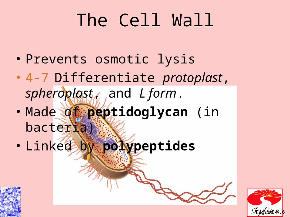
Figure 4.6
The Cell Wall
• Prevents osmotic lysis
• 4-7 Differentiate protoplast, spheroplast, and L form.
• Made of peptidoglycan (in bacteria)
• Linked by polypeptides

Gram-Positive Bacterial Cell Wall
Figure 4.13b

Gram-Negative Bacterial Cell Wall
Figure 4.13c

• Thick peptidoglycan
• Teichoic acids
Gram-positiveCell Wall
Figure 4.13b–c
Thin peptidoglycan Outer membrane Periplasmic space
Gram-positiveCell Wall

Gram neg
• Lipoprotein phospholipid outer membrane surrounding a thin peptidoglycan
• Makes gram neg resistant to– Phagocytosis– Antibiotics– Chemical reactions– Enzymes (lysozyme)– Has lipid A endotoxin– O polysaccaride antigen O157:H7 E.c.

Gram-Negative Outer Membrane
Figure 4.13c

How the gram stain works to differentiate between G+ and G-

The Gram Stain
Table 4.1
(a) Gram-Positive (b) Gram-Negative

The Gram Stain Mechanism
• Crystal violet-iodine crystals form in cell
• Gram-positive– Alcohol dehydrates peptidoglycan– CV-I crystals do not leave
• Gram-negative– Alcohol dissolves outer membrane and leaves
holes in peptidoglycan– CV-I washes out

• 2-ring basal body
• Disrupted by lysozyme
• Penicillin sensitive
Gram-PositiveCell Wall
Figure 4.13b–c
4-ring basal body Endotoxin Tetracycline sensitive
Gram-NegativeCell Wall


Nontypical cell walls
• Mycoplasma (acid fast) do not have ppt containing cell wall.
• Archaea contain another chemical called pseudomurein

Figure 24.8
Atypical Cell Walls
• Acid-fast cell walls– Like gram-positive– Waxy lipid (mycolic acid) bound to
peptidoglycan– Mycobacterium– Nocardia

Atypical Cell Walls
• Mycoplasmas– Lack cell walls– Sterols in plasma membrane
• Archaea– Wall-less or– Walls of pseudomurein (lack NAM and D-amino
acids)

Damage to the Cell Wall
• Lysozyme digests disaccharide in peptidoglycan• Penicillin inhibits peptide bridges in peptidoglycan• Protoplast is a wall-less cell• Spheroplast is a wall-less gram-positive cell
– Protoplasts and spheroplasts are susceptible to osmotic lysis
• L forms are wall-less cells that swell into irregular shapes

Plasma membrane
• Defines the living and nonliving parts of the cell– Everything on the inside is living– Everything on the outside is not living
• Is selectively permeable
• Workspace for enzymes of metabolic reactions

Plasma Membrane• Phospholipid bilayer
• Peripheral proteins
• Integral proteins
• Transmembrane proteins
Figure 4.14b

PM Workspace
– Nutrient breakdown– Energy production– Photosynthesis– Afforded by mesosomes which are regular
infoldings of the plasma membrane
• Weaknesses: destroyed by actions of alcohols, detergents and polymyxins

• Membrane is as viscous as olive oil.
• Proteins move to function
• Phospholipids rotate and move laterally
Fluid Mosaic Model
Figure 4.14b

• Damage to the membrane by alcohols, quaternary ammonium (detergents) and polymyxin antibiotics causes leakage of cell contents.
Plasma Membrane
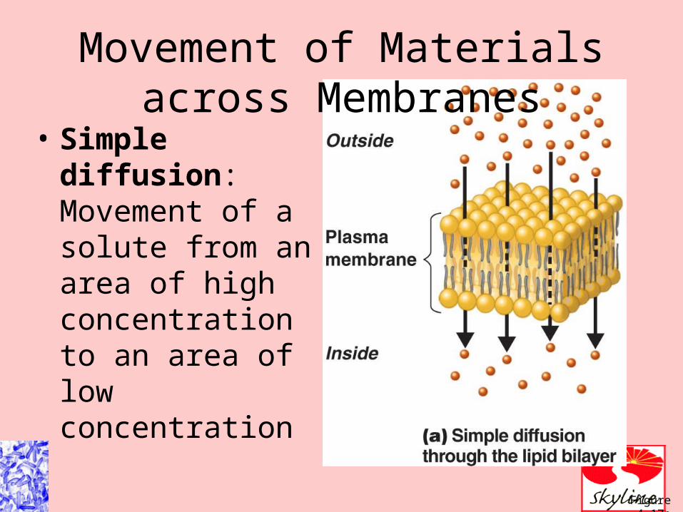
Figure 4.17a
Movement of Materials across Membranes
• Simple diffusion: Movement of a solute from an area of high concentration to an area of low concentration

Figure 4.17b-c
Movement of Materials across Membranes
• Facilitated diffusion: Solute combines with a transporter protein in the membrane

ANIMATION Passive Transport: Principles of Diffusion
ANIMATION Passive Transport: Special Types of Diffusion
Movement of Materials across Membranes

Figure 4.18a
Movement of Materials across Membranes
• Osmosis: The movement of water across a selectively permeable membrane from an area of high water to an area of lower water concentration
• Osmotic pressure: The pressure needed to stop the movement of water across the membrane

Figure 4.17d
Movement of Materials across Membranes
• Through lipid layer
• Aquaporins (water channels)

Figure 4.18a–b
The Principle of Osmosis

Figure 4.18c–e
The Principle of Osmosis

ANIMATION Active Transport: Overview
ANIMATION Active Transport: Types
Movement of Materials across Membranes
• Active transport: Requires a transporter protein and ATP
• Group translocation: Requires a transporter protein and PEP

Cytoplasm's
• The liquid component of the cell within the PM
• Mostly water, dissolved ions, DNA ribosomes and inclusions
• Concept of homeostasis

Nuclear area
• Contains the bacterial chromosome
• Bacteria may also have plasmids with up to 25% of the genetic materials
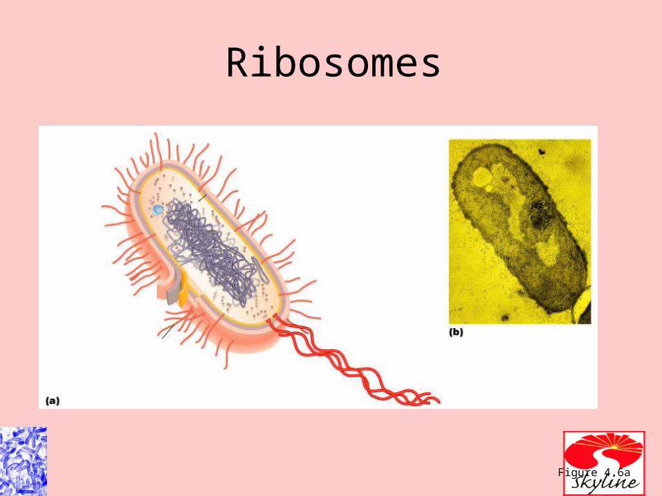
Ribosomes
Figure 4.6a

Ribosomes
Figure 4.19
Inclusions
• Typically reserve deposits of excess materials like inorganic phosphate
• Polysaccharide granules
• Lipids
• Sulfur
• Gas
• iron

Figure 4.19
The Prokaryotic Ribosome
• Protein synthesis
• 70S– 50S + 30S subunits

Figure 4.20
Magnetosomes
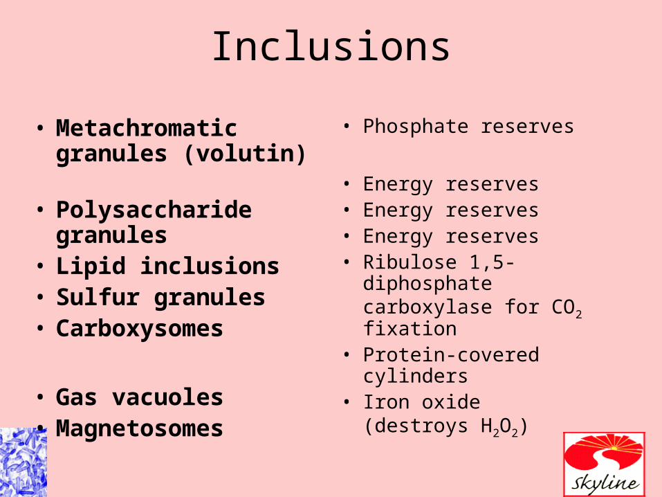
Inclusions
• Metachromatic granules (volutin)
• Polysaccharide granules
• Lipid inclusions• Sulfur granules• Carboxysomes
• Gas vacuoles• Magnetosomes
• Phosphate reserves
• Energy reserves• Energy reserves• Energy reserves• Ribulose 1,5-diphosphate
carboxylase for CO2 fixation
• Protein-covered cylinders• Iron oxide
(destroys H2O2)

Endospores
• Resting and waiting stage
• Resistant to drying and other harsh conditions


The Eukaryotic cell


Comparison
• Flagella and cilia tubulin (9/2) arrangement
• Cell wall of different materials
• Glycocalyx
• Plasma membrane


organelles
• Nucleus• ER• 80s ribosomes• Golgi complex• Lysozymes• Vacuoles• Mitochondria• Chloroplasts• Peroxisomes
Organelles
• 4-18 Define organelle.
• 4-19 Describe the functions of the nucleus, endoplasmic reticulum, Golgi complex, lysosomes, vacuoles, mitochondria, chloroplasts, peroxisomes, and centrosomes.

Organelles
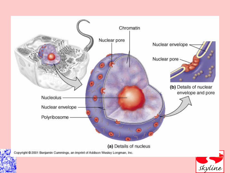
• Nucleus: Contains chromosomes
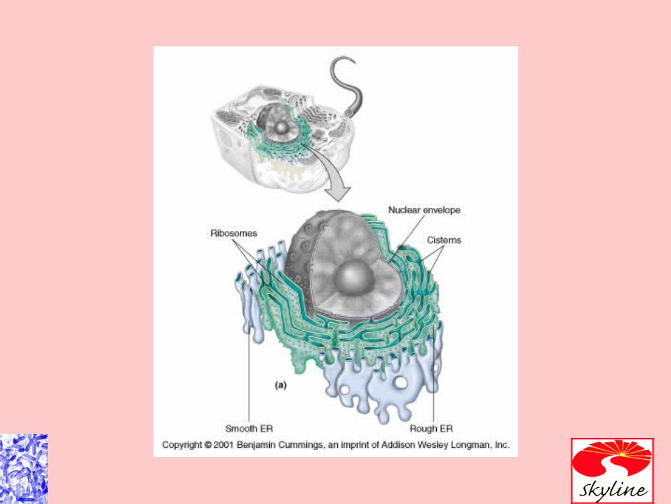
• ER: Transport network
• Golgi complex: Membrane formation and secretion
• Lysosome: Digestive enzymes
• Vacuole: Brings food into cells and provides support

Organelles
• Mitochondrion: Cellular respiration
• Chloroplast: Photosynthesis
• Peroxisome: Oxidation of fatty acids; destroys H2O2
• Centrosome: Consists of protein fibers and centrioles

Figure 4.24
The Eukaryotic Nucleus

Figure 4.24a–b
The Eukaryotic Nucleus

Figure 4.25
Rough Endoplasmic Reticulum

Figure 4.25a
Detailed Drawing of Endoplasmic Reticulum

Figure 4.25b
Micrograph of Endoplasmic Reticulum

Figure 4.26
Golgi Complex

Figure 4.22b
Lysosomes and Vacuoles

Figure 4.27
Mitochondria

Figure 4.28
Chloroplasts
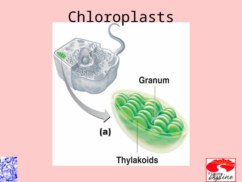
Figure 4.28a
Chloroplasts

Figure 4.28b
Chloroplasts

Figure 4.22b
Peroxisome and Centrosome

• Not membrane-bound:– Ribosome Protein synthesis– Centrosome Consists of protein
fibers and centrioles– Centriole Mitotic spindle formation
Eukaryotic Cell

Evolution of eukaryotes
• Endosymbiotic theory

Membrane activity
• Diffusion
• Osmosis
• Passive diffusion
• Facilitated diffusion
• Active transport
• Know the relationships


• Active transport of substances requires a transporter protein and ATP.
• Group translocation of substances requires a transporter protein and PEP.
Movement Across Membranes