CH 25. Monomer of Nucleic Acids A molecular complex of three types of subunit molecules 1.Phosphate...
51
CH 25 DNA
-
Upload
helena-rodgers -
Category
Documents
-
view
223 -
download
0
Transcript of CH 25. Monomer of Nucleic Acids A molecular complex of three types of subunit molecules 1.Phosphate...

CH 25DNA

Monomer of Nucleic Acids
A molecular complex of three types of subunit molecules
1. Phosphate
2. Pentose sugar
3. Nitrogen-containing base
NUCLEOTIDES
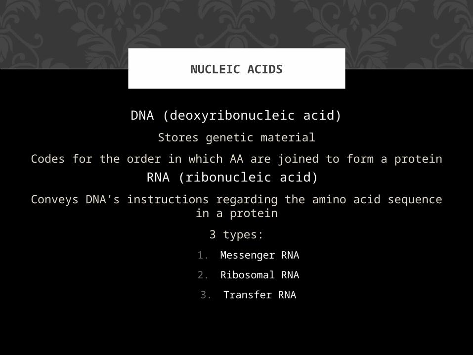
DNA (deoxyribonucleic acid)
Stores genetic material
Codes for the order in which AA are joined to form a protein
RNA (ribonucleic acid)
Conveys DNA’s instructions regarding the amino acid sequence in a protein
3 types:
1. Messenger RNA
2. Ribosomal RNA
3. Transfer RNA
NUCLEIC ACIDS

DNA RNA
Sugar Deoxyribose Ribose
Bases Adenine, Guanine, Thymine, Cytosine
Adenine, Guanine, Uracil, Cytosine
Strands Double Stranded (with base pairing) Single Stranded
Helix Yes No
NUCLEIC ACIDS

DNA is double-stranded with
complementary base pairing
A-TC-G
DNA STRUCTURE
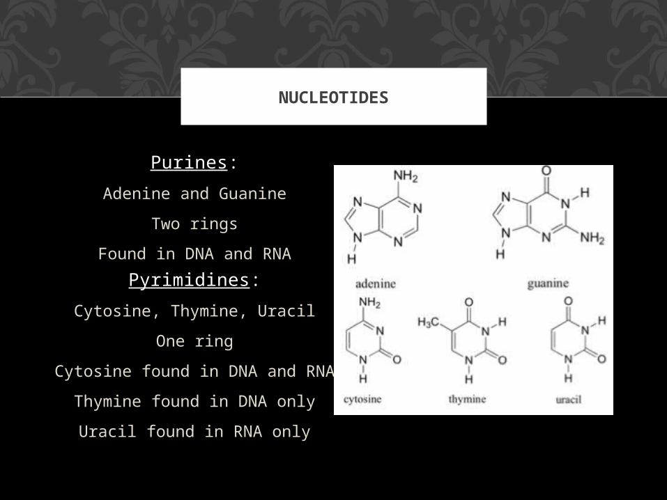
Purines:
Adenine and Guanine
Two rings
Found in DNA and RNA
Pyrimidines:
Cytosine, Thymine, Uracil
One ring
Cytosine found in DNA and RNA
Thymine found in DNA only
Uracil found in RNA only
NUCLEOTIDES

In the mid-1900s, scientists knew that chromosomes, made up of DNA (deoxyribonucleic acid) and proteins, contained genetic information.However, they did not know whether the DNA or
the proteins was the actual genetic material.
HISTORY

Various researchers showed that DNA was the genetic material when they performed an experiment with a T2
virus.
By using different radioactively labeled components, they demonstrated that only the virus DNA entered a bacterium
to take over the cell and produce new viruses.
HISTORY

VIRAL DNA IS LABELED
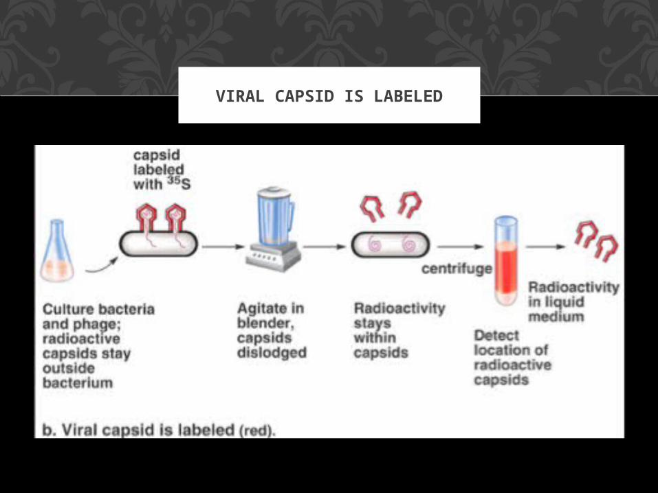
VIRAL CAPSID IS LABELED

The structure of DNA was determined by James Watson and Francis Crick in the early 1950s.
They deduced the following:• DNA has a twisted, ladder-like structure
(double helix)
• The sugar-phosphate molecules make up the sides of the ladder and the bases make up the
rungs
• Since A bonds with T and G with C, the rungs have a constant width
(purine paired with a pyrimidine)
DNA STRUCTURE
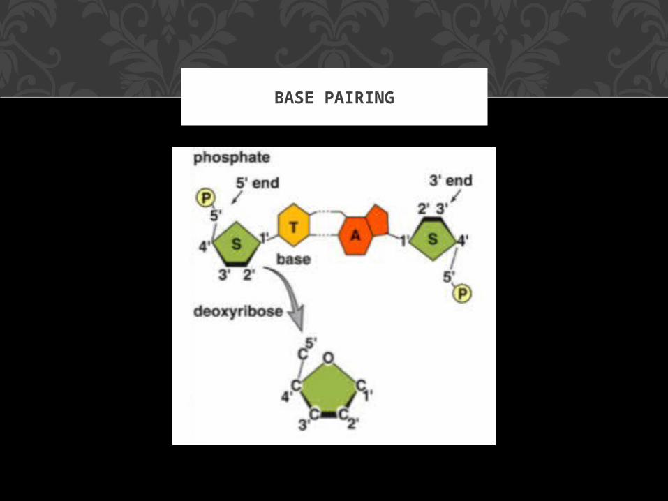
BASE PAIRING
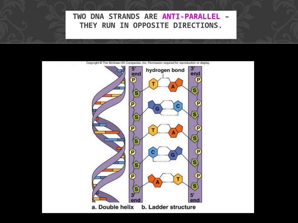
TWO DNA STRANDS ARE ANTI-PARALLEL – THEY RUN IN OPPOSITE
DIRECTIONS.
DNA replication occurs during chromosome duplication;
an exact copy of the DNA is produced with the aid of
DNA polymerase (an enzyme)
Hydrogen bonds between bases break and enzymes “unzip” the molecule.
Each old strand of nucleotides serves as a template for each new strand.
REPLICATION

New nucleotides move into complementary positions are joined by DNA polymerase.
The process is semiconservative because each new double helix is composed of an old strand of
nucleotides from the parent molecule and one newly-formed strand.
REPLICATION
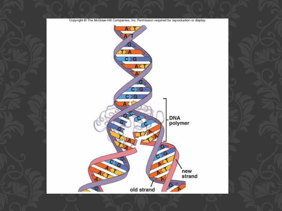

LADDER CONFIGURATION & DNA REPLICATION
1. ‘DNA: Structure, function and replication’ - WS
2. ‘DNA’ notes booklet3. ‘Protein Synthesis’ – WS
4. CH 2 Review Q’s
TO WORK ON:
25.2GENE EXPRESSION

A gene is a segment of DNA that specifies the amino acid sequence of a protein.
Gene expression occurs when gene activity leads to a protein product in the cell.
A gene does not directly control protein synthesis; instead, it passes its genetic information on to RNA, which is more
directly involved in protein synthesis.
GENE EXPRESSION
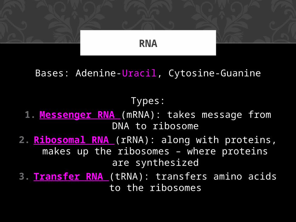
Bases: Adenine-Uracil, Cytosine-Guanine
Types:
1. Messenger RNA (mRNA): takes message from DNA to ribosome
2. Ribosomal RNA (rRNA): along with proteins, makes up the ribosomes – where proteins are
synthesized
3. Transfer RNA (tRNA): transfers amino acids to the ribosomes
RNA
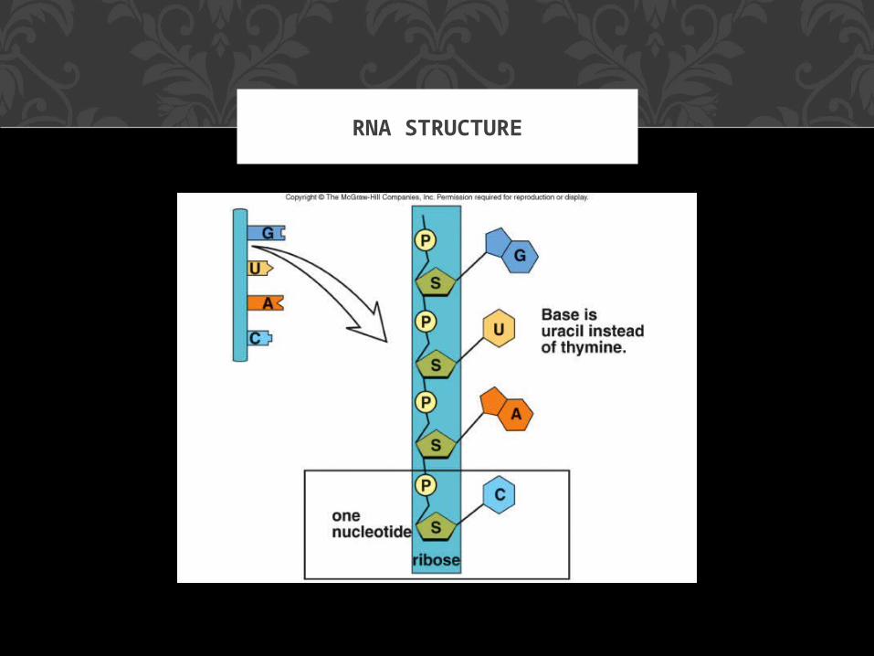
RNA STRUCTURE
1. Transcription: makes an RNA molecule complementary to a portion of DNA
2. Translation: occurs when the sequence of bases of mRNA directs the sequence of amino
acids in a polypeptide
PROTEIN SYNTHESIS

DNA specifies the synthesis of proteins because it contains a triplet code: every three bases stand for
one amino acid.
Each three-letter unit of an mRNA molecule is called a codon.
Most amino acids have more than one codon; there are 20 amino acids with a possible 64 different
triplets.
The code is nearly universal among living organisms.
THE GENETIC CODE

MRNA CODONS

GENE EXPRESSION
During transcription in the nucleus, a segment of DNA unwinds and unzips, and the DNA serves
as a template for mRNA formation.
RNA polymerase joins the RNA nucleotides so that the codons in mRNA are complementary to
the triplet code in DNA.
TRANSCRIPTION


DNA contains exons and introns.
Before mRNA leaves the nucleus, it is processed and the introns are excised so that only the exons
are expressed.
The splicing of mRNA is done by ribozymes, organic catalysts composed of RNA, not protein.
Primary mRNA is processed into mature mRNA.
PROCESSING OF MRNA


Translation is the second step by which gene expression leads to protein synthesis.
During translation, the sequence of codons in mRNA specifies the order of amino acids in a
protein.
Translation requires several enzymes and two other types of RNA: transfer RNA and
ribosomal RNA.
TRANSLATION

During translation, transfer RNA (tRNA) molecules attach to their own particular amino
acid and travel to a ribosome.
Through complementary base pairing between anticodons of tRNA and codons of mRNA, the
sequence of tRNAs and their amino acids form the sequence of the polypeptide.
TRNA

TNA: AMINO ACID CARRIER
Ribosomal RNA, also called structural RNA, is made in the nucleolus.
Proteins made in the cytoplasm move into the nucleus and join with ribosomal RNA to form the
subunits of ribosomes.
A large subunit and small subunit of a ribosome leave the nucleus and join in the
cytoplasm to form a ribosome just prior to protein synthesis.
RRNA

A ribosome has a binding site for mRNA as well as binding sites for two tRNA molecules at a time.
As the ribosome moves down the mRNA molecule, new tRNAs arrive, and a polypeptide forms and
grows longer.
Translation terminates once the polypeptide is fully formed; the ribosome separates into two subunits
and falls off the mRNA.
Several ribosomes may attach and translate the same mRNA, therefore the name polyribosome.
RIBOSOMES

POLYRIBOSOME

During translation, the codons of an mRNA base-pair with tRNA anticodons.
Protein translation requires these steps:1) Chain initiation
2) Chain elongation3) Chain termination.
Enzymes are required for each step, and the first two steps require energy.
TRANSLATION: 3 STEPS

During chain initiation, a small ribosomal subunit, the mRNA, an initiator tRNA, and a large
ribosomal unit bind together.First, a small ribosomal subunit attaches to the
mRNA near the start codon.The anticodon of tRNA, called the initiator RNA,
pairs with this codon.Then the large ribosomal subunit joins.
CHAIN INITIATION

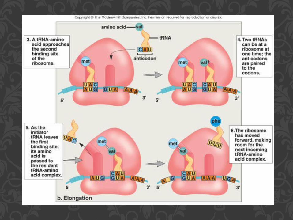
The initiator tRNA passes its amino acid to a tRNA-amino acid complex that has come to the
second binding site.
The ribosome moves forward and the tRNA at the second binding site is now at the first site, a
sequence called translocation.
The previous tRNA leaves the ribosome and picks up another amino acid before returning.
CHAIN ELONGATION
Chain termination occurs when a stop-codon sequence is reached.
The polypeptide is enzymatically cleaved from the
last tRNA by a release factor, and the ribosome falls away from the mRNA molecule.
A newly synthesized polypeptide may function alone or become part of a protein.
CHAIN TERMINATION


DNA in the nucleus contains a triplet code; each group of three bases stands for one amino acid.
During transcription, an mRNA copy of the DNA template is made.
The mRNA is processed before leaving the nucleus.
The mRNA joins with a ribosome, where tRNA carries the amino acids into position during
translation.
REVIEW


A gene mutation is a change in the sequence of bases within a gene.
MUTATIONS

Frameshift mutations involve the addition or removal of a base during the formation of mRNA; these change the genetic message by shifting the
“reading frame.”
FRAMESHIFT MUTATION

The change of just one nucleotide causing a codon change can cause the wrong amino acid to be inserted in a
polypeptide; this is a point mutation.
In a silent mutation, the change in the codon results in the same amino acid.
If a codon is changed to a stop codon, the resulting protein may be too short to function; this is a nonsense
mutation.
If a point mutation involves the substitution of a different amino acid, the result may be a protein that cannot reach
its final shape; this is a missense mutation.
An example is Hbs which causes sickle-cell disease.
POINT MUTATIONS

SICKLE CELL DISEASE

Mutations can be spontaneous or caused by environmental influences called mutagens.
Mutagens include radiation (X-rays, UV radiation), and organic chemicals (in cigarette smoke and
pesticides).
DNA polymerase proofreads the new strand against the old strand and detects mismatched
pairs, reducing mistakes to one in a billion nucleotide pairs replicated.
CAUSE & REPAIR OF MUTATIONS

Transposons are specific DNA sequences that move from place to place within and between
chromosomes.
These so-called jumping genes can cause a mutation to occur by altering gene expression.
It is likely all organisms, including humans, have transposons.
TRANSPOSONS: JUMPING GENES