Ch 151 Sensation & Perception Ch. 15: The Chemical Senses © Takashi Yamauchi (Dept. of Psychology,...
32
ch 15 1 Sensation & Perception Ch. 15: The Chemical Senses © Takashi Yamauchi (Dept. of Psychology, Texas A&M University) Main topics Olfactory system odors Neural codes Neural receptors Cortical structure Taste
-
Upload
eleanor-moody -
Category
Documents
-
view
221 -
download
0
Transcript of Ch 151 Sensation & Perception Ch. 15: The Chemical Senses © Takashi Yamauchi (Dept. of Psychology,...

ch 15 1
Sensation & PerceptionCh. 15: The Chemical Senses
© Takashi Yamauchi (Dept. of Psychology, Texas A&M University)
Main topicsOlfactory system
odors
Neural codes
Neural receptors
Cortical structure
Taste
ch 15 2
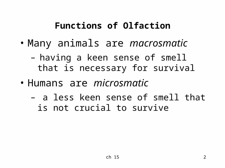
Functions of Olfaction
• Many animals are macrosmatic– having a keen sense of smell that is
necessary for survival
• Humans are microsmatic – a less keen sense of smell that is not
crucial to survive

ch 15 3
Detecting Odors - continued
• Rats are 8 to 50 times more sensitive to odors than humans
• Dogs are 300 to 10,000 times more sensitive
• The difference lies in the number of receptors they each have
– Humans have 10 million and dogs have 1 billion olfactory receptors

ch 15 4
Detecting Odors
• Measuring the detection threshold
– Yes/no procedure - participants are given trials with odors along with “blank” trials
• They respond by saying yes or no
– Forced-choice - two trials are given, one with odorant and one without
• Participant indicates which smells strongest
ch 15 5
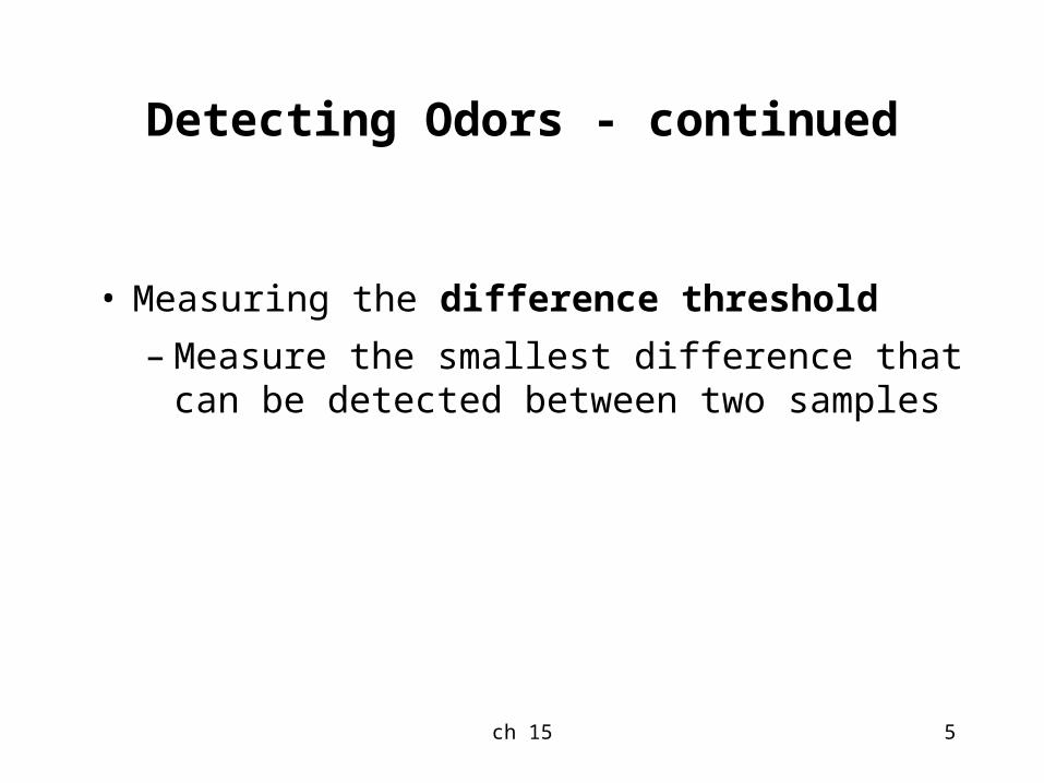
Detecting Odors - continued
• Measuring the difference threshold
– Measure the smallest difference that can be detected between two samples

ch 15 6Table 15.1 Human odor detection thresholds

ch 15 7
Identifying Odors
• Humans can discriminate among 100,000 odors but they cannot label them accurately
– This appears to be caused by an inability to retrieve the name from memory, from a lack of sensitivity

ch 15 8
The Puzzle of Olfactory Quality
• Researchers have found it difficult to map perceptual experience onto physical attributes of odorants.
• Linking chemical structure to types of smells
• Some molecules with similar shapes had very different smells
• Some similar smells came from molecules with different shapes

ch 15 9
Structure of the Olfactory System
• Olfactory mucosa is located at the top of the nasal cavity
– Odorants are carried along the mucosa coming in contact with the sensory neurons
– Cilia of these neurons contain the receptors
– Humans have about 350 types of receptors.
– Signals are carried to the glomeruli in the olfactory bulb
Sensory neurons
receptors
glomeruli

ch 15 10
Structure of the Olfactory System - continued
– Signals are sent to
• Primary olfactory (piriform) cortex in the temporal lobe
• Secondary olfactory (orbitofrontal) cortex in the frontal lobe
• Amygdala deep in the cortex

ch 15 11
Activating Receptor Neurons
• Calcium imaging method
– Soak up the receptor neurons with a chemical.
– This causes the receptors fluoresce with a green glow when exposed to ultraviolelet light.
– When the receptors are responding to ordorants, they take up a lot of calium (Ca++) inside the receptor.
– The increase in Ca++ decreases this fluorescence.

ch 15 12
Activating Receptor Neurons - continued
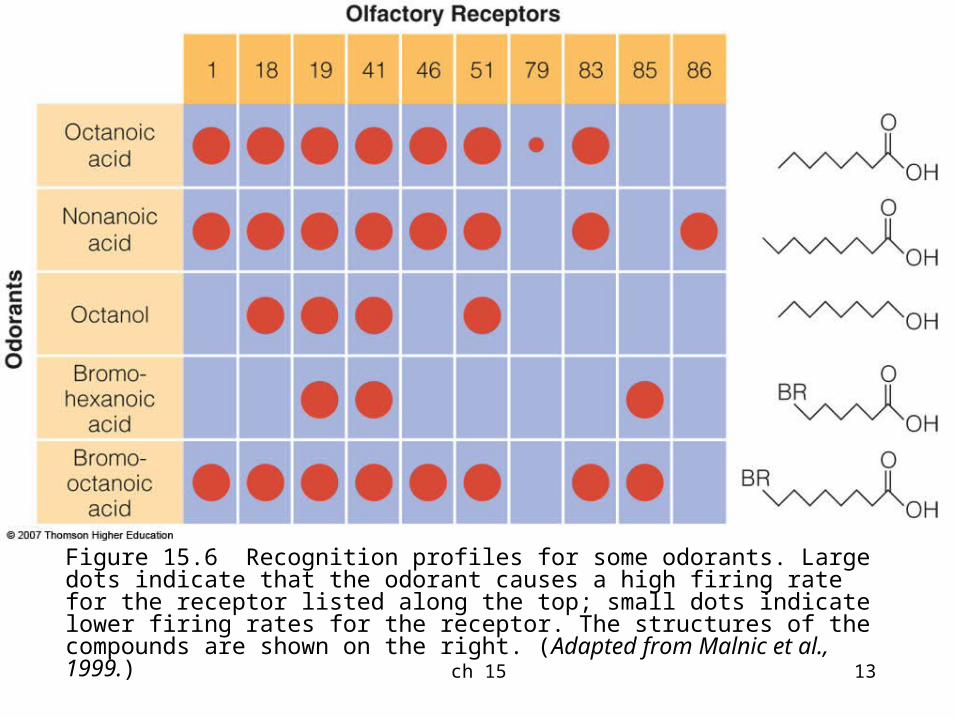
• Combinatorial code for odor
– Odorants are coded by combinations of olfactory receptors
– Specific receptors may be part of the code for multiple odorants.
ch 15 13
Figure 15.6 Recognition profiles for some odorants. Large dots indicate that the odorant causes a high firing rate for the receptor listed along the top; small dots indicate lower firing rates for the receptor. The structures of the compounds are shown on the right. (Adapted from Malnic et al., 1999.)

ch 15 14
Combinatorial coding?
• Remember distributed coding in face representation?
• Representing color by S, M, L cones.
• Even language is combinatorial.
• Combining 26 alphabets many many words.

ch 15 15
Activating the Olfactory Bulb
• Olfactory mucosa is divided into 4 zones
– Each zone contains a variety of different receptors
– Specific types of receptors are found in only one zone
– Odorants tend to activate neurons within a particular zone

ch 15 16
Activating the Olfactory Bulb - continued
• Optical imaging method
– Cortical cells consume oxygen when activated
– Red light is used to determine the amount of oxygen in the cells
– More oxygen reflects less red light

ch 15 17
Figure 15.9 Areas in the rat olfactory bulb that are activated by various chemicals: (a) a series of carbolic acids: (b) a series of aliphatic alcohols. (Uchida, N., Takahaski, Y. K., Tanifuji, M., & Mori, K. (2000). Odor maps in the mammalian olfactory bulb: Domain organization and odorant structural features. Nature Neuroscience, 3, 1035-1043.)

ch 15 18
Patterns of activation in the rat olfactory bulb (Linster, et al., 2001)
• 2-deoxyglucose (2DG) technique– 2DG, which contains
glucose, is ingested into an animal
– Animal is exposed to different chemicals
– Neural activation is measured by amount of radioactivity present
• This technique shows the pattern of neural activation is related to both chemical structure and to perception
Figure 15.10 These molecules have the same chemical formula, but the molecular group at the bottom is rotated to a different position. The black arrows indicate that the two forms of liminone activate similar areas in the olfactory bulb.
The pattern of activation on the OB is related to functional groups and structures of the chemicals as well as their perceived odors

ch 15 19
• Somewhat similar to– Retinotopic map V1– Tonotopic map A1– Topological locations specify the
characteristics of stimuli (light, sounds, ordorants)

ch 15 20
Taste

ch 15 21
Functions of Taste
• Sweetness is usually associated with substances that have nutritive value
• Bitter is usually associated with substances that are potentially harmful
• Salty taste indicates the presence of sodium
• However, there is not a perfect connection between tastes and function of substances

ch 15 22
Basic Taste Qualities
• Five basic taste qualities:
– Salty
– Sour
– Sweet
– Bitter
– Umami - described as meaty, brothy or savory and associated with MSG

ch 15 23
Human Tongue (from wikipedia)
The surface of the tongue is very bumpy: many ridges and valleys.
This structure is called Papillae (singular: papilla)
There are 4 types of papillae:
Filiform, Fungiform, Circmvallate, foliate

ch 15 24
Tongue
• papillae:– Filiform - shaped
like cones and located over entire surface
– Fungiform - shaped like mushrooms and found on sides and tip
– Foliate - series of folds on back and sides
– Circumvallate - shaped like flat mounds in a trench located at back
circumvllate
foliate
filiform
fungiform

ch 15 25
Structure of the Taste System - continued
• Taste buds are located in papallae except for filiform
– Tongue contains approximately 10,000 taste buds
– Each taste bud has taste cells with tips that extend into the taste pore
– Transduction occurs when chemicals contact the receptor sites on the tips
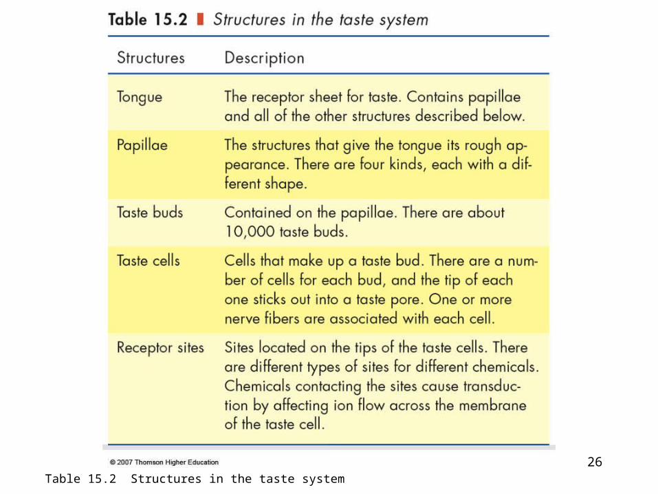
ch 15 26Table 15.2 Structures in the taste system

ch 15 27
Structure of the Taste System - continued
• Signals from taste cells travel along a set of pathways:
– Chorda tympani nerve from front and sides of tongue
– Glossopharyngeal nerve from back of tongue
– Vagus nerve from mouth and throat
– Superficial petronasal nerve from soft palate

ch 15 28
Structure of the Taste System - continued
• These pathways make connections in the nucleus of solitary tract in the spinal cord
• Then they travel to the thalamus
• Followed by areas in the frontal lobe:
– Insula
– Frontal opervulum cortex
– Orbital frontal cortex

ch 15 29
Neural Coding for Taste - continued
• Evidence exists for both specificity and distributed coding
• Some researchers suggest that the neural system for taste may function like the visual system for color
• Currently there is no agreed upon explanation for the neural system for taste

ch 15 30
The Perception of Flavor
• Combination of smell, taste, and other sensations (such as burning of hot peppers)
• Odor stimuli from food in the mouth reaches the olfactory mucosa through the retronasal route
• The taste of most compounds is influenced by olfaction, but a few, such as MSG are not

ch 15 31
The Physiology of Flavor Perception
• Responses from taste and smell are first combined in the orbital frontal cortex (OFC)
• OFC also receives input from the primary somatosensory cortex and the inferotemporal cortex in the visual what pathway– Bimodal neurons in this
area respond to taste and smell as well as taste and vision
– Firing of these neurons is also affected by the level of hunger of the animal for a specific food
Wikipedia.org
Ellis, Logan, & Dixon “human cross-section”

ch 15 32
Figure 15.22 The orbital frontal cortex (OFC) receives inputs from vision, olfaction, and touch, as shown. It is the first area where signals from the taste and smell systems meet. (Adapted from E. T. Rolls (2000). The orbitofrontal cortex and reward. Cerebral Cortex, 10, 284-294, Fig. 2. Reprinted with permission from Oxford University Press.)