Ch 14- Brain & Cranial Nerves. Protection 1.Bones of the cranium 2.Cranial meninges- Are continuous...
44
Ch 14- Brain & Cranial Nerves
-
Upload
shanon-spencer -
Category
Documents
-
view
218 -
download
1
Transcript of Ch 14- Brain & Cranial Nerves. Protection 1.Bones of the cranium 2.Cranial meninges- Are continuous...
Ch 14- Brain & Cranial Nerves
Protection1. Bones of the cranium
2. Cranial meninges- Are continuous with spinal meninges• Protect the brain from cranial trauma (head injury resulting from impact with another
object)• 3 layers
• Pia mater- • Arachnoid mater- • Dura mater
• 2 layers – outer layer fused to periosteum of cranial bones• Outer and inner layer separated by gap where fluids and BVs are located• Dural folds: Inward folding of inner layer of dura mater
• Provides additional stabilization and support (seatbelt)• Falx celebri • Tentorium cerebelli • Falx cerebelli
• Dural sinuses: large collecting veins within dural folds, includes superior/inferior sagittal sinus and transverse sinus
Severe Head Injuries
• Bleeding in cranial cavity can result in:• Epidural hemorrhage- cranial artery breaks and blood is forced between dura
mater and cranial cavity =
• Cranial vein break = delay in symptoms; fatal if not treated
• Subdural hemorrhage: blood enters inner layer of dura mater from small vein/dural sinuses; delay in symptoms• Pool of blood forming outside of damaged vessel =
P ro te c tio n
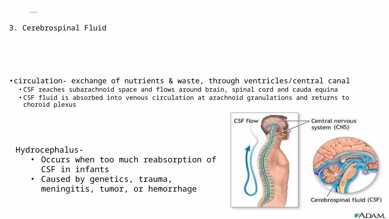
3. Cerebrospinal Fluid
• circulation- exchange of nutrients & waste, through ventricles/central canal• CSF reaches subarachnoid space and flows around brain, spinal cord and cauda equina• CSF fluid is absorbed into venous circulation at arachnoid granulations and returns to choroid plexus
Hydrocephalus- • Occurs when too much reabsorption of CSF in infants• Caused by genetics, trauma, meningitis, tumor, or
hemorrhage

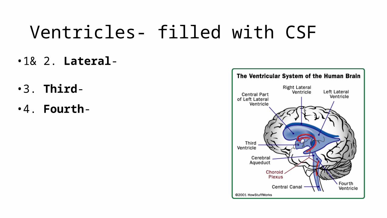
Ventricles- filled with CSF
•1& 2. Lateral-
•3. Third-
•4. Fourth-
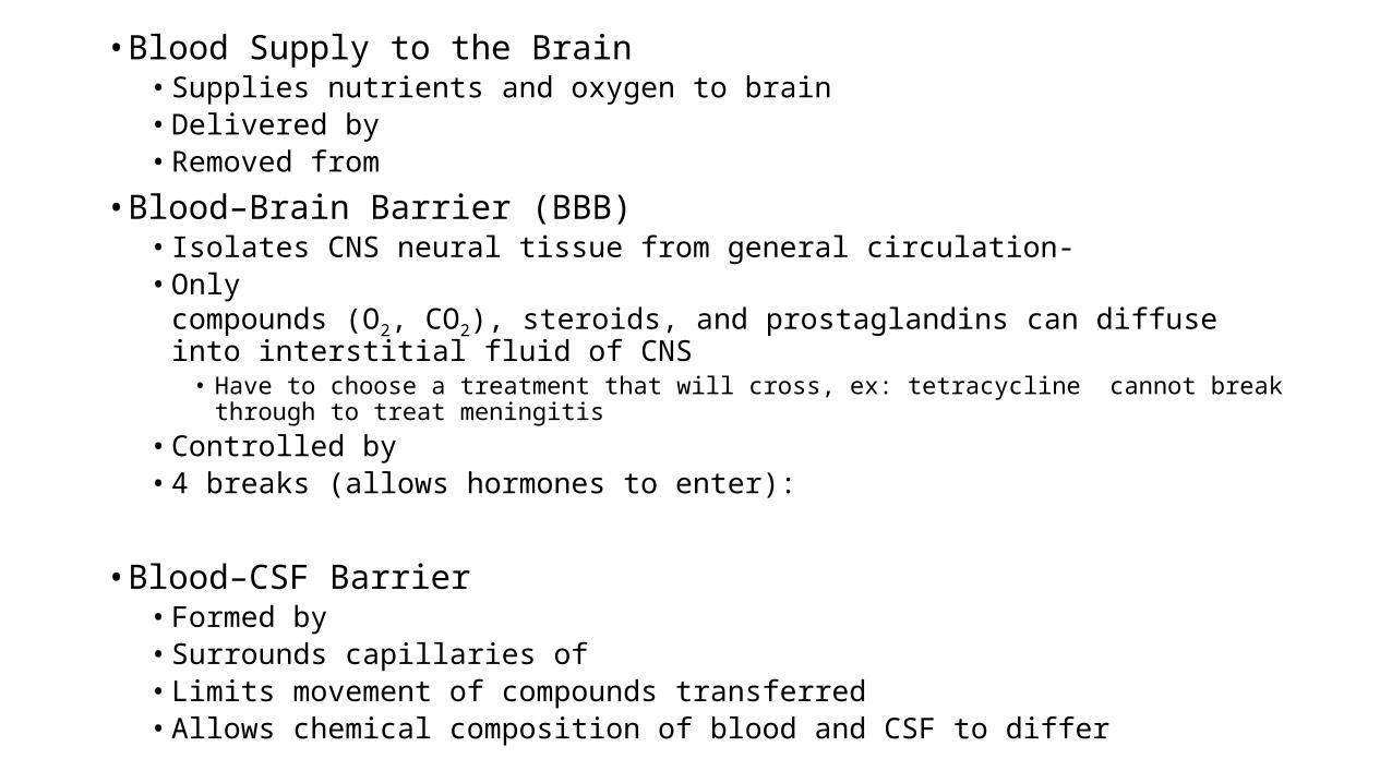
• Blood Supply to the Brain• Supplies nutrients and oxygen to brain• Delivered by • Removed from
• Blood–Brain Barrier (BBB)• Isolates CNS neural tissue from general circulation- • Only compounds (O2, CO2), steroids, and
prostaglandins can diffuse into interstitial fluid of CNS• Have to choose a treatment that will cross, ex: tetracycline cannot break through to treat
meningitis• Controlled by • 4 breaks (allows hormones to enter):
• Blood–CSF Barrier• Formed by • Surrounds capillaries of • Limits movement of compounds transferred• Allows chemical composition of blood and CSF to differ
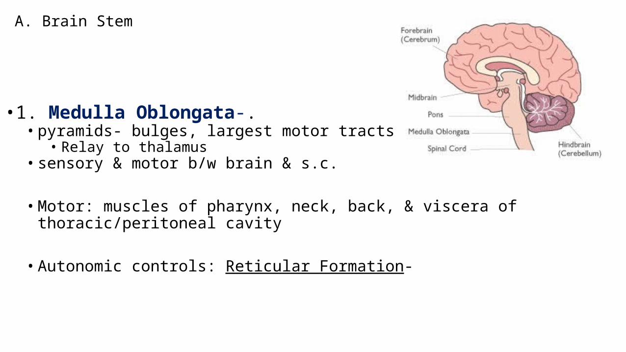
A. Brain Stem
•1. Medulla Oblongata-.• pyramids- bulges, largest motor tracts cross over
• Relay to thalamus• sensory & motor b/w brain & s.c.
• Motor: muscles of pharynx, neck, back, & viscera of thoracic/peritoneal cavity
• Autonomic controls: Reticular Formation-
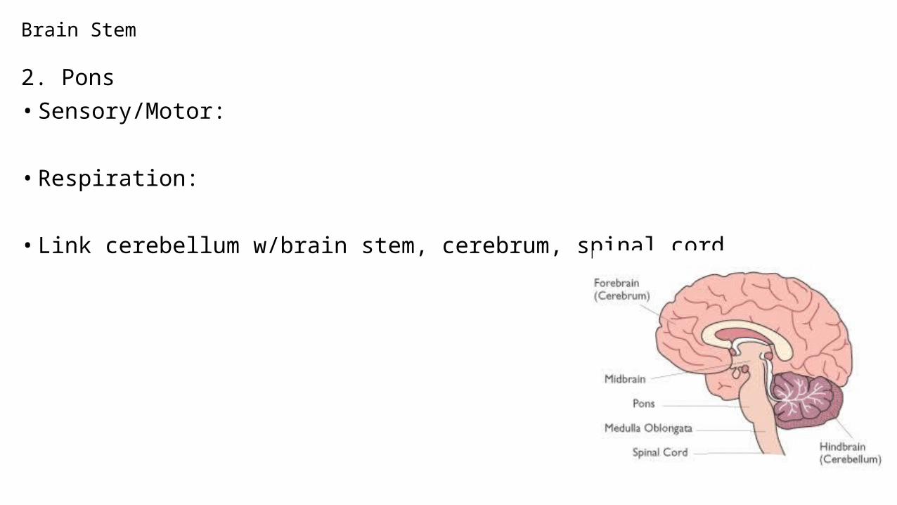
Brain Stem
2. Pons• Sensory/Motor:
• Respiration:
• Link cerebellum w/brain stem, cerebrum, spinal cord
Brain Stem
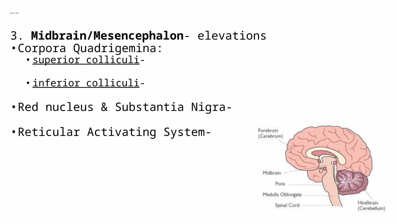
3. Midbrain/Mesencephalon- elevations• Corpora Quadrigemina:• superior colliculi-
• inferior colliculi-
• Red nucleus & Substantia Nigra-
• Reticular Activating System-
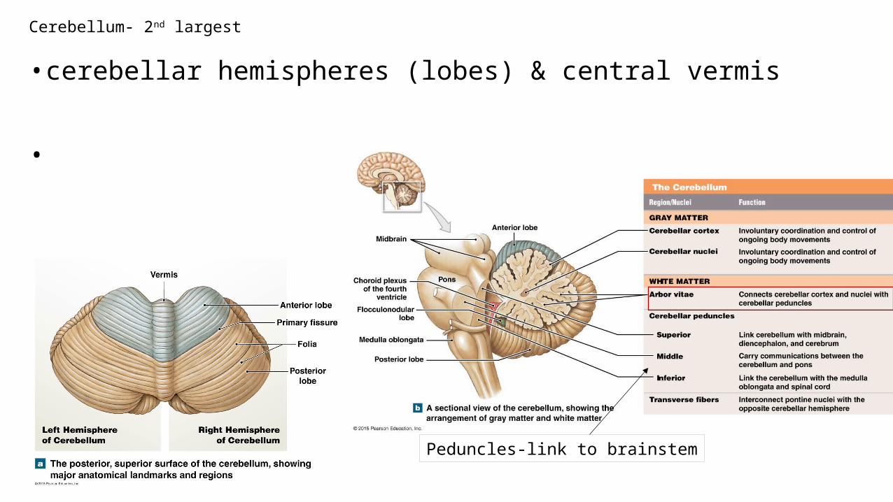
Cerebellum- 2nd largest
• cerebellar hemispheres (lobes) & central vermis
•
Peduncles-link to brainstem
Problems with Cerebellum
• Ataxia- • Inability to sit or stand without assistance• From trauma, stroke, drugs (alcohol)
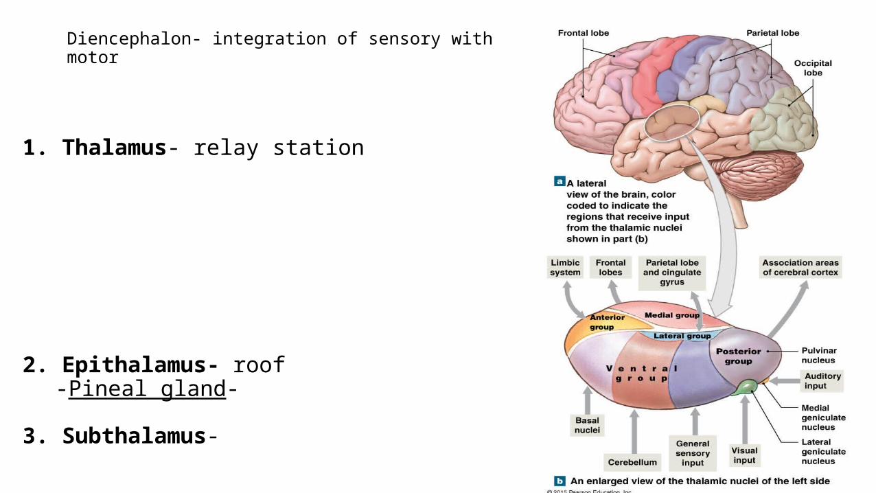
Diencephalon- integration of sensory with motor
1. Thalamus- relay station
2. Epithalamus- roof-Pineal gland-
3. Subthalamus-
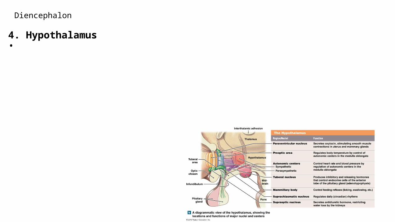
Diencephalon
4. Hypothalamus•
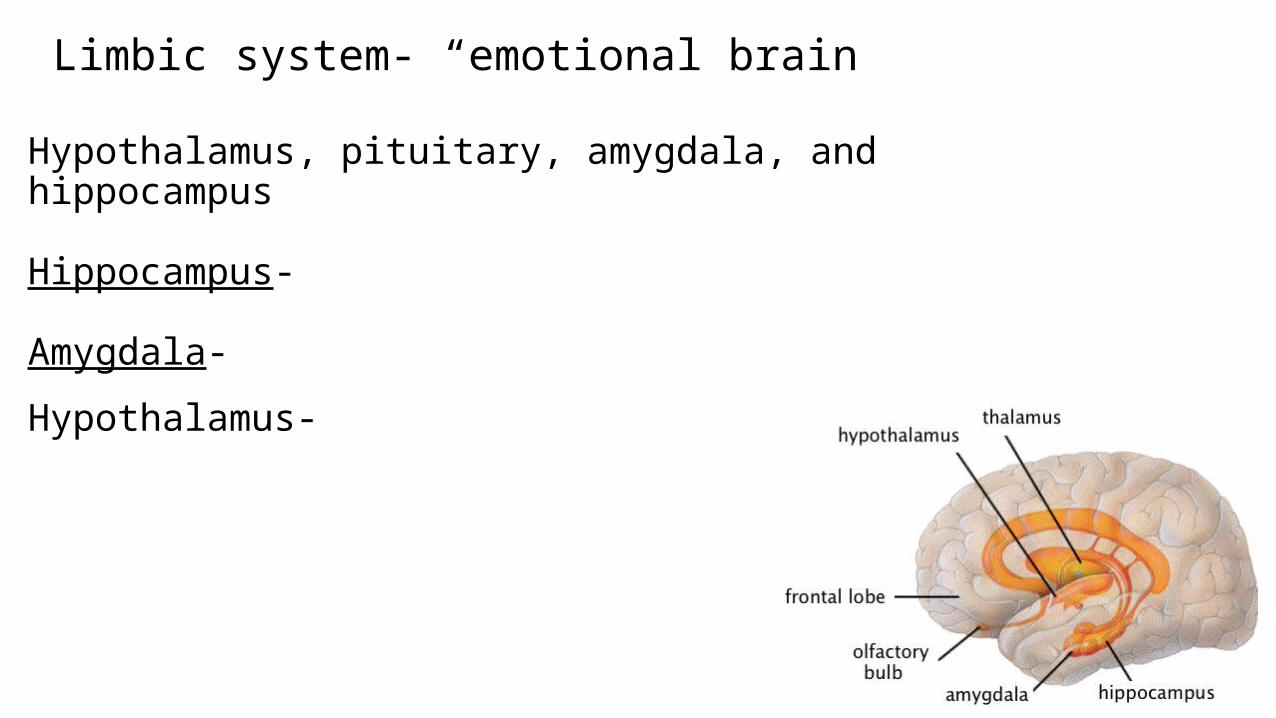
Limbic system- “emotional brain”
Hypothalamus, pituitary, amygdala, and hippocampus
Hippocampus-
Amygdala-
Hypothalamus-

Charles Whitman• engineering student and retired U.S.
Marine• He killed his wife and mother before going
to the top of the University of Texas tower and opened fire on persons crossing the campus and on nearby streets. He ended up killing 16 people and wounded 31, before being killed by police officers. The shooting spree lasted 96 minutes. • Post-mortem revealed a brain tumor near
his amygdala. • http://
www.youtube.com/watch?v=TtOQnM3LQkE
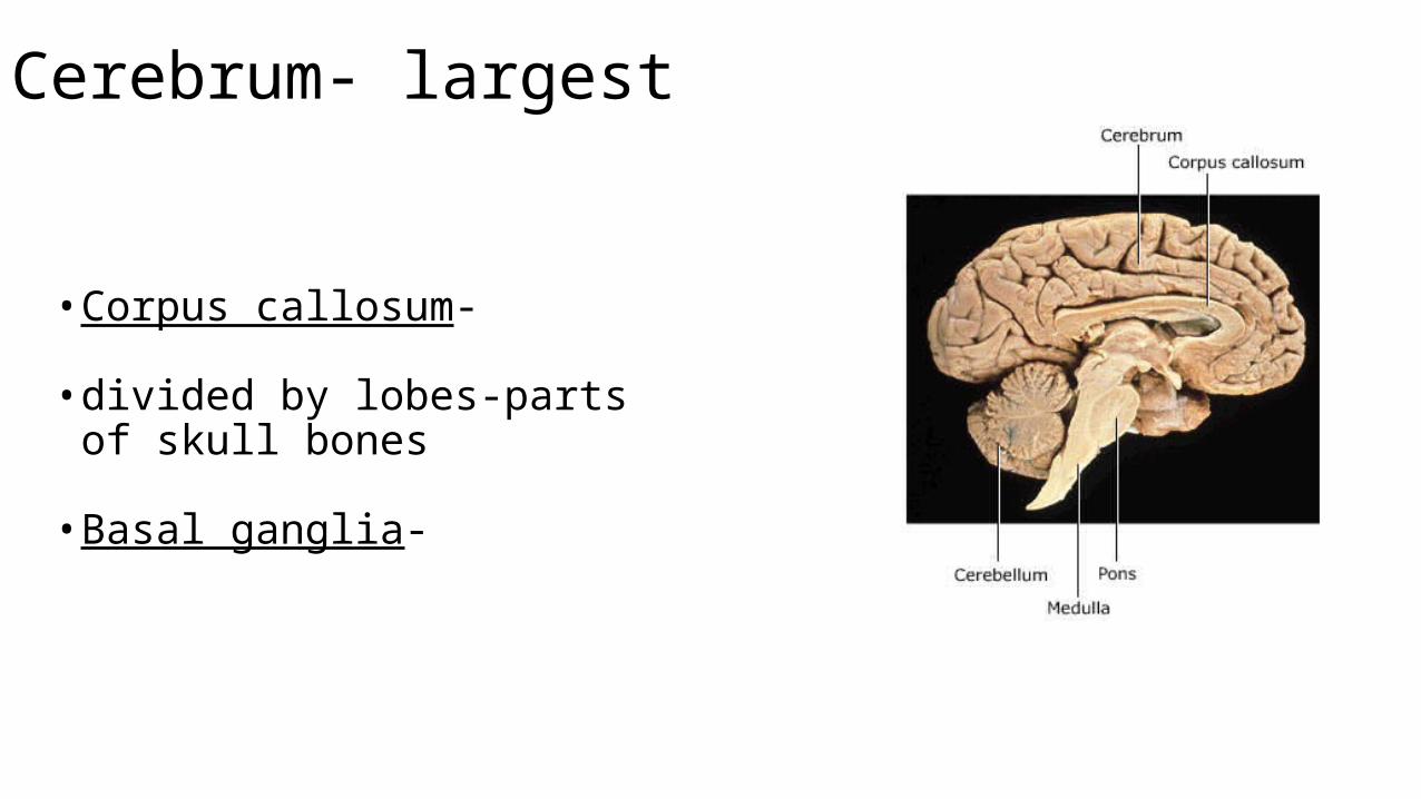
Cerebrum- largest
• Corpus callosum-
• divided by lobes-parts of skull bones
• Basal ganglia-
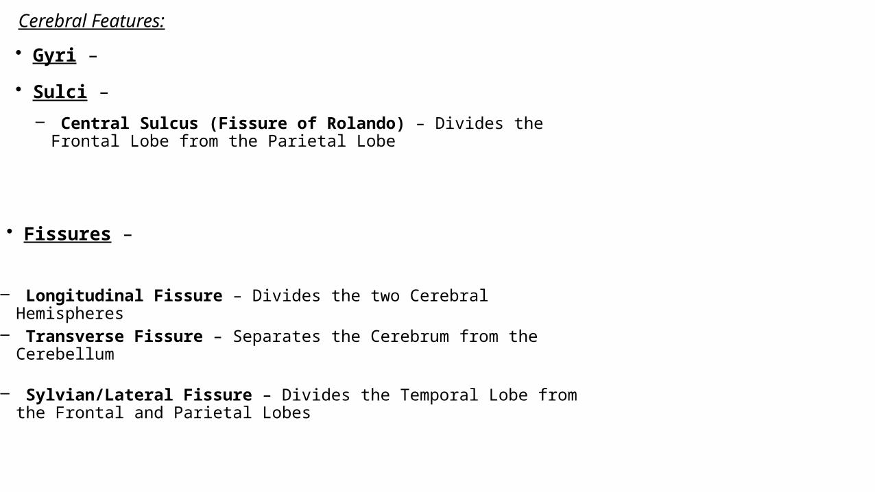
Cerebral Features:
• Sulci –
– Central Sulcus (Fissure of Rolando) – Divides the Frontal Lobe from the Parietal Lobe
• Fissures –
– Longitudinal Fissure – Divides the two Cerebral Hemispheres
– Transverse Fissure – Separates the Cerebrum from the Cerebellum
– Sylvian/Lateral Fissure – Divides the Temporal Lobe from the Frontal and Parietal Lobes
• Gyri –
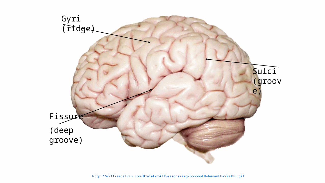
Gyri (ridge)
Fissure
(deep groove)
Sulci (groove)
http://williamcalvin.com/BrainForAllSeasons/img/bonoboLH-humanLH-viaTWD.gif
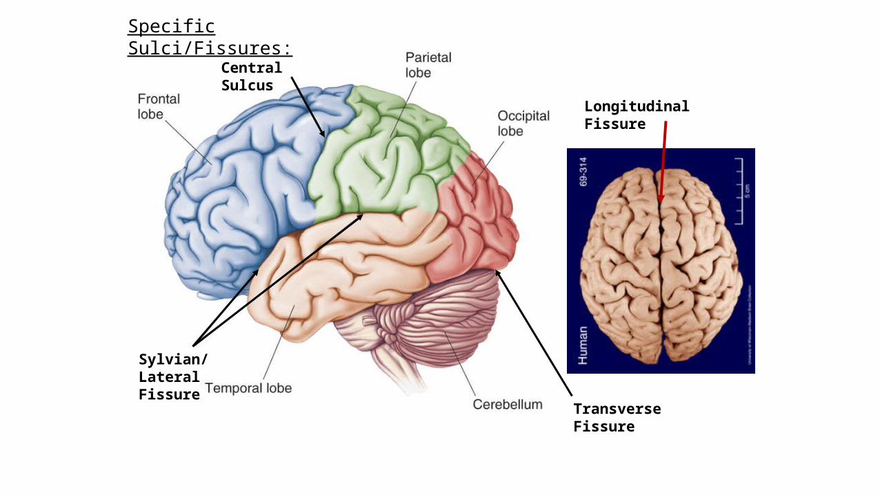
Longitudinal Fissure
Transverse Fissure
Sylvian/Lateral Fissure
Central Sulcus
Specific Sulci/Fissures:
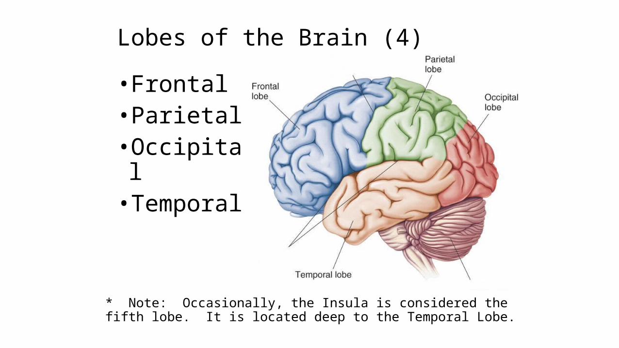
Lobes of the Brain (4)
• Frontal•Parietal•Occipital• Temporal
* Note: Occasionally, the Insula is considered the fifth lobe. It is located deep to the Temporal Lobe.
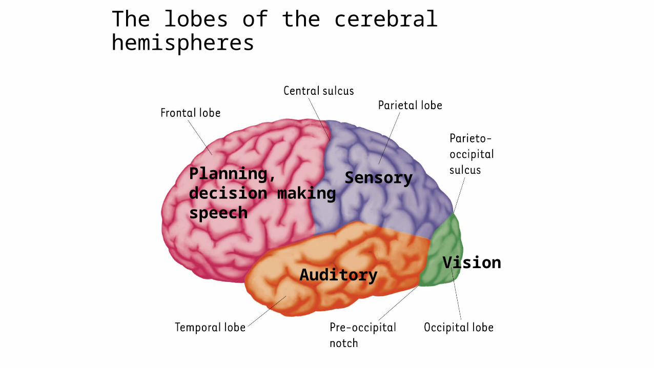
The lobes of the cerebral hemispheres
Planning, decision making speech
Sensory
AuditoryVision

Lobes of the Brain - Frontal
• The Frontal Lobe of the brain is located deep to the Frontal Bone of the skull.
• It plays an integral role in the following functions/actions:
Modified from: http://www.bioon.com/book/biology/whole/image/1/1-8.tif.jpg
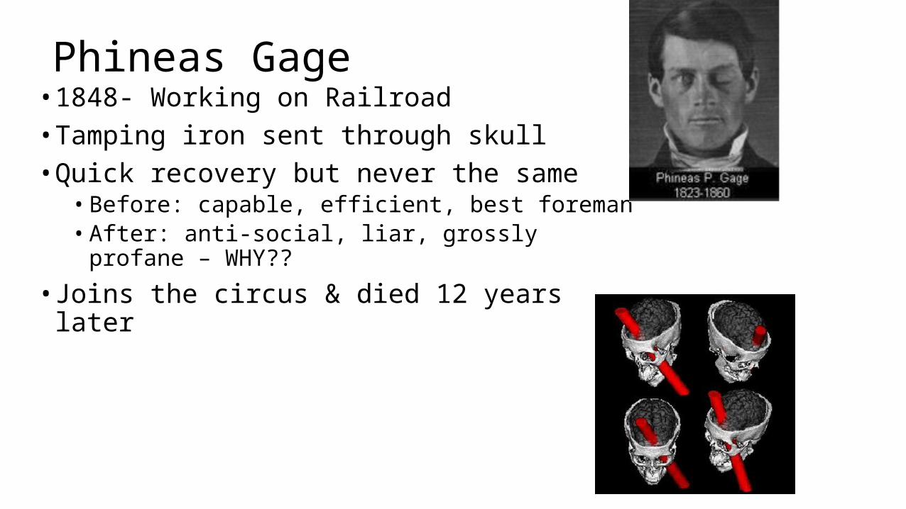
Phineas Gage• 1848- Working on Railroad• Tamping iron sent through skull• Quick recovery but never the same• Before: capable, efficient, best foreman• After: anti-social, liar, grossly profane – WHY??
• Joins the circus & died 12 years later
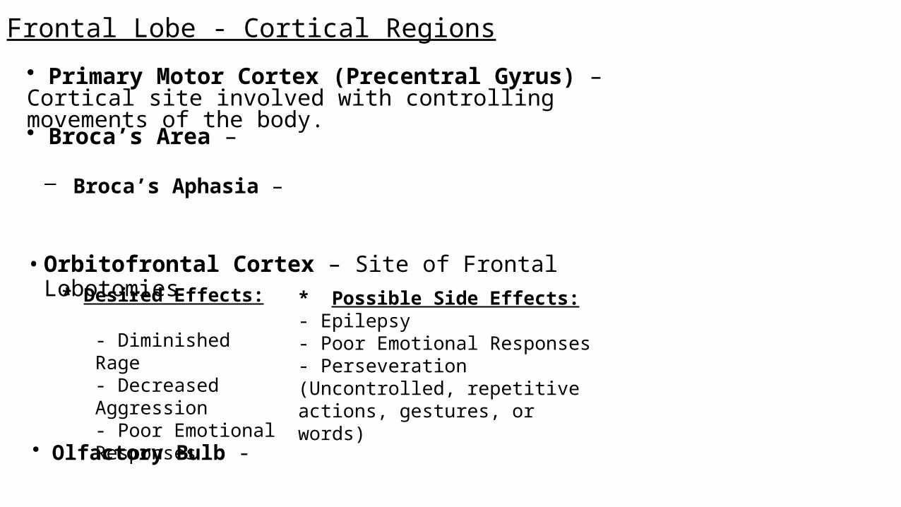
Frontal Lobe - Cortical Regions
• Orbitofrontal Cortex – Site of Frontal Lobotomies
• Primary Motor Cortex (Precentral Gyrus) – Cortical site involved with controlling movements of the body.
• Broca’s Area –
– Broca’s Aphasia –
• Olfactory Bulb -
* Desired Effects:- Diminished Rage- Decreased Aggression- Poor Emotional Responses
* Possible Side Effects:- Epilepsy- Poor Emotional Responses- Perseveration (Uncontrolled, repetitive actions, gestures, or words)
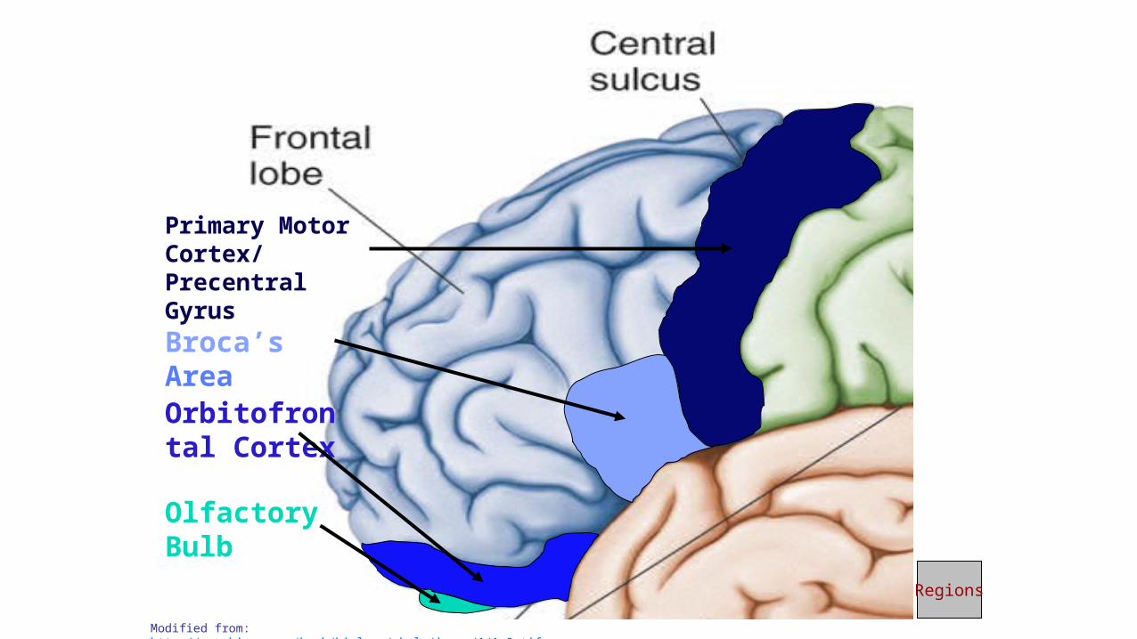
Primary Motor Cortex/ Precentral Gyrus
Broca’s Area
Orbitofrontal Cortex
Olfactory Bulb
Modified from: http://www.bioon.com/book/biology/whole/image/1/1-8.tif.jpg
Regions
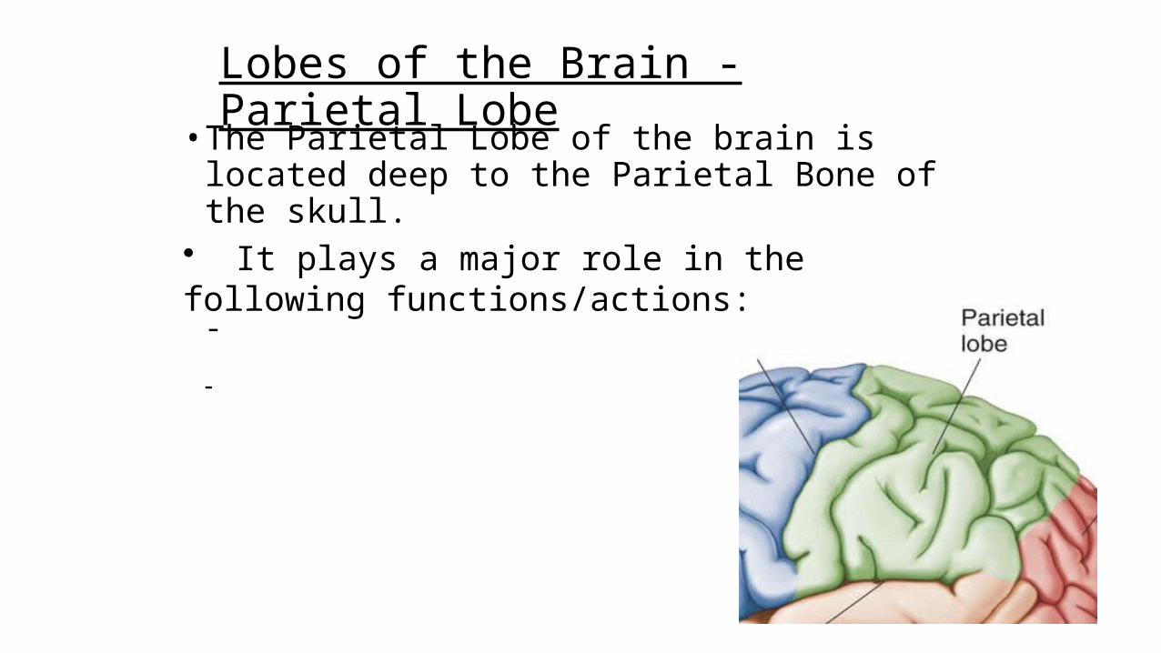
Lobes of the Brain - Parietal Lobe
• The Parietal Lobe of the brain is located deep to the Parietal Bone of the skull.
• It plays a major role in the following functions/actions:
-
-
Parietal Lobe - Cortical Regions
• Primary Somatosensory Cortex (Postcentral Gyrus) – Site involved with processing of tactile and proprioceptive information.
• Somatosensory Association Cortex - Assists with the integration and interpretation of sensations relative to body position and orientation in space. May assist with visuo-motor coordination.
• Primary Gustatory Cortex – Primary site involved with the interpretation of the sensation of Taste.
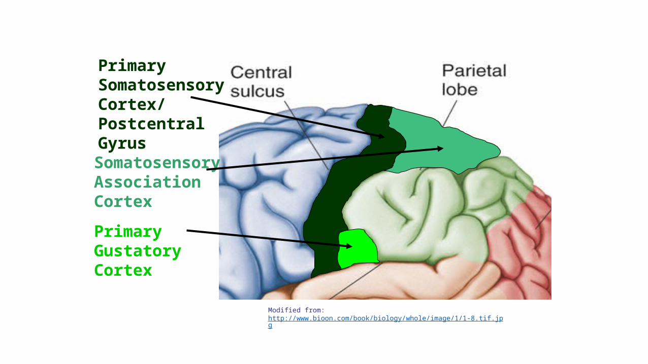
Primary Somatosensory Cortex/ Postcentral Gyrus
Primary Gustatory Cortex
Somatosensory Association Cortex
Modified from: http://www.bioon.com/book/biology/whole/image/1/1-8.tif.jpg
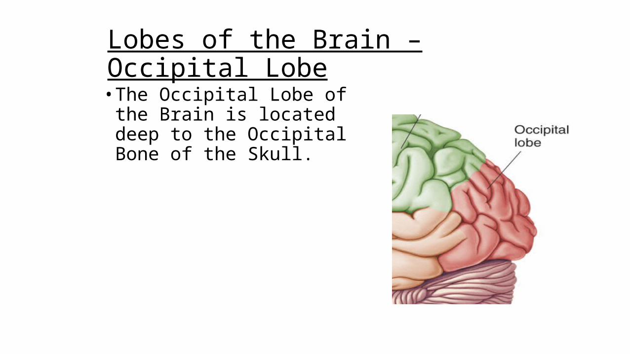
Lobes of the Brain – Occipital Lobe• The Occipital Lobe of the Brain
is located deep to the Occipital Bone of the Skull.
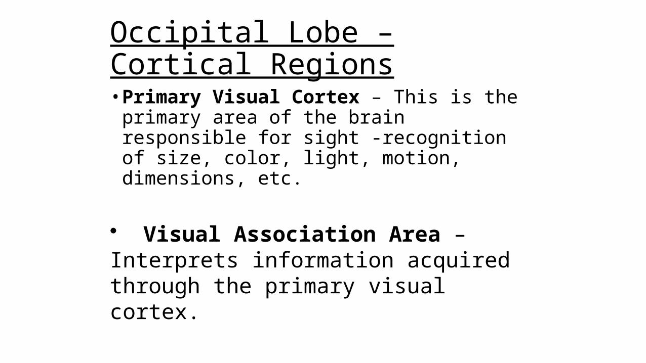
Occipital Lobe – Cortical Regions• Primary Visual Cortex – This is the primary area of
the brain responsible for sight -recognition of size, color, light, motion, dimensions, etc.
• Visual Association Area – Interprets information acquired through the primary visual cortex.
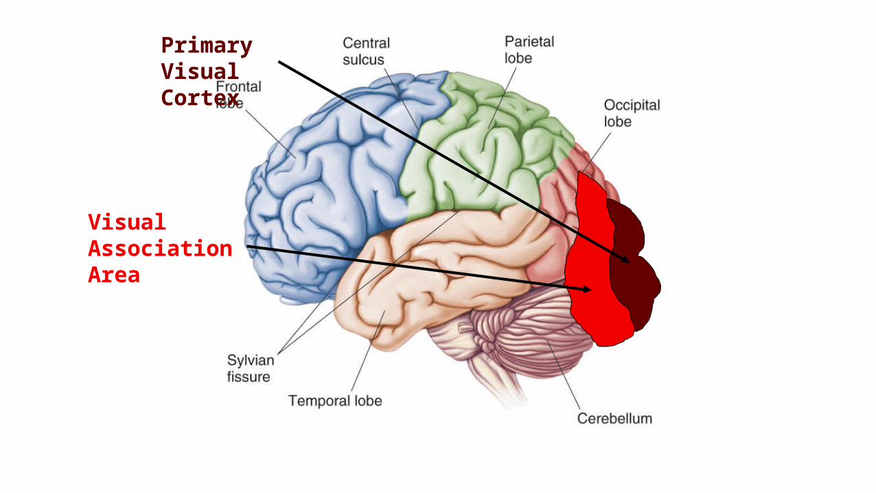
Primary Visual Cortex
Visual Association Area
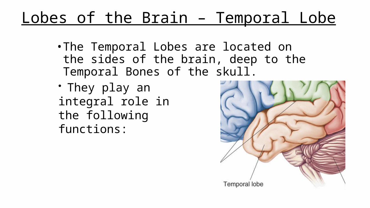
Lobes of the Brain – Temporal Lobe
• The Temporal Lobes are located on the sides of the brain, deep to the Temporal Bones of the skull.
• They play an integral role in the following functions:
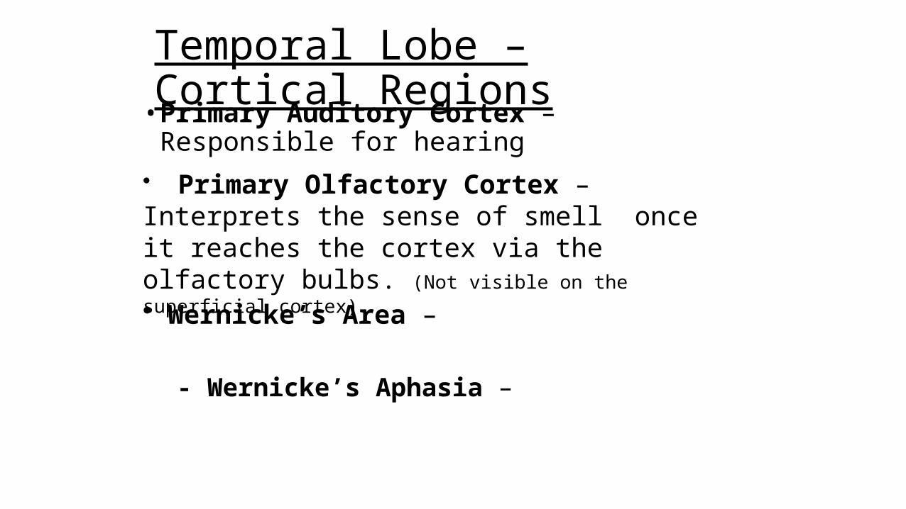
Temporal Lobe – Cortical Regions• Primary Auditory Cortex – Responsible for hearing
• Primary Olfactory Cortex – Interprets the sense of smell once it reaches the cortex via the olfactory bulbs. (Not visible on the superficial cortex)
• Wernicke’s Area –
- Wernicke’s Aphasia –
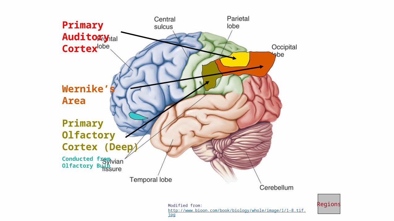
Primary Auditory Cortex
Wernike’s Area
Primary Olfactory Cortex (Deep)Conducted from Olfactory Bulb
RegionsModified from: http://www.bioon.com/book/biology/whole/image/1/1-8.tif.jpg
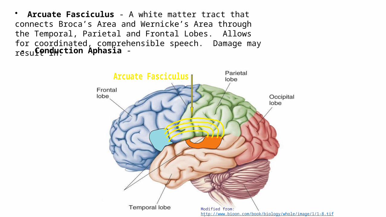
• Arcuate Fasciculus - A white matter tract that connects Broca’s Area and Wernicke’s Area through the Temporal, Parietal and Frontal Lobes. Allows for coordinated, comprehensible speech. Damage may result in:
- Conduction Aphasia -
Modified from: http://www.bioon.com/book/biology/whole/image/1/1-8.tif.jpg
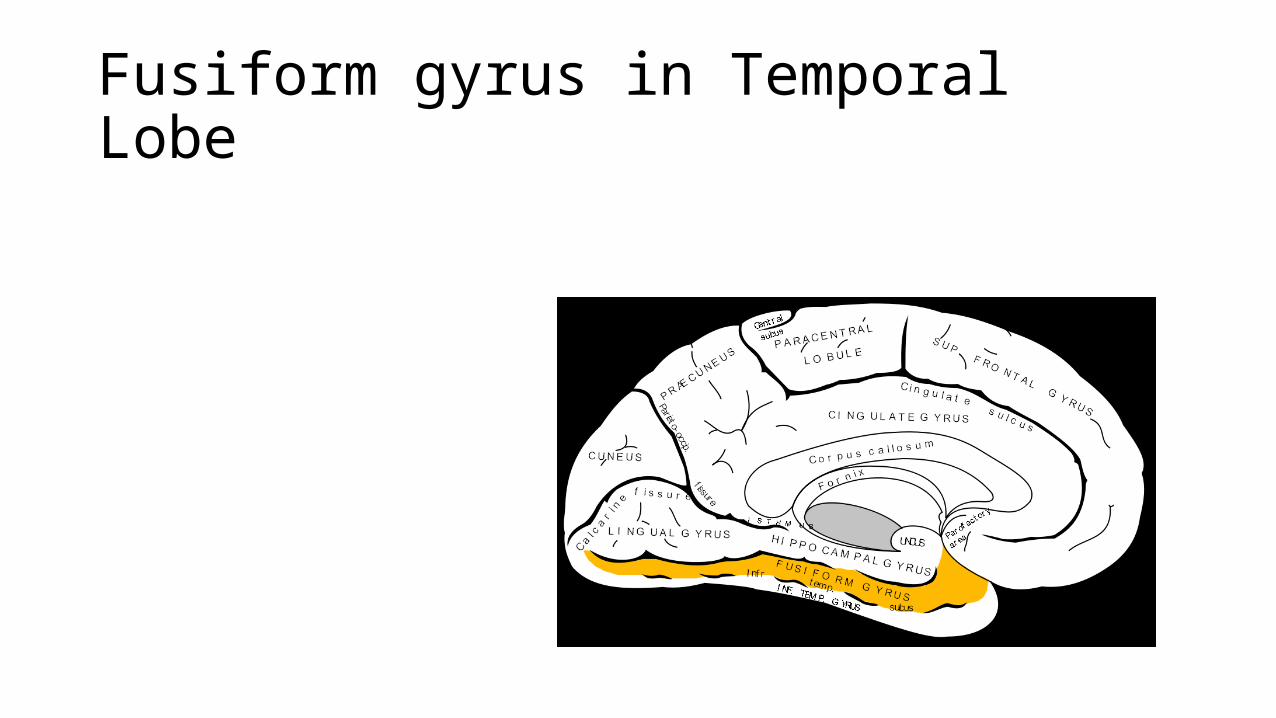
Fusiform gyrus in Temporal Lobe
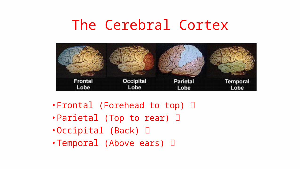
The Cerebral Cortex
• Frontal (Forehead to top) • Parietal (Top to rear) • Occipital (Back) • Temporal (Above ears)
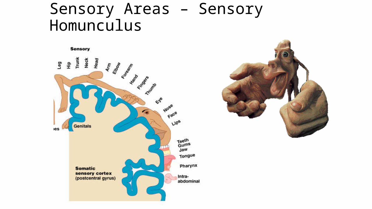
Sensory Areas – Sensory Homunculus
Hemispheric Lateralization
Left Right
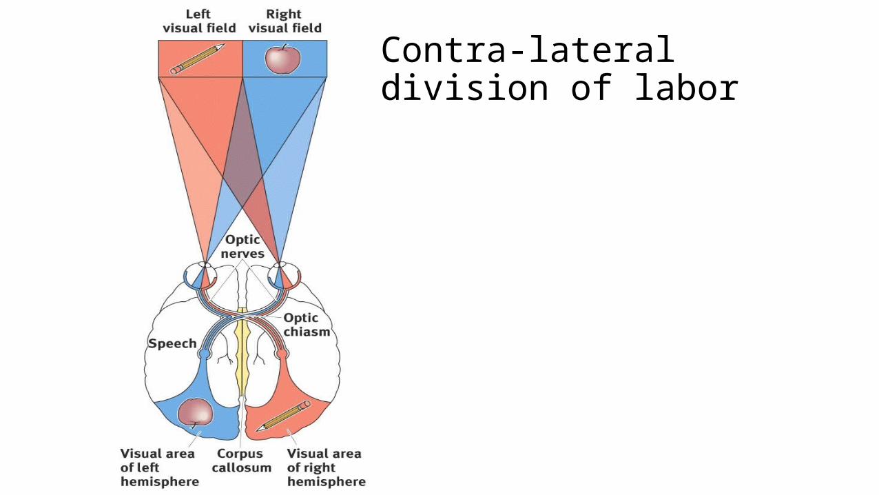
Contra-lateral division of labor
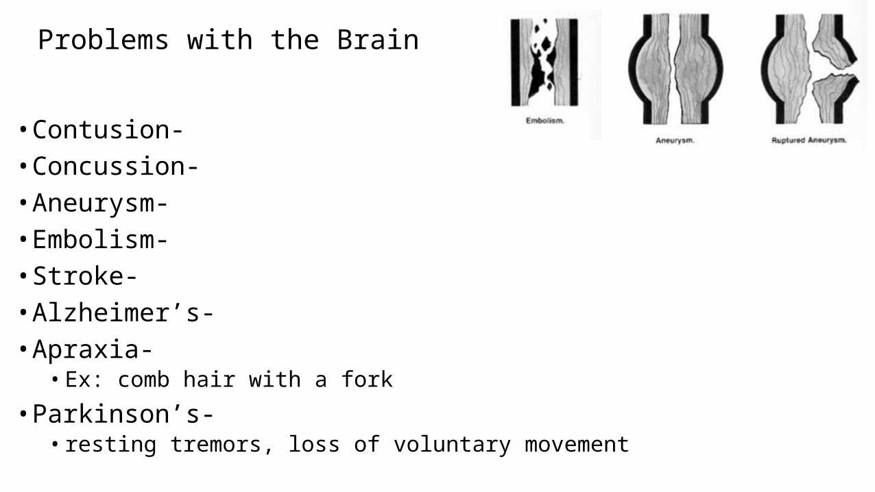
Problems with the Brain
• Contusion- • Concussion- • Aneurysm- • Embolism- • Stroke- • Alzheimer’s- • Apraxia- • Ex: comb hair with a fork
• Parkinson’s- • resting tremors, loss of voluntary movement
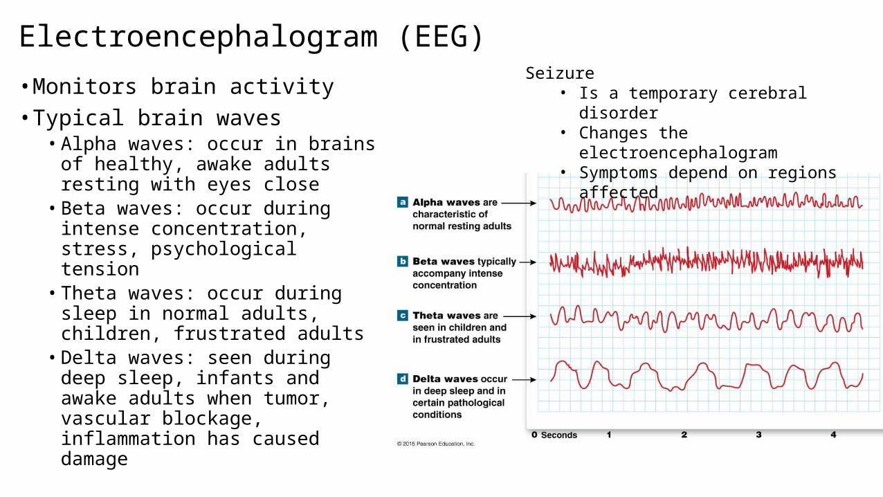
Electroencephalogram (EEG)
• Monitors brain activity• Typical brain waves• Alpha waves: occur in brains of
healthy, awake adults resting with eyes close• Beta waves: occur during intense
concentration, stress, psychological tension• Theta waves: occur during sleep in
normal adults, children, frustrated adults• Delta waves: seen during deep
sleep, infants and awake adults when tumor, vascular blockage, inflammation has caused damage
Seizure• Is a temporary cerebral disorder• Changes the electroencephalogram• Symptoms depend on regions affected
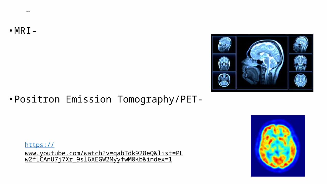
Imaging
• MRI-
• Positron Emission Tomography/PET-
https://www.youtube.com/watch?v=qabTdk928eQ&list=PLw2fLCAnU7j7Xr_9sl6XEGW2MyyfwM0Kb&index=1