Ch 13 Lecture Outline Thursday
47
Copyright © 2010 Pearson Education, Inc. Structure of a Nerve Cordlike organ of the PNS Bundle of myelinated and unmyelinated peripheral axons enclosed by connective tissue
-
Upload
raul-reynoso -
Category
Documents
-
view
219 -
download
0
Transcript of Ch 13 Lecture Outline Thursday

8/8/2019 Ch 13 Lecture Outline Thursday
http://slidepdf.com/reader/full/ch-13-lecture-outline-thursday 1/47
Copyright © 2010 Pearson Education, Inc.
Structure of a Nerve
Cordlike organ of the PNS
Bundle of myelinated and unmyelinated
peripheral axons enclosed by connective
tissue

8/8/2019 Ch 13 Lecture Outline Thursday
http://slidepdf.com/reader/full/ch-13-lecture-outline-thursday 2/47
Copyright © 2010 Pearson Education, Inc.
Structure of a Nerve
Connective tissue coverings include:
Endoneurium²loose connective tissue that
encloses axons and their myelin sheaths
Perineurium²coarse connective tissue that
bundles fibers into fascicles
Epineurium²tough fibrous sheath around a
nerve

8/8/2019 Ch 13 Lecture Outline Thursday
http://slidepdf.com/reader/full/ch-13-lecture-outline-thursday 3/47
Copyright © 2010 Pearson Education, Inc. Figure 13.3b
Blood
vessels
Fascicle
Epineur ium
Per ineur iumEndoneur ium
Axon
Myelin sheath
(b)

8/8/2019 Ch 13 Lecture Outline Thursday
http://slidepdf.com/reader/full/ch-13-lecture-outline-thursday 4/47
Copyright © 2010 Pearson Education, Inc.
Classif ication of Nerves
Most nerves are mixtures of afferent and efferent
fibers and somatic and autonomic (visceral) fibers
Pure sensory (afferent) or motor (efferent) nerves are
rare Types of fibers in mixed nerves:
Somatic afferent and somatic efferent
Visceral afferent and visceral efferent
Peripheral nerves classified as cranial or spinal
nerves

8/8/2019 Ch 13 Lecture Outline Thursday
http://slidepdf.com/reader/full/ch-13-lecture-outline-thursday 5/47

8/8/2019 Ch 13 Lecture Outline Thursday
http://slidepdf.com/reader/full/ch-13-lecture-outline-thursday 6/47
Copyright © 2010 Pearson Education, Inc.
Cranial Nerves
Twelve pairs of nerves associated with the
brain
Most are mixed in function; two pairs are
purely sensory
Each nerve is identified by a number
(I through XII) and a name
³On occasion, our trusty truck acts f unny²very
good vehicle anyhow´

8/8/2019 Ch 13 Lecture Outline Thursday
http://slidepdf.com/reader/full/ch-13-lecture-outline-thursday 7/47
Copyright © 2010 Pearson Education, Inc. Figure 13.5 (a)
Frontal lobe
Temporal lobe
Infundibulum
Facialnerve (VII)
Vestibulo-
cochlear
nerve (VIII)Glossopharyngeal
nerve (IX)Vagus nerve (X)
Accessory nerve (XI)
Hypoglossal nerve (XII)
(a)
Filaments of
olfactory
nerve (I)
Olfactor y bulb
Olfactor y tract
Optic chiasma
Optic nerve
(II)
Optic tractOculomotor nerve (III)Trochlear
nerve (IV) Trigeminal
nerve (V)
Abducensnerve (VI)
Cerebellum
Medulla
oblongata

8/8/2019 Ch 13 Lecture Outline Thursday
http://slidepdf.com/reader/full/ch-13-lecture-outline-thursday 8/47
Copyright © 2010 Pearson Education, Inc. Figure 13.5 (b)
*PS = parasympathetic(b)
Cranial nerves
I ² VI
I
II
III
IV
V
VI
Olfactor y
Optic
Oculomotor
Trochlear
Tr igeminal
Abducens
Yes (smell)
Yes (vision)
No
No
Yes (general
sensation)
No
No
No
Yes
Yes
Yes
Yes
No
No
Yes
No
No
No
Cranial nerves
VII ² XII
Sensory
f unction
Motor
f unction
PS*
fibers
Sensory
f unction
Motor
f unction
PS*
fibers
VII
VIII
IX
X
XI
XII
Facial
Vestibulocochlear
Glossophar yngeal
Vagus
Accessor y
Hypoglossal
Yes (taste)
Yes (hear ing
and balance)
Yes (taste)
Yes (taste)
No
No
Yes
Some
Yes
Yes
Yes
Yes
Yes
No
Yes
Yes
No
No

8/8/2019 Ch 13 Lecture Outline Thursday
http://slidepdf.com/reader/full/ch-13-lecture-outline-thursday 9/47
Copyright © 2010 Pearson Education, Inc.
I: The Olfactor y Nerves
Arise from the olfactory
receptor cells of nasalcavity
Pass through the
cribriform plate of theethmoid bone
Fibers synapse in the
olfactory bulbs
Pathway terminates in the
primary olfactory cortex
Purely sensory (olfactory)function

8/8/2019 Ch 13 Lecture Outline Thursday
http://slidepdf.com/reader/full/ch-13-lecture-outline-thursday 10/47
Copyright © 2010 Pearson Education, Inc.
II: The Optic Nerves Arise from the retinas
Pass through the opticcanals, converge and
partially cross over at the
optic chiasma
Optic tracts continue to the
thalamus, where they
synapse
Optic radiation fibers run
to the occipital (visual)
cortex
Purely sensory (visual) function

8/8/2019 Ch 13 Lecture Outline Thursday
http://slidepdf.com/reader/full/ch-13-lecture-outline-thursday 11/47
Copyright © 2010 Pearson Education, Inc.
III: The Oculomotor Nerves Fibers extend from the
ventral midbrainthrough the superior
orbital fissures to the
extrinsic eye muscles
Functions in raising the
eyelid, directing the
eyeball, constricting the
iris (parasympathetic),and controlling lens
shape

8/8/2019 Ch 13 Lecture Outline Thursday
http://slidepdf.com/reader/full/ch-13-lecture-outline-thursday 12/47
Copyright © 2010 Pearson Education, Inc.
IV: The Trochlear Nerves Fibers from the
dorsal midbrainenter the orbits via
the superior orbital
fissures to innervate
the superior oblique
muscle
Primarily a motor
nerve that directsthe eyeball

8/8/2019 Ch 13 Lecture Outline Thursday
http://slidepdf.com/reader/full/ch-13-lecture-outline-thursday 13/47
Copyright © 2010 Pearson Education, Inc.
V: The Tr igeminal Nerves Largest cranial nerves; fibers
extend from pons to face Three divisions
Ophthalmic (V1) passesthrough the superior orbital
fissure
Maxillary (V2) passes throughthe foramen rotundum
Mandibular (V3) passes
through the foramen ovale
Convey sensory impulses frovarious areas of the face (V1)and (V2), and supplies motor
fibers (V3) for mastication

8/8/2019 Ch 13 Lecture Outline Thursday
http://slidepdf.com/reader/full/ch-13-lecture-outline-thursday 14/47

8/8/2019 Ch 13 Lecture Outline Thursday
http://slidepdf.com/reader/full/ch-13-lecture-outline-thursday 15/47
Copyright © 2010 Pearson Education, Inc.
VII: The Facial Nerves Fibers from the pons travel
through the internal acousticmeatuses, and emerge through
the stylomastoid foramina to the
lateral aspect of the face
Chief motor nerves of the facewith 5 major branches
Motor functions include facial
expression, parasympathetic
impulses to lacrimal andsalivary glands
Sensory function (taste) from
the anterior two-thirds of the
tongue

8/8/2019 Ch 13 Lecture Outline Thursday
http://slidepdf.com/reader/full/ch-13-lecture-outline-thursday 16/47
Copyright © 2010 Pearson Education, Inc.
VIII: The Vestibulocochlear Nerves
Afferent fibers from the hearing
receptors (cochlear division)and equilibrium receptors
(vestibular division) pass from
the inner ear through the
internal acoustic meatuses,
and enter the brain stem at the
pons-medulla border
Mostly sensory function; smallmotor component for
adjustment of sensitivity of
receptors

8/8/2019 Ch 13 Lecture Outline Thursday
http://slidepdf.com/reader/full/ch-13-lecture-outline-thursday 17/47
Copyright © 2010 Pearson Education, Inc.
IX: The Glossophar yngeal Nerves Fibers from the medulla
leave the skull via the jugular foramen and run to thethroat
Motor functions: innervate
part of the tongue andpharynx for swallowing, andprovide parasympatheticfibers to the parotid salivaryglands
Sensory functions: fibers conducttaste and general sensoryimpulses from the pharynx andposterior tongue, and impulses
from carotid chemoreceptors and

8/8/2019 Ch 13 Lecture Outline Thursday
http://slidepdf.com/reader/full/ch-13-lecture-outline-thursday 18/47
Copyright © 2010 Pearson Education, Inc.
X: The Vagus Nerves The only cranial nerves that
extend beyond the head and
neck region
Fibers from the medulla exit the
skull via the jugular foramen
Most motor fibers are
parasympathetic fibers that helpregulate the activities of the
heart, lungs, and abdominal
viscera
Sensory fibers carry impulses from
thoracic and abdominal viscera,
baroreceptors, chemoreceptors,
and taste buds of posterior tongue
and pharynx

8/8/2019 Ch 13 Lecture Outline Thursday
http://slidepdf.com/reader/full/ch-13-lecture-outline-thursday 19/47

8/8/2019 Ch 13 Lecture Outline Thursday
http://slidepdf.com/reader/full/ch-13-lecture-outline-thursday 20/47
Copyright © 2010 Pearson Education, Inc.
XII: The Hypoglossal Nerves Fibers from the
medulla exit theskull via the
hypoglossal canal
Innervate extrinsicand intrinsic
muscles of the
tongue that
contribute toswallowing and
speech

8/8/2019 Ch 13 Lecture Outline Thursday
http://slidepdf.com/reader/full/ch-13-lecture-outline-thursday 21/47
Copyright © 2010 Pearson Education, Inc.
Spinal Nerves
31 pairs of mixed nerves named according to
their point of issue from the spinal cord
8 cervical (C1 ±C8)
12 thoracic (T1 ±T12)
5 Lumbar (L1 ±L5)
5 Sacral (S1 ±S5) 1 Coccygeal (C0)

8/8/2019 Ch 13 Lecture Outline Thursday
http://slidepdf.com/reader/full/ch-13-lecture-outline-thursday 22/47

8/8/2019 Ch 13 Lecture Outline Thursday
http://slidepdf.com/reader/full/ch-13-lecture-outline-thursday 23/47
Copyright © 2010 Pearson Education, Inc.
Spinal Nerves: Roots
Each spinal nerve connects to the spinal cord
via two roots
Ventral roots
Contain motor (efferent) fibers from the ventral
horn motor neurons
Fibers innervate skeletal muscles)

8/8/2019 Ch 13 Lecture Outline Thursday
http://slidepdf.com/reader/full/ch-13-lecture-outline-thursday 24/47
Copyright © 2010 Pearson Education, Inc.
Spinal Nerves: Roots
Dorsal roots
Contain sensory (afferent) fibers from sensory
neurons in the dorsal root ganglia
Conduct impulses from peripheral receptors
Dorsal and ventral roots unite to form spinal
nerves, which then emerge from the vertebral
column via the intervertebral foramina

8/8/2019 Ch 13 Lecture Outline Thursday
http://slidepdf.com/reader/full/ch-13-lecture-outline-thursday 25/47
Copyright © 2010 Pearson Education, Inc. Figure 13.7 (a)
Dorsal rootganglion
Gray matter
White matter
Ventral root
Dorsal root
Dorsal and
ventral rootletsof spinal nerve
Dorsal ramusof spinal nerve
Ventral ramus
of spinal nerve
Sympathetic trunkganglion
Spinal nerve
Rami communicantes
Anterior view showing spinal cor d, associated nerves, and vertebrae.
The dorsal and ventral roots arise medially as rootlets and join
laterally to form the spinal nerve.

8/8/2019 Ch 13 Lecture Outline Thursday
http://slidepdf.com/reader/full/ch-13-lecture-outline-thursday 26/47
Copyright © 2010 Pearson Education, Inc.
Spinal Nerves: Rami
Each spinal nerve branches into mixed rami
Dorsal ramus
Larger ventral ramus
Meningeal branch
Rami communicantes (autonomic pathways)
join to the ventral rami in the thoracic region

8/8/2019 Ch 13 Lecture Outline Thursday
http://slidepdf.com/reader/full/ch-13-lecture-outline-thursday 27/47
Copyright © 2010 Pearson Education, Inc.
Spinal Nerves: Rami
All ventral rami except T2 ±T12 form interlacing
nerve networks called plexuses (cervical,
brachial, lumbar, and sacral)
The back is innervated by dorsal rami viaseveral branches
Ventral rami of T2 ±T12 as intercostal nerves
supply muscles of the ribs, anterolateralthorax, and abdominal wall

8/8/2019 Ch 13 Lecture Outline Thursday
http://slidepdf.com/reader/full/ch-13-lecture-outline-thursday 28/47
Copyright © 2010 Pearson Education, Inc. Figure 13.7 (b)
Dorsal ramus
Ventral ramus
Intercostal nerve
Spinal nerve
Rami communicantes
Dorsal root
ganglion Dorsal rootVentral root
Sympathetic trunk
ganglion
Ster num
(b) Cross section of thorax showing the main roots and
branches of a spinal nerve.
Branches of intercostal
nerve Lateral cutaneous Anter ior cutaneous

8/8/2019 Ch 13 Lecture Outline Thursday
http://slidepdf.com/reader/full/ch-13-lecture-outline-thursday 29/47
Copyright © 2010 Pearson Education, Inc.
Cervical Plexus
Formed by ventral rami of C1 ±C4
Innervates skin and muscles of the neck, ear,
back of head, and shoulders
Phrenic nerve
Major motor and sensory nerve of the
diaphragm (receives fibers from C3
±C5
)

8/8/2019 Ch 13 Lecture Outline Thursday
http://slidepdf.com/reader/full/ch-13-lecture-outline-thursday 30/47
Copyright © 2010 Pearson Education, Inc. Figure 13.8
Hypoglossal
nerve (XII)
C1
C2
C3
C4
C5
Segmental
branches
Lesser occipitalnerveGreater aur icular
nerve
Ansa cervicalis
Phrenic nerve
Supraclavicular
nerves
Accessor y nerve (XI)
Transverse
cervical nerve
Ventral
rami:
Ventral rami

8/8/2019 Ch 13 Lecture Outline Thursday
http://slidepdf.com/reader/full/ch-13-lecture-outline-thursday 31/47
Copyright © 2010 Pearson Education, Inc. Table 13.3

8/8/2019 Ch 13 Lecture Outline Thursday
http://slidepdf.com/reader/full/ch-13-lecture-outline-thursday 32/47
Copyright © 2010 Pearson Education, Inc.
Brachial Plexus
Formed by ventral rami of C5 ±C8 and T1 (and often
C4 and T2)
It gives rise to the nerves that innervate the upper
limb Major branches of this plexus:
Roots²five ventral rami (C5 ±T1)
Trunks²upper, middle, and lower Divisions²anterior and posterior
Cords²lateral, medial, and posterior

8/8/2019 Ch 13 Lecture Outline Thursday
http://slidepdf.com/reader/full/ch-13-lecture-outline-thursday 33/47

8/8/2019 Ch 13 Lecture Outline Thursday
http://slidepdf.com/reader/full/ch-13-lecture-outline-thursday 34/47
Copyright © 2010 Pearson Education, Inc. Figure 13.9 (d)
Anter ior
divisions
(d) Flowchart summarizing relationships within the
brachial plexus
Major terminal
branches
(peripheral nerves)
Cor ds Divisions Tr unksRoots
(ventral
rami)
MusculocutaneousMedian
Ulnar
Radial
Axillar y
Anter ior
Poster ior
Anter ior
Poster ior
Poster ior
Anter ior
Upper
Middle
Lower
Lateral
Medial
Poster ior
Poster ior
divisions
Trunks Roots
C5
C6
C7
C8
T1

8/8/2019 Ch 13 Lecture Outline Thursday
http://slidepdf.com/reader/full/ch-13-lecture-outline-thursday 35/47
Copyright © 2010 Pearson Education, Inc.
Brachial Plexus: Nerves
Axillary²innervates the deltoid, teres minor, and skinand joint capsule of the shoulder
Musculocutaneous²innervates the biceps brachiiand brachialis and skin of lateral forearm
Median²innervates the skin, most flexors andpronators in the forearm, and some intrinsic musclesof the hand
Ulnar²supplies the flexor carpi ulnaris, part of theflexor digitorum profundus, most intrinsic muscles of the hand, and skin of medial aspect of hand
Radial²innervates essentially all extensor muscles,supinators, and posterior skin of limb

8/8/2019 Ch 13 Lecture Outline Thursday
http://slidepdf.com/reader/full/ch-13-lecture-outline-thursday 36/47
Copyright © 2010 Pearson Education, Inc. Figure 13.9 (c)
Median nerve
Musculocutaneous nerve
Radial nerveHumerus
Ulna
Ulnar nerveMedian nerve
Radius
Radial nerve (superf icial branch)
Superf icial branch of ulnar nerveDorsal branch of ulnar nerve
Digital branch of ulnar nerve
Muscular branch
Digital branch
(c) The major nerves of the upper limb
Axillary
nerveAnter ior
divisions
Poster ior
divisions
Trunks Roots

8/8/2019 Ch 13 Lecture Outline Thursday
http://slidepdf.com/reader/full/ch-13-lecture-outline-thursday 37/47
Copyright © 2010 Pearson Education, Inc. Table 13.4
8/8/2019 Ch 13 Lecture Outline Thursday
http://slidepdf.com/reader/full/ch-13-lecture-outline-thursday 38/47
Copyright © 2010 Pearson Education, Inc.
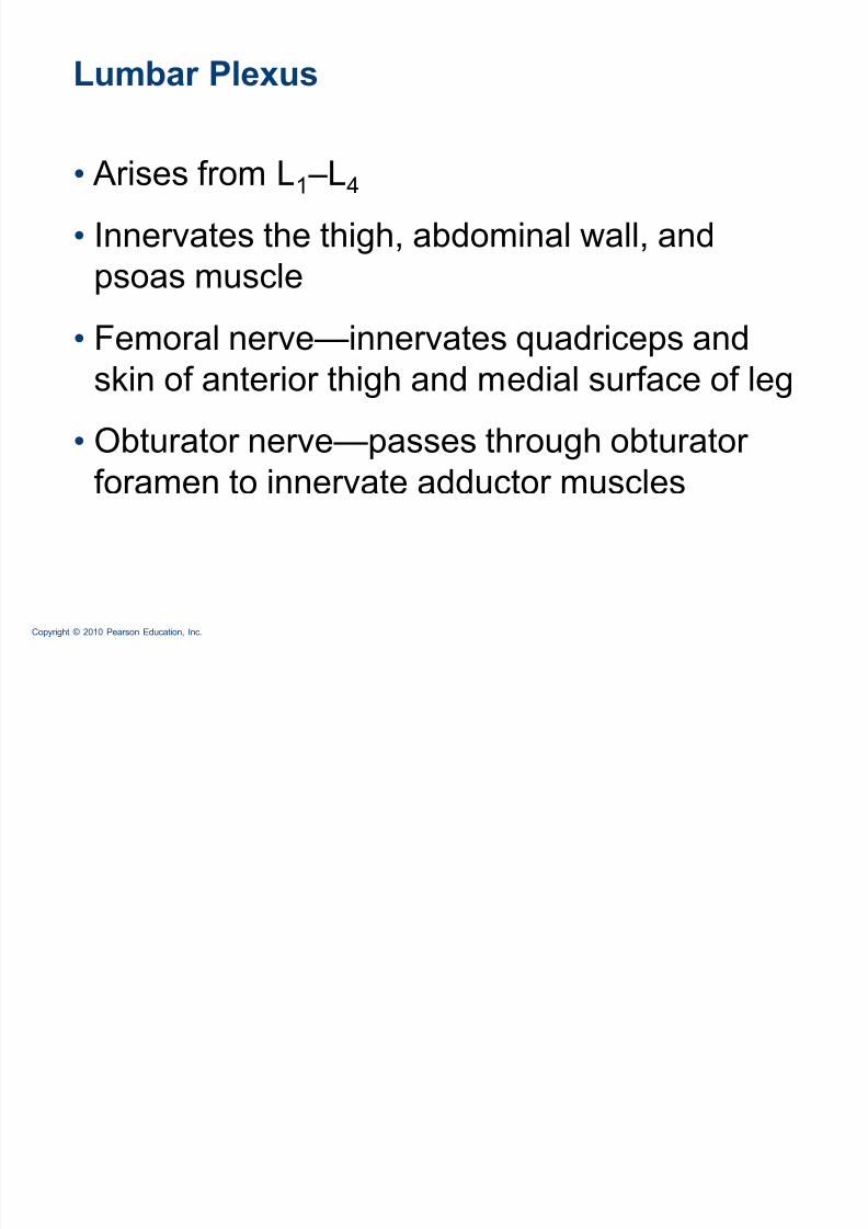
Lumbar Plexus
Arises from L1 ±L4
Innervates the thigh, abdominal wall, and
psoas muscle
Femoral nerve²innervates quadriceps and
skin of anterior thigh and medial surface of leg
Obturator nerve²passes through obturator
foramen to innervate adductor muscles

8/8/2019 Ch 13 Lecture Outline Thursday
http://slidepdf.com/reader/full/ch-13-lecture-outline-thursday 39/47
Copyright © 2010 Pearson Education, Inc. Figure 13.10
(a) Ventral rami and major branches
of the lumbar plexus
Iliohypogastr ic
L1
L2
L3
L4
L5
Ilioinguinal
Genitofemoral
Lateral femoral
cutaneous
Obturator
Femoral
Lumbosacral
trunk
Lateral femoral
cutaneous
Anter ior femoralcutaneous
Saphenous
Obturator
Iliohypogastr icIlioinguinal
Femoral
Ventral ramiVentral
rami:
(b) Distribution of the major nerves from
the lumbar plexus to the lower limb

8/8/2019 Ch 13 Lecture Outline Thursday
http://slidepdf.com/reader/full/ch-13-lecture-outline-thursday 40/47
Copyright © 2010 Pearson Education, Inc. Table 13.5

8/8/2019 Ch 13 Lecture Outline Thursday
http://slidepdf.com/reader/full/ch-13-lecture-outline-thursday 41/47
Copyright © 2010 Pearson Education, Inc.
Sacral Plexus
Arises from L4 ±S4
Serves the buttock, lower limb, pelvic structures, and
perineum
Sciatic nerve
Longest and thickest nerve of the body
Innervates the hamstring muscles, adductor magnus,
and most muscles in the leg and foot Composed of two nerves: tibial and common fibular

8/8/2019 Ch 13 Lecture Outline Thursday
http://slidepdf.com/reader/full/ch-13-lecture-outline-thursday 42/47
Copyright © 2010 Pearson Education, Inc. Figure 13.11 (a)
Super ior gluteal
LumbosacraltrunkInfer ior
gluteal
Commonf ibular Tibial
Poster ior femoral
cutaneousPudendal
Sciatic
Ventral rami and major branches
of the sacral plexus
L4
L5
S1
S2
S3
S4
S5
Co1
Ventral rami Ventral rami:

8/8/2019 Ch 13 Lecture Outline Thursday
http://slidepdf.com/reader/full/ch-13-lecture-outline-thursday 43/47
Copyright © 2010 Pearson Education, Inc. Figure 13.11 (b)
Super ior glutealInfer ior gluteal
Common f ibular
Deep f ibular
Superf icial f ibular
Plantar branches
Tibial
Sural (cut)
Poster ior femoral
cutaneous
Pudendal
Sciatic
(b) Distribution of the major nerves from
the sacral plexus to the lower limb

8/8/2019 Ch 13 Lecture Outline Thursday
http://slidepdf.com/reader/full/ch-13-lecture-outline-thursday 44/47
Copyright © 2010 Pearson Education, Inc. Table 13.6

8/8/2019 Ch 13 Lecture Outline Thursday
http://slidepdf.com/reader/full/ch-13-lecture-outline-thursday 45/47
Copyright © 2010 Pearson Education, Inc.
Innervation of Skin
Dermatome: the area of skin innervated by
the cutaneous branches of a single spinal
nerve
All spinal nerves except C1 participate indermatomes
Most dermatomes overlap, so destruction of a
single spinal nerve will not cause completenumbness

8/8/2019 Ch 13 Lecture Outline Thursday
http://slidepdf.com/reader/full/ch-13-lecture-outline-thursday 46/47
Copyright © 2010 Pearson Education, Inc. Figure 13.12
C2
C3
C4
C5T1
T2
T2T3T4T5
C6
C8C7 C7
C6
T6
T7
T8
T9
T10
T11T12
L1
S2S3
L1
L2
L3
L4
L5
L2
L3
L4
L5
S1
C5
C6
C8
T2
C5
C6
S1
Anterior
view
C2
C3
C4
C5
C6C7C8
C8 C8
C7 C7
T1
T2
T3T4T5T6T7T8T9
T10
T11
T12
L1L2
L3
S1
(b) Posterior
view
L5S2
S1
S1
S3
S2 S1S2
S4
S5
L5L5
L4
L5L5
L4
C6 C6
C5
L4
L3
L2
L1
L4

8/8/2019 Ch 13 Lecture Outline Thursday
http://slidepdf.com/reader/full/ch-13-lecture-outline-thursday 47/47
Innervation of Joints
Hilton¶s law: Any nerve serving a muscle that
produces movement at a joint also innervates
the joint and the skin over the joint