Ch. 10: The Muscular System: Three things that a muscle will be assigned to perform: 1.) Agonist:...
81
• Ch. 10: The Muscular System: • Three things that a muscle will be assigned to perform: 1.) Agonist: Prime mover, main muscle or muscles doing the intended job or performance. 2.) Antagonists: Muscle or a group of muscles that is associated with agonist in whatever movement the agonist does the antagonist does the opposite of. - If the agonist is flexing the antagonist is extending. 3.) Synergist: Muscle or muscles that aid in the movement of either the agonist or the antagonist muscles.
-
Upload
angel-cook -
Category
Documents
-
view
217 -
download
0
Transcript of Ch. 10: The Muscular System: Three things that a muscle will be assigned to perform: 1.) Agonist:...
• Ch. 10: The Muscular System:• Three things that a muscle will be assigned to
perform:
1.) Agonist: Prime mover, main muscle or muscles doing the intended job or performance.
2.) Antagonists: Muscle or a group of muscles that is associated with agonist in whatever movement the agonist does the antagonist does the opposite of.
- If the agonist is flexing the antagonist is extending.
3.) Synergist: Muscle or muscles that aid in the movement of either the agonist or the antagonist muscles.

• The Muscular System:• Just like the skeletal system, the muscular system is
divided into two groups:– Axial muscle group: Head, thoracic, spine.– Appendicular muscle group: Arms, hands, legs, feet.

• The Muscular System:• The purpose of the muscular system is to
do work.• In order to complete this task the muscles
attaching to the bones allows them to act as levers.
• Three different types of levers.– 1st class lever– 2nd class lever– 3rd class lever

• The Muscular System:• Levers
– 1st class lever: fulcrum or pivot point is in the middle, resistance on one side and the effort is on the other side.
- tilting your chin back, spine is the pivot, resistance is your chin, effort is the
muscles attached to the base of the head.Load
Fulcrum
Effort
EffortLoad
Fulcrum
EffortLoad
Fulcrum

• The Muscular System:• Levers
– 2nd class lever: fulcrum is at one end, the load is in the middle, effort is at the other end.
- Lifting your heel off the ground, pivot is your toes, resistance is the heel, effort is your calf muscles.
Fulcrum Effort
Load
Fulcrum
Load
Effort

• The Muscular System:• Levers
– 3rd class lever: fulcrum is at one end, effort is in the middle, resistance is at the other end.
- Doing a bicep curl, pivot is your elbow, resistance is in your hand, effort is coming from your forearm (distal).
Fulcrum
EffortLoad
FulcrumLoad
Effort

• The Muscular System:• Frontalis:• O: Galea aponeurotica
(sheath on the mid head)• I: Above the eyebrow• F: Raise eyebrows

• The Muscular System:• Occipitalis:• O: External protuberance
of the occipital bone• I: Galea aponeurotica• F: Retract scalp

• The Muscular System:• Temporalis:• O: Temporal lines of the
skull (suture sites)• I: Coronoid process• F: Elevate mandible

• The Muscular System:• Orbicularis oculi:• O: Medial portion of the
eye orbit• I: Skin around the eyelid• F: Close and open eye
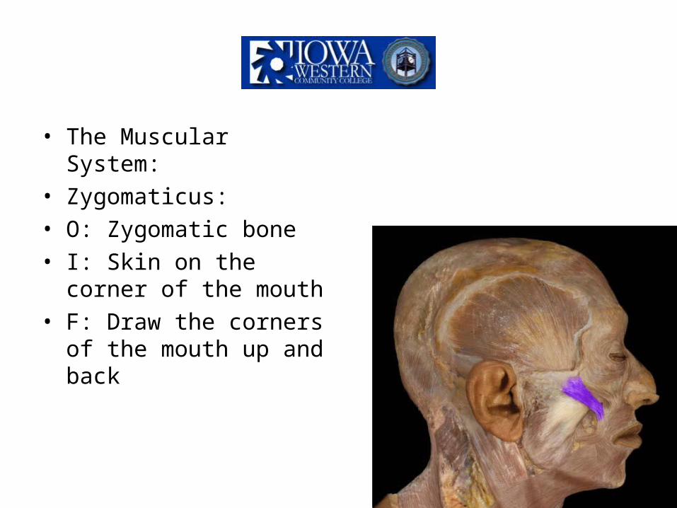
• The Muscular System:• Zygomaticus:• O: Zygomatic bone• I: Skin on the corner of
the mouth• F: Draw the corners of
the mouth up and back

• The Muscular System:• Masseter:• O: Zygomatic arch• I: Lateral surface of the
mandible• F: Elevate mandible
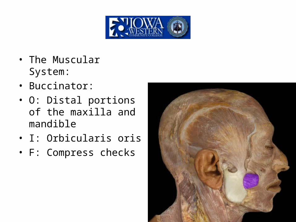
• The Muscular System:• Buccinator:• O: Distal portions of the
maxilla and mandible• I: Orbicularis oris• F: Compress checks
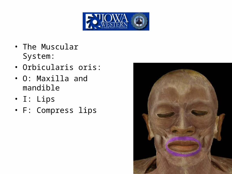
• The Muscular System:• Orbicularis oris:• O: Maxilla and mandible• I: Lips• F: Compress lips

• The Muscular System:
• Sternocleidomastoid:• O: Superior aspect of
sternum and clavicle• I: Mastoid process• F: flex the neck, allows
the head to pivot
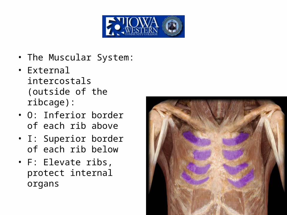
• The Muscular System:• External intercostals
(outside of the ribcage):• O: Inferior border of each
rib above• I: Superior border of each
rib below• F: Elevate ribs, protect
internal organs

• The Muscular System:• Internal intercostals
(inside the ribcage):• O: Superior border of the
rib below• I: Inferior border of the rib
above• F: Depress rib

• The Muscular System:• External oblique (outside
ab muscles):• O: Lower portion of the
eighth rib• I: Iliac crest• F: Compress abdomen

• The Muscular System:• Internal oblique (inside
body cavity ab muscle):• O: Iliac crest• I: Lower portion of the
ribs, xiphoid process• F: Compress the
abdomen

• The Muscular System:• Transverse abdominous
(inner most ab muscle):• O: Cartilage of lower ribs,
iliac crest• I: Pubis• F: Compress abdomen

• The Muscular System:• Rectus abdominous (front
portion of abs):• O: Superior aspect of the
pubis symphysis• I: Inferior surface of ribs
5-7, xiphoid process• F: Depress ribs

• The Muscular System:• Serratus anterior:• O: Lateral side of ribs one
through nine• I: Anterior surface of
scapula, medial border• F: protract scapula
against the chest wall, boxers muscle

• The Muscular System:• Pectoralis Major:• Fan shaped muscle that is
superficial on the anterior side of the thoracic cavity
• O: Sternum (body), clavicle
• I: Greater tubercle of the humerus
• F: Adduct of the arm, helps with throwing and flexion of the arm

• The Muscular System:• Pectoralis Minor:• Lies deep to the
pectoralis major.• O: Ribs 3-5
• I: Coracoid process of the scapula
• F: Raise ribs 3-5, depress glenoid cavity

• The Muscular System:• Deltoid:• Thick three bodied
muscles• O: Clavicle, acromion
spine of the scapula (posterior side)
• I: Proximal head of the humerus
• F: Abduction, forward flexion, and backward flexion
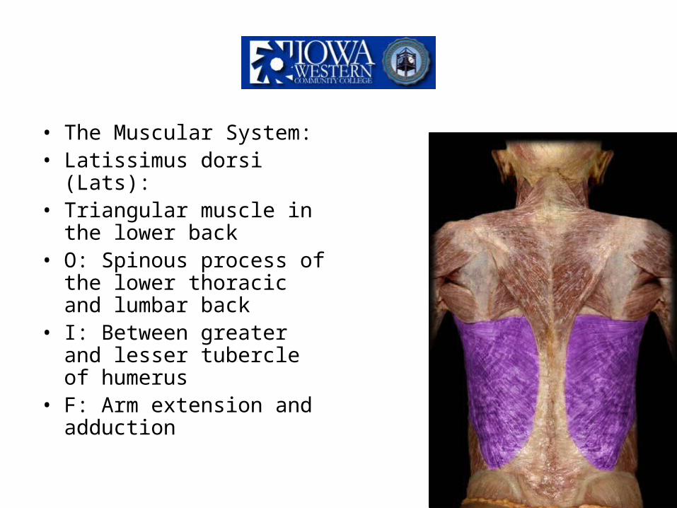
• The Muscular System:• Latissimus dorsi (Lats):• Triangular muscle in the
lower back• O: Spinous process of the
lower thoracic and lumbar back
• I: Between greater and lesser tubercle of humerus
• F: Arm extension and adduction

• The Muscular System:• Trapezius• Upper back muscles• O: External protuberance
of the occipital bone, C7 and all spinous process of thoracic vertebrae.
• I: Acromion spine of scapula, lateral clavicle
• F: Raise scapula, stabilize head
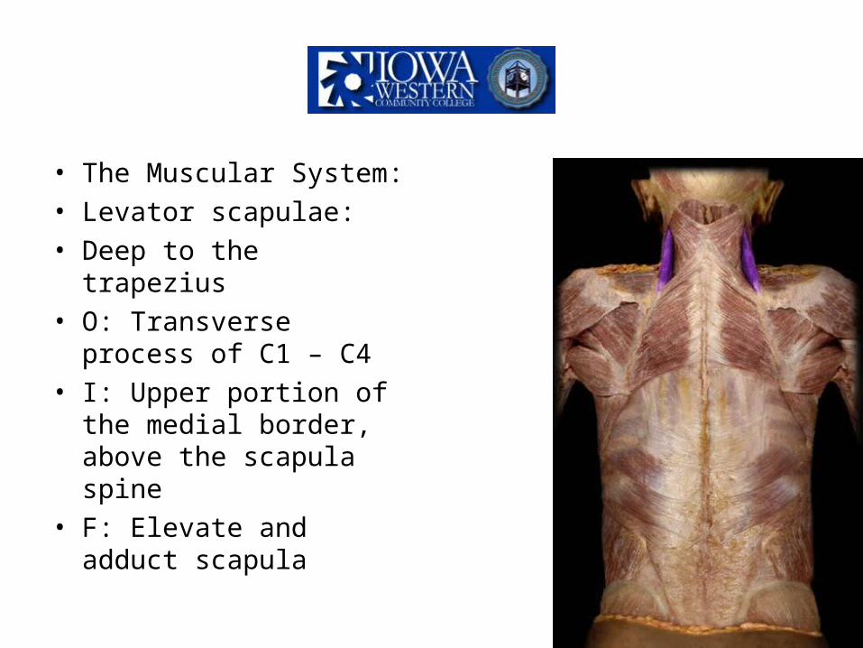
• The Muscular System:• Levator scapulae:• Deep to the trapezius• O: Transverse process of
C1 – C4• I: Upper portion of the
medial border, above the scapula spine
• F: Elevate and adduct scapula

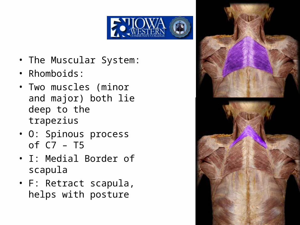
• The Muscular System:• Rhomboids:• Two muscles (minor and
major) both lie deep to the trapezius
• O: Spinous process of C7 – T5
• I: Medial Border of scapula
• F: Retract scapula, helps with posture

• The Muscular System:• Rotator cuff:• Subscapularis:• Anterior muscle under the
scapula• O: Ventral (front) surface
of scapula, subscapularis fossa
• I: Lesser tubercle of humerus
• F: Assists in arm movement


• The Muscular System:• Rotator cuff:• Supraspinatus:• Sits above the spine of
the scapula• O: Medial border,
superior aspect of the scapula, supraspinous fossa
• I: Greater tubercle of humerus
• F: Stabilize the shoulder


• The Muscular System:• Rotator cuff:• Infraspinatus:• Sits below the spine of
the scapula• O: Medial border, inferior
aspect of the scapula, infraspinous fossa
• I: Greater tubercle of the humerus
• F: Stabilize humerus and aid in ROM


• The Muscular System:• Rotator cuff:• Teres minor:• Small round muscle• O: Lateral, dorsal border
of scapula• I: Greater tubercle of
humerus• F: Stabilize humerus and
aid in ROM

• The Muscular System:• Rotator cuff:• Teres major:• Thick rounded, inferior to
teres minor• O: posterior surface of
scapula• I: groove of humerus• F: adducts humerus,
external rotation of arm

• The Muscular System:• Biceps brachii:• Anterior surface of upper
arm• Composed of two heads
(Biceps)• O: Coracoid process• I: Radial tuberosity• F: Flexion of the arm
(flexion of the radius)

• The Muscular System:• Brachialis:• Lies underneath the
biceps brachii• O: Distal half of the
humerus• I: Coronoid process of the
ulna (anterior elbow)• F: Flexion of the arm
(flexion of the ulna)

• The Muscular System:• Brachioradialis:• Superficial muscle on the
lateral side of the arm• O: Distal end of the
humerus• I: Styloid process of
radius• F: Synergist muscle for
flexion of the arm

• The Muscular System:• Triceps brachii:• Posterior surface of the
upper arm• Composed of three heads
(Triceps)• O: Superior, lateral
border of the scapula• I: Olecranon process• F: Extension of the arm
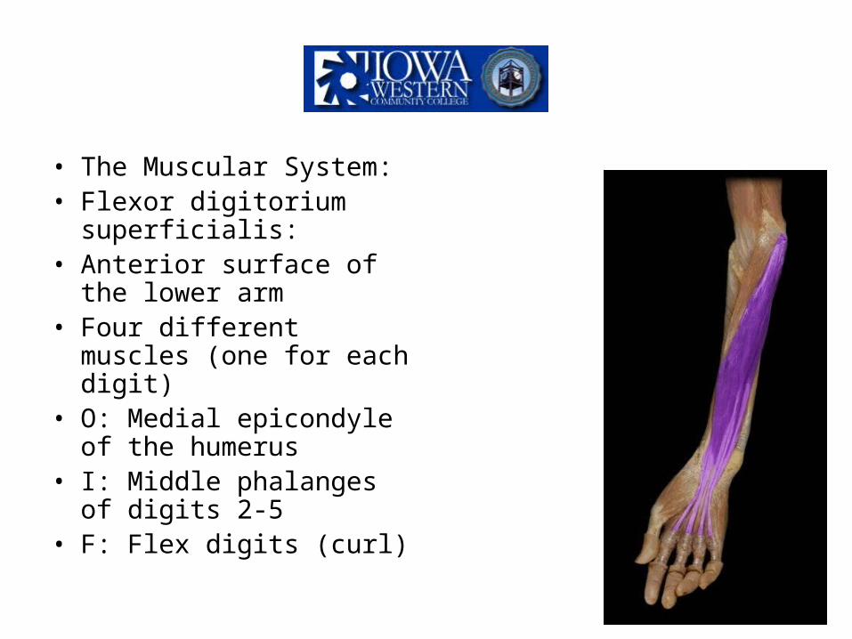
• The Muscular System:• Flexor digitorium
superficialis:• Anterior surface of the
lower arm• Four different muscles
(one for each digit)• O: Medial epicondyle of
the humerus• I: Middle phalanges of
digits 2-5• F: Flex digits (curl)

• The Muscular System:• Flexor pollicis longus:• Anterior surface of the
lower arm• One muscle• O: Anterior surface of the
distal radius• I: Distal phalanges of the
thumb, anterior side• F: Flex the thumb
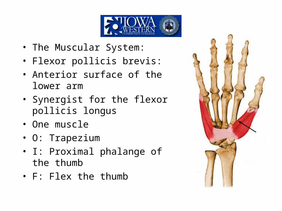
• The Muscular System:• Flexor pollicis brevis:• Anterior surface of the lower arm• Synergist for the flexor pollicis
longus• One muscle• O: Trapezium• I: Proximal phalange of the thumb• F: Flex the thumb

• The Muscular System:• Extensor digitorium
superficialis• Posterior surface of the
lower arm• Four muscles, one for
each digit• O: Lateral epicondyle of
humerus• I: Distal phalanges of
digits 2-5• F: Extend each digit

• The Muscular System:• Supinator:• Anterior surface of the
forearm. Lies deep to the flexor digit.
• O: Lateral epicondyle of the humerus
• I: upper 1/3 of the radius• F: Supination of the
forearm into the ACP
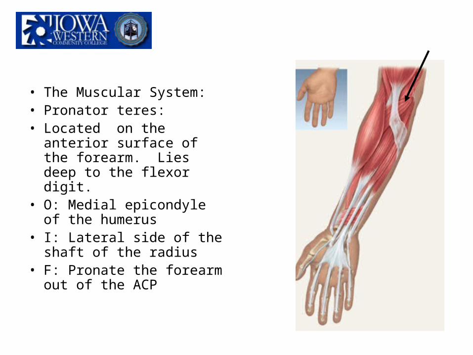
• The Muscular System:• Pronator teres:• Located on the anterior
surface of the forearm. Lies deep to the flexor digit.
• O: Medial epicondyle of the humerus
• I: Lateral side of the shaft of the radius
• F: Pronate the forearm out of the ACP

• The Muscular System:• Flexor carpi radialis:• Located on the anterior
surface of the forearm. Superficial to the flex. digit.
• O: Medial epicondyle of the humerus
• I: 2nd and 3rd metacarpals• F: flex wrist

• The Muscular System:• Flexor carpi ulnaris:• Located on the anterior
surface of the forearm. Superficial to the flex. digit.
• O: medial epicondyle of the humerus, medial side of the olecranon process
• I: pisiform, hamate, 5th metacarpal
• F: flex the wrist

• The Muscular System:• Extensor carpi radialis
longus:• Located on the posterior
surface of the forearm.• O: Lateral surface of the
humerus superior to the condyle
• I: 2nd metacarpal• F: Extend the wrist

• The Muscular System:• Extensor carpi ulnaris:• Located posterior surface
of the forearm• O: Lateral epicondyle of
the humerus, shaft of the ulna
• I: 5th metacarpal• F: Extends the wrist

• The Muscular System:• Extensor pollicus longus:• Posterior surface of lower
arm• One muscle• O: Posterior distal surface
of radius and ulna• I: Distal phalange of
thumb, posterior side• F: Extend thumb

• The Muscular System:• Thigh and Pelvis • Ilopsoas:• Aids in ROM (flexion).• O: Iliac crest, lateral
surface of the sacrum• I: Lesser trochanter on
the femur• F: Flex of the hip when
trunk is in a solid position.

• The Muscular System:• Thigh and Pelvis • Adductor longus:• Main adductor muscle• Covers the middle portion
of the adductor magnus• O: Pubis synthesis• I: Medial shaft of the
femur• F: Flex the leg, laterally
rotates leg, adduction

• The Muscular System:• Thigh and Pelvis• Adductor magnus:• Synergist to the adductor
longus• Large triangular with a broad
insertion point• Lies deep to the adductor
longus• O: Ischial tuberosity• I: Medial side of the femur
(proximal head, shaft, distal head)
• F: Aid in adduction of leg

• The Muscular System:• Thigh and Pelvis• Adductor brevis:• Synergist for the add. Longus• Lies deep to the add. Longus• Superficial to the add. Magnus• O: Ischium• I: Above the add. Longus on
the shaft of the femur• F: Aid in adduction
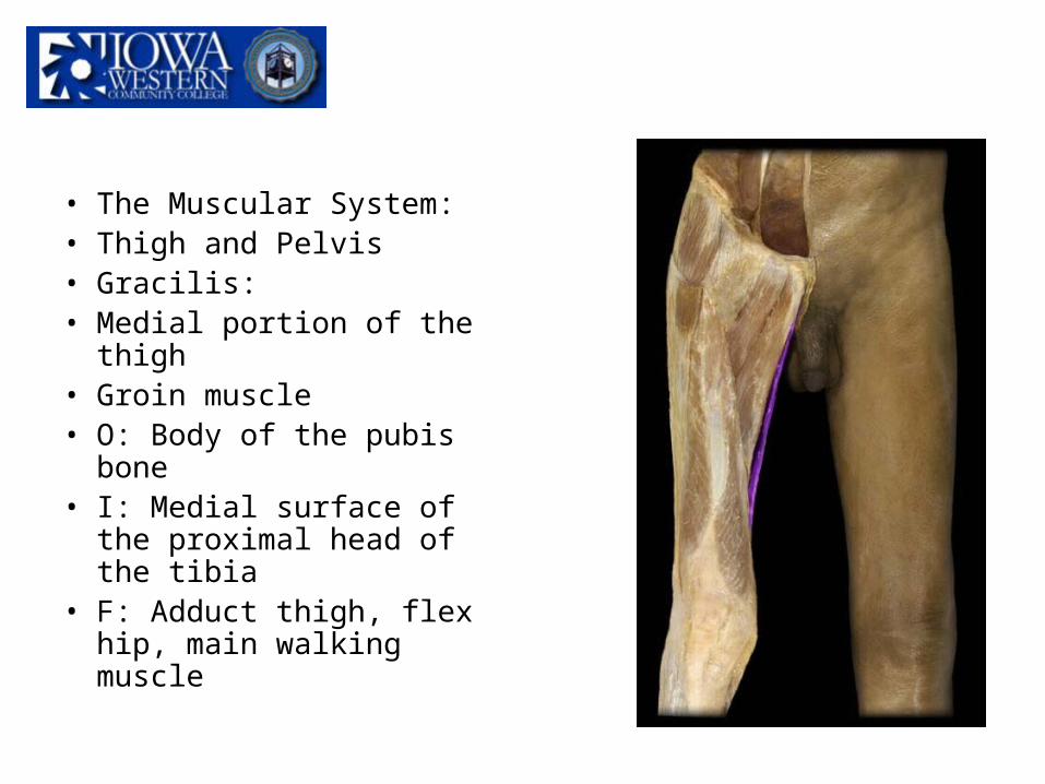
• The Muscular System:• Thigh and Pelvis• Gracilis:• Medial portion of the thigh• Groin muscle• O: Body of the pubis
bone• I: Medial surface of the
proximal head of the tibia• F: Adduct thigh, flex hip,
main walking muscle

• The Muscular System:• Thigh and Pelvis• Sartorius:• Longest muscle in the
body• O: Anterior portion of the
iliac crest• I: Medial aspect of the
proximal head of the tibia• F: Flex knee, laterally
rotate knee
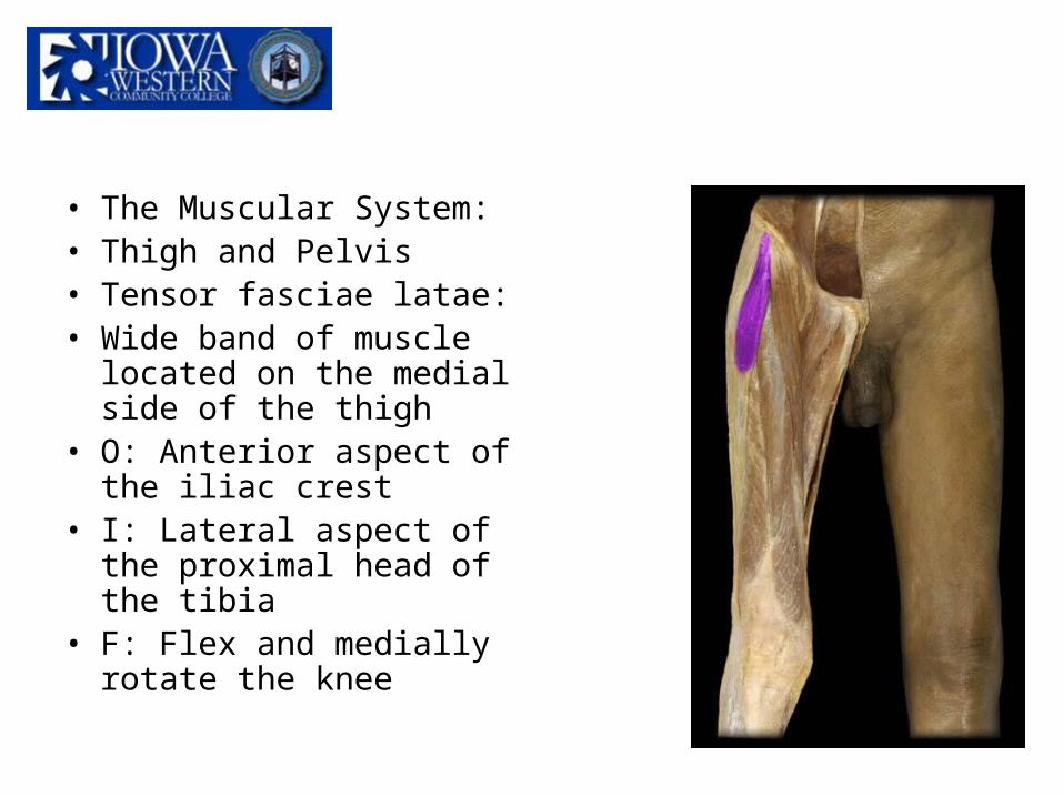
• The Muscular System:• Thigh and Pelvis• Tensor fasciae latae:• Wide band of muscle
located on the medial side of the thigh
• O: Anterior aspect of the iliac crest
• I: Lateral aspect of the proximal head of the tibia
• F: Flex and medially rotate the knee

• The Muscular System:• Thigh and Pelvis• Quadriceps• Located on the anterior side of the leg.• Made up of four muscles
– Rectus femoris – Vastus lateralis– Vastus medialis– Vastus intermedius
• Extends knee

• The Muscular System:• Thigh and Pelvis• Rectus femoris:• Superficial muscle in the
thigh• Largest quad muscle• O: Anterior and inferior
iliac crest• I: Superior border of the
patella• F: Extend knee, flex the
thigh, stabilize patella

• The Muscular System:• Thigh and Pelvis• Vastus lateralis:• Lateral side of the thigh• Synergist to the rectus
femoris• O: Greater trochanter of
the femur• I: Lateral border of the
patella• F: Extend knee, stabilize
patella
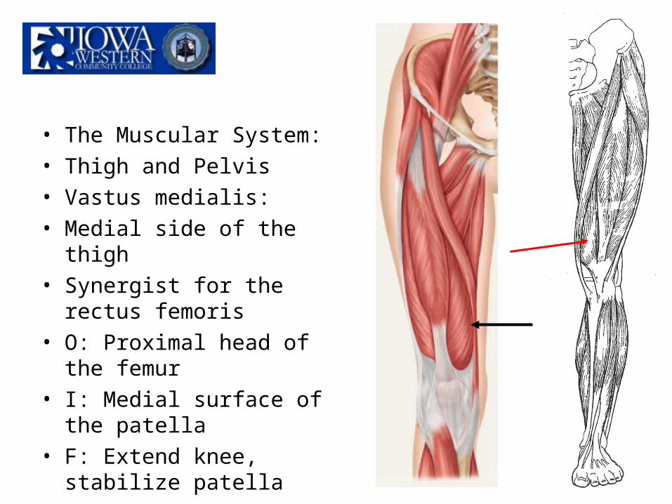
• The Muscular System:• Thigh and Pelvis• Vastus medialis:• Medial side of the thigh• Synergist for the rectus
femoris• O: Proximal head of the
femur• I: Medial surface of the
patella • F: Extend knee, stabilize
patella

• The Muscular System:• Thigh and Pelvis• Vastus intermedius:• Lies deep to the rectus
femoris• O: Proximal head of the
femur• I: Superior border of he
patella (underneath the rectus femoris insertion)
• F: Extend knee, stabilize patella

• The Muscular System:• Thigh and Pelvis• Tibialis anterior:• Anterior portion of the leg• Next to the tibia crest• O: Lateral condyle of the
tibia• I: 1st cuneiform (medial)
and the first metatarsal• F: Dorsiflexion

• The Muscular System:• Thigh and Pelvis• Extensor digitorium
longus:• Lateral side of the lower
leg• O: Lateral condyle of the
tibia and the majority (3/4) of the proximal fibula
• I: Middle and distal phalanges of the digits 2-5
• F: Extend toes

• The Muscular System:• Thigh and Pelvis• Extensor hallucis longus:• Medial side of the lower
leg• Great toe (big toe)• O: Medial fibula shaft• I: Distal phalange of the
great toe• F: Extend great toe

• The Muscular System:• Thigh and Pelvis• Hamstrings:• Located on the posterior side of the thigh• Flexes the knee• Made up of three muscles:
– Semimembranous– Biceps femoris– Semitendinous

• The Muscular System:• Thigh and Pelvis• Semimembranous:• Lies on the medial portion
of the thigh• Lies deep to the
semitendinous• Larger than the
semitendinous• O: Ischium• I: Medial condyle of the
tibia• F: Flex knee

• The Muscular System:• Thigh and Pelvis• Semitendinous:• Lies on the medial portion
of the thigh• Superficial to the
semimembranous• O: Ischium• I: Medial, superior tibial
shaft• F: Flex knee

• The Muscular System:• Thigh and Pelvis• Biceps femoris:• Lies on the lateral portion
of the thigh• O: Ischium and the
proximal head of the femur
• I: Head of the fibula and lateral condyle of the tibia
• F: Flex knee

• The Muscular System:• Thigh and Pelvis• Buttock muscles:• Cushioning and protection for the pelvic girdle.• Three muscles that make up the buttock
muscles.– Gluteus maximus– Gluteus medius– Gluteus minimus

• The Muscular System:
• Thigh and Pelvis• Gluteus maximus:• Main bulk of the
buttocks, superficial• O: Ilium, sacrum,
coccyx• I: Proximal head of
the femur• F: Extend leg when
walking and climbing
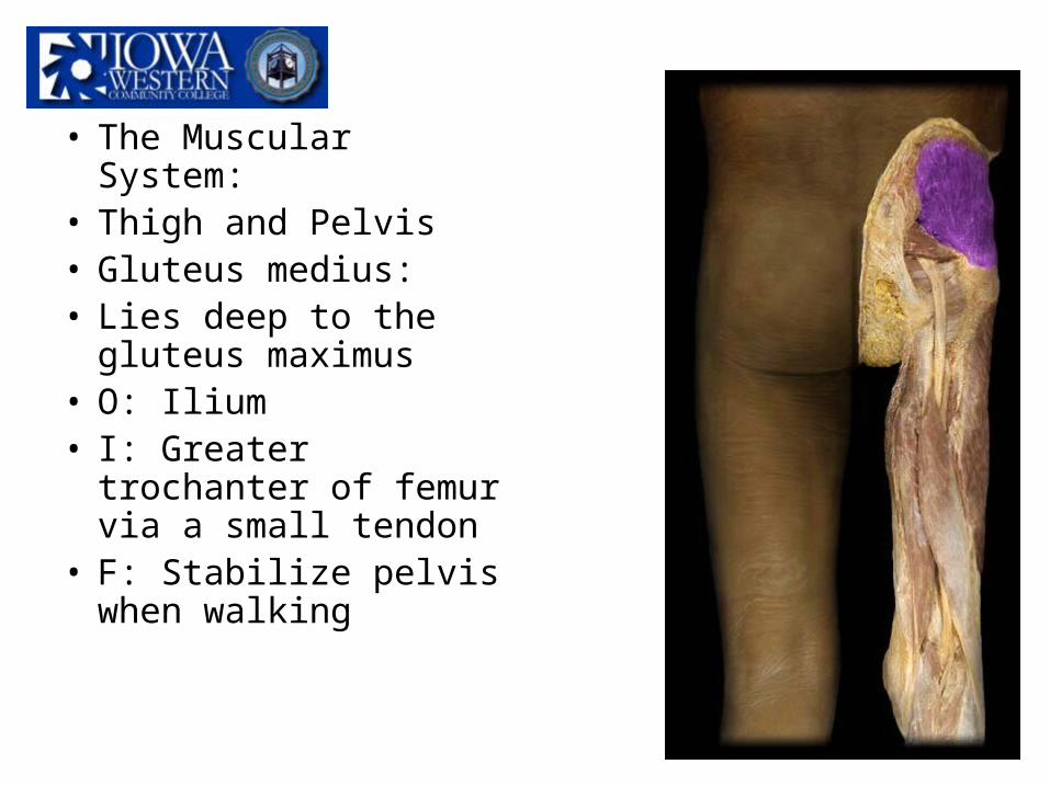
• The Muscular System:
• Thigh and Pelvis• Gluteus medius:• Lies deep to the
gluteus maximus• O: Ilium• I: Greater trochanter
of femur via a small tendon
• F: Stabilize pelvis when walking

• The Muscular System:• Thigh and Pelvis• Gluteus minimus:• Lies deep to the gluteus
medius• Main synergist buttock
muscle• O: Ilium• I: Anterior border of the
greater trochanter of the femur
• F: Help stabilize the pelvis

• The Muscular System:• Thigh and Pelvis• Gastronemius:• Superficial muscle• O: Medial and lateral
condyle of the femur• I: Calcaneus• F: Plantar flexion

• The Muscular System:• Thigh and Pelvis• Soleus:• Lies deep to the
gastronemius• Synergist for the
gastronemius• O: Proximal head of the
tibia and fibula• I: Calcaneus (underneath
the insertion of the gastronemius)
• F: Plantar flexion

• The Muscular System:
• Thigh and Pelvis• Flexor digitorium
longus:• Medial portion of the
lower leg• O: Medial condyle of
the tibia (posterior)• I: Distal phalanges of
digits 2-5• F: Flex toes

• The Muscular System:• Thigh and Pelvis• Flexor hallucis longus:• Great toe (big toe)• Medial portion of the
lower leg• O: Middle of the fibula
shaft• I: Distal phalange of the
great toe• F: Flex the great toe