CF® Dyes - Biotium · Reactive dyes for bioconjugation are generally susceptible to hydrolysis,...
32
CF® Dyes Next Generation Fluorescent Dyes Introduction to CF® Dyes Quick reference table ... p. 2 Overview of CF® dyes ... p. 3 CF® dyes FAQs ... p. 4 CF® Dyes Technical Information Technical profiles by CF® dye color ... pp. 5-19 CF® dyes for super-resolution microscopy ... pp. 20-21 Selected CF® dye product references ... p. 22 Reactive Dyes and Labeling Kits Mix-n-Stain™ Antibody Labeling Kits ... p. 23 Mix-n-Stain™ Small Ligand Labeling Kits ... p. 23 Reactive dyes ... p. 24 CF® Dye Conjugates Bioconjugates ... p. 25 Labeled nucleotides ... p. 25 Primary antibody conjugates ... p. 26 Anti-biotin, anti-GFP, and anti-tag antibodies .... p. 27 Secondary antibodies ... p. 27 Highly cross-adsorbed secondary antibodies ... pp. 28-29 F(ab’)2 fragments ... p. 29 Isotype-specific secondary antibodies ... p. 29 Related Products and Accessories EverBrite™ antifade mounting media, counterstains, TrueBlack® autofluorescence quencher and more ... pp. 30-31
Transcript of CF® Dyes - Biotium · Reactive dyes for bioconjugation are generally susceptible to hydrolysis,...
CF® Dyes Next Generation Fluorescent Dyes
Introduction to CF® DyesQuick reference table ... p. 2Overview of CF® dyes ... p. 3CF® dyes FAQs ... p. 4
CF® Dyes Technical InformationTechnical profiles by CF® dye color ... pp. 5-19CF® dyes for super-resolution microscopy ... pp. 20-21Selected CF® dye product references ... p. 22
Reactive Dyes and Labeling KitsMix-n-Stain™ Antibody Labeling Kits ... p. 23Mix-n-Stain™ Small Ligand Labeling Kits ... p. 23Reactive dyes ... p. 24
CF® Dye ConjugatesBioconjugates ... p. 25Labeled nucleotides ... p. 25Primary antibody conjugates ... p. 26Anti-biotin, anti-GFP, and anti-tag antibodies .... p. 27Secondary antibodies ... p. 27Highly cross-adsorbed secondary antibodies ... pp. 28-29F(ab’)2 fragments ... p. 29Isotype-specific secondary antibodies ... p. 29
Related Products and AccessoriesEverBrite™ antifade mounting media, counterstains, TrueBlack® autofluorescence quencher and more ... pp. 30-31

2 • www.biotium.com
CF® dye Page λEx (nm)
λEm (nm) Excitation * Replacement for Features and applications
CF®350 5 347 448 UV Alexa Fluor® 350, AMCA, DyLight® 350
• Brightest blue fluorescent conjugates for 350 nm excitation • Highly water-soluble and pH insensitive
CF®405S 5 404 431 405 nm Alexa Fluor® 405, Cascade Blue®, DyLight® 405 • Better compatibility with common instruments
CF®405M 5 408 452 405 nm BD Horizon™ V450, eFluor® 450, Pacific Blue®
• More photostable than Pacific Blue® dye with less green spill-over • Excellent choice for super-resolution imaging by SIM**
CF®405L 6 395 545 405 nm Pacific Orange® • 405 nm excitable orange fluorescent dye for multicolor detection
CF®430 6 426 498 405 nm Pacific Green®, BD Horizon™ V500, Krome Orange™
• Photostable 405 nm excitable green dye • Perfect match for the CFP filter set
CF®440 6 440 515 405 nm Alexa Fluor® 430 • Photostable 405 nm excitable green dyeCF®450 7 450 538 405 nm Unique dye • Green dye with unique spectral properties
CF®488A 7 490 515 488 nm ATTO 488, Alexa Fluor® 488,
Cy®2, DyLight® 488, FAM, FITC, Fluorescein
• Less non-specific binding and less red spill-over than Alexa Fluor® 488 • Very photostable • Compatible with super-resolution imaging by TIRF**
CF®514 8 516 548 488 nm Alexa Fluor® 514 • Green dye that can be separated from CF®488A by spectral unmixing
CF®532 9 527 558 532 nm Alexa Fluor® 532, ATTO 532 • Significantly brighter than Alexa Fluor® 532CF®535ST 9 535 568 532 nm Unique dye for STORM** • Orange dye designed for STORM super-resolution microscopy**
CF®543 10 541 560 532, 543, or 546 nm
Alexa Fluor® 546, Tetramethylrhodamine (TAMRA) • Significantly brighter than Alexa Fluor® 546
CF®555 10 555 565 532, 543, 546,, 555, or 568 nm
Alexa Fluor® 555, ATTO 550, Cy®3, DyLight® 549, TRITC
• Brighter than Cy®3 • Validated in multicolor super-resolution imaging by STORM**
CF®568 11 562 583 532, 543, 546, 555, or 568 nm
Alexa Fluor® 568, ATTO 565, Rhodamine Red
• Optimized for the 568 nm line of the Ar-Kr mixed-gas • Brighter and more photostable than Alexa Fluor 568 • Compatible with TIRF and multicolor STORM**
CF®594 12 593 614 532, 543, 546, 555, or 568 nm
Alexa Fluor® 594, ATTO 594, DyLight® 594, Texas Red®
• Yields the brightest conjugates among spectrally similar dyes • Extremely photostable
CF®594ST 12 593 614 532, 543, 546, 555, or 568 nm Unique dye for STORM** • Specifically designed for super-resolution imaging by STORM**
CF®620R 13 617 639 633 or 635 nm LightCycler® Red 640 • Highly fluorescent dye with unique spectral properties
CF®633 14 630 650 633 or 635 nm Alexa Fluor® 633, Alexa Fluor® 647, Cy®5, DyLight® 633
• Yields the brightest antibody conjugates among spectrally similar dyes • Far more photostable than Alexa Fluor® 647 • Compatible with super-resolution TIRF, FIONA, and gSHRImP**
CF®640R 15 642 662 633, 635, or 640 nm
Alexa Fluor® 647, ATTO 647N, Cy®5, DyLight® 649
• Has the best photostability among dyes with Cy®5-like spectra • Yields highly fluorescent protein conjugates • Compatible with TIRF and FLIMP super-resolution techniques**
CF®647 16 650 665 633, 635, or 640 nm
Alexa Fluor® 647, ATTO 647N, Cy®5, DyLight® 649
• Brighter than Cy®5 • Compatible with multicolor super-resolution imaging by STORM**
CF®660C 17 667 685 633, 635, or 640 nm Alexa Fluor® 660 • Much brighter and more photostable than Alexa Fluor® 660
• Compatible with multicolor super-resolution imaging by STORM**
CF®660R 17 663 682 633, 635, or 640 nm Alexa Fluor® 660 • Brighter than Alexa Fluor® 660
• The most photostable 660 nm dye
CF®680 18 681 698 680 or 685 nm Alexa Fluor® 680, Cy®5.5, DyLight® 680, IRDye® 680LT
• The brightest among spectrally similar 680 nm dyes • Validated in multicolor STORM and dual-color 3D super-resolution imaging** • Compatible with LI-COR® Odyssey® System
CF®680R 18 680 701 680 or 685 nm Alexa Fluor® 680, Cy®5.5, DyLight® 680, IRDye® 680LT
• The most photostable 680 nm dye • Suitable for labeling nucleic acids and small biomolecules • Compatible with LI-COR® Odyssey® System • Compatible with STED and single molecule spectroscopy**
CF®750 19 755 777 680 or 685 nm Alexa Fluor® 750, Cy®7, DyLight® 750, IRDye® 750
• Exceptionally bright and stable • Highly water soluble without bearing excessive charge • Validated in super-resolution imaging by STORM**
CF®770 19 770 797 785 nm DyLight® 800, IRDye® 800CW, ZW800-1
• Exceptionally bright and stable • Compatible with LI-COR® Odyssey® System
CF®790 19 784 806 785 nm Alexa Fluor® 790 • Exceptionally bright and stable • Highly water soluble without bearing excessive charge
CF®800 19 797 816 785 nm Spectrally similar to Indocyanine green • Unique long wavelength near-infrared dye
CF® Dyes Quick Reference TableVi
sible
spec
trum
Near
-infra
red
Far-r
ed
Alexa Fluor, Cascade Blue, Pacific Blue, and Texas Red are registered trademarks of Invitrogen; ATTO dyes are products of ATTO-TEC GmbH; BD Horizon is a trademark of BD Biosciences; Cy® is a registered trademark of GE Healthcare; DyLight is a registered trademark of Thermo Fisher Scientific; eFluor is a registered trademark of eBioscience; IRDye and Odyssey are registered trademarks of LI-COR Bioscience; Krome Orange is a trademark of Beckman Coulter; LightCycler is a registered trademark of Roche Applied Science.
*Visible and far-red dyes can be excited by a UV light source for epifluorescence microscopy. **See pp. 20-21 for more information about CF® dyes for super-resolution microscopy

www.biotium.com • 3
Next-generation fluorescent dyes
CF® dyes are a series of highly water-soluble fluorescent dyes spanning the visible and near-infrared (IR) spectrum for labeling biomolecules, especially proteins and nucleic acids. Developed by scientists at Biotium using new breakthrough chemistries, CF® dyes rival or exceed the quality of other commercial dyes, such as Alexa Fluor® dyes, due to the following novel features. Novel rhodamine chemistry
Rhodamine dyes are known for their excellent photostability and good fluorescence quantum yield; consequently several of the Alexa Fluor® dyes bear the rhodamine core structure. Unfortunately, traditional rhodamine chemistry makes it difficult to extend the fluorescence wavelength to the far-red region and even more challenging in the near-IR region, especially for water-soluble dyes for bioconjugation. Recently, Biotium scientists discovered a new way to prepare novel rhodamine dyes of any fluorescence color from green to near-IR. The new chemistry is a key element in the development of many of our CF® dyes, particularly our far-red CF® dyes, which are not only bright and water-soluble but also extremely photostable.
Excellent labeling efficiency
Reactive dyes for bioconjugation are generally susceptible to hydrolysis, which can cause problems for shipping, handling and storage and result in lower labeling efficiency. Heavily sulfonated dyes, such as the Alexa Fluor® dyes, DyLight® dyes and IRDyes® are particularly hygroscopic, worsening the hydrolysis problem. For example, the percent of active Alexa Fluor® 488 succinimidyl ester (SE) could be well below 50% by the time of application (according to Life Technologies’ Alexa Fluor® 488 microscale labeling kit product information sheet, Invitrogen). In contrast, all of Biotium’s amine-reactive CF® dyes have a relatively stable form of SE, which is more resistant to hydrolysis than the SE in many of the Alexa Fluor® dyes. Accordingly, CF® dye SE products generally give consistently higher labeling efficiency, thus providing users a better value.
Mix-n-Stain™ antibody labeling technology
Biotium has developed a breakthrough antibody labeling technology with CF® dyes — Mix-n-Stain™ antibody labeling kits. With this technology, you merely need to mix your antibody with the reaction buffer and CF® dye provided in the kit, and in 30 minutes you will have an optimally labeled CF® dye-antibody conjugate ready for immunostaining. The labeling technology provides unprecedented convenience for antibody labeling. Mix-n-Stain™ labeled antibodies can be used for multicolor immunostaining, allowing staining with multiple primary antibodies from the same host species when pre-labeled primary antibodies are not available.
CF® Dyes for Super-Resolution Microscopy
Recent publications comparing synthetic dyes for super-resolution imaging have shown CF® dyes give the best performance for multiple methods. The superior brightness, photostability, and photochemical switching properties of certain CF® dyes are ideal for 3-D SIM, 3-D STORM, and other super-resolution and single-molecule imaging techniques. See pp. 20-21 for more information.
Unrivaled near-infrared dyes
Near-IR dyes are typically much larger in size than dyes in the visible range. The large size often results in serious problems of low dye solubility, dye aggregation and poor fluorescence quantum yield. To overcome the problems, many commercial near-IR dyes, such as the near-IR Alexa Fluor® dyes, DyLight® dyes and IRDyes®, are prepared by placing a number of negatively charged sulfonate group on the dyes. While sulfonation improves dye solubility and fluorescence quantum yield to some degree, it creates another even more serious problem: non-specific binding of the bioconjugates prepared from the dyes. For example, conjugation to a highly negatively charged dye can dramatically alter an antibody’s isoelectric point (iP), which is essential for maintaining specific antibody-antigen interaction. With this insight, Biotium scientists devised a revolutionary new approach to near-IR dye design that results in superior physical properties of the dyes without introducing an excessive amount of negative charge.
Biotium’s near-IR CF® dyes are based on the core structure of either cyanine dyes or rhodamine dyes. Those core structures are modified such that the intramolecular mobility of the dyes is restricted, which leads to higher quantum yield and better water solubility without adding excessive charge. As a result, near-IR CF® dyes are much brighter and more photostable than any other near-IR dyes. Most importantly, antibodies labeled with near-IR CF® dyes™ give far better signal-to-noise ratio in immunostaining compared with antibody conjugates prepared with other commercial near-IR dyes.
Multi-color flexibility
Biotium currently offers 28 CF® dyes with additional colors in development. The CF® dye product line includes reactive CF® dyes, labeling kits, CF®-labeled secondary antibodies and streptavidin, and many other CF®-labeled biomolecules.
Alexa Fluor is a registered trademark of Invitrogen; DyLight® is a registered trademark of Thermo Fisher Scientific; IRDye is a registered trademark of Li-COR Bioscience.
CF® dyes and Mix-n-Stain antibody labeling technology are covered by pending U.S. and international patents. We welcome inquiries about licensing the use of our dyes, trademarks or technologies; email us at [email protected].
CF® Dyes Overview
350 450 550 650 750 850 950Wavelength (nm)
CF350CF405SCF405MCF405LCF430CF440CF488ACF514CF532CF543CF555CF568CF594CF620RCF633CF640RCF647CF660CCF660RCF680CF680RCF750CF770CF790CF800
Emission spectra of CF® dyes

4 • www.biotium.com
CF® DyesFrequently Asked Questions
Question AnswerWhat does the CF in CF® dyes stand for? CF® initially was an abbreviation for Cyanine-based Fluorescent dyes. These were the first patented CF® dyes based on cyanine dye
structures. Since then, our CF® dye patent portfolio has expanded to include four different fluorescent dye core structures that cover the fluorescence spectrum from UV to NIR.
What are the chemical structures of CF® dyes? The exact chemical structures of CF® dyes are currently confidential but will be fully disclosed at a later stage when pending patents become granted. In general terms, the structure of a CF® dye may be divided into two parts: a) dye core structure (i.e. the aromatic ring skeleton that defines the dye’s color or absorption/emission wavelengths), and b) core structure-modifying elements. At present, CF® dyes bear the core structures of coumarin, pyrene, rhodamine or cyanine dyes. Blue fluorescent CF® dyes are based on coumarin or pyrene dye core structure, while green to near-IR CF® dyes are based on either cyanine or rhodamine dye core structures. Core structure-modifying elements refer to various chemical attachments to the core structure and are a key aspect of the CF® dye invention that makes CF® dyes superior to other commercial dyes.
What are the quantum yields of CF® dyes? The quantum yield of a fluorescent dye can vary widely depending on the dye’s micro-environment if the dye is attached to a protein or other molecule. A good way to compare the relative quantum yields of different dyes is to plot the total fluorescence of the labeled proteins as a function of degree of labeling by the dyes, as we have done with CF® dyes and other commercial dyes in the dye description pages in this guide.
How stable are CF® dyes? There are three aspects to dye stability: 1) chemical stability of the dye core structure; 2) stability of the reactive group; and 3) photostability of the dye. CF® dyes bear the core structures of coumarin, pyrene, rhodamine or cyanine dyes, all of which are known to have excellent chemical stability. In general, the dyes are far more stable than the antibodies or other biomolecules they label. CF® dyes are also stable enough for labeled nucleic acids to be used in PCR or nucleic acid hybridization, where high temperature is involved. Reactive CF® dyes comprise a reactive group used in bioconjugation. Among the various reactive groups, only amine-reactive succinimidyl ester (SE) and thiol-reactive maleimide groups are susceptible to hydrolysis and therefore are moisture-sensitive. CF® dye SE products are relatively more stable than other commercial SE dyes. This is because CF® SE dyes are derived from aliphatic carboxylic groups, which results in a more stable SE form, while other commercial SE dyes usually are derived from aromatic carboxylic acid groups that yield a less stable SE form. Photostability refers to the dye’s ability to withstand photobleaching. Photobleaching is mainly a concern when dyes are subjected to intense illumination for an extended period of time, such as during confocal microscopy. Among the four types of core structures, rhodamine is the most photostable, followed by cyanine, pyrene and coumarin cores. The structure-modifying groups and the way they are attached to the dye cores are a key innovative aspect of CF® dye technologies that contributes to the superior photostability of CF® dyes over that of other commercial dyes. In general, rhodamine-based CF® dyes, whose wavelengths range from green to the near-IR region, offer the best photostability, making these dyes ideal for microscopy applications.
Are CF® dyes sensitive to pH? CF® dyes are chemically stable within the pH range of at least 2 –11. The fluorescence of most CF® dyes is relatively insensitive to pH, except for that of CF®405M, CF®568, CF®620R, and CF®633. The fluorescence of these four CF® dyes becomes weaker when pH drops below 4.5.
Are CF® dyes fixable? CF® dyes can tolerate formaldehyde fixation. However, whether a CF® dye-labeled probe is fixable will depend on the fixability of the probe itself. Proteins with free amine groups that bind other proteins generally are formaldehyde-fixable.
What is the difference between CF®405s, CF®405M, and CF®405L?
All three of these dyes can be excited by the 405 nm laser (or UV mercury lamp). They differ in their emission wavelengths. CF®405S has the shortest blue fluorescence emission at 431 nm, while CF®405M has longer wavelength blue fluorescence emission at 452 nm. CF®405L has orange fluorescence emission at 545 nm. We recommend choosing the dye that best fits your instrument’s detection settings (see pp. 5-6 for more information).
For several CF® dye colors, there is an R form and a C form, both having similar absorption and emission spectra. In such a case, which of the two CF® dyes should I choose?
Rhodamine-based CF® dyes (designated R) generally have better photostability but weaker fluorescence than their cyanine-based equivalents (designated C). Therefore, rhodamine-based near-IR CF® dyes are a better choice for microscopy, while cyanine-based CF® dyes are more ideal for flow cytometry, Western blotting, and other applications where photobleaching is less of a concern. Another factor to consider is the size of the dyes. Some of the cyanine-based near-IR CF® dyes are much larger than the rhodamine-based equivalents. For antibody labeling, either version of the CF® dyes is suitable. However, for applications where the dye size may cause a steric problem, the smaller dye may be a better choice.
How soluble are CF® dyes? CF® dyes are highly water soluble (>100 mg/mL). They are also very soluble in other polar solvents, such as DMSO, DMF, methanol and ethanol. However, CF® dyes are poorly soluble or insoluble in non-polar solvents.
What are the charges on CF® dyes? Most CF® dyes carry 1-2 negative charges while a few cyanine-based near-IR CF® dyes carry 3-4 negative charges. However, the more negatively charged CF® dyes comprise unique structural features that shield the negative charges such that the biomolecules (such as antibodies) the dyes label do not lose specificity due to the excessive negative charges.
Can CF® dyes be used for STORM? Several CF® dyes have been validated in super-resolution imaging by STORM, as well as other super-resolution techniques. Biotium also offers dyes specifically designed for STORM imaging. See pp. 20-21 for more information.
What are the major applications of CF® dyes? CF® dyes are ideal for protein labeling because of their high water solubility, which reduces fluorescence quenching. They are also useful for labeling oligonucleotides that require multiple copies of a dye for maximal fluorescence, such as the preparation of FISH probes, where water soluble dyes can minimize fluorescence quenching. Finally, CF® dyes make excellent polar tracers that can be used for visualizing the morphology or long-term tracing of neurons.

www.biotium.com • 5
CF®350A bright UV-excitable blue fluorescent dye
Technical Summary
Abs/Em Maxima: 347/448 nmExtinction coefficient: 18,000Molecular weight: ~ 496Excitation source: UVReplaces: Alexa Fluor® 350, AMCA, DyLight® 350
250 300 350 400 450 500 550
Emission
Abso
rptio
n
Wavelength (nm)
CF350
Figure 1. Absorption and emission spectra of CF®350 goat anti-mouse conjugate in PBS.
CF®
350
Figure 2. HeLa cells stained with mouse anti-tubulin antibody and CF®350 goat anti-mouse IgG (cyan).
CF®405S and CF®405MImproved brightness and photostability for the 405 nm laser line
Technical Summary
CF®405SAbs/Em Maxima: 404/431 nmExtinction coefficient: 33,000Molecular weight: ~ 1,169Excitation laser line: 405 nmReplaces: Alexa Fluor® 405, Cascade Blue®, DyLight® 405
CF®405MAbs/Em Maxima: 408/452 nmExtinction coefficient: 41,000Molecular weight: ~ 503Excitation laser line: 405 nmReplaces: Pacific Blue®, BD Horizon™ V450
Figure 2. Intracellular staining of Jurkat cells was performed with mouse anti-CD3 or isotype control followed by goat anti-mouse IgG conjugated to Alexa Fluor 405 (AF405) or CF®405S. Fluorescence was analyzed on a BD LSR II flow cytometer with 405 nm excitation and 450/50 nm emission filter. Bars represent the relative fluorescence of the geometric means of the cell populations.
0
4
8
12
16
1.5 5.4 6.4 1.6 4.8 7.2
Rel
ativ
e Fl
uore
scen
ce
Isotype Control
Mouse Anti-CD3
DOL:
AF405 CF405S
0
20
40
60
80
100
0 15 30 45 60
Nor
mal
ized
Flu
ores
cenc
e
Time (s)
CF405MPacific Blue
Figure 3. Photostability of CF®405M and Pacific Blue. CF®405M and Pacific Blue dye solutions were continuously exposed to mercury arc lamp microscope excitation with a DAPI filter set. Images were captured every 5 seconds for one minute. Fluorescence intensity was normalized to time 0.
CF®
405S & CF®
405M
Figure 1. Absorption and emission spectra of CF®405S and CF®405M goat anti-mouse conjugates in PBS.
Features • Brighter and more photostable than AMCA• Direct replacement for Alexa Fluor® 350• Highly water soluble and pH-insensitive
320 370 420 470 520
EmissionAb
sorp
tion
Wavelength (nm)
- - -CF405SCF405M
Features• CF®405S: Brighter than Alexa Fluor® 405• CF®405M: More photostable than Pacific Blue®, with less
spill-over in the green channel• CF®405M: an excellent choice for super-resolution imaging
by SIM (see pp. 20-21)
6 • www.biotium.com
CF®
430
& C
F®44
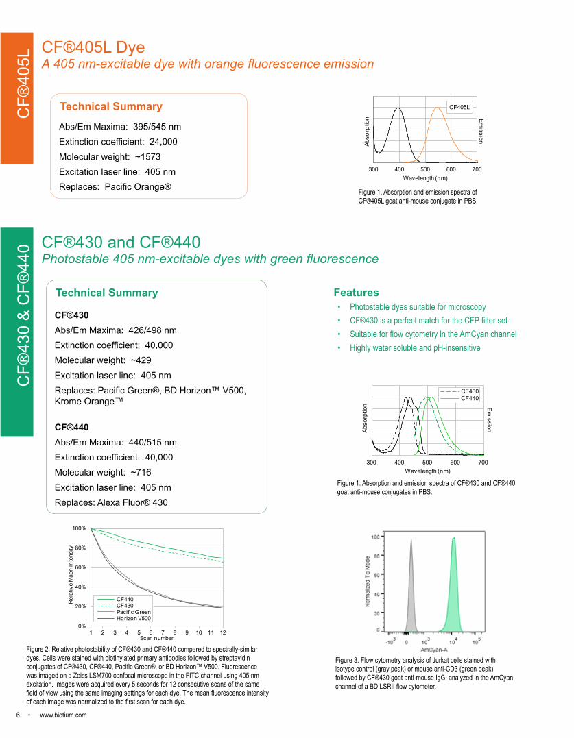
0 CF®430 and CF®440Photostable 405 nm-excitable dyes with green fluorescence
Technical Summary
CF®430Abs/Em Maxima: 426/498 nmExtinction coefficient: 40,000Molecular weight: ~429Excitation laser line: 405 nmReplaces: Pacific Green®, BD Horizon™ V500, Krome Orange™
CF®440Abs/Em Maxima: 440/515 nmExtinction coefficient: 40,000Molecular weight: ~716Excitation laser line: 405 nmReplaces: Alexa Fluor® 430
Features• Photostable dyes suitable for microscopy• CF®430 is a perfect match for the CFP filter set• Suitable for flow cytometry in the AmCyan channel• Highly water soluble and pH-insensitive
300 400 500 600 700
Abso
rptio
n
Wavelength (nm)
CF430CF440
Emission
0%
20%
40%
60%
80%
100%
1 2 3 4 5 6 7 8 9 10 11 12
Rel
ativ
e M
aen
Inte
nsity
Scan number
CF440CF430Pacific GreenHorizon V500
Figure 1. Absorption and emission spectra of CF®430 and CF®440 goat anti-mouse conjugates in PBS.
Figure 2. Relative photostability of CF®430 and CF®440 compared to spectrally-similar dyes. Cells were stained with biotinylated primary antibodies followed by streptavidin conjugates of CF®430, CF®440, Pacific Green®, or BD Horizon™ V500. Fluorescence was imaged on a Zeiss LSM700 confocal microscope in the FITC channel using 405 nm excitation. Images were acquired every 5 seconds for 12 consecutive scans of the same field of view using the same imaging settings for each dye. The mean fluorescence intensity of each image was normalized to the first scan for each dye.
Figure 3. Flow cytometry analysis of Jurkat cells stained with isotype control (gray peak) or mouse anti-CD3 (green peak) followed by CF®430 goat anti-mouse IgG, analyzed in the AmCyan channel of a BD LSRII flow cytometer.
CF®405L DyeA 405 nm-excitable dye with orange fluorescence emission
CF®
405L
Technical Summary
Abs/Em Maxima: 395/545 nm Extinction coefficient: 24,000Molecular weight: ~1573Excitation laser line: 405 nmReplaces: Pacific Orange®
300 400 500 600 700
Emission
Abso
rptio
n
Wavelength (nm)
CF405L
Figure 1. Absorption and emission spectra of CF®405L goat anti-mouse conjugate in PBS.
www.biotium.com • 7
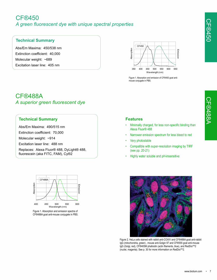
CF®488AA superior green fluorescent dye
400 450 500 550 600
EmissionAb
sorp
tion
Wavelength (nm)
CF488A
Technical Summary
Abs/Em Maxima: 490/515 nmExtinction coefficient: 70,000Molecular weight: ~914Excitation laser line: 488 nmReplaces: Alexa Fluor® 488, DyLight® 488, fluorescein (aka FITC, FAM), Cy®2
Figure 1. Absorption and emission spectra of CF®488A goat anti-mouse conjugate in PBS.
CF®
488AFeatures• Minimally charged, for less non-specific binding than
Alexa Fluor® 488• Narrower emission spectrum for less bleed to red• Very photostable• Compatible with super-resolution imaging by TIRF
(see pp. 20-21)• Highly water soluble and pH-insensitive
Figure 2. HeLa cells stained with rabbit anti-COXIV and CF®488A goat anti-rabbit IgG (mitochondria, green) , mouse anti-Golgin 97 and CF®555 goat anti-mouse IgG (Golgi, red), CF®405M phalloidin (actin filaments, blue), and RedDot™2 (nuclei, magenta). See p. 30 for more information on RedDot™2.
CF®
450CF®450A green fluorescent dye with unique spectral properties
Technical Summary
Abs/Em Maxima: 450/538 nmExtinction coefficient: 40,000Molecular weight: ~689Excitation laser line: 405 nm
350 400 450 500 550 600 650Wavelength (nm)
CF450
Abso
rptio
n Emission
Figure 1. Absorption and emission of CF®450 goat anti-mouse conjugate in PBS.
8 • www.biotium.com
CF®514Alternative green fluorescent dye
350 450 550 650Wavelength (nm)
Abso
rptio
n
CF488ACF514
Emission
Technical Summary
Abs/Em Maxima: 516/548 nmExtinction coefficient: 105,000Molecular weight: ~1216Excitation laser line: 488 nmReplaces: Alexa Fluor® 514
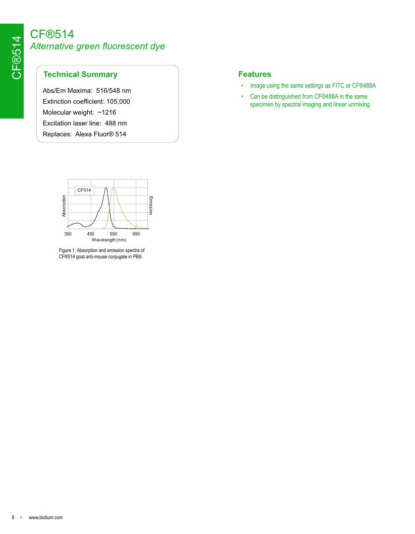
Figure 1. Absorption and emission spectra of CF®514 goat anti-mouse conjugate in PBS.
CF®
514
Features• Image using the same settings as FITC or CF®488A• Can be distinguished from CF®488A in the same
specimen by spectral imaging and linear unmixing
www.biotium.com • 9
CF®532 A bright orange fluorescent dye for the 532 nm laser
Technical Summary
Abs/Em Maxima: 527/558 nmExtinction coefficient: 96,000Molecular weight: ~ 685Excitation laser line: 532 nmDirect replacement for: Alexa Fluor® 532, Atto 532
300 400 500 600 700
Abso
rptio
n
Wavelength (nm)
Emission
CF532
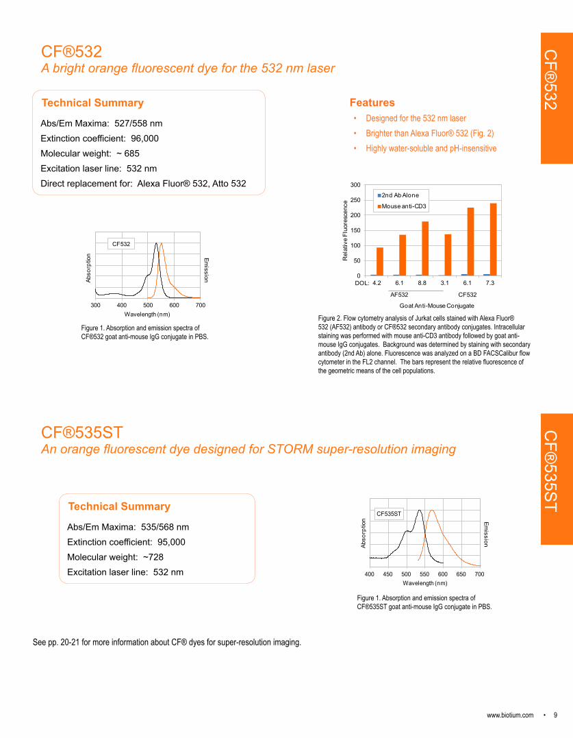
Figure 1. Absorption and emission spectra of CF®532 goat anti-mouse IgG conjugate in PBS.
0
50
100
150
200
250
300
4.2 6.1 8.8 3.1 6.1 7.3
Rel
ativ
e Fl
uore
scen
ce
Goat Anti-Mouse Conjugate
2nd Ab Alone
Mouse anti-CD3
DOL:
AF532 CF532
Figure 2. Flow cytometry analysis of Jurkat cells stained with Alexa Fluor® 532 (AF532) antibody or CF®532 secondary antibody conjugates. Intracellular staining was performed with mouse anti-CD3 antibody followed by goat anti-mouse IgG conjugates. Background was determined by staining with secondary antibody (2nd Ab) alone. Fluorescence was analyzed on a BD FACSCalibur flow cytometer in the FL2 channel. The bars represent the relative fluorescence of the geometric means of the cell populations.
Features• Designed for the 532 nm laser • Brighter than Alexa Fluor® 532 (Fig. 2)• Highly water-soluble and pH-insensitive
CF®
532
CF®535STAn orange fluorescent dye designed for STORM super-resolution imaging
Technical Summary
Abs/Em Maxima: 535/568 nmExtinction coefficient: 95,000Molecular weight: ~728Excitation laser line: 532 nm
Figure 1. Absorption and emission spectra of CF®535ST goat anti-mouse IgG conjugate in PBS.
400 450 500 550 600 650 700Wavelength (nm)
CF535ST
Abso
rptio
n Emission
See pp. 20-21 for more information about CF® dyes for super-resolution imaging.
CF®
535ST

10 • www.biotium.com
CF®555A bright and photostable orange-red dye
Technical Summary
Abs/Em Maxima: 555/565 nmExtinction coefficient: 150,000Molecular weight: ~ 901Excitation laser line: 532 nm or 568 nmDirect replacement for: Alexa Fluor® 555, ATTO 550, Cy®3, DyLight® 549, Rhodamine
Figure 2. Frozen section of rat testis stained with mouse anti-tubulin and CF®488A goat anti-mouse (min x rat) (microtubules, green), CF®555 Mix-n-Stain labeled mouse anti-ZO1 (tight junctions, red) and CF®640R phalloidin (actin filaments, cyan). See p. 23 for more information on Mix-n-Stain™ antibody labeling kits.
450 500 550 600 650
Emission
Abso
rptio
n
Wavelength (nm)
CF555
Figure 1. Absorption and emission spectra of CF®555 goat anti-mouse conjugate in PBS.
Features• Brighter than Cy®3• Highly water-soluble• Validated in multicolor STORM super-resolution imaging
(see pp. 20-21)
CF®
555
CF®543 An orange fluorescent dye ideal for the 543 nm laser
Technical Summary
Abs/Em Maxima: 541/560 nmExtinction coefficient: 100,000Molecular weight: ~ 870Excitation laser line: 532 nm, 543 nm, or 546 nmDirect replacement for: Alexa Fluor® 546, TAMRA
450 500 550 600 650
EmissionAb
sorp
tion
Wavelength (nm)
CF543
Figure 1. Absorption and emission spectra of CF®543 goat anti-mouse conjugate in PBS.
0
20
40
60
80
100
2 4 6 8 10 12 14
Rel
ativ
e Fl
uore
scen
ce
Degree of Labeling (DOL)
CF543AF546
Figure 2. Relative fluorescence of CF®543 and Alexa Fluor® 546 (AF546) goat anti-mouse conjugates as a function of the number of dye molecules per protein (degree of labeling).
CF®
543
Features• Optimized for the 543 nm laser• Yields the brightest conjugates among spectrally similar dyes• Highly water-soluble and pH-insensitive

www.biotium.com • 11
Features• Yields much brighter antibody conjugates than Alexa
Fluor® 568 • Extremely photostable• Excellent choice for multi-color imaging with CF®488A and
CF®640R• Compatible with TIRF and multicolor STORM super-
resolution imaging (see pp. 20-21)
Technical Summary
Abs/Em Maxima: 562/583 nmExtinction coefficient: 100,000Molecular weight: ~ 714Excitation laser line: 532 nm or 568 nmDirect replacement for: Alexa Fluor® 568, ATTO 565, Rhodamine Red
450 500 550 600 650 700
Emission
Abso
rptio
n
Wavelength (nm)
CF568
0
20
40
60
80
100
0 60 120 180 240 300N
orm
aliz
ed F
luor
esce
nce
Time (s)
CF568AF568
Figure 3. Photostability of CF®568 and Alexa Fluor® 568 (AF568) streptavidin conjugates. Intracellular staining of Jurkat cells was performed using anti-CD3-biotin followed by streptavidin-CF®568 or streptavidin-AF568. Cells were continuously exposed to mercury arc lamp microscope excitation with a Cy3 filter set. Images were captured every 15 seconds for 5 minutes and fluorescence intensity was normalized to time 0.
0
50
100
150
200
Rel
ativ
e Fl
uore
scen
ce
Goat Anti-Mouse Conjugate
IsotypeCD3
AF568DOL 2.9
AF568DOL 4.2
CF568DOL 2.2
CF568DOL4.1
Figure 2. Intracellular staining of Jurkat cells was performed using mouse anti-CD3 or isotype control followed by goat anti-mouse IgG conjugates. Fluorescence was analyzed on a BD FACSCalibur flow cytometer in the FL2 channel. Bars represent the relative fluorescence of the geometric means of the cell populations.
CF®568Outshines Alexa Fluor®568
Figure 1. Absorption and emission spectra of CF®568 goat anti-mouse conjugate in PBS.
CF®
568
Figure 4. MCF-7 cells stained with CF®568 monoclonal anti-Ep-CAM (clone EGP40/826) at 5 ug/mL (red). Nuclei are counterstained with Hoechst 33342 (blue). See p. 26 for more information on primary antibody conjugates.
12 • www.biotium.com
CF®594Truly the brightest deep red dye
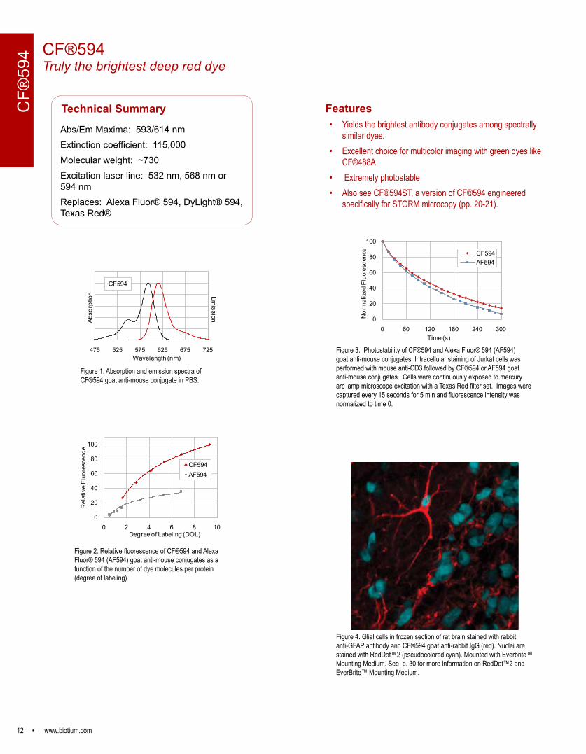
Features• Yields the brightest antibody conjugates among spectrally
similar dyes. • Excellent choice for multicolor imaging with green dyes like
CF®488A• Extremely photostable• Also see CF®594ST, a version of CF®594 engineered
specifically for STORM microcopy (pp. 20-21).
Technical Summary
Abs/Em Maxima: 593/614 nmExtinction coefficient: 115,000Molecular weight: ~730Excitation laser line: 532 nm, 568 nm or 594 nmReplaces: Alexa Fluor® 594, DyLight® 594, Texas Red®
0
20
40
60
80
100
0 2 4 6 8 10
Rel
ativ
e Fl
uore
scen
ce
Degree of Labeling (DOL)
CF594AF594
Figure 2. Relative fluorescence of CF®594 and Alexa Fluor® 594 (AF594) goat anti-mouse conjugates as a function of the number of dye molecules per protein (degree of labeling).
0
20
40
60
80
100
0 60 120 180 240 300
Nor
mal
ized
Flu
ores
cenc
e
Time (s)
CF594AF594
Figure 3. Photostability of CF®594 and Alexa Fluor® 594 (AF594) goat anti-mouse conjugates. Intracellular staining of Jurkat cells was performed with mouse anti-CD3 followed by CF®594 or AF594 goat anti-mouse conjugates. Cells were continuously exposed to mercury arc lamp microscope excitation with a Texas Red filter set. Images were captured every 15 seconds for 5 min and fluorescence intensity was normalized to time 0.
475 525 575 625 675 725
EmissionAb
sorp
tion
Wavelength (nm)
CF594
Figure 1. Absorption and emission spectra of CF®594 goat anti-mouse conjugate in PBS.
Figure 4. Glial cells in frozen section of rat brain stained with rabbit anti-GFAP antibody and CF®594 goat anti-rabbit IgG (red). Nuclei are stained with RedDot™2 (pseudocolored cyan). Mounted with Everbrite™ Mounting Medium. See p. 30 for more information on RedDot™2 and EverBrite™ Mounting Medium.
CF®
594

www.biotium.com • 13
CF®620RA bright and photostable far-red dye
Features• Highly water-soluble • Highly fluorescent and extremely photostable• Absorption/emission at 617/639 nm for use in FRET or
multi-color detection
Technical Summary
Abs/Em Maxima: 617/639 nmExtinction coefficient: 115,000Molecular weight: ~ 738Excitation laser line: 633 nm or 635 nmReplaces: LightCycler® Red 640
500 550 600 650 700 750
EmissionAb
sorp
tion
Wavelength (nm)
CF620R
Figure 1. Absorption and emission spectra of CF®620R free acid in PBS.
CF®
620R
14 • www.biotium.com
CF®
633
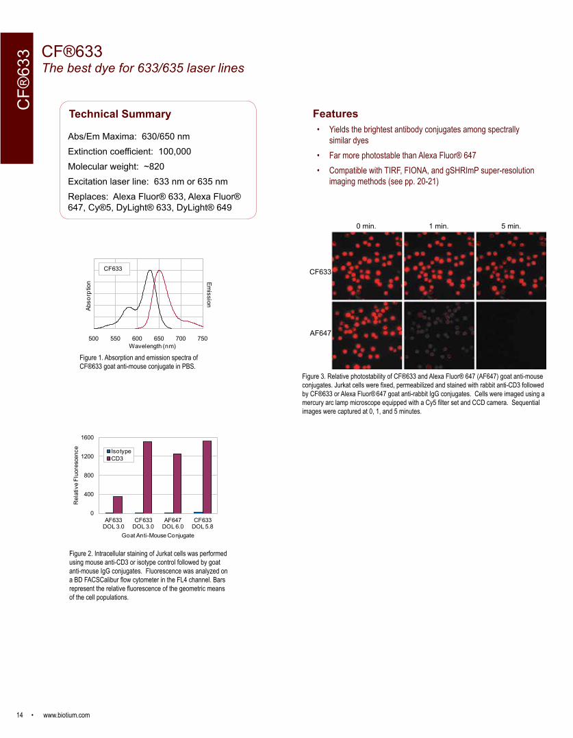
Features• Yields the brightest antibody conjugates among spectrally
similar dyes • Far more photostable than Alexa Fluor® 647• Compatible with TIRF, FIONA, and gSHRImP super-resolution
imaging methods (see pp. 20-21)
Figure 2. Intracellular staining of Jurkat cells was performed using mouse anti-CD3 or isotype control followed by goat anti-mouse IgG conjugates. Fluorescence was analyzed on a BD FACSCalibur flow cytometer in the FL4 channel. Bars represent the relative fluorescence of the geometric means of the cell populations.
CF®633The best dye for 633/635 laser lines
Technical Summary
Abs/Em Maxima: 630/650 nmExtinction coefficient: 100,000Molecular weight: ~820Excitation laser line: 633 nm or 635 nmReplaces: Alexa Fluor® 633, Alexa Fluor® 647, Cy®5, DyLight® 633, DyLight® 649
500 550 600 650 700 750
Emission
Abso
rptio
n
Wavelength (nm)
CF633
Figure 3. Relative photostability of CF®633 and Alexa Fluor® 647 (AF647) goat anti-mouse conjugates. Jurkat cells were fixed, permeabilized and stained with rabbit anti-CD3 followed by CF®633 or Alexa Fluor® 647 goat anti-rabbit IgG conjugates. Cells were imaged using a mercury arc lamp microscope equipped with a Cy5 filter set and CCD camera. Sequential images were captured at 0, 1, and 5 minutes.
Figure 1. Absorption and emission spectra of CF633 goat anti-mouse conjugate in PBS.
0 min. 1 min. 5 min.
CF633
AF647
0
400
800
1200
1600
AF633 DOL 3.0
CF633 DOL 3.0
AF647 DOL 6.0
CF633 DOL 5.8
Rel
ativ
e Fl
uore
scen
ce
Goat Anti-Mouse Conjugate
IsotypeCD3
Figure 1. Absorption and emission spectra of CF®633 goat anti-mouse conjugate in PBS.
www.biotium.com • 15
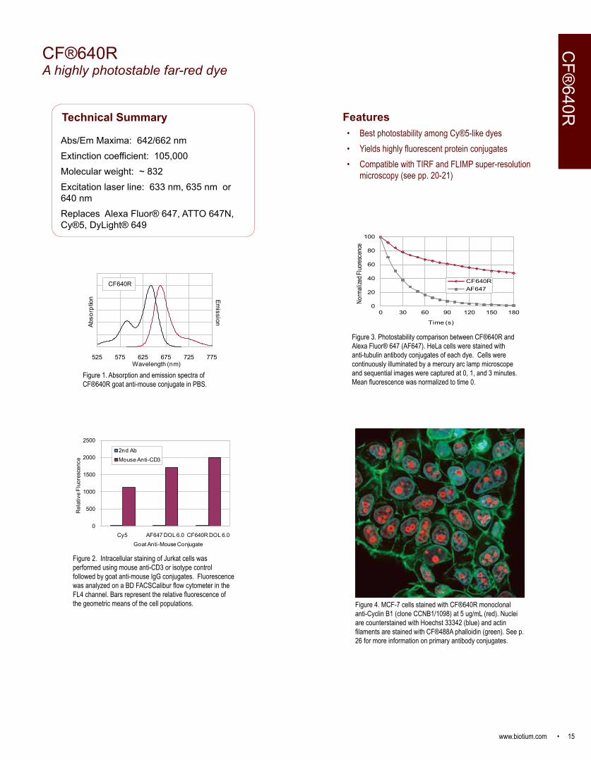
CF®640RA highly photostable far-red dye
Technical Summary
Abs/Em Maxima: 642/662 nmExtinction coefficient: 105,000Molecular weight: ~ 832Excitation laser line: 633 nm, 635 nm or 640 nmReplaces Alexa Fluor® 647, ATTO 647N, Cy®5, DyLight® 649
Figure 2. Intracellular staining of Jurkat cells was performed using mouse anti-CD3 or isotype control followed by goat anti-mouse IgG conjugates. Fluorescence was analyzed on a BD FACSCalibur flow cytometer in the FL4 channel. Bars represent the relative fluorescence of the geometric means of the cell populations.
0
500
1000
1500
2000
2500
Cy5 AF647 DOL 6.0 CF640R DOL 6.0
Rel
ativ
e Fl
uore
scen
ce
Goat Anti-Mouse Conjugate
2nd AbMouse Anti-CD3
525 575 625 675 725 775
Emission
Abso
rptio
n
Wavelength (nm)
CF640R
Figure 1. Absorption and emission spectra of CF®640R goat anti-mouse conjugate in PBS.
Features• Best photostability among Cy®5-like dyes• Yields highly fluorescent protein conjugates • Compatible with TIRF and FLIMP super-resolution
microscopy (see pp. 20-21)
Figure 3. Photostability comparison between CF®640R and Alexa Fluor® 647 (AF647). HeLa cells were stained with anti-tubulin antibody conjugates of each dye. Cells were continuously illuminated by a mercury arc lamp microscope and sequential images were captured at 0, 1, and 3 minutes. Mean fluorescence was normalized to time 0.
CF®
640R
0
20
40
60
80
100
0 30 60 90 120 150 180
Norm
alize
d Fluo
resce
nce
Time (s)
CF640RAF647
Figure 4. MCF-7 cells stained with CF®640R monoclonal anti-Cyclin B1 (clone CCNB1/1098) at 5 ug/mL (red). Nuclei are counterstained with Hoechst 33342 (blue) and actin filaments are stained with CF®488A phalloidin (green). See p. 26 for more information on primary antibody conjugates.

16 • www.biotium.com
CF®647A highly fluorescent far-red dye
Technical Summary
Abs/Em Maxima: 650/665 nmExtinction coefficient: 240,000Molecular weight: ~ 1058Excitation laser line: 633 nm, 635 nm or 640 nmReplaces: Cy®5, Alexa Fluor® 647, DyLight® 649
550 600 650 700 750
Emission
Abso
rptio
n
Wavelength (nm)
CF647
Figure 1. Absorption and emission spectra of CF®647 goat anti-mouse conjugate in PBS.
CF®
647
Figure 3. Cultured rat hippocampal neurons microinjected with CF®647 hydrazide (red) and stained with SynaptoGreen™ C4 (FM1-43) (green, synaptic vesicles). Image courtesy of Professor Guosong Liu, Tsinghua University, Beijing, China.
Features• Brighter than Cy®5• Highly water soluble and pH insensitive• Validated in multi-color super-resolution imaging by STORM
(see pp. 20-21)
Figure 2. Intracellular staining of Jurkat cells with CF®647 monoclonal anti-nucleollin (clone 365-2) (pink) or CF®647 IgG1 isotype control (blue) at 1 ug/tube, compared to unstained cellls (yellow). Cells were analyzed in the APC channel of a BD LSRII flow cytometer. See p. 26 for more information on primary antibody conjugates.
www.biotium.com • 17
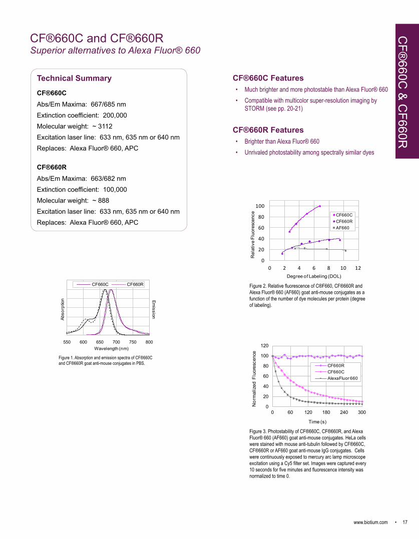
CF®660C and CF®660R Superior alternatives to Alexa Fluor® 660
0
20
40
60
80
100
0 2 4 6 8 10 12
Rel
ativ
e Fl
uore
scen
ce
Degree of Labeling (DOL)
CF660CCF660RAF660
Figure 2. Relative fluorescence of C®F660, CF®660R and Alexa Fluor® 660 (AF660) goat anti-mouse conjugates as a function of the number of dye molecules per protein (degree of labeling).
0
20
40
60
80
100
120
0 60 120 180 240 300
Nor
mal
ized
Flu
ores
cenc
e
Time (s)
CF660RCF660CAlexaFluor 660
Figure 3. Photostability of CF®660C, CF®660R, and Alexa Fluor® 660 (AF660) goat anti-mouse conjugates. HeLa cells were stained with mouse anti-tubulin followed by CF®660C, CF®660R or AF660 goat anti-mouse IgG conjugates. Cells were continuously exposed to mercury arc lamp microscope excitation using a Cy5 filter set. Images were captured every 10 seconds for five minutes and fluorescence intensity was normalized to time 0.
Technical Summary
CF®660CAbs/Em Maxima: 667/685 nmExtinction coefficient: 200,000Molecular weight: ~ 3112Excitation laser line: 633 nm, 635 nm or 640 nmReplaces: Alexa Fluor® 660, APC
CF®660RAbs/Em Maxima: 663/682 nmExtinction coefficient: 100,000Molecular weight: ~ 888Excitation laser line: 633 nm, 635 nm or 640 nmReplaces: Alexa Fluor® 660, APC
550 600 650 700 750 800
Emission
Abso
rptio
n
Wavelength (nm)
CF660C CF660R
Figure 1. Absorption and emission spectra of CF®660C and CF®660R goat anti-mouse conjugates in PBS.
CF®660C Features• Much brighter and more photostable than Alexa Fluor® 660• Compatible with multicolor super-resolution imaging by
STORM (see pp. 20-21)
CF®660R Features• Brighter than Alexa Fluor® 660• Unrivaled photostability among spectrally similar dyes
CF®
660C & C
F660R
18 • www.biotium.com
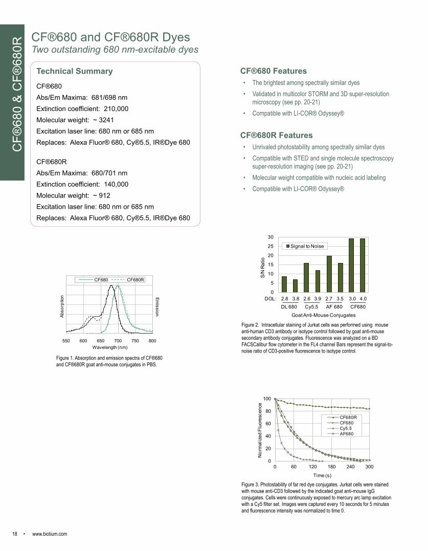
CF®680 and CF®680R DyesTwo outstanding 680 nm-excitable dyes
Figure 3. Photostability of far red dye conjugates. Jurkat cells were stained with mouse anti-CD3 followed by the indicated goat anti-mouse IgG conjugates. Cells were continuously exposed to mercury arc lamp excitation with a Cy5 filter set. Images were captured every 10 seconds for 5 minutes and fluorescence intensity was normalized to time 0.
Figure 2. Intracellular staining of Jurkat cells was performed using mouse anti-human CD3 antibody or isotype control followed by goat anti-mouse secondary antibody conjugates. Fluorescence was analyzed on a BD FACSCalibur flow cytometer in the FL4 channel Bars represent the signal-to-noise ratio of CD3-positive fluorescence to isotype control.
0
20
40
60
80
100
0 60 120 180 240 300
Nor
mal
ized
Flu
ores
cenc
e
Time (s)
CF680RCF680Cy5.5AF680
0
5
10
15
20
25
30
S/N
Rat
io
Goat Anti-Mouse Conjugates
Signal to Noise
DOL: 2.8 3.8 2.6 3.9 2.7 3.5 3.0 4.0DL 680 Cy5.5 AF 680 CF680
Technical Summary
CF®680Abs/Em Maxima: 681/698 nmExtinction coefficient: 210,000Molecular weight: ~ 3241Excitation laser line: 680 nm or 685 nmReplaces: Alexa Fluor® 680, Cy®5.5, IR®Dye 680
CF®680RAbs/Em Maxima: 680/701 nmExtinction coefficient: 140,000Molecular weight: ~ 912Excitation laser line: 680 nm or 685 nmReplaces: Alexa Fluor® 680, Cy®5.5, IR®Dye 680
550 600 650 700 750 800
Emission
Abso
rptio
n
Wavelength (nm)
CF680 CF680R
Figure 1. Absorption and emission spectra of CF®680 and CF®680R goat anti-mouse conjugates in PBS.
CF®
680
& C
F®68
0R
CF®680 Features• The brightest among spectrally similar dyes• Validated in multicolor STORM and 3D super-resolution
microscopy (see pp. 20-21)• Compatible with LI-COR® Odyssey®
CF®680R Features• Unrivaled photostability among spectrally similar dyes• Compatible with STED and single molecule spectroscopy
super-resolution imaging (see pp. 20-21)• Molecular weight compatible with nucleic acid labeling• Compatible with LI-COR® Odyssey®

www.biotium.com • 19
CF®750, CF®770, CF®790, and CF®800Unrivaled Near-Infrared Dyes
Technical Summary
CF®750Abs/Em Maxima: 755/777 nmExtinction coefficient: 250,000Molecular weight: ~ 3009Excitation laser line: 633, 635, 680 or 685 nmReplaces: Alexa Fluor® 750, Cy®7, DyLight® 750
CF®770Abs/Em Maxima: 770/797 nmExtinction coefficient: 220,000Molecular weight: ~ 3138Excitation laser line: 785 nmReplaces: DyLight™ 800, IRDye 800CW
CF®790Abs/Em Maxima: 784/806 nmExtinction coefficient: 210,000Molecular weight: ~ 3267Excitation laser line: 785 nmReplaces: Alexa Fluor® 790
CF®800Abs/Em Maxima: 797/816Extinction coefficient: 210,000Molecular weight: ~3334Excitation laser line: 785 nmSpectrally similar to: Indocyanine Green
Figure 2. Tumors in mice were imaged using an IVIS® imaging system (Caliper Life Sciences) 24 hours, 48 hours, and 96 hours after IV injection of Avastin conjugated to CF®750. Images courtesy of Caliper Life Sciences.
Figure 3. Near-infrared western blotting with CF® dyes compared to spectrally similar dyes. A. Two-fold dilutions of HeLa cell lysate containing from 2 ug to 0.125 ug total protein per lane were separated by SDS-PAGE, transferred to a PVDF membrane, and probed with mouse anti-tubulin and rabbit anti-COXIV antibodies. Secondary detection was performed with either IRDye® 680LT goat anti-mouse (red) and IRDye® 800CW goat anti-rabbit (green) (LI-COR®; lanes 1-6) or CF®680 goat anti-mouse (red) and CF®770 goat anti-rabbit (green) (lanes 7-12) at the same final concentrations. Membranes were scanned using an Odyssey® infrared imaging system. Quantitation of the bands showed approximately 1.5-2-fold higher fluorescence intensity of CF® dye secondary antibodies compared to IRDye® secondary antibodies. B. Western blots of HeLa cell lysate (lanes 2 and 4) were probed with mouse anti-tubulin antibody followed by goat anti-mouse conjugated to Alexa Fluor® 790 (AF790, left) or CF®790 (right). CF®790 does not introduce excessive negative charge to antibody conjugates, which can increase non-specific binding.
Features• Exceptionally bright and stable• Ideal for in vivo imaging• Compatible with LI-COR® Odyssey®• Superior signal-to-noise for bioconjugates• CF®750 validated in STORM microscopy (see pp. 20-21)
Figure 1. Absorption and emission spectra of near-IR CF® dye goat anti-mouse conjugates in PBS.
Near-IR
CF®
Dyes
IRDye, LI-COR, Odyssey, and In-Cell Western are trademarks or registered trademarks of LI-COR, Inc. in the United States and other countries. IVIS is a registered trademark of Caliper LIfe Sciences.
600 650 700 750 800 850 900
Emission
Abso
rptio
n
Wavelength (nm)
CF750
600 650 700 750 800 850 900
Emission
Abso
rptio
n
Wavelength(nm)
CF770
600 650 700 750 800 850 900
Emission
Abso
rptio
n
Wavelength(nm)
CF790
600 650 700 750 800 850 900
Abso
rptio
n
Wavelength (nm)
Emission
CF800
IRDye® 680 LT/800 CW CF680/ CF7701 2 3 4 5 6 7 8 9 10 11 12
A CF790AF 7901 2 3 4
B

20 • www.biotium.com
Recent publications comparing synthetic dyes for super-resolution imaging have shown CF® dyes give the best performance for multiple methods. The superior brightness, photostability, and photochemical switching properties of certain CF® dyes are ideal for 3-D SIM, 3-D STORM, and other super-resolution and single molecule imaging techniques. Biotium’s CF®405M has been found to be the brightest and most photostable short wavelength fluorescent dye for SIM. Six CF® dyes spanning the visible red, far-red, and near-infrared spectra have been validated for STORM, including three color imaging with CF®568, CF®647, and CF®680. See Lehmann et al. 2015, and a full list of references for CF® dye single-molecule imaging applications on page 21. Biotium offers a wide selection of CF® dye labeled secondary antibodies, other conjugates, and labeling kits; visit www.biotium.com for our full selection of products.
Figure 1. Comparison of conventional wide-field microscopy (left) with STORM (right) using CF® dye conjugates. Fixed cells were stained with mouse anti-tubulin antibody followed by CF® dye conjugated anti-mouse secondary antibody (top row: CF®647, middle row: CF®660C, bottom row: CF®680). For STORM, samples were sealed in buffer that contained 5% (w/v) glucose, 100 mM cysteamine, 0.8 mg/mL glucose oxidase, and 40 µg/mL catalase, in Tris-HCl (pH 7.5). Samples were imaged on a Nikon Ti-Eclipse w/ PFS microscope with a CFI Plan Apo Lambda 100x oil objective. Dye molecules were photoswitched and imaged using a 647 nm laser; a 405 nm laser was used to assist dye reactivation to the emitting state. Emission was collected with an Andor iXon Ultra 897 EMCCD camera for a total of 100,000 frames per image at a frame rate of 110 Hz.
STORM microscopy images courtesy of Sam Kenny and Professor Ke Xu, College of Chemistry, University of California, Berkeley.
Figure 2. CF®568 (left) produces better images than Cy®3b (right) in 3-D STORM microscopy. Fixed cells were stained with mouse anti-tubulin antibody followed by dye-conjugated anti-mouse secondary antibodies. See Figure 1 legend for imaging conditions. Dye molecules were photoswitched and imaged using a 560 nm laser; a 405 nm laser was used to assist dye reactivation to the emitting state.
Wide-field microscopy STORMCF®647
CF®660C
CF®680
CF®568 Cy®3b
2 mm 2 mm
500 nm 500 nm
-600
z
(nm
)
+
600
FIONA: Fluorescence Imaging with One Nanometer Accuracy; FLImP: Fluorophore localization imaging with photobleaching; SHRImP: Single-molecule high-resolution imaging with photobleaching; SIM: Structured illumination microscopy; STED: Stimulated emission depletion; STORM: Stochastical optical reconstruction microscopy; TIRF: Total internal reflection fluorescence
CF® Dyes for Super-Resolution ImagingSu
per-R
esol
utio
n Im
agin
g

www.biotium.com • 21
CF® Dyes for Super-Resolution Imaging Super-Resolution Im
aging
CF® Dye Abs/Em maxima
Extinction coefficient
Super resolution application
References
CF®405S 404/431 nm 33,000 SIM Demmerle et al. (2017). Nature Protocols 12, 988–1010.Essig et al. (2017). Immunity https://doi.org/10.1016/j.immuni.2017.11.008
CF®405M 408/452 nm 41,000 SIM Kraus, F. et al. (2017). Nat Protoc 12, 1011-1028. doi:nprot.2017.020Markaki, Y. et al. (2013). Methods Mol Biol 950, 43-64.Miron, E. et. al. (2016). In: Mark C. Leake (ed.), Methods in Molecular Biology, vol. 1431, 127-140.Ohgomori, T. et al. (2017). Eur J Neurosci 46, 2001-2014. doi:10.1111/ejn.13650
CF®488A 490/515 nm 70,000 STED, TIRF Angelov, B. & Angelova, A. (2017). Nanoscale 9, 9797-9804. doi:10.1039/c7nr03454g (STED)Zanetti-Domingues, L.C. et al. (2013). PLoS ONE 8(9): e74200. (TIRF)
CF®535ST 535/568 nm 95,000 STORM Collaborator communication; contact [email protected] for more information.CF®555 555/565 nm 150,000 Multicolor STORM Lehmann, M. et al. (2015). J Biophotonics DOI 10.1002/jbio.201500119CF®568 562/583 nm 100,000 Multicolor STORM,
SIM, TIRFGong, Y.-N. et al. (2017). Cell Cycle, 1-13. doi:10.1080/15384101.2017.1371889 (STORM)Gorur, A. et al. (2017). J Cell Biol 216, 1745-1759. doi:10.1083/jcb.201702135 (STORM)Heller, J. (2017). OM&P 3, 48-58, doi:doi:10.20388/omp2017.002.0045 (STORM)Jorgans, D.M. et al. (2017). J Cell Sci 2017 130: 177-189. doi: 10.1242/jcs.190967 (STORM)Karanasios, E. et al. (2016). Nat Commun 7: 12420. DOI: 10.1038/ncomms12420 (STORM)Kraus, F. et al. (2017). Nat Protoc 12, 1011-1028, doi:nprot.2017.020 (SIM)Lehmann, M. et al. (2015). J Biophotonics DOI 10.1002/jbio.201500119 (STORM)Lim, A. et al. (2017). Mol Biol Cell doi:mbc.E16-12-0820 (SIM)Turkowyd, B. et al. (2016). Anal Bioanal Chem DOI 10.1007/s00216-016-9781-8 (STORM)Zanetti-Domingues, L.C. et al. (2013). PLoS ONE 8(9): e74200.(TIRF)Zhang, M. et al. (2015). eLife 2015;10.7554/eLife.11205 (STORM)
CF®594ST 593/614 nm 115,000 STORM Collaborator communication; contact [email protected] for more information. Note: CF®594ST is a unique dye designed for STORM. Our original CF®594 is not suitable for STORM.
CF®633 630/650 nm 100,000 FIONA, gSHRImP, Single molecule tracking, TIRF
Bosch, P. J. et al. (2014). Biophys J 107, 803-814. (TIRF)Huang, T. et al. (2018). Biophysical Journal 114, 301–310. (Single Molecule Tracking)Kim, H. J., and Selvin, P. R. (2013). SpringerReference Encyclopedia of Biophysics. (FIONA)Simonson, P. D. et al. (2011). Nano Lett 11, 5090-5096. DOI:10.1021/nl203560r (gSHRImP)Zanetti-Domingues, L.C. et al. (2013). PLoS ONE 8(9): e74200. (TIRF)Zhang, R. et al. (2017). eLife 2017;6:e30959. (TIRF)
CF®640R 642/662 nm 105,000 FLImP, SIM, TIRF Bosch, P. J. et al. (2014). Biophys J 107, 803-814. (TIRF)Loh, L. N. (2017). MBio 8, doi:mBio.02030-16 (SIM)Martin-Fernandez, M. L. et al. (2013). J Microsc 252, 16-22. (TIRF)Needham, S.R. et al. (2015). Biochem Soc Trans 43, 309–314. (FLImP)Needham, S.R. et al. (2016). Nat Commun 7, 13307. doi:ncomms13307 (FLImP)Zanetti-Domingues, L.C. et al. (2013). PLoS ONE 8(9): e74200. (TIRF)Zanetti-Domingues, L.C. et al. (2015). Prog Biophys Mol Biol. doi:S0079-6107(15)00047-4 (FLImP)
CF®647 650/665 nm 240,000 Multicolor STORM Gong, Y.-N. et al. (2017). Cell Cycle, 1-13. doi:10.1080/15384101.2017.1371889Lehmann, M. et al. (2015). J Biophotonics DOI 10.1002/jbio.201500119Olivier, N. et al. (2013). Biomed Opt Express 4, 885-899.Turkowyd, B. et al. (2016). Anal Bioanal Chem DOI 10.1007/s00216-016-9781-8
CF®660C 667/685 nm 200,000 Multicolor STORM Turkowyd, B. et al. (2016). Anal Bioanal Chem DOI 10.1007/s00216-016-9781-8Zhang, Z., et al. (2015). Nature Methods doi:10.1038/nmeth.3528.
CF®680 681/698 nm 210,000 Dual-Color 3D SMLM, Multicolor STORM
Früh, S.M. et al. (2015). Nature Communications 6, 7275. (STORM)Glebov, O.O. et al. (2017). Cell Rep 18, 2715-2728. doi:S2211-1247(17)30279-6 (STORM)Gorur, A. et al. (2017). J Cell Biol 216, 1745-1759. doi:10.1083/jcb.201702135 (STORM)Lehmann, M. et al. (2015). J Biophotonics DOI 10.1002/jbio.201500119 (STORM)Platonova, E. et al. (2015). ACS Chem. Biol.10(6),1411–1416. (STORM)Platonova, E. et al. (2015). Methods doi: http://dx.doi.org/10.1016/j.ymeth.2015.06.018. (STORM)Salvador-Gallego, R. et al. (2016). EMBO J. DOI 10.15252/embj.201593384. (STORM)Shrestha, R. L. et al. (2017). Nat Commun 8, 150. doi:10.1038/s41467-017-00209-z (STORM)Turkowyd, B. et al. (2016). Anal Bioanal Chem DOI 10.1007/s00216-016-9781-8 (SMLM)Winterflood, C.M. et al. (2015). Biophys J. 109, 3–6. (SMLM)Zhang, Z., et al. (2015). Nature Methods doi:10.1038/nmeth.3528. (STORM)
CF®680R 680/701 nm 140,000 Multicolor STORM, Single-molecule spectroscopy, Single molecule tracking, STED
Conley, G. et al. (2017). Science Advances 3(10), e1700969 DOI:10.1126/sciadv.1700969 (STORM)Görlitz, F. et al. (2014). Progress Electromagnetics Res 147, 57-68. (STED)Huang, T. et al. (2018). Biophysical Journal 114, 301–310. (Single Molecule Tracking)König, I. et al. (2015). Nature Methods doi:10.1038/nmeth.3475 (Single molecule spectroscopy)Winter, F.R. et al. (2017). Scientific Reports 7, 46492. DOI: 10.1038/srep46492 (STED)
CF®750 755/777 nm 250,000 STORM Collaborator communication; contact [email protected] for more information.
22 • www.biotium.com
Visit our website for more CF® dye references and applications
Selected CF® Dye ReferencesC
F® D
ye R
efer
ence
s
In Cell Western®Andriamihaja, M. et al., Free Radical Biol Med 85, 219 (2015).Audebert, M. et al., Toxicol Appl Pharmacol 260, 58 (2012).Jamin, E. L. et al., PLoS One 8, e58591 (2013).Khoury, L. et al., Environ Mol Mutagen 54, 737 (2013).Khoury, L. et al., Mutagenesis 31, 83 (2016).
Near-infrared western blottingAnnahazi, A. et al., Am J Gastroenterol 108, 1392 (2013).Avelar, G. M. et al., Curr Biol 24, 1234 (2014).Babelova, A. et al., PLoS One 8, e80328 (2013).Dabek, M. et al., Inflamm Bowel Dis 17, 1409 (2011).Gauthier, T. et al., Mol Nutr Food Res 57, 1026 (2013).Huc, L. et al., Toxicol In Vitro 26, 709 (2012).Kohler, C. et al., BMC Microbiol 12, 210 (2012).Liang, Y. et al., Nat Immunol 14, 858 (2013).Lucioli, J. et al., Toxicon 66, 31 (2013).Martinsen, A. et al., Cell Calcium 52, 413 (2012).Moussa, L. et al., Clin Nutr 32, 51 (2012).Nebot-Vivinus, M. et al., World J Gastroenterol 20, 6832 (2014).Oliveira, A. F. et al., J Neurosci 34, 776 (2014).Olivier, I. et al., Inflamm Bowel Dis 17, 747 (2011).Ottenheijm, C. A. et al., Brain 136, 1718 (2013).Wu, C. L. et al., PLoS One 7, e34999 (2012).
In vivo imaging Abe, S. et al., J Immunol 190, 6239 (2013).Alawieh, A. et al., J of Neuroinflamm 12, 1 (2015).Alfonso-Loeches, S. et al., Glia 60, 948 (2012).Darniot, M. et al., Antiviral Res 93, 364 (2012).Fan, Q. et al., Biomed Res Int 2014, 459676 (2014).Gao, F. et al., ACS Nano 9, 4976 (2015).Herz, C. et al., J Cell Mol Med, (2014).Li, X. et al., J Pharmacol Exp Ther 351, 206 (2014).Li, X. et al., Eur J Pharm Sci 52, 132 (2014).Matsuo, H. et al., Int J Nanomed 7, 3341 (2012).Moore, L. et al., Adv Mater 25, 3532 (2013).Suemizu, H. et al., PLoS One 8, e82708 (2013).Sun, X. et al., Eur J Nucl Med Mol Imaging 41, 1428 (2014).Sun, Y. et al., Biotechniques 52, 1 (2012).Takeyama, H. et al., Surg Endosc 28, 1984 (2014).Zeiderman, M. R. et al., J Surg Res 190, 111 (2014).Annahazi, A. et al., Am J Gastroenterol 108, 1392 (2013).Avelar, G. M. et al., Curr Biol 24, 1234 (2014).Babelova, A. et al., PLoS One 8, e80328 (2013).Dabek, M. et al., Inflamm Bowel Dis 17, 1409 (2011).Huc, L. et al., Toxicol In Vitro 26, 709 (2012).Kohler, C. et al., BMC Microbiol 12, 210 (2012).Liang, Y. et al., Nat Immunol 14, 858 (2013).Lucioli, J. et al., Toxicon 66, 31 (2013).Martinsen, A. et al., Cell Calcium 52, 413 (2012).Moussa, L. et al., Clin Nutr 32, 51 (2012).Oliveira, A. F. et al., J Neurosci 34, 776 (2014).Olivier, I. et al., Inflamm Bowel Dis 17, 747 (2011).Ottenheijm, C. A. et al., Brain 136, 1718 (2013).Wu, C. L. et al., PLoS One 7, e34999 (2012).
Immunofluorescence microscopyAhmed, S. M. et al., J Cell Biol 199, 951 (2012).Akhmetshina, A. et al., Nat Commun 3, 735 (2012).Al Tanoury, Z. et al., J Cell Sci 127, 521 (2014).Amann, R. et al., J Virol 87, 1618 (2012).Azzedine, H. et al., Hum Mol Genet 22, 4224 (2013).Barboro, P. et al., PLoS One 8, e79212 (2013).Blankenburg, S. et al., Neuropharmacol 88, 134 (2014).Bosch, P. J. et al., Biophys J 107, 803 (2014).Chen, T. et al., J Am Chem Soc 135, 11595 (2013).Fan, Y. et al., Nat Cell Biol 16, 445 (2014).Felix-Ortiz, A. C. et al., Neuron 79, 658 (2013).Feng, X. et al., Nat Commun 5, 4487 (2014).Feng, X. et al., Cell Death Differ 21, 397 (2014).Gan, E. S. et al., Nat Commun 5, 5098 (2014).Hoepflinger, M. C. et al., J Exp Bot 64, 5553 (2013).Huh, D. et al., Nat Protoc 8, 2135 (2013).Inutsuka, A. et al., Neuropharmacol 85, 451 (2014).Ise, H. et al., Glycobiology 22, 788 (2012).Jalewa, J. et al., PLoS One 9, e88003 (2014).Kanno, T. et al., Free Radic Biol Med, (2012).Kapoor, N. et al., PLoS One 8, e53657 (2013).Kawase, A. et al., J Nat Med 68, 395 (2013).Kern, F. et al., Biochem J 455, 217 (2013).Kiyono, M. et al., Planta 235, 841 (2012).Kohara, K. et al., Nat Neurosci 17, 269 (2014).Komura, K. et al., Glycobiology 22, 1741 (2012).Lee, A. T. et al., J Neurosci 34, 11519 (2014).Li, W. et al., J Neurosci 33, 8423 (2013).Lim, A. K. et al., Development 140, 3819 (2013).Lindberg, O. R. et al., PLoS One 7, e46380 (2012).Lozada, A. F. et al., J Neurosci 32, 7651 (2012).Ma, L. et al., PLoS One 7, e51777 (2012).Markaki, Y. et al., Methods Mol Biol 950, 43 (2013).Neumann, K. et al., Immunity 40, 389 (2014).Ouyang, D. Y. et al., Autophagy 9, 20 (2013).Perez Bay, A. E. et al., J Cell Sci 127, 4457 (2014).Perez Bay, A. E. et al., EMBO J 32, 2125 (2013).Persson, A. et al., Glia 61, 790 (2013).Qian, M. et al., J Virol 87, 3571 (2013).Qu, X. et al., Biomaterials 34, 9812 (2013).Rohde, J. et al., PLoS One 8, e83802 (2013).Schneider, K. et al., Nucleic Acids Res 41, 4860 (2013).Shiheido, H. et al., PLoS One 7, e38878 (2012).Shiimori, M. et al., Mol Cell Biol, (2012).Stasevich, T. J. et al., Methods 70, 77 (2014).Tai, L. H. et al., Nat Neurosci 15, 1281 (2012).Thege, F. I. et al., Lab Chip 14, 1775 (2014).Tokunaga, M. et al., Chem Biol 20, 935 (2013).Wang, Y. et al., PLoS One 9, e89751 (2014).Wieduwild, R. et al., J Am Chem Soc 135, 2919 (2013).Witteveldt, J. et al., Nucleic Acids Res 42, 3314 (2014).Yang, R. et al., Nanomedicine 9, 636 (2012).Yates, J. L. et al., J Immunol 191, 1240 (2013).Yu, L. et al., PLoS One 8, e71988 (2013).Zhang, X. et al., Nucleic Acids Res 41, e152 (2013).Zurla, C. et al., PLoS One 6, e19727 (2011).
Mix-n-Stain™ Antibody Labeling KitsAhi, Y. S. et al., J Gen Virol 94, 1325 (2013). Anderson, M. S. et al., J Virol 88, 9111 (2014). Arul, M. et al., Cytotechnology 66, 481 (2013). Deo, D. I. et al., Biomacromolecules 15, 2555 (2014). Ersek, B. et al., J Immunol 189, 1602 (2012). Galletti, G. et al., Lab Chip 14, 147 (2014). Gorham, J. B. et al., Food Hydrocolloids 52, 952 (2016). Hirtz, M. et al., Adv Mat Interfaces, n/a (2016). Home, P. et al., Proc Natl Acad Sci U S A 109, 7362 (2012). Kesik, M. et al., RSC Advances 5, 83361 (2015). Lim, A. K. et al., Development 140, 3819 (2013). Lin, T.-B. et al., J Pineal Res, n/a (2016). Mikosik, A. et al., Immun Ageing 10, 27 (2013). Miyagishima, S. Y. et al., BMC Plant Biol 14, 57 (2014). Montesinos-Rongen, M. et al., J Immunol 195, 1312 (2015). Osteikoetxea, X. et al., PLoS One 10, e0121184 (2015). Park, K. S. et al., Nat Med 17, 1504 (2011).Ruger, M. et al., BMC Microbiol 14, 56 (2014). Ruger, M. et al., Cytometry A 81, 1055 (2012). Sainski, A. M. et al., J Cell Biol 207, 159 (2014). Tschumper, R. C. et al., Blood Cancer J 3, e112 (2013).Wienand, K. et al., PLoS One 10, e0120734 (2015). Xu, Z. et al., Proc Natl Acad Sci U S A 110, 13097 (2013).Yamamoto, S. et al., Dev Biol 393, 33 (2014).Yamashita, A. et al., PLoS One 9, e86426 (2014).Yang, Y. et al., Free Radic Biol Med 53, 437 (2012).Zolnerciks, J. K. et al., FASEB J 28, 4335 (2014).
Flow cytometryEndo, H. et al., Cell Death Dis 5, e1027 (2014).Gielen, P. R. et al., Neuropharmacology, (2013).Giuntini, S. et al., Infect Immun 80, 187 (2012).Kesik, M. et al., RSC Advances 5, 83361 (2015).Li, X. et al., Eur J Pharm Sci 52, 132 (2014).Mahassni, S. H. et al., Saudi J. Biol Sci 20, 131 (2013).Mikosik, A. et al., Immun Ageing 10, 27 (2013).Osteikoetxea, X. et al., PLoS One 10, e0121184 (2015).Ruger, M. et al., BMC Microbiol 14, 56 (2014).Ruger, M. et al., Cytometry A 81, 1055 (2012).Sainski, A. M. et al., J Cell Biol 207, 159 (2014).Skindersoe, M. E. et al., Cytometry A 81, 430 (2012).Wienand, K. et al., PLoS One 10, e0120734 (2015).Zeiderman, M. R. et al., J Surg Res 190, 111 (2014).
Fluorescence polarization assayAntolín-Urbaneja, J. C. et al.,Optical Sensors, vol. 8774.Di Giovanni, S. et al., Anal Meth 4, 3558 (2012).Pennacchio, A. et al., Food Chem 190, 381 (2016).Varriale, A. et al., J Agric Food Chem 63, 9159 (2015).
Super-resolution microscopySee pp. 20-21
In-Cell Western is a registered trademark of LI-COR, Inc. in the United States and other countries.
CF® dyes and conjugates have been cited in hundreds of publications, with new references published every day. Visit www.biotium.com/downloads for a more comprehensive list of CF® dye references by color and application.
www.biotium.com • 23
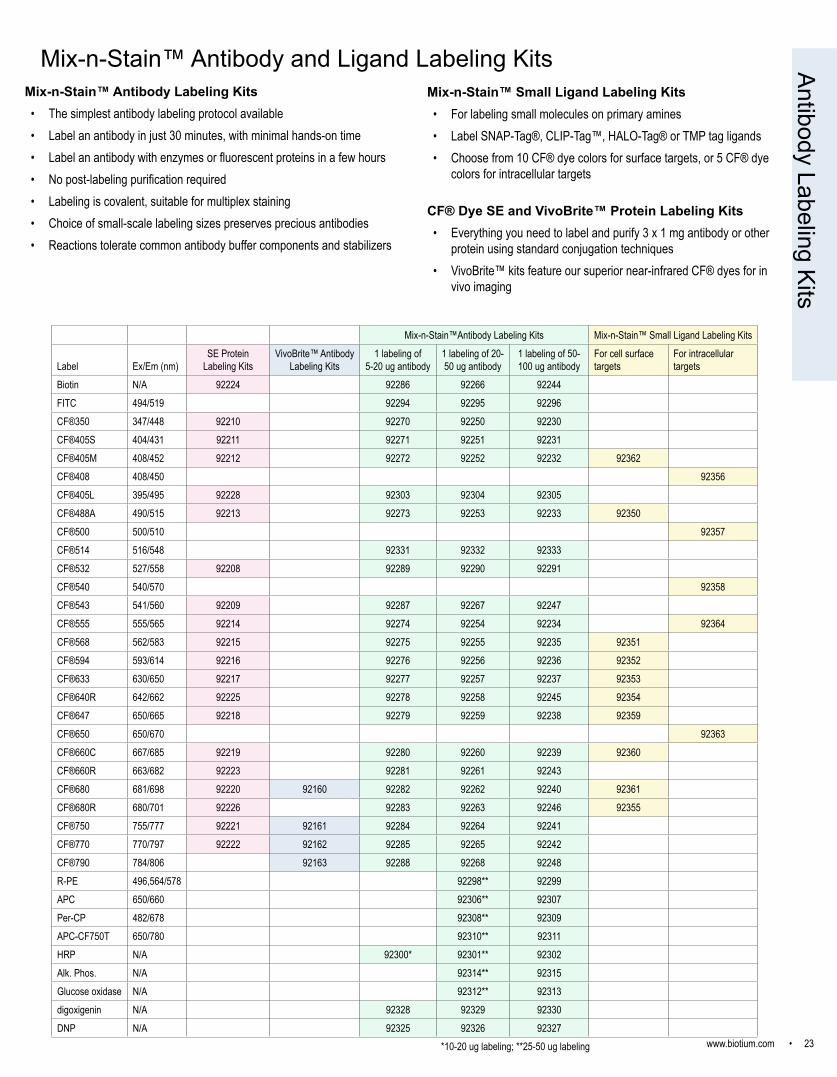
Mix-n-Stain™ Antibody Labeling Kits• The simplest antibody labeling protocol available• Label an antibody in just 30 minutes, with minimal hands-on time• Label an antibody with enzymes or fluorescent proteins in a few hours• No post-labeling purification required• Labeling is covalent, suitable for multiplex staining• Choice of small-scale labeling sizes preserves precious antibodies• Reactions tolerate common antibody buffer components and stabilizers
Antibody Labeling Kits
Mix-n-Stain™ Small Ligand Labeling Kits• For labeling small molecules on primary amines• Label SNAP-Tag®, CLIP-Tag™, HALO-Tag® or TMP tag ligands • Choose from 10 CF® dye colors for surface targets, or 5 CF® dye
colors for intracellular targets
Mix-n-Stain™Antibody Labeling Kits Mix-n-Stain™ Small Ligand Labeling Kits
Label Ex/Em (nm)SE Protein
Labeling KitsVivoBrite™ Antibody
Labeling Kits1 labeling of
5-20 ug antibody1 labeling of 20-50 ug antibody
1 labeling of 50-100 ug antibody
For cell surface targets
For intracellular targets
Biotin N/A 92224 92286 92266 92244FITC 494/519 92294 92295 92296CF®350 347/448 92210 92270 92250 92230CF®405S 404/431 92211 92271 92251 92231CF®405M 408/452 92212 92272 92252 92232 92362CF®408 408/450 92356CF®405L 395/495 92228 92303 92304 92305CF®488A 490/515 92213 92273 92253 92233 92350CF®500 500/510 92357CF®514 516/548 92331 92332 92333CF®532 527/558 92208 92289 92290 92291CF®540 540/570 92358CF®543 541/560 92209 92287 92267 92247CF®555 555/565 92214 92274 92254 92234 92364CF®568 562/583 92215 92275 92255 92235 92351CF®594 593/614 92216 92276 92256 92236 92352CF®633 630/650 92217 92277 92257 92237 92353CF®640R 642/662 92225 92278 92258 92245 92354CF®647 650/665 92218 92279 92259 92238 92359CF®650 650/670 92363CF®660C 667/685 92219 92280 92260 92239 92360CF®660R 663/682 92223 92281 92261 92243CF®680 681/698 92220 92160 92282 92262 92240 92361CF®680R 680/701 92226 92283 92263 92246 92355CF®750 755/777 92221 92161 92284 92264 92241CF®770 770/797 92222 92162 92285 92265 92242CF®790 784/806 92163 92288 92268 92248R-PE 496,564/578 92298** 92299APC 650/660 92306** 92307Per-CP 482/678 92308** 92309APC-CF750T 650/780 92310** 92311HRP N/A 92300* 92301** 92302Alk. Phos. N/A 92314** 92315Glucose oxidase N/A 92312** 92313digoxigenin N/A 92328 92329 92330DNP N/A 92325 92326 92327
CF® Dye SE and VivoBrite™ Protein Labeling Kits• Everything you need to label and purify 3 x 1 mg antibody or other
protein using standard conjugation techniques• VivoBrite™ kits feature our superior near-infrared CF® dyes for in
vivo imaging
*10-20 ug labeling; **25-50 ug labeling
Mix-n-Stain™ Antibody and Ligand Labeling Kits

24 • www.biotium.com
Reactive group/unit size
Alkyne0.5 mg
Amine1 mg
Aminooxy1 mg
Azide0.5 mg
BCN0.5 mg
Hydrazide1 mg
Maleimide1 umol
MTS1 mg
Picolyl azide0.5 mg
SE1 umol
Tyramide**0.5 mg
Reacts with
Azides, picolyl azides
Activated carboxylic
acids
Aldehydes & ketones
Alkynes, BCN
Azides Polar tracer* Thiols Thiols Alkynes Primary amines;lysine
residues
HRP substrate
CF®350 92035 92050 92151 92020 92109 92170CF®405S 92036 92055 92113 92183 92030 92110 92197CF®405M 92093 92056 92092 92114 92021 92111CF®405L 92046 92112 92198CF®430 92118 92117CF®440 92124 92123CF®450 96012 96011CF®488A 92086 92037 92051 92080 92075 92152 92022 92097 92187 92120 92171CF®500 96026CF®514 92103 92199CF®532 92180 92045 92104CF®543 92181 92044 92098 92105 92172CF®555 92087 92038 92081 92153 92023 92130 96021CF®568 92088 92039 92057 92082 92076 92154 92024 92188 92131 92173CF®570 96015 96014CF®583 96017 96016CF®594 92089 92040 92052 92083 92077 92158 92025 92099 92189 92132 92174CF®620R 92033 92106 92194CF®633 92041 92053 92156 92026 92133CF®640R 92091 92043 92058 92085 92078 92157 92034 92096 92190 92108 92175CF®647 92090 92042 92084 92136 92027 92191 92135 96022CF®650 96027CF®660C 92095 92094 92028 96001 92137CF®660R 96004 96010 92059 92182 96024 92031 92134 92195CF®680 96005 92119 92029 96003 92139CF®680R 96006 92054 92079 96025 92032 96007 92107 92196CF®750 92102 92142CF®770 92065 92192 92150CF®790 92155*CF®800 92128 92127*
CF® Dye Reactive DyesA wide selection of colors and functional groups for dye conjugation
* For conjugation to aldehyde or ketone groups, we recommend using CF® dye aminooxy forms. * Unit size 0.25 umol
Don’t see what you’re looking for?We regularly add new CF® dye products to our catalog according to customer demand. Be sure to check our website for updates. If you are looking for a CF® dye product not listed in our catalog, please let us know. We may be able to add it as a new product, or perform a custom synthesis for you.Visit www.biotium.com to see our full selection of reactive biotin reagents and traditional reactive dyes, as well as sets of size- and charge-matched dyes.
Rea
ctiv
e D
yes
** Visit www.biotium.com to see our Tyramide Amplification Kits, containing CF® dye tyramide and HRP secondary antibodies.
www.biotium.com • 25
CF® Dye Bioconjugates
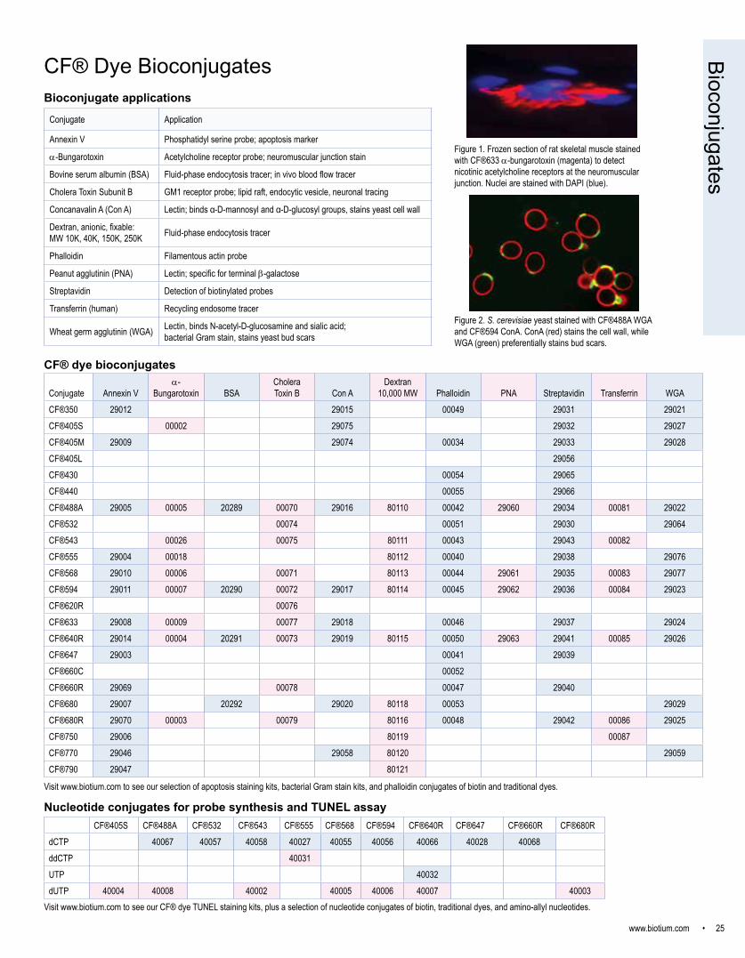
Conjugate Application
Annexin V Phosphatidyl serine probe; apoptosis marker
a-Bungarotoxin Acetylcholine receptor probe; neuromuscular junction stain
Bovine serum albumin (BSA) Fluid-phase endocytosis tracer; in vivo blood flow tracer
Cholera Toxin Subunit B GM1 receptor probe; lipid raft, endocytic vesicle, neuronal tracing
Concanavalin A (Con A) Lectin; binds α-D-mannosyl and α-D-glucosyl groups, stains yeast cell wall
Dextran, anionic, fixable: MW 10K, 40K, 150K, 250K Fluid-phase endocytosis tracer
Phalloidin Filamentous actin probe
Peanut agglutinin (PNA) Lectin; specific for terminal b-galactose
Streptavidin Detection of biotinylated probes
Transferrin (human) Recycling endosome tracer
Wheat germ agglutinin (WGA) Lectin, binds N-acetyl-D-glucosamine and sialic acid; bacterial Gram stain, stains yeast bud scars
Figure 1. Frozen section of rat skeletal muscle stained with CF®633 a-bungarotoxin (magenta) to detect nicotinic acetylcholine receptors at the neuromuscular junction. Nuclei are stained with DAPI (blue).
Conjugate Annexin Va-
Bungarotoxin BSACholera Toxin B Con A
Dextran 10,000 MW Phalloidin PNA Streptavidin Transferrin WGA
CF®350 29012 29015 00049 29031 29021CF®405S 00002 29075 29032 29027CF®405M 29009 29074 00034 29033 29028CF®405L 29056CF®430 00054 29065CF®440 00055 29066CF®488A 29005 00005 20289 00070 29016 80110 00042 29060 29034 00081 29022CF®532 00074 00051 29030 29064CF®543 00026 00075 80111 00043 29043 00082CF®555 29004 00018 80112 00040 29038 29076CF®568 29010 00006 00071 80113 00044 29061 29035 00083 29077CF®594 29011 00007 20290 00072 29017 80114 00045 29062 29036 00084 29023CF®620R 00076CF®633 29008 00009 00077 29018 00046 29037 29024CF®640R 29014 00004 20291 00073 29019 80115 00050 29063 29041 00085 29026CF®647 29003 00041 29039CF®660C 00052CF®660R 29069 00078 00047 29040CF®680 29007 20292 29020 80118 00053 29029CF®680R 29070 00003 00079 80116 00048 29042 00086 29025CF®750 29006 80119 00087CF®770 29046 29058 80120 29059CF®790 29047 80121
CF®405S CF®488A CF®532 CF®543 CF®555 CF®568 CF®594 CF®640R CF®647 CF®660R CF®680RdCTP 40067 40057 40058 40027 40055 40056 40066 40028 40068ddCTP 40031UTP 40032dUTP 40004 40008 40002 40005 40006 40007 40003
Visit www.biotium.com to see our CF® dye TUNEL staining kits, plus a selection of nucleotide conjugates of biotin, traditional dyes, and amino-allyl nucleotides.
Visit www.biotium.com to see our selection of apoptosis staining kits, bacterial Gram stain kits, and phalloidin conjugates of biotin and traditional dyes.
Nucleotide conjugates for probe synthesis and TUNEL assay
Bioconjugate applications
CF® dye bioconjugates
Bioconjugates
Figure 2. S. cerevisiae yeast stained with CF®488A WGA and CF®594 ConA. ConA (red) stains the cell wall, while WGA (green) preferentially stains bud scars.

26 • www.biotium.com
Primary antibody conjugates
AA, ForssmanACTHAdenosine Monophosphate Deaminase 3AFPAlkaline PhosphataseAMACR / p504SAndrogen ReceptorArginase 1
BBaxBCL-10bcl-2bcl-6bcl-xBeta CateninBeta-2 MicroglobulinBiotinBlood Group ABlood Group Antigen ABlood Group Antigen BBovine Serum AlbuminBrdUBromodeoxyuridine
CCA19-9Caldesmon, HMWCalgranulin BCalponin-1CalprotectinCarbonic Anhydrase IXCarcinoembryonic AntigenCD10CD100CD104CD106 / VCAM1CD117CD11aCD11bCD11cCD13CD14CD146 / Mucin 18CD147CD15 / FUT4 / Lewis xCD16CD16 / Fc-gamma Receptor IIICD171 / L1CAMCD176 / T-F AgCD18CD19CD195 / CCR-5CD1aCD1bCD2CD20CD21CD22CD25CD26
CD27CD28CD282 / TLR2CD284 / TLR4CD30CD31 / PECAM-1CD32CD33CD34CD35 / CR1CD36CD37CD38CD3eCD4CD41aCD43CD44 StandardCD45 / LCACD45RACD45RBCD45ROCD46CD47CD48CD5CD50CD53CD54CD54 / ICAM-1CD55CD56 / NCAMCD57 / B3GAT1CD59CD6CD61CD63CD66CD66, panCD68CD7CD70CD71CD74CD79aCD8CD84CD86CD8ACD8BCD90CD90 / Thy1CD95CD98CD99Cdc20CDw17CDw60CDw75CELA3BChromogranin ACMV-p65c-Mybc-Myc
Complement C4dCreatine PhosphokinaseCyclin A2Cyclin B1Cyclin D1Cytochrome CCytokeratin 10Cytokeratin 10/13Cytokeratin 14Cytokeratin 17Cytokeratin 18Cytokeratin 19Cytokeratin 5/8Cytokeratin 6Cytokeratin 7Cytokeratin 8Cytokeratin 8/18Cytokeratin, AcidicCytokeratin, BasicCytokeratin, HMWCytokeratin, LMWCytokeratin, multiCytokeratin, pan
DDOG-1Double Stranded DNA
EE-Cadherin / CD324EGFREMI1Eosinophil PeroxidaseEp-CAM / CD326Erythrocyte SpecificEstrogen ReceptorEstrogen Receptor beta 1
FFascin-1FGF23FibronectinFOXP3FSH beta
GGFAPGITR / Tnfrsf18GLG1Glucose Regulated Protein 94Glycophorin A / CD235aGlypican-3GM-CSFGnRH-ReceptorGolgi Complexgp100 / Melanosome Granulocyte MarkerGCSFGranzyme B
HHCG-alpha
HCG-betaHCG-intactHelicobacter pyloriHeparan Sulphate ProteoglycanHepatocyte Specific AntigenHepPar-1HER-2 / CD340HIF1?Histiocytoma MarkerHistone H1HLA-Aw32 & HLA-A25HLA-BHLA-DRBHSP27HSP60Human Nuclear AntigenHuman Nucleolar AntigenHuman Papillomavirus 16
IIDH1IgA ImmunoglobulinIgA Secretory ComponentIGF-1IgGIgG ImmunoglobulinIgM ImmunoglobulinIL-6InsulinInterferon alpha 1Interferon alpha-2Interferon gammaInvolucrinIPO-38Isotype control, mouse IgG1, kIsotype control, mouse IgG2a, kIsotype control, mouse IgG2b, k
KKappa Light ChainKsp-Cadherin / CDH16Ku-Holo
LLambda Light ChainLamininLEC ChemokineLewis ALewis BLiver CanuliculiLung Specific AntigenLuteinizing Hormone beta
MMacrophage Specific AntigenMAGE A1Major Vault ProteinMALT-1MAP3K1MART-1 / Melan-AMCAM / MUC18 / Mucin 18 / CD146Melanoma MarkerMHC I
MHC IIMilk Fat GlobulinMiTFMitochondriaMitochondrial MarkerMoesinMRP-14MUC18 / Mucin 18 / CD146MUC2MUC5ACMucin 1 / EMA / Episialin / CD227Mucin 3Mucin 5ACMucin 6Muscle Specific ActinMyeloid-Associated Differentiation MarkerMyoD1MyogeninMyosin, Smooth Muscle Heavy Chain
NNapsin-ANeurofilamentNeurofilament, phosphoNGFRNKX2.2Nuclear AntigenNuclear MembraneNuclear Membrane MarkerNucleolar / NucleoliNucleolinNuMA
OODC-1
Pp21 / WAF1p24-HIVp27 / KIP1p34 / cdk1p40p53p55;50 EBV-Early Antigenp57 / KIP2p57Kip2PAX6PAX7PCNAPD1 / PDCD1 / CD279pgp9.5PhosphotyrosinePLAPPlasma Cell MarkerPLGFPmel17 / gp100 / SILVPodocalyxinProgesteroneProgesterone ReceptorProlactin Receptor
Prostate Specific AntigenProximal Nephrogenic Antigen pS2PSAPTENPTHPTH / Parathyroid Hormone
RRabiesRetinol Binding Protein-1
SS100S100A9SHBGSmall Cell Lung CancerSmooth Muscle ActinSOX10SUMO-1SUMO-2SUMO-2/3
TTAG-72 / CA72.4TenascinTestosteroneTGFalphaTGF-betaThomsen-Friedenreich Ag. Thymidylate SynthaseThyroglobulinTIMP3TNF alphaTopoisomerase I, MTTOX3TRAcPTransgelin / SM22-alphaTransglutaminase IITRIM29TRP1TTF-1 / NKX2.1Tyrosinase
UUACA / NuclingUGT1A9UPK3A
VVEGF-AVEGFR1 / Flt-1VEGFR2 / Flk-1 Vimentin
Wvon Willebrand FactorWT1
ZZAP70
Prim
ary
Antib
odie
s
Our collection of monoclonal primary antibodies is constantly growing, visit www.biotium.com to see the most up-to-date offerings. Available in these formats:• Purified: 200 ug/mL in PBS with 0.05% BSA & 0.05% azide, 100 uL (20 ug) or 500 uL (100 ug) unit size• Purified, BSA-free: 1 mg/ml in PBS with 0.05% azide, ready-to-use for Mix-n-Stain™ labeling (see p. 23) or other conjugation, 50 uL (50 ug) unit size• Conjugates: Your choice of 12 CF® dye colors for microscopy, flow cytometry, or near-infrared western blot, plus R-PE, APC, and PerCP; 100 ug/mL in PBS with 0.05% BSA & 0.05% azide, 100 uL (10 ug) or 500 uL (50 ug) unit size
www.biotium.com • 27
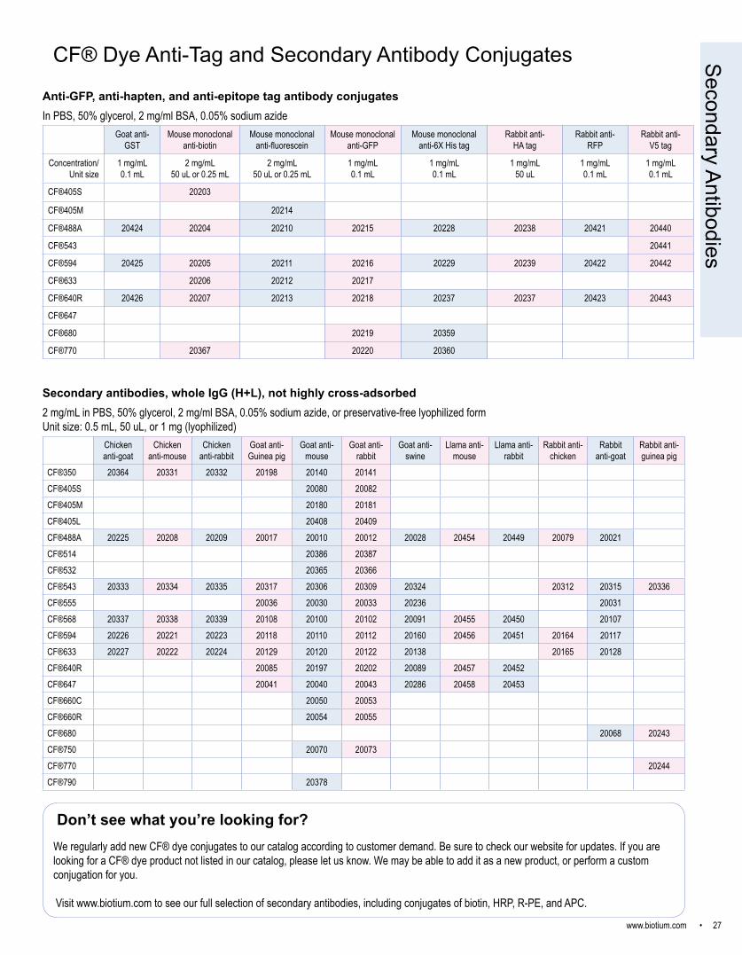
Secondary antibodies, whole IgG (H+L), not highly cross-adsorbed2 mg/mL in PBS, 50% glycerol, 2 mg/ml BSA, 0.05% sodium azide, or preservative-free lyophilized formUnit size: 0.5 mL, 50 uL, or 1 mg (lyophilized)
CF® Dye Anti-Tag and Secondary Antibody Conjugates
Anti-GFP, anti-hapten, and anti-epitope tag antibody conjugatesIn PBS, 50% glycerol, 2 mg/ml BSA, 0.05% sodium azide
Chicken anti-goat
Chicken anti-mouse
Chicken anti-rabbit
Goat anti-Guinea pig
Goat anti-mouse
Goat anti-rabbit
Goat anti-swine
Llama anti-mouse
Llama anti-rabbit
Rabbit anti-chicken
Rabbit anti-goat
Rabbit anti-guinea pig
CF®350 20364 20331 20332 20198 20140 20141CF®405S 20080 20082CF®405M 20180 20181CF®405L 20408 20409CF®488A 20225 20208 20209 20017 20010 20012 20028 20454 20449 20079 20021CF®514 20386 20387CF®532 20365 20366CF®543 20333 20334 20335 20317 20306 20309 20324 20312 20315 20336CF®555 20036 20030 20033 20236 20031CF®568 20337 20338 20339 20108 20100 20102 20091 20455 20450 20107CF®594 20226 20221 20223 20118 20110 20112 20160 20456 20451 20164 20117CF®633 20227 20222 20224 20129 20120 20122 20138 20165 20128CF®640R 20085 20197 20202 20089 20457 20452CF®647 20041 20040 20043 20286 20458 20453CF®660C 20050 20053CF®660R 20054 20055CF®680 20068 20243CF®750 20070 20073CF®770 20244CF®790 20378
Goat anti-GST
Mouse monoclonal anti-biotin
Mouse monoclonal anti-fluorescein
Mouse monoclonal anti-GFP
Mouse monoclonal anti-6X His tag
Rabbit anti-HA tag
Rabbit anti-RFP
Rabbit anti-V5 tag
Concentration/ Unit size
1 mg/mL0.1 mL
2 mg/mL50 uL or 0.25 mL
2 mg/mL50 uL or 0.25 mL
1 mg/mL0.1 mL
1 mg/mL0.1 mL
1 mg/mL50 uL
1 mg/mL0.1 mL
1 mg/mL0.1 mL
CF®405S 20203
CF®405M 20214
CF®488A 20424 20204 20210 20215 20228 20238 20421 20440
CF®543 20441
CF®594 20425 20205 20211 20216 20229 20239 20422 20442
CF®633 20206 20212 20217
CF®640R 20426 20207 20213 20218 20237 20237 20423 20443
CF®647
CF®680 20219 20359
CF®770 20367 20220 20360
Don’t see what you’re looking for?We regularly add new CF® dye conjugates to our catalog according to customer demand. Be sure to check our website for updates. If you are looking for a CF® dye product not listed in our catalog, please let us know. We may be able to add it as a new product, or perform a custom conjugation for you.
Visit www.biotium.com to see our full selection of secondary antibodies, including conjugates of biotin, HRP, R-PE, and APC.
Secondary Antibodies
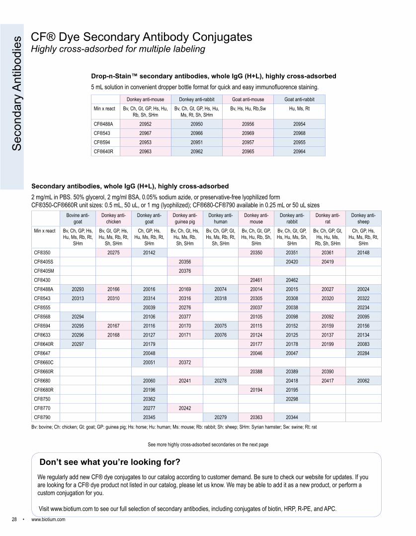
28 • www.biotium.com
Secondary antibodies, whole IgG (H+L), highly cross-adsorbed2 mg/mL in PBS. 50% glycerol, 2 mg/ml BSA, 0.05% sodium azide, or preservative-free lyophilized formCF®350-CF®660R unit sizes: 0.5 mL, 50 uL, or 1 mg (lyophilized); CF®680-CF®790 available in 0.25 mL or 50 uL sizes
Bovine anti-goat
Donkey anti-chicken
Donkey anti-goat
Donkey anti-guinea pig
Donkey anti-human
Donkey anti-mouse
Donkey anti-rabbit
Donkey anti-rat
Donkey anti-sheep
Min x react Bv, Ch, GP, Hs, Hu, Ms, Rb, Rt,
SHm
Bv, Gt, GP, Hs, Hu, Ms, Rb, Rt,
Sh, SHm
Ch, GP, Hs, Hu, Ms, Rb, Rt,
SHm
Bv, Ch, Gt, Hs, Hu, Ms, Rb,
Sh, SHm
Bv, Ch, GP, Gt, Hs, Ms, Rb, Rt,
Sh, SHm
Bv, Ch, Gt, GP, Hs, Hu, Rb, Sh,
SHm
Bv, Ch, Gt, GP, Hs, Hu, Ms, Sh,
SHm
Bv, Ch, GP, Gt, Hs, Hu, Ms,
Rb, Sh, SHm
Ch, GP, Hs, Hu, Ms, Rb, Rt,
SHmCF®350 20275 20142 20350 20351 20361 20148CF®405S 20356 20420 20419CF®405M 20376CF®430 20461 20462CF®488A 20293 20166 20016 20169 20074 20014 20015 20027 20024CF®543 20313 20310 20314 20316 20318 20305 20308 20320 20322CF®555 20039 20276 20037 20038 20234CF®568 20294 20106 20377 20105 20098 20092 20095CF®594 20295 20167 20116 20170 20075 20115 20152 20159 20156CF®633 20296 20168 20127 20171 20076 20124 20125 20137 20134CF®640R 20297 20179 20177 20178 20199 20083CF®647 20048 20046 20047 20284CF®660C 20051 20372CF®660R 20388 20389 20390CF®680 20060 20241 20278 20418 20417 20062CF®680R 20196 20194 20195CF®750 20362 20298CF®770 20277 20242CF®790 20345 20279 20363 20344
CF® Dye Secondary Antibody ConjugatesHighly cross-adsorbed for multiple labeling
Bv: bovine; Ch: chicken; Gt: goat; GP: guinea pig; Hs: horse; Hu: human; Ms: mouse; Rb: rabbit; Sh: sheep; SHm: Syrian hamster; Sw: swine; Rt: rat
See more highly cross-adsorbed secondaries on the next page
Don’t see what you’re looking for?We regularly add new CF® dye conjugates to our catalog according to customer demand. Be sure to check our website for updates. If you are looking for a CF® dye product not listed in our catalog, please let us know. We may be able to add it as a new product, or perform a custom conjugation for you.
Visit www.biotium.com to see our full selection of secondary antibodies, including conjugates of biotin, HRP, R-PE, and APC.
Seco
ndar
y An
tibod
ies
Drop-n-Stain™ secondary antibodies, whole IgG (H+L), highly cross-adsorbed5 mL solution in convenient dropper bottle format for quick and easy immunofluorence staining.
Donkey anti-mouse Donkey anti-rabbit Goat anti-mouse Goat anti-rabbitMin x react Bv, Ch, Gt, GP, Hs, Hu,
Rb, Sh, SHmBv, Ch, Gt, GP, Hs, Hu,
Ms, Rt, Sh, SHmBv, Hs, Hu, Rb,Sw Hu, Ms, Rt
CF®488A 20952 20950 20956 20954CF®543 20967 20966 20969 20968CF®594 20953 20951 20957 20955CF®640R 20963 20962 20965 20964

www.biotium.com • 29
Goat anti-chicken
Goat anti-human
Goat anti-mouse
Goat anti-mouse (min x rat)
Goat anti-rabbit
Goat anti-rat
Rabbit anti-human
Rabbit anti-mouse
Rabbit anti-rat
Rabbit anti-sheep
Bv, Gt, GP, Hs, Hu, Ms, Rb, Rt, Sh, SHm
Bv, Hs, Ms Bv, Hs, Hu, Rb, Sw
Bv, Ch, Gt, GP Hs Hu Rb Rt,
Sh, SHm
Hu, Ms, Rt Bv, Hs, Hu, Rb
Ms Hu Hu Hu
CF®350 20143 20144 20147 20149CF®405SCF®405M 20375 20182 20340 20373 20374CF®430 20459 20460CF®488A 20020 20022 20018 20302 20019 20023 20071 20026 20025 20172CF®543 20311 20319 20299 20328 20300 20321 20307 20323CF®555 20034 20320 20231 20232 20233 20235CF®568 20104 20097 20101 20301 20103 20096 20093 20094CF®594 20114 20154 20111 20303 20113 20155 20072 20158 20157 20173CF®633 20126 20132 20121 20341 20123 20133 20066 20136 20135 20174CF®640R 20084 20081 20175 20304 20176 20088 20200 20201CF®647 20044 20280 20281 20282 20283 20285CF®660C 20371 20052 20356 20369 20370CF®660RCF®680 20287 20065 20067 20069 20061CF®680R 20192 20193CF®750 20463CF®770 20288 20077 20078 20383CF®790 20342 20343
Bv: bovine; Ch: chicken; Gt: goat; GP: guinea pig; Hs: horse; Hu: human; Ms: mouse; Rb: rabbit; Sh: sheep; SHm: Syrian hamster; Sw: swine; Rt: rat
Secondary antibodies, F(ab’)2 fragments2 mg/mL, unit size: 0.25 mL or 50 uL
Goat anti-mouse, F(ab’)2
Goat anti-rabbit, F(ab’)2
CF®350 20145 20146CF®488A 20011 20013CF®543 20329 20330CF®555 20032 20035CF®568 20109 20099CF®594 20119 20153CF®633 20130 20131CF®640R 20086 20087CF®647 20042 20045CF®680 20063 20064
Goat anti-mouse isotype-specific antibodiesHighly cross-adsorbed for multiple labeling (min X Bv, Hu, Rb), 2 mg/mL, unit size: 0.25 mL or 50 uL
Goat anti-mouse IgG1
Goat anti-mouse IgG2a
Goat anti-mouse IgG2b
CF®350 20245 20255 20265CF®405S 20380 20381 20382CF®488A 20246 20256 20266CF®543 20325 20356 20327CF®555 20247 20257 20267CF®568 20248 20258 20268CF®594 20249 20259 20269CF®633 20250 20260 20270CF®640R 20251 20261 20271CF®647 20252 20262 20272CF®680 20253 20263 20273CF®770 20254 20264 20274
Goat anti-human isotype-specfic antibodies2 mg/mL, unit size: 0.25 mL or 50 uL
Goat anti-human IgA
(alpha chain)
Goat anti-human IgM (mu chain)
CF®488A 20428 20347CF®594 20429 20348CF®640R 20349CF®633 20427CF®647 20346CF®680 20384CF®770 20385
Secondary antibodies, whole IgG (H+L) , highly cross-adsorbed (continued from p. 28)2 mg/mL in PBS. 50% glycerol, 2 mg/ml BSA, 0.05% sodium azide, or preservative-free lyophilized formCF®350-CF®660R unit sizes: 0.5 mL, 50 uL, or 1 mg (lyophilized); CF®680-CF®790 available in 0.25 mL or 50 uL sizes
Secondary AntibodiesCF® Dye Secondary Antibody ConjugatesHighly cross-adsorbed, F(ab’)2 fragments, and isotype-specific secondary antibodies

30 • www.biotium.com
Related Products and AccessoriesBuffers, mounting media, counterstains and more
FITC Photostability, Wet-Set Mounting Media
FITC Photostability, Hardset Mounting Media
Cy®5 Photostability, Wet-Set Mounting Media
0
20
40
60
80
100
120
0 20 40 60 80 100 120
Nor
mal
ized
Flu
ores
cenc
e
Time (s)
EverBriteVectashieldSlowFade GoldPBS 0
20
40
60
80
100
120
0 20 40 60 80 100 120
Nor
mal
ized
Flu
ores
cenc
e
Time (s)
EverBriteVectashieldSlowFade GoldPBS
0
20
40
60
80
100
120
0 20 40 60 80 100 120
Nor
mal
ized
Flu
ores
cenc
e
Time (sec)
EverBrite HSVectashield HSProlong GoldPBS
Cy®5 Photostability, Hardset Mounting Media
0
20
40
60
80
100
120
0 20 40 60 80 100 120
Nor
mal
ized
Flu
ores
cenc
e
Time (sec)
EverBrite HSVectashield HSProlong GoldPBS
Figure 2. Photostability of HeLa cell immunofluorescence staining in various mounting media. A major advantage of EverBrite™ mounting medium is that, unlike Vectashield®, it does not decrease the fluorescence of cyanine-based fluorophores. Fluorescence values for Cy®5 in Vectashield® media are normalized to PBS time 0 to illustrate the drop in fluorescence of cyanine dyes caused by Vectashield®.
Figure 1. HeLa cells stained with rabbit anti-COX IV and CF®488A goat anti-rabbit (mitochondria, green), and mouse anti-Golgin 97 and CF®555 goat anti-mouse (Golgi, cyan) Actin filaments are stained with CF®405 phalloidin (blue) and nuclei are stained with RedDot™2 (red).
Figure 3. CoverGrip™ Coverslip Sealant brush bottle and refill bottle.
Acce
ssor
ies
RedDot™1 and RedDot™2 Far-Red Nuclear StainsRedDot™1• Replaces Draq®5 for live cell nuclear staining• Cell normalization for In Cell Western®• Cell cycle analysis by flow cytometry
RedDot™2• Replaces Draq®7 for dead cell staining and fixed
cell staining• More specific than Draq®7 for fixed cell nuclear
counterstaining
EverBrite™ Mounting Media• Superior antifade protection• Compatible with CF® dyes and other dyes• Compatible with cyanine-based dyes (Cy® dyes,
Alexa Fluor® 647 and DyLight® 649), unlike VectaShield®
• Available in wet-set or hardset formulations• With or without DAPI
CoverGrip™ Coverslip Sealant• Designed specifically for sealing coverslip edges• Won’t mix with aqueous mounting medium like
nail polish can• Made with natural Limonene solvent• 15 mL brush bottle, or 100 mL refill SuperHT Pap Pens
• Create hydrophobic barriers on glass slides to separate specimens and conserve antibodies
• Insoluble in aqueous buffers, detergents, alcohol, or acetone, removable with xylene
• Stable at temperatures up to 120oC• SuperHT Mini Pap Pen: 2.5 mm tip, ~400
applications• SuperHT Pap Pen: 4 mM tip, ~800 applications
Figure 4. SuperHT Pap Pens.
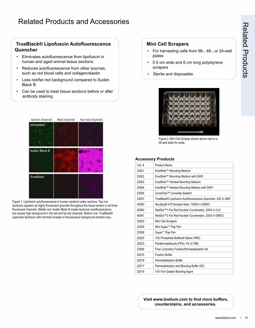
www.biotium.com • 31
Related Products and Accessories
Cat. # Product Name23001 EverBrite™ Mounting Medium23002 EverBrite™ Mounting Medium with DAPI23003 EverBrite™ Hardset Mounting Medium23004 EverBrite™ Hardset Mounting Medium with DAPI23005 CoverGrip™ Coverslip Sealant23007 TrueBlack® Lipofuscin Autofluorescence Quencher, 20X in DMF40083 NucSpot® 470 Nuclear Stain, 1000X in DMSO40060 RedDot™1 Far Red Nuclear Counterstain, 200X in H2O40061 RedDot™2 Far Red Nuclear Counterstain, 200X in DMSO22003 Mini Cell Scrapers22005 Mini Super HT Pap Pen22006 Super HT Pap Pen22020 10X Phosphate-Buffered Saline (PBS)22023 Paraformaldehyde (PFA), 4% in PBS23006 Flow Cytometry Fixation/Permeabilization Kit22015 Fixation Buffer22016 Permeabilization Buffer22017 Permeabilization and Blocking Buffer (5X)22010 10X Fish Gelatin Blocking Agent
Accessory Products
Figure 2. Mini Cell Scraper shown above next to a 48-well plate for scale.
Related Products
Mini Cell Scrapers• For harvesting cells from 96-, 48-, or 24-well
plates• 0.5 cm wide and 6 cm long polystyrene
scrapers• Sterile and disposable
TrueBlack® Lipofuscin Autofluorescence Quencher• Eliminates autofluorescence from lipofuscin in
human and aged animal tissue sections• Reduces autofluorescence from other sources,
such as red blood cells and collagen/elastin• Less red/far-red background compared to Sudan
Black B• Can be used to treat tissue sections before or after
antibody staining
Figure 1. Lipofuscin autofluorescence in human cerebral cortex sections. Top row: lipofuscin appears as highly fluorescent granules throughout the tissue section in all three fluorescent channels. Middle row: Sudan Black B masks lipofuscin autofluorescence, but causes high background in the red and far-red channels. Bottom row: TrueBlack® quenches lipofuscin with minimal increase in fluorescence background (bottom row).
Visit www.biotium.com to find more buffers, counterstains, and accessories.
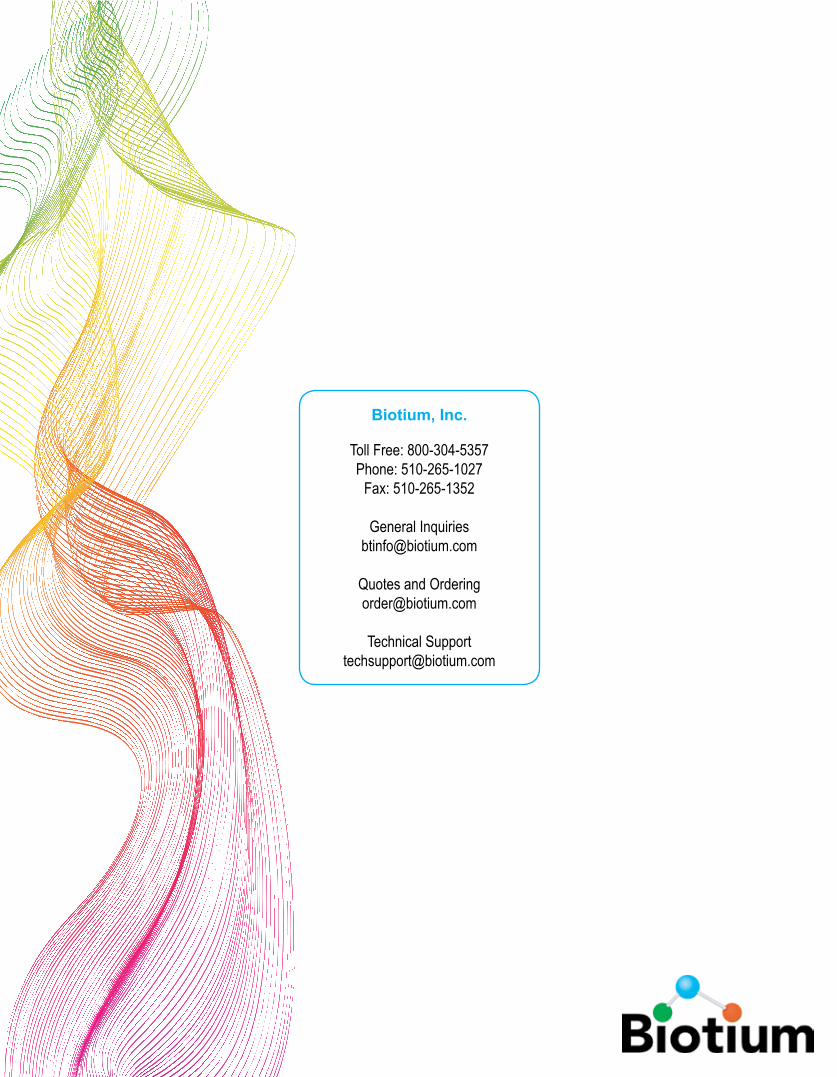
Biotium, Inc.
Toll Free: 800-304-5357Phone: 510-265-1027
Fax: 510-265-1352
General [email protected]
Quotes and [email protected]
Technical [email protected]


![Radiosynthesis and Bioconjugation of [18 F]FPy5yne - Triumf](https://static.fdocuments.us/doc/165x107/6203aba2da24ad121e4c1a6b/radiosynthesis-and-bioconjugation-of-18-ffpy5yne-triumf.jpg)