
CEREBROVASCULAR DISEASES - · PDF fileCerebrovascular diseases •Brain disorders caused by...
40
CEREBROVASCULAR DISEASES By: Shifaa’ AlQa’qa’
Transcript of CEREBROVASCULAR DISEASES - · PDF fileCerebrovascular diseases •Brain disorders caused by...

CEREBROVASCULAR DISEASES
By: Shifaa’ AlQa’qa’
mohammad
Sticky Note
Sarah Awaisheh

Cerebrovascular diseases
• Brain disorders caused by pathologic processes involving blood vessels
• 3 pathogenic mechanisms
(1) thrombotic occlusion,
(2) embolic occlusion,
(3) vascular rupture/Hemorrhage
me
= means pathologic changes in the BVs supplying the brain
me
Vessels in the brain can be affected by three mechanisms:
me
me
me
me
First two result in blocking the BV while last one result with rupture and bleeding
me
These three will lead to infarction and stroke..

• Stroke:
is the clinical term for acute-onset neurologic deficits resulting from hemorrhagic or obstructive vascular lesions.
transient ischemic attack (TIA) ????
me
It is a medical condition in which poor blood flow to the brain results in brain death. Hemorrhage, ischemia, infarction all these leads to stroke.
me
Symptoms :
me
Symptoms depend on which part has been occluded, but mainly: Paralysis on one part (hemiparalysis) Difficulty in speak and swallowing Decrease level of consciousness Loss of vision on one side (hemilateral)
me
If the symptoms last less than 24h it is TIAIt is caused by a transient ischemia , so the symptoms are reversible
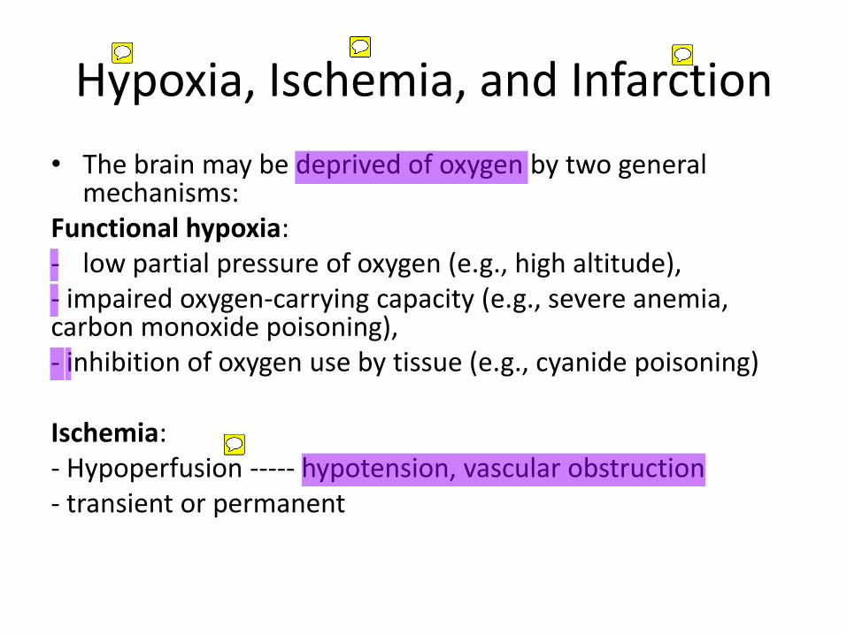
Hypoxia, Ischemia, and Infarction
• The brain may be deprived of oxygen by two general mechanisms:
Functional hypoxia: - low partial pressure of oxygen (e.g., high altitude), - impaired oxygen-carrying capacity (e.g., severe anemia, carbon monoxide poisoning), - inhibition of oxygen use by tissue (e.g., cyanide poisoning)
Ischemia: - Hypoperfusion ----- hypotension, vascular obstruction - transient or permanent
me
Good perfusion with low oxygen concentration
me
Decrease blood supply
me
End result of sustained ischemia. (Necrosis)
me
me
—>
me
Things that decrease o2 without decreasing blood supply:
me
me
me
me
Ischemia is divided according the cause into: Global ischemia ( due to systemic hypotension) Focal ischemia ( due to vascular obstruction) It can be transient like in TIA or permanent and leads to stroke
me

Global Cerebral Ischemia
• occur in the setting of severe systemic hypotension, usually when systolic pressures fall below 50 mm Hg, as in cardiac arrest, shock, and severe hypotension.
• Neurons are more susceptible to hypoxic injury than are glial cells
• The most susceptible neurons (vulnerable areas) are: - The pyramidal cells of the hippocampus and neocortex - Purkinje cells of the cerebellum
- In severe global cerebral ischemia, widespread neuronal
death occurs irrespective of regional vulnerability
me
Affecting all the brain die to systemic hypotension
me
me
me
me
me
me
Areas that are affected even with mild ischemia:

• MORPHOLOGY:
the brain is swollen, wide gyri, narrowed sulci.
The cut surface shows poor demarcation between gray and white matter
me
me
me
me
me
Due to edema makes the boundaries unclear

• Irreversible ischemic injury---- infarction Early changes (12 to 24 hours): - acute neuronal cell change - the reaction to tissue damage begins with infiltration by neutrophils Subacute changes (24 hours to 2 weeks) - necrosis of tissue - influx of macrophages - vascular proliferation, - reactive gliosis Repair (after 2 weeks) - removal of all necrotic tissue - Loss of organized CNS structure - gliosis
me
First 12h of global ischemia the brain is normal
me
me
me
me
me
me
me
(Red neurons, pyknosis..)
me
me
me
Normally there is no neutrophils in the brain but in case of infarction they’re seen And the give a clue that there’s infarction and it is has been since 12-24h
me
me
This is seen in global ischemia due to pseudolaminar necrosis , which means that the areas that is necrotic is uneven with the gliaiotic areas resulting in layering of gliaosis and necrosis

me
me
Blood vessels surrounded by neutrophils this means there is an ischemia(12 to 24 hours)
me
me
Red neurons

• The distribution of neuronal loss and gliosis in the neocortex typically is uneven with preservation of some layers and devastation of others—a pattern termed pseudolaminar necrosis.

• Border zone (“watershed”) infarcts:
- wedge shaped
- regions of the brain and spinal cord that lie at the most distal portions of arterial territories.
- In the cerebral hemispheres, the border zone between the anterior and the middle cerebral artery distributions is at greatest risk.
me
me
This is an infarction at the end of arterial supply btw the ant. And middle cerebral artery and they are also vulnerable for infarction and they become with wedge-shaped
me
me
me

• The clinical outcome varies with the severity and duration of the insult:
Mild insult---- only a transient postischemic
confusional state, with eventual complete recovery
Severe insult ---- vegetative state, brain death--- mechanical ventilation---- brain autolysis
me
me
The end result of ischemia may be:
me
me
me
If the ischemia was sever - - - leads to brain damage in the form of vegetative state or brain death
me
Vegetative stateالمريض بكون زي النبات فقط بصحى و بنام و يأكل و يتنفسBut he is unaware and bedridden Here the main injury is in the cerebrum while the cerebellum and brain stem are normal thus the respiratory and cvs functions are tho.-No need for mechanical ventilation
me
Here the patient will lose motor reflexes and respiratory functions and the EEG of brain is flat showing no activity of the brain-Needs mechanical ventilation which lead to brain autolysis

Focal Cerebral Ischemia
• Cerebral arterial occlusion ----- focal ischemia ------ infarction in the distribution of the compromised vessel.
• modified by collateral blood flow -------- the circle of Willis or cortical-leptomeningeal anastomoses
me
The main cause of Focal:
me
me
Accessory blood vessel between two arteries, once the artery is occluded it opens to compensate and they’re more effective with age thus old patient with MI is less severe than young one.
me
me
These are the most common places for collateral BVs in the brainAnd collaterals are not found in deep parts of the brain thus the deep part are more susceptible for ischemia


• By contrast, there is little if any collateral flow to structures such as the thalamus, basal ganglia, and deep white matter, which are supplied by deep penetrating vessels.
me
Deep parts have no or little collateral:

• Causes:
- Emboli ---------- more common
- Thrombosis
me
Of focal ischemia
me
Occlusion by:
me
It is dislodge of an embolus or clot that moves toward different parts of the body -Origin of it outside the brain
me
Occurs with atherosclerosis patient when a plaque starts to form and develops into clot

• Emboli:
- Cardiac mural thrombi (myocardial dysfunction, valvular disease, and atrial fibrillation)
- Arterial thromboemboli (atheromatous plaques within the carotid arteries or aortic arch)
- Venous emboli/paradoxical embolism (thromboemboli from deep leg veins and fat emboli)
me
Most common causes of brain emboli:
me
It is a thrombosis in the heart dislodges and goes to the brain via the arterial blood supply
me
A thrombi that has been lodged and caused thromboemboli
me
In normal people venous thrombi goes to the lungs but we can have a venous thrombi going to the brain if a person has a shunt btween the Rt. and Lt. sides of the heart which is abnormal The name of this emboli is : parodoxial embolism

• The territory of the middle cerebral artery, a direct extension of the internal carotid artery, is most frequently affected by embolic infarction.
• Emboli tend to lodge where vessels branch or in areas of stenosis, usually caused by atherosclerosis.
me
Most common affected area in the emboli:
me
me
The emboli likes to settle down in the abnormal places like stenosis or atherosclerosis

• Thrombosis:
- Thrombotic occlusions usually are superimposed on atherosclerotic plaques
- common sites are:
The carotid bifurcation,
The origin of the middle cerebral artery,
At either end of the basilar artery.
me
The 2nd cause of focal ischemia ( causes occlusion )
me
me
Of thrombosis:

• Infarcts can be divided into two broad groups: Nonhemorrhagic infarcts--- Hemorrhagic infarcts--- result from reperfusion of ischemic tissue, either through collaterals or after dissolution of emboli ----- multiple, petechial hemorrhages Thrombolytic therapies????
me
Now the end result of the occlusion is infarction, it could be:
me
me
me
Occurs due to reperfusion injury either due to the patient has been given a treatment or the emboli has been automatically dislodged
me
me
In the hemorrhage infarction the brain comes in the form of : petechial hemorrhage ( very small in size 1mm and multiple )
me
It is important to differentiate btw the hemorrhage and non bcz of the treatment If we give a hemorrhage patient a thrombolytic treatment this will deteriorate the state while it is normal in non hemorrhage


• MORPHOLOGY: Macroscopic appearance of a nonhemorrhagic infarct: first 6 hours---no change 48 hours--- tissue is pale, soft, and swollen days 2 to 10--- the brain turns gelatinous and friable, and the boundary between normal and abnormal tissue becomes more distinct as edema resolves in the adjacent viable tissue. day 10 to week 3--- the tissue liquefies, eventually leaving a fluid-filled cavity lined by dark gray tissue, which gradually expands as dead tissue is resorbed
me
me
me
me
Edema
me
me
me
The boundaries of necrosis is unclear unless the edema resolves
me
me
me
The tissue of the brain become liquid and from here we came up with liquefactive necrosis
me
me
me
The liquid will make a cavity filled with a fluid ( cyst ) with a dark gray color
me
Grossly


• Microscopically: After the first 12 hours: - ischemic neuronal change (red neurons) - cytotoxic and vasogenic edema . Endothelial and glial cells, mainly
astrocytes, swell - myelinated fibers begin to disintegrate. Up to 48 hours: neutrophilic emigration 2 to 3 weeks: - Macrophages - astrocytes at the edges of the lesion progressively enlarge, divide, and
develop a prominent network of cytoplasmic extensions After several months: - the striking astrocytic nuclear and cytoplasmic enlargement regresses - dense feltwork of glial fibers admixed with new capillaries lining the cavity
wall
me
On the microscope 🔬:
me
me
The astrocytes will disappear leaving fibers

• Hemorrhagic infarction:
+ blood extravasation
me
On microscope 🔬: the hemorrhage infarction appears like non hemorrhage but with blood extravasation ( we see RBCs on the microscope)

Intracranial Hemorrhage
• intracerebral hemorrhage (intraventricular, intraparenchymal)
• subarachnoid hemorrhage
• epidural hemorrhage
• subdural hemorrhage
me
Bleeding within the skull or brain
me
Classified according the anatomical site into:
me
** It is a serious condition bcz as you know blood when builds up it can increase the ICP


Primary Brain Parenchymal Hemorrhage
• Spontaneous ----- nontraumatic
• peak incidence at about 60 years of age
• rupture of a small intraparenchymal vessels
me
me
—>
me
Not secondary to trauma , it is spontaneous in the brain
me
me
Cause:
me
me
me
Due to hypertension

• Hypertension is the leading underlying cause
• Hypertensive intraparenchymal hemorrhages typically occur in the
basal ganglia, thalamus, pons, cerebellum
me
me
اكثر مناطق بصير فيهم bleeding بسبب الـ hypertension
me
me
me

• it can affect small regions and be clinically silent
• can be clinically devastating when it affects large portions of the brain or extends into the ventricular system
me
me
me
me

• MORPHOLOGY:
extravasated blood
cavity with a brown, discolored rim
Anoxic neuronal and glial changes
Edema
pigment- and lipid-laden macrophages
reactive astrocytes
me
The macrophages are pigmented ( hemosiderein )
me
me

Cerebral Amyloid Angiopathy
• is a disease in which amyloidogenic peptides (beta amyloid), typically the same ones found in Alzheimer disease, deposit in the walls of medium- and small-caliber meningeal and cortical vessels.
• Amyloid deposition weakens vessel walls and increases the risk of hemorrhages
me
It is one of the causes that leads to primary intraparynchemal hemorrhage Bet Amyloid will deposit on the BVs walls in the brain making the walls thin and weakens results in ruptured and hemorrhage
me
me
me

• CAA-associated hemorrhages often occur in the lobes of the cerebral cortex (lobar hemorrhages) ----- Primary intraparenchymal hemorrhages
me

Hypertensive Cerebrovascular Disease
• Hypertension --- hyaline arteriolar sclerosis ----weakened wall---- vulnerable to rupture
• arteries and arterioles that supply:
the basal ganglia,
the hemispheric white matter,
and the brain stem
me
The arteries that are susceptible to develop hyaline arteriole sclerosis:
me
Features or changes of the arteries when there’s hypertension: hyaline arteriole sclerosis which makes the artery weak and susceptible to be ruptured

• In some instances, minute aneurysms (Charcot-Bouchard microaneurysms) form in vessels less than 300 μm in diameter.
me
me
me
This is an important lesionNow aneurysm is a dilation in the BV making the wall thin and weakens-in hypertensive this aneurysm is micro less than 300 micrometer -it gives an indication that the patient has had hypertension

• Pathologic brain processes are related to hypertension:
- Intracerebral hemorrhage
- Lacunar infarcts
- slit hemorrhage
- Acute hypertensive encephalopathy
me
me
See next slides...

• Lacunar infarcts:
- are small cavitary infarcts
- found most commonly in:
deep gray matter (basal ganglia and thalamus), the deep white matter,
the pons
- caused by occlusion of a single penetrating branch of a large cerebral artery
me
me
ال infarction بكون على شكل lacunae ( فجوة )
me
me
me
me
me
me
me
It occluded the branches of the main artery not the main artery it self


• Slit hemorrhage:
- Rupture of the small-caliber penetrating vessels
- hemorrhages resorb, leaving behind a slitlike cavity
me
ال hemorrhage لما يصيرله absorption بعمل شكل زي ال slit
me
me

• Acute hypertensive encephalopathy:
- sudden sustained rises in diastolic blood pressure to greater than 130 mm Hg
- increased intracranial pressure
- global cerebral dysfunction
headaches, confusion, vomiting, convulsions, coma
- brain edema,
- transtentorial or tonsillar herniation
- Petechiae and fibrinoid necrosis of arterioles in the gray and white matter may be seen microscopically
me
Occurs when there’s sudden increase in the BP
me
me
me
me
me
me
Symptoms:
me
me
If not treated can lead to:
me
me
me
The end result of it fibrinoid necrosis in arterioles.
me
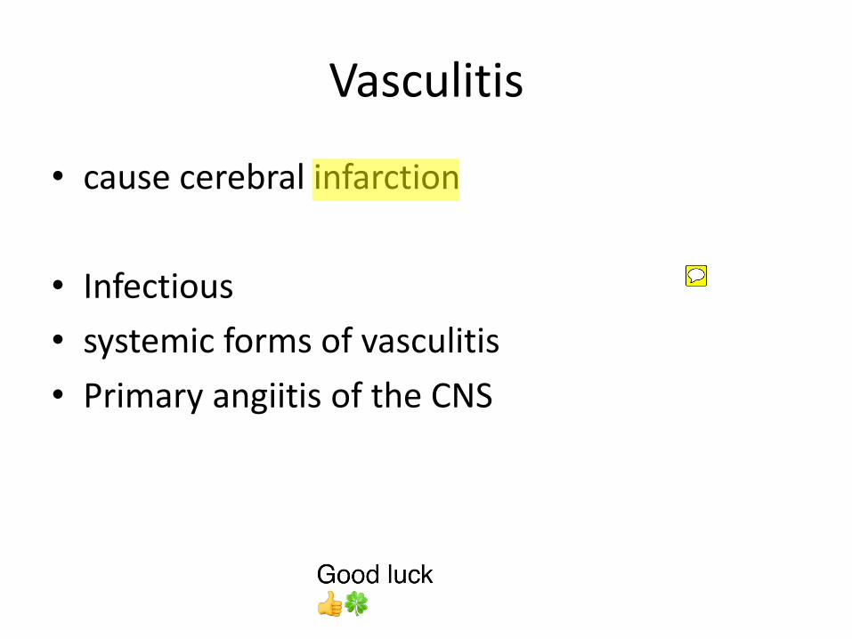
Vasculitis
• cause cerebral infarction
• Infectious
• systemic forms of vasculitis
• Primary angiitis of the CNS
me
One of the things that cause a weak BVs
me
Can lead to:
me
me
Can be primary - - - vasculitis that mainly affecting brain BVs like in autoimmune Secondary - - - associated with systemic diseases like SLE or polyarteritis nodosa OR secondary to infection like in TB , fungi or opportunistic infection.
me
Good luck 👍🍀