Cerebrospinal Fluid Penetration and Combination Therapy of ...
17
Journal of Personalized Medicine Article Cerebrospinal Fluid Penetration and Combination Therapy of Entrectinib for Disseminated ROS1/NTRK-Fusion Positive Pediatric High-Grade Glioma Lisa Mayr 1,2,3, † , Armin S. Guntner 4, † , Sibylle Madlener 1,3 , Maria T. Schmook 5 , Andreas Peyrl 1 , Amedeo A. Azizi 1 , Karin Dieckmann 6 , Dominik Reisinger 1 , Natalia M. Stepien 1 , Kathrin Schramm 7,8 , Anna Laemmerer 1,2,3 , David T. W. Jones 7,8 , Jonas Ecker 9 , Felix Sahm 10,11 , Till Milde 7,9 , Kristian W. Pajtler 7,12,13 , Mirjam Blattner-Johnson 7,8 , Miroslav Strbac 14 , Christian Dorfer 15 , Thomas Czech 15 , Dominik Kirchhofer 1,2,3 , Lisa Gabler 2,3 , Walter Berger 2,3 , Christine Haberler 16 , Leonhard Müllauer 17 , Wolfgang Buchberger 4 , Irene Slavc 1 , Daniela Lötsch-Gojo 2,3,15, * , ‡ and Johannes Gojo 1,3,7,12, * , ‡ 1 Department of Pediatrics and Adolescent Medicine and Comprehensive Center for Pediatrics, Medical University of Vienna, 1090 Vienna, Austria; [email protected] (L.M.); [email protected] (S.M.); [email protected] (A.P.); [email protected] (A.A.A.); [email protected] (D.R.); [email protected] (N.M.S.); [email protected] (A.L.); [email protected] (D.K.); [email protected] (I.S.) 2 Department of Medicine I, Institute of Cancer Research, Medical University of Vienna, 1090 Vienna, Austria; [email protected] (L.G.); [email protected] (W.B.) 3 Comprehensive Cancer Center-Central Nervous System Tumors Unit, Medical University of Vienna, 1090 Vienna, Austria 4 Institute of Analytical Chemistry, Johannes Kepler University, 4020 Linz, Austria; [email protected] (A.S.G.); [email protected] (W.B.) 5 Division of Neuroradiology and Musculoskeletal Radiology, Department of Biomedical Imaging and Image-Guided Therapy, Medical University of Vienna, 1090 Vienna, Austria; [email protected] 6 Department of Radiotherapy, Medical University of Vienna, 1090 Vienna, Austria; [email protected] 7 Hopp Children’s Cancer Center Heidelberg (KiTZ), 69120 Heidelberg, Germany; [email protected] (K.S.); [email protected] (D.T.W.J.); [email protected] (T.M.); [email protected] (K.W.P.); [email protected] (M.B.-J.) 8 Pediatric Glioma Research Group, German Cancer Research Center (DKFZ), 69120 Heidelberg, Germany 9 Clinical Cooperation Unit Pediatric Oncology, Hopp Children’s Cancer Center Heidelberg (KiTZ), 69120 Heidelberg, Germany; [email protected] 10 Department of Neuropathology, Institute of Pathology, University Hospital Heidelberg, 69120 Heidelberg, Germany; [email protected] 11 Clinical Cooperation Unit Neuropathology, German Consortium for Translational Cancer Research (DKTK), German Cancer Research Center (DKFZ), 69120 Heidelberg, Germany 12 Division of Pediatric Neurooncology, German Cancer Research Center (DKFZ), 69120 Heidelberg, Germany 13 Department of Pediatric Oncology, Hematology, and Immunology, University Hospital Heidelberg, 69120 Heidelberg, Germany 14 Department of Laboratory Medicine and Pathology, Tree Top Hospital, Hulhumale 23000, Maldives; [email protected] 15 Department of Neurosurgery, Medical University of Vienna, 1090 Vienna, Austria; [email protected] (C.D.); [email protected] (T.C.) 16 Division of Neuropathology and Neurochemistry, Department of Neurology, Medical University of Vienna, 1090 Vienna, Austria; [email protected] J. Pers. Med. 2020, 10, 290; doi:10.3390/jpm10040290 www.mdpi.com/journal/jpm
Transcript of Cerebrospinal Fluid Penetration and Combination Therapy of ...

Journal of
Personalized
Medicine
Article
Cerebrospinal Fluid Penetration and CombinationTherapy of Entrectinib for DisseminatedROS1/NTRK-Fusion Positive PediatricHigh-Grade Glioma
Lisa Mayr 1,2,3,†, Armin S. Guntner 4,† , Sibylle Madlener 1,3, Maria T. Schmook 5,Andreas Peyrl 1 , Amedeo A. Azizi 1 , Karin Dieckmann 6, Dominik Reisinger 1,Natalia M. Stepien 1, Kathrin Schramm 7,8, Anna Laemmerer 1,2,3 , David T. W. Jones 7,8,Jonas Ecker 9, Felix Sahm 10,11, Till Milde 7,9 , Kristian W. Pajtler 7,12,13,Mirjam Blattner-Johnson 7,8, Miroslav Strbac 14, Christian Dorfer 15 , Thomas Czech 15,Dominik Kirchhofer 1,2,3, Lisa Gabler 2,3, Walter Berger 2,3 , Christine Haberler 16 ,Leonhard Müllauer 17 , Wolfgang Buchberger 4, Irene Slavc 1 , Daniela Lötsch-Gojo 2,3,15,*,‡
and Johannes Gojo 1,3,7,12,*,‡
1 Department of Pediatrics and Adolescent Medicine and Comprehensive Center for Pediatrics,Medical University of Vienna, 1090 Vienna, Austria; [email protected] (L.M.);[email protected] (S.M.); [email protected] (A.P.);[email protected] (A.A.A.); [email protected] (D.R.);[email protected] (N.M.S.); [email protected] (A.L.);[email protected] (D.K.); [email protected] (I.S.)
2 Department of Medicine I, Institute of Cancer Research, Medical University of Vienna, 1090 Vienna, Austria;[email protected] (L.G.); [email protected] (W.B.)
3 Comprehensive Cancer Center-Central Nervous System Tumors Unit, Medical University of Vienna,1090 Vienna, Austria
4 Institute of Analytical Chemistry, Johannes Kepler University, 4020 Linz, Austria;[email protected] (A.S.G.); [email protected] (W.B.)
5 Division of Neuroradiology and Musculoskeletal Radiology, Department of Biomedical Imaging andImage-Guided Therapy, Medical University of Vienna, 1090 Vienna, Austria;[email protected]
6 Department of Radiotherapy, Medical University of Vienna, 1090 Vienna, Austria;[email protected]
7 Hopp Children’s Cancer Center Heidelberg (KiTZ), 69120 Heidelberg, Germany;[email protected] (K.S.); [email protected] (D.T.W.J.);[email protected] (T.M.); [email protected] (K.W.P.);[email protected] (M.B.-J.)
8 Pediatric Glioma Research Group, German Cancer Research Center (DKFZ), 69120 Heidelberg, Germany9 Clinical Cooperation Unit Pediatric Oncology, Hopp Children’s Cancer Center Heidelberg (KiTZ),
69120 Heidelberg, Germany; [email protected] Department of Neuropathology, Institute of Pathology, University Hospital Heidelberg,
69120 Heidelberg, Germany; [email protected] Clinical Cooperation Unit Neuropathology, German Consortium for Translational Cancer Research (DKTK),
German Cancer Research Center (DKFZ), 69120 Heidelberg, Germany12 Division of Pediatric Neurooncology, German Cancer Research Center (DKFZ), 69120 Heidelberg, Germany13 Department of Pediatric Oncology, Hematology, and Immunology, University Hospital Heidelberg,
69120 Heidelberg, Germany14 Department of Laboratory Medicine and Pathology, Tree Top Hospital, Hulhumale 23000, Maldives;
[email protected] Department of Neurosurgery, Medical University of Vienna, 1090 Vienna, Austria;
[email protected] (C.D.); [email protected] (T.C.)16 Division of Neuropathology and Neurochemistry, Department of Neurology, Medical University of Vienna,
1090 Vienna, Austria; [email protected]
J. Pers. Med. 2020, 10, 290; doi:10.3390/jpm10040290 www.mdpi.com/journal/jpm

J. Pers. Med. 2020, 10, 290 2 of 17
17 Department of Pathology, Medical University of Vienna, 1090 Vienna, Austria;[email protected]
* Correspondence: [email protected] (D.L.-G.); [email protected] (J.G.)† Co-first author.‡ Co-last author.
Received: 18 November 2020; Accepted: 16 December 2020; Published: 18 December 2020 �����������������
Abstract: Targeting oncogenic fusion-genes in pediatric high-grade gliomas (pHGG) with entrectinibhas emerged as a highly promising therapeutic approach. Despite ongoing clinical studies, todate, no reports on the treatment of cerebrospinal fluid (CSF) disseminated fusion-positive pHGGexist. Moreover, clinically important information of combination with other treatment modalitiessuch as intrathecal therapy, radiotherapy and other targeted agents is missing. We report on ourclinical experience of entrectinib therapy in two CSF disseminated ROS1/NTRK-fusion-positivepHGG cases. Combination of entrectinib with radiotherapy or intrathecal chemotherapy appearsto be safe and has the potential to act synergistically with entrectinib treatment. In addition,we demonstrate CSF penetrance of entrectinib for the first time in patient samples suggesting targetengagement even upon CSF dissemination. Moreover, in vitro analyses of two novel cell modelsderived from one case with NTRK-fusion revealed that combination therapy with either a MEK(trametinib) or a CDK4/6 (abemaciclib) inhibitor synergistically enhances entrectinib anticancereffects. In summary, our comprehensive study, including clinical experience, CSF penetrance andin vitro data on entrectinib therapy of NTRK/ROS1-fusion-positive pHGG, provides essential clinicaland preclinical insights into the multimodal treatment of these highly aggressive tumors. Our datasuggest that combined inhibition of NTRK/ROS1 and other therapeutic vulnerabilities enhances theantitumor effect, which should be followed-up in further preclinical and clinical studies.
Keywords: NTRK fusion; ROS1 fusion; entrectinib; radiotherapy; CSF penetrance; targeted therapies;trametinib; abemaciclib
1. Introduction
Molecular profiling has significantly improved diagnosis and prognostic prediction of pediatricbrain tumors and has therefore already been implemented in the current as well as the upcomingWHO classification of tumors of the central nervous system (CNS) [1]. Gene fusions, includingthe neurotrophic tyrosine kinase (NTRK) family or c-ros oncogene 1 (ROS1), are relatively rare,yet their therapeutic impact has been proven in multiple solid tumor types [2–4]. NTRK gene fusionsoccur with a prevalence of less than 1% across all tumor types [5,6]. With respect to brain tumors,the estimated prevalence of NTRK-fusions is 0.55 to 2% for gliomas and neuroepithelial tumors [5].In the subgroup of infant hemispheric glioma (IHG), nearly two-thirds of cases harbor molecularalterations of anaplastic lymphoma kinase (ALK), NTRK, ROS1 or tyrosine-protein kinase MET [7,8].Adult and infant NTRK-fused gliomas are primarily located in the hemispheres with high-gradehistology, whereas in older children, a more diverse anatomic distribution and low to high-gradehistologic grades are found [9]. Adjuvant chemotherapy (CT) after surgery is the first approach ininfants and allows delay or even avoidance of radiotherapy (RT) with a 5-year overall survival (OS)of 25.0% and 42.9% in ROS1- and NTRK-driven tumors, respectively [7,10,11]. In older pediatricpatients, treatment consisting of surgery, RT and CT is applied, resulting in long-term survival ratesaround 10% [12]. Concomitant genomic alterations of NTRK-fused gliomas are more frequent in adultpatients and high-grade tumors and include, among others, CDKN2A/B loss, TERT promoter mutation,TP53 mutation/biallelic inactivation/loss, PTEN loss/mutation/biallelic inactivation, EGFR amplification,ATRX mutation, RB1 loss, and PIK3CA mutation [9].
J. Pers. Med. 2020, 10, 290 3 of 17
As ROS1 or NTRK alterations are also major oncogenic drivers in other solid tumor types,targeted therapies have already been developed and evaluated within clinical studies. Larotrectinib andentrectinib, two highly specific TRK inhibitors, are FDA and EMA approved in patients with tumorsharboring a TRK fusion or a ROS1-fusion in the case of entrectinib [13]. However, neoplasms in thebrain are protected by the blood brain barrier (BBB), and the ability to pass this tissue layer is of utmostimportance for targeted therapies [14]. Moreover, the penetrance of cerebrospinal fluid (CSF) maybe particularly relevant for the treatment of leptomeningeal disseminated tumors. We have recentlyshown that CSF concentrations of small molecules can be reliably detected via samples obtained froman Ommaya reservoir, but data for CSF penetrance of TRK inhibitors in brain tumor patients is stilllacking [15]. Entrectinib was designed to cross the BBB and displayed an objective response rate of79% across different adult and pediatric solid tumors as well as efficacy in CNS tumors [16].
To date, no reports on the treatment of leptomeningeal dissemination in fusion-positive pediatrichigh-grade glioma (pHGG) exist. Moreover, clinically important information of potential combinationswith other treatment modalities such as intrathecal therapy, RT and other targeted agents is missing.Here we report on our experience in treating two pediatric patients with leptomeningeal disseminatedpHGG with entrectinib. Moreover, we demonstrate CSF penetration of entrectinib for the first time in areal-world setting in one patient and provide insights into resistance patterns and emerging therapeuticvulnerabilities upon entrectinib treatment of pediatric glioma.
2. Methods and Materials
2.1. Patient Samples and Characteristics
Both cases were treated at the Department of Pediatrics and Adolescent Medicine of the MedicalUniversity of Vienna (MUV) (one was referred at progression for entrectinib treatment after previoustreatment at other centers). Clinical information was obtained from patient charts. The extent ofsurgical resection was defined on postoperative magnetic resonance imaging (MRI) performed within48 h as gross total resection (GTR, no obvious residual tumor), partial resection (PR, 10–50% residualtumor) and biopsy (>50% residual tumor). The study was approved by the local institutional reviewboard of the Medical University of Vienna (EK Nr. 1244/2016). Informed consent was obtained for allpatients and/or legal representatives.
2.2. Histopathology
The histopathological diagnoses were assessed by experienced neuropathologists according tothe 2016 WHO classification. For diagnostic purposes, a routine histopathological examination onformalin-fixed paraffin-embedded (FFPE) tissue was performed, including immunohistochemical(IHC) analysis with vimentin, GFAP, S100, EMA, CKAE1/3, Desmin, Olig2, MAP2, CD31, CD34,CD99, p53, bcl-2, NCAM, IDH1, BCAT1, ATRX, BCOR, STAT6, BAF47 (INI1), SMARCA4 (BRG1),EGFR, PD-L1.
2.3. Next-Generation Sequencing (NGS)
In case 2, NGS was performed on the tissue of the left frontal metastasis with oncominecomprehensive assay v3 (Thermo Fisher Scientific, Waltham, MA, USA) according to the manufacturer’sinstructions [17]. DNA and RNA extracted from FFPE tissue were used. Oncomine comprehensiveassay v3 detects single nucleotide variants (SNV), copy number variations (CNV), gene fusions,and indels from 161 unique cancer-associated genes [17].
FoundationOne® CDx (F1CDx, Foundation Medicine, Inc., Cambridge, UK) analysis wasconducted with the biopsy of the left fronto-median metastasis of case 2. F1CDx is an NGS-basedin vitro diagnostic device for detection of substitutions, insertion and deletion alterations (indels),and copy number alterations (CNAs) in 324 genes and select gene rearrangements, as well as genomicsignatures including microsatellite instability (MSI) and tumor mutational burden (TMB) using DNA

J. Pers. Med. 2020, 10, 290 4 of 17
isolated from FFPE tumor tissue specimens [18]. Moreover, the left fronto-median metastasis of case 2was analyzed at the Deutsches Krebsforschungszentrum (DKFZ) through the individualized therapyfor relapsed Malignancies in childhood (INFORM) registry [19]. The first relapse of case 1 was analyzedvia the Pediatric Targeted Therapy 2.0 (PTT 2.0) registry study (NCT-2016-041 4) at the DKFZ.
2.4. High-Performance Liquid Chromatography-Mass Spectrometry (HPLC-MS) Analysis
The quantitation of entrectinib was performed with a modified version of the general assaydescribed earlier by our group [15]. First, CSF specimens were treated with a five-fold surplus ofmethanol. After homogenization and centrifugation, 7 µL of supernatants were submitted to HPLC-MSanalysis using a 1260 Infinity II HPLC (Agilent Technologies, Santa Clara, CA, USA) hyphenated to a6460 triple quadrupole mass spectrometer (Agilent Technologies). For chromatography, a Poroshell120 column (EC-C18, 2.7 µm, 3 × 150 mm, Agilent Technologies) in combination with a gradientsystem of acetonitrile and water (both modified with 0.1% formic acid) was used. Sensitivity, as well asspecificity of the assay, were assured in method development. Possible matrix effects were compensatedwith matrix-matched calibration.
2.5. Cell Models
Tumor tissues for analyses and establishment of patient-derived cell models were derived frompatients treated at the MUV. The primary gliosarcoma cell lines originating from case 2 VBT247 (primarytumor at diagnosis) and VBT363 (3rd recurrence under treatment with entrectinib) were cultured inRPMI-1640 medium (Sigma-Aldrich, St. Louis, MO, USA) supplemented with 10% fetal calf serum(FCS, Gibco, Thermo Fisher Scientific) at 37 ◦C in a 5% CO2 incubator. The cell models were regularlychecked for mycoplasma contamination.
2.6. ATP Assay
Cells were plated in triplicates (4 × 10⁴ cells/mL) in 100 µL growth medium per well in 96-wellplates and allowed to attach for 24 h. All drugs were purchased from Selleck Chemicals (Houston,TX, USA). Entrectinib (0 to 10 µM), trametinib (0 to 10 µM), everolimus (0 to 50 µM) and abemaciclib(0 to 10 µM) were added alone or in different combination regimens in 100 µL growth medium with10% FCS and cells were exposed for 72 h. The proportion of viable cells was determined by ATP assayfollowing the manufacturer’s recommendations (“CellTiter-Glo® luminescent cell viability assay”,Promega, Madison, WI, USA). Luminescence was measured at 1000 nm at the Tecan infinite 200Pro(Zurich, Switzerland). Raw data were analyzed using GraphPad Prism 8.0 software (GraphPadSoftware Inc., La Jolla, CA, USA). Results are given as mean +/− SD and were normalized to untreatedcontrol cells. Cytotoxicity was expressed as IC50-values calculated from full dose–response curves(drug concentrations inducing a 50% reduction of the cell number in comparison to the untreatedcontrol cells). The interaction between the activities of combined drugs is expressed by the combinationindex (CI) as published by Chou [20] using CalcuSyn software (Biosoft, Ferguson, MO, USA). CI < 0.9,CI = 0.9–1.2 or CI > 1.2 represent synergism, additive effects and antagonism, respectively.
2.7. Colony Formation Assay
A low-density of cells (1 × 10⁴ cells per well) was seeded in 500 µL growth medium in triplicates in24-well plates. Following 24 h of recovery, 1 µM entrectinib, 1 µM trametinib, 1 µM abemaciclib, 5 µMeverolimus or different combination treatments were applied and repeated after 7 days incubationtime. On day 14 of exposure, cells were washed twice with 1×PBS, fixed with methanol at 4 ◦C andstained with crystal violet (10 µg crystal violet per 10 mL PBS). Photomicrographs were taken usinga Nikon D3200 camera, and densitometric quantification of the images was analyzed with ImageJsoftware (Image J2, Wayne Rasband, NIH, Bethesda, MD, USA).

J. Pers. Med. 2020, 10, 290 5 of 17
2.8. Statistical Analysis
Statistical analysis was performed using GraphPad Prism 8.0. All experiments were carried outindependently at least three times. All data are expressed as mean +/− S.D. Statistical significance ofdifferences was analyzed by using one-way ANOVA. A p-value < 0.05 was considered statisticallysignificant. Throughout the study the following classification is used: *, p < 0.05; **, p < 0.01 ***,p < 0.001, ****, p < 0.0001.
3. Results
3.1. Clinical Characteristics and Response to Entrectinib
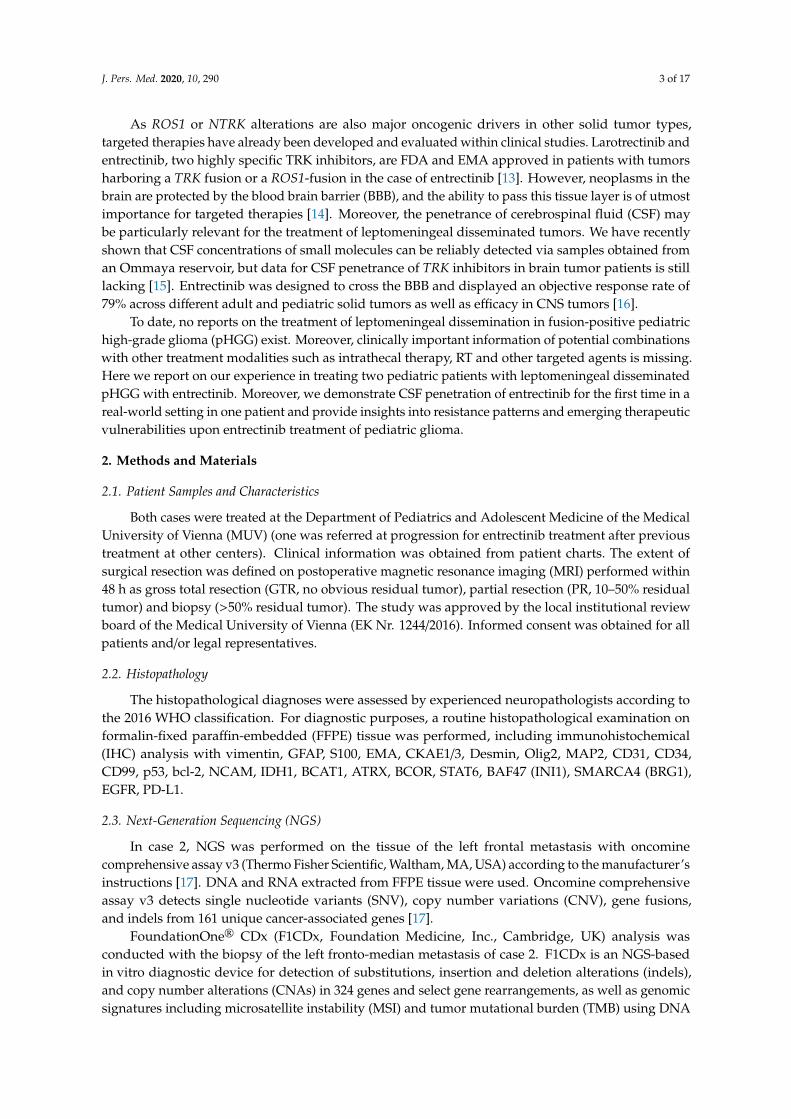
Case 1 (overview in Figure 1, including H&E and p53 IHC staining in Figure S1A,B), diagnosedwith an IHG at two years of age, received a GTR and consecutive follow-up as first-line therapy. Due toa local recurrence detected six months after diagnosis, the patient was transferred to the HeidelbergUniversity Hospital and was treated with PR and CT according to the HIT-MED Guidance protocol(ClinicalTrials.gov NCT02417324). Following two courses of CT, tumor progression was observed,and therapy was switched to local proton irradiation (54 Gy to the tumor bed) and concomitant oraltemozolomide (TMZ) (75 mg/m2). Molecular analysis of tumor tissue revealed a ROS1:ARCN1 fusion,and therapy with a ROS1-inhibitor was therefore suggested but not feasible due to the unavailabilityof matching clinical studies in Europe at this time point. Consequently, the patient was referred backto the external center. Six months later, the patient developed focal seizures prompting a GTR ofthe residual tumor. Histopathological examination revealed 15% vital tumor cells. Four weeks afterre-operation, the cranial follow-up MRI revealed extensive CSF dissemination in both lateral ventricles(Figure 1). At this time, the patient was referred to the Medical University of Vienna for treatment withentrectinib and received a follow-up MRI of the craniospinal axis only six weeks later, just before his firstappointment in Vienna. Within this limited timeframe, the tumor had rapidly progressed, now showingprogressive CSF dissemination in both lateral ventricles, the fourth ventricle, and the spinal dural sac.Treatment with entrectinib (400 mg daily) was immediately initiated and well-tolerated. Subsequently,tumor growth was drastically reduced and showed partial response. However, regular surveillanceMRIs revealed slow tumor progression over the following months. Due to the previous detectionof mitogen-activated kinase (MAPK) pathway activation in comprehensive molecular profiling andan inactivating PTEN mutation as well as the loss of chromosome 10, therapy was augmented withtrametinib and everolimus. The therapeutic approach with entrectinib, trametinib and everolimusresulted in an almost stable disease. Unfortunately, the patient developed a massive drug rash andtherapy with trametinib and everolimus had to be terminated after 14 weeks of trametinib and 10 weeksof everolimus. Subsequently, the patient was treated with local RT (whole ventricular dose 36 Gyand 42 Gy focal to the metastasis in the dural sac) and concomitant entrectinib. Eight months afterRT, the patient currently shows stable disease with entrectinib monotherapy and remains in goodclinical condition.
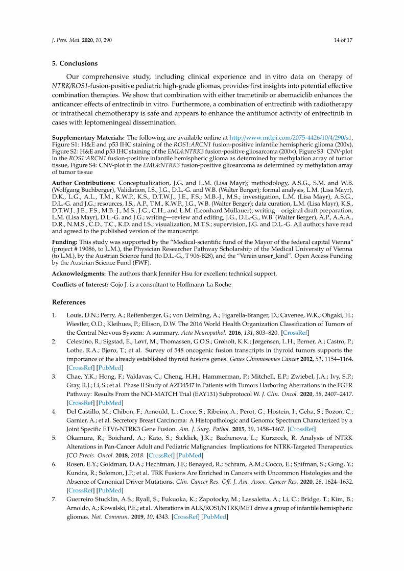
Case 2 (overview depicted in Figure 2, including H&E and p53 IHC staining in Figure S2A,B),presented at the age of nine years with a left parieto-occipital solid cystic tumor, diagnosed asgliosarcoma. GTR was followed by two cycles of ICE (ifosfamide, carboplatin, etoposide) [21] and focalRT (59.4 Gy to the tumor bed) as first-line therapy. Adjuvant CT was changed to TMZ (150–200 mg/m2
on 5 consecutive days every 28 days) because of prolonged aplasia and was augmented withintraventricular therapy via an Ommaya reservoir consisting of etoposide, aqueous cytarabine andtopotecan every other week [22–25]. Ten months after the initial diagnosis, metastasis in the left frontallobe occurred, and treatment consisting of GTR followed by focal RT (59.4 Gy to the tumor bed) wasapplied. Intrathecal therapy was discontinued during RT. However, two weeks following re-irradiation,the patient reported massive, constant and irrepressible pain in his right leg. MRI revealed distinctcerebral and spinal leptomeningeal dissemination with the biggest metastasis located at the levelof thoracic vertebra 9 (Th9), causing compression of the spinal cord. Again, focal RT was applied

J. Pers. Med. 2020, 10, 290 6 of 17
(five times 5 Gy) only to the symptomatic lesion at level Th9. Further molecular analysis of thetumor tissue revealed an EML4:NTRK3 fusion. Treatment was switched to entrectinib (600 mg daily)augmented with intrathecal therapy every other week. No side effects were observed, and the clinicalcondition substantially improved after RT and upon treatment with entrectinib making it even possiblefor the patient to exert sports only eight weeks after RT and initiation of entrectinib. Neuroimagingrevealed a regression of the cerebral and spinal leptomeningeal deposits (Figure 2 depicts the responsein the cervical region that had not been irradiated). Five months after the initiation of entrectinib,the patient developed ataxia and dizziness. Further leptomeningeal dissemination with new lesionsin the left medial superior frontal gyrus, right postcentral, bilateral cerebellar and basal brainstemregions was observed (Figure 2). In order to detect further potential therapeutic targets, biopsy andsubsequent molecular analysis of the left fronto-medial metastasis was performed. Unfortunately,the patient succumbed to his disease due to the rapid tumor progression.J. Pers. Med. 2020, 10, x FOR PEER REVIEW 6 of 18
Figure 1. Case 1, an infantile hemispheric glioma harboring a ROS1:ARCN1 fusion. Progression and therapy response of metastases in the lateral ventricles depicted in axial T2 weighted magnetic resonance images with cerebrospinal fluid (CSF) suppression (a–e). The arrows indicate tumor (red, progression; blue, response/stable disease). The timeline indicates the different treatment strategies and interventions. The frontobasal metastasis (a) was resected.
Case 2 (overview depicted in Figure 2, including H&E and p53 IHC staining in Figure S2A,B), presented at the age of nine years with a left parieto-occipital solid cystic tumor, diagnosed as gliosarcoma. GTR was followed by two cycles of ICE (ifosfamide, carboplatin, etoposide) [21] and focal RT (59.4 Gy to the tumor bed) as first-line therapy. Adjuvant CT was changed to TMZ (150–200 mg/m2 on 5 consecutive days every 28 days) because of prolonged aplasia and was augmented with intraventricular therapy via an Ommaya reservoir consisting of etoposide, aqueous cytarabine and topotecan every other week [22–25]. Ten months after the initial diagnosis, metastasis in the left frontal lobe occurred, and treatment consisting of GTR followed by focal RT (59.4 Gy to the tumor bed) was applied. Intrathecal therapy was discontinued during RT. However, two weeks following re-irradiation, the patient reported massive, constant and irrepressible pain in his right leg. MRI revealed distinct cerebral and spinal leptomeningeal dissemination with the biggest metastasis located at the level of thoracic vertebra 9 (Th9), causing compression of the spinal cord. Again, focal RT was applied (five times 5 Gy) only to the symptomatic lesion at level Th9. Further molecular analysis of the tumor tissue revealed an EML4:NTRK3 fusion. Treatment was switched to entrectinib (600 mg daily) augmented with intrathecal therapy every other week. No side effects were observed, and the clinical condition substantially improved after RT and upon treatment with entrectinib making it even possible for the patient to exert sports only eight weeks after RT and initiation of entrectinib. Neuroimaging revealed a regression of the cerebral and spinal leptomeningeal deposits (Figure 2 depicts the response in the cervical region that had not been irradiated). Five months after the initiation of entrectinib, the patient developed ataxia and dizziness. Further leptomeningeal dissemination with new lesions in the left medial superior frontal gyrus, right postcentral, bilateral
Figure 1. Case 1, an infantile hemispheric glioma harboring a ROS1:ARCN1 fusion. Progressionand therapy response of metastases in the lateral ventricles depicted in axial T2 weighted magneticresonance images with cerebrospinal fluid (CSF) suppression (a–e). The arrows indicate tumor (red,progression; blue, response/stable disease). The timeline indicates the different treatment strategies andinterventions. The frontobasal metastasis (a) was resected.
3.2. Molecular Diagnostics and Next Generation Sequencing
With respect to case 1, NGS within the PTT2.0 registry study (NCT-2016–0414) revealed aROS1:ARCN1 fusion in the IHG. The tumor was negative for IDH1(R132) mutation and MGMTunmethylated. Furthermore, a hot spot mutation in the PTEN gene and a heterozygous loss(LOH) of chromosome 10, causing a combined loss of PTEN function in the tumor was detected.Immunohistochemical analyses showed an aberrant activation of the MAPK pathway. Table 1 shows an

J. Pers. Med. 2020, 10, 290 7 of 17
overview of histopathology, molecular characteristics and next-generation sequencing of our patients.The respective copy number plot derived from methylation array analysis is depicted in Figure S3.
J. Pers. Med. 2020, 10, x FOR PEER REVIEW 7 of 18
cerebellar and basal brainstem regions was observed (Figure 2). In order to detect further potential therapeutic targets, biopsy and subsequent molecular analysis of the left fronto-medial metastasis was performed. Unfortunately, the patient succumbed to his disease due to the rapid tumor progression.
Figure 2. Case 2, a gliosarcoma harboring an EML4:NTRK3 fusion. Therapy response and progression of metastases under treatment with entrectinib is depicted in axial T1 weighted contrast-enhanced magnetic resonance images of the brain and sagittal images of the cervical spine. The red arrows indicate the primary tumor (a) and metastases (b,c,e,f), the blue arrow the regression of the perimedullary metastasis (d). The timeline indicates the different treatment strategies and interventions.
3.2. Molecular Diagnostics and Next Generation Sequencing
With respect to case 1, NGS within the PTT2.0 registry study (NCT-2016–0414) revealed a ROS1:ARCN1 fusion in the IHG. The tumor was negative for IDH1(R132) mutation and MGMT unmethylated. Furthermore, a hot spot mutation in the PTEN gene and a heterozygous loss (LOH) of chromosome 10, causing a combined loss of PTEN function in the tumor was detected. Immunohistochemical analyses showed an aberrant activation of the MAPK pathway. Table 1 shows an overview of histopathology, molecular characteristics and next-generation sequencing of our patients. The respective copy number plot derived from methylation array analysis is depicted in Figure S3.
Table 1. Histopathologic, molecular characteristics and next-generation sequencing.
Case
Disease Status Location Histology
Gene-Fusion
Mutations MGMT
Chromosomal Deletions
Molecular Findings
Cell Line
1 1st
recurrence
right hemispher
e IHG
ROS/ARCN1 PTEN
unmeth. 10
MAPK-activation -
2 primary tumor
left occipital
gliosarcoma
EML4-NTRK3
MRE11A (VUS)
unmeth.
CDKN2A/B - VBT24
7
Figure 2. Case 2, a gliosarcoma harboring an EML4:NTRK3 fusion. Therapy response and progressionof metastases under treatment with entrectinib is depicted in axial T1 weighted contrast-enhancedmagnetic resonance images of the brain and sagittal images of the cervical spine. The red arrows indicatethe primary tumor (a) and metastases (b,c,e,f), the blue arrow the regression of the perimedullarymetastasis (d). The timeline indicates the different treatment strategies and interventions.
Table 1. Histopathologic, molecular characteristics and next-generation sequencing.
Case Disease Status Location Histology Gene-Fusion Mutations MGMT ChromosomalDeletions
MolecularFindings
CellLine
1 1st recurrence right hemisphere IHG ROS/ARCN1 PTEN unmeth. 10 MAPK-activation -
2 primary tumor left occipital gliosarcoma EML4-NTRK3 MRE11A(VUS) unmeth. CDKN2A/B - VBT247
2 3rd recurrence leftfronto-median gliosarcoma EML4-NTRK3 INSR, NF2 unmeth. 5 p, 8 p, 20, 22,
CDKN2A/B
AURKC, IGF1,TGFB3
overexpressionVBT363
In case 2, Oncomine Comprehensive v3 analysis of the local left frontal gliosarcoma recurrencedetected an EML4:NTRK3 fusion, a CDKN2A/B loss and an MRE11A alteration of unknown significance.Analysis of the left fronto-median metastasis that developed under treatment with entrectinib via theINFORM registry at the DKFZ revealed the already known EML4:NTRK3 fusion without resistancemutation, the homozygous CDKN2A/B deletion already detected in the primary tumor, an INSRmutation (p. D601Y, VAF = 0.4), an NF2 splicing mutation (c.522 + 1G > C, VAF = 0.66, resulting inexon-skipping), and AURKC, IGF1 and TGFB3 mRNA overexpression. Analysis of the same biopsyvia F1CDx confirmed the homozygous CDKN2A/B loss and the NF2 mutation. The respective copynumber plot derived from methylation array analysis is depicted in Figure S4.
3.3. CSF Penetration of Entrectinib
As already mentioned, penetrance of the brain is of essential importance in the treatment ofCNS tumors, but so far, evidence for CSF penetration of entrectinib has been limited to preclinical

J. Pers. Med. 2020, 10, 290 8 of 17
studies [26]. Using our previously published approach [15], we analyzed entrectinib CSF penetrationin 5 samples of case 2 obtained before administration of intrathecal therapy. We found entrectinibconcentrations in the low nM range, which increased during ongoing therapy (Figure 3). As case 1 hasno Ommaya reservoir and underwent no surgery at the MUV, CSF samples were not available of thispatient for HPLC-MS analysis.
J. Pers. Med. 2020, 10, x FOR PEER REVIEW 8 of 18
2 3rd
recurrence
left fronto-median
gliosarcoma
EML4-NTRK3
INSR, NF2 unmeth
.
5 p, 8 p, 20, 22,
CDKN2A/B
AURKC, IGF1, TGFB3
overexpression
VBT363
In case 2, Oncomine Comprehensive v3 analysis of the local left frontal gliosarcoma recurrence detected an EML4:NTRK3 fusion, a CDKN2A/B loss and an MRE11A alteration of unknown significance. Analysis of the left fronto-median metastasis that developed under treatment with entrectinib via the INFORM registry at the DKFZ revealed the already known EML4:NTRK3 fusion without resistance mutation, the homozygous CDKN2A/B deletion already detected in the primary tumor, an INSR mutation (p. D601Y, VAF = 0.4), an NF2 splicing mutation (c.522 + 1G > C, VAF = 0.66, resulting in exon-skipping), and AURKC, IGF1 and TGFB3 mRNA overexpression. Analysis of the same biopsy via F1CDx confirmed the homozygous CDKN2A/B loss and the NF2 mutation. The respective copy number plot derived from methylation array analysis is depicted in Figure S4.
3.3. CSF Penetration of Entrectinib
As already mentioned, penetrance of the brain is of essential importance in the treatment of CNS tumors, but so far, evidence for CSF penetration of entrectinib has been limited to preclinical studies [26]. Using our previously published approach [15], we analyzed entrectinib CSF penetration in 5 samples of case 2 obtained before administration of intrathecal therapy. We found entrectinib concentrations in the low nM range, which increased during ongoing therapy (Figure 3). As case 1 has no Ommaya reservoir and underwent no surgery at the MUV, CSF samples were not available of this patient for HPLC-MS analysis.
Figure 3. CSF penetrance of entrectinib. Entrectinib concentration (nM) detected in the cerebrospinal fluid of case 2 by high-performance liquid chromatography-mass spectrometry (HPLC-MS) as previously described [15]. Entrectinib levels increase over time of intake. Individual points depict entrectinib concentrations in independent samples obtained via an Ommaya reservoir before administration of intrathecal therapy.
3.4. Impact of Entrectinib and Combination with Targeted Therapies on NTRK-Fusion Positive pHGG Cell Viability and Proliferation
Under standard culture conditions, treatment with increasing concentrations of entrectinib for 72 h caused a dose-dependent decrease of cell viability in both gliosarcoma cell lines that were derived from two consecutive surgeries of case 2, VBT247 (primary diagnosis) and the 3rd recurrence under treatment with entrectinib (VBT363); growth curves and IC50 values are depicted in Figure 4A,E. Interestingly, the antiproliferative effect of entrectinib showed no difference in the tumor cell model VBT247 derived from the primary tumor as compared to VBT363, the cell model established following clinical resistance to entrectinib. As both cell lines harbor a homozygous loss of CDKN2A/B we tested the effect of the CDK4/6 inhibitor abemaciclib on cell viability (Figure 4B,E). VBT247 and
conc
entra
tion
(nM
)
Figure 3. CSF penetrance of entrectinib. Entrectinib concentration (nM) detected in the cerebrospinalfluid of case 2 by high-performance liquid chromatography-mass spectrometry (HPLC-MS) as previouslydescribed [15]. Entrectinib levels increase over time of intake. Individual points depict entrectinibconcentrations in independent samples obtained via an Ommaya reservoir before administration ofintrathecal therapy.
3.4. Impact of Entrectinib and Combination with Targeted Therapies on NTRK-Fusion Positive pHGG CellViability and Proliferation
Under standard culture conditions, treatment with increasing concentrations of entrectinib for72 h caused a dose-dependent decrease of cell viability in both gliosarcoma cell lines that were derivedfrom two consecutive surgeries of case 2, VBT247 (primary diagnosis) and the 3rd recurrence undertreatment with entrectinib (VBT363); growth curves and IC50 values are depicted in Figure 4A,E.Interestingly, the antiproliferative effect of entrectinib showed no difference in the tumor cell modelVBT247 derived from the primary tumor as compared to VBT363, the cell model established followingclinical resistance to entrectinib. As both cell lines harbor a homozygous loss of CDKN2A/B we testedthe effect of the CDK4/6 inhibitor abemaciclib on cell viability (Figure 4B,E). VBT247 and VBT363showed a distinct sensitivity against CDK4/6 inhibition. In contrast, both gliosarcoma cell models werecomparably insensitive to treatment with trametinib and everolimus with IC50 values above 10 µMclearly exceeding clinically achievable doses in the short-term exposure as depicted in Figure 4C–E.
The impact of a combined application of the NTRK inhibitor entrectinib with other targetedtherapies (e.g., abemaciclib, trametinib and everolimus) was tested for short- (72 h) and long-termexposure (14 days) by ATP and clonogenicity assays, respectively. NTRK inhibition distinctlysynergized with trametinib in VBT247 and VBT363 cells (growth curves and combination indicesshown in Figure 5A,B). In contrast, entrectinib generally antagonized the effect of abemaciclib in lowconcentrations but indeed synergized with abemaciclib in higher concentrations (5 µM abemaciclib)depicted in Figure 5C,D. Last, entrectinib showed additive to synergistic effects when combined witheverolimus in VBT247 and VBT363 cells (Figure 5E,F).

J. Pers. Med. 2020, 10, 290 9 of 17
J. Pers. Med. 2020, 10, x FOR PEER REVIEW 9 of 18
VBT363 showed a distinct sensitivity against CDK4/6 inhibition. In contrast, both gliosarcoma cell models were comparably insensitive to treatment with trametinib and everolimus with IC50 values above 10 µM clearly exceeding clinically achievable doses in the short-term exposure as depicted in Figure 4C–E.
Figure 4. Effect of single-agent targeted therapies in EML4:NTRK3 positive cell lines. The impact on cell viability of (A) entrectinib, (B) abemaciclib, (C) trametinib and (D) everolimus on the primary tumor (VBT247) and the 3rd recurrence under treatment with entrectinib (VBT363) was tested after 72 h incubation by ATP assay in triplicates. (E). The inhibitory effect is expressed as IC50 values calculated from full dose–response curves (drug concentrations inducing a 50% reduction of the cell number in comparison to the untreated control cells).
The impact of a combined application of the NTRK inhibitor entrectinib with other targeted therapies (e.g., abemaciclib, trametinib and everolimus) was tested for short- (72 h) and long-term exposure (14 days) by ATP and clonogenicity assays, respectively. NTRK inhibition distinctly synergized with trametinib in VBT247 and VBT363 cells (growth curves and combination indices shown in Figure 5A,B). In contrast, entrectinib generally antagonized the effect of abemaciclib in low
viab
ility
(fol
d co
ntro
l)
viab
ility
(fol
d co
ntro
l)
viab
ility
(fol
d co
ntro
l)
D
E IC50; µM
Cell line
Targeted therapy VBT247 VBT363
Entrectinib 4.55 4.56Trametinib >10 >10Abemaciclib 3.16 2.90Everolimus 37.78 38.08
B
C
Avi
abili
ty (f
old
cont
rol)
Figure 4. Effect of single-agent targeted therapies in EML4:NTRK3 positive cell lines. The impact oncell viability of (A) entrectinib, (B) abemaciclib, (C) trametinib and (D) everolimus on the primarytumor (VBT247) and the 3rd recurrence under treatment with entrectinib (VBT363) was tested after 72 hincubation by ATP assay in triplicates. (E). The inhibitory effect is expressed as IC50 values calculatedfrom full dose–response curves (drug concentrations inducing a 50% reduction of the cell number incomparison to the untreated control cells).
In order to test antiproliferative effects in more detail, we performed a long-term exposureclonogenicity assay. Inhibitory effects were already detectable at 1 µM entrectinib and 1 µM abemaciclibin both gliosarcoma cell lines as depicted in Figure 6A,B. Furthermore, the sensitizing effect of trametiniband everolimus was distinctly increased in the clonogenicity assay and was already detectable at 1 µMtrametinib and 5 µM everolimus (Figure 6A,B). The strong effect of trametinib alone was not furtherincreased when combined with entrectinib.

J. Pers. Med. 2020, 10, 290 10 of 17
J. Pers. Med. 2020, 10, x FOR PEER REVIEW 10 of 18
concentrations but indeed synergized with abemaciclib in higher concentrations (5 µM abemaciclib) depicted in Figure 5C,D. Last, entrectinib showed additive to synergistic effects when combined with everolimus in VBT247 and VBT363 cells (Figure 5E,F).
Figure 5. Effect of entrectinib and combination treatment on cell viability and toxicity. Impact of entrectinib treatment in combination with (A) trametinib, (C) abemaciclib, (E) everolimus on cell
1 100.0
0.5
1.0
1.5 VBT363
Entrectinib µM
Control
0.5µM Trametinib
1µM Trametinib
5µM Trametinib
0.1µM Trametinib
0.1 0.5 5
viab
ility
(fol
d co
ntro
l)vi
abili
ty (f
old
cont
rol)
viab
ility
(fol
d co
ntro
l)
0.0 0.5 1.0 1.50.1
1
10
Entrectinib µM
CI v
alue
0.5 µM 1 µM 5 µM Trametinib
VBT363
SYNERGISTIC
ADDITIVE
ANTAGO
NISTIC
0.0 0.5 1.0 1.50.1
1
10
Entrectinib µM
0.5 µM 1 µM 5 µM Abemaciclib
VBT363
ADDITIVE
A B
C D
E
1 100.0
0.5
1.0
1.5 VBT247
Entrectinib µM
viab
ility
(fol
d co
ntro
l) Control
0.5µM Abemaciclib
1µM Abemaciclib
5µM Abemaciclib
0.1 0.5 50.0 0.5 1.0 1.5
0.1
1
10
Entrectinib µM0.5 µM 1 µM 5 µM Abemaciclib
VBT247
ADDITIVE
0.0 0.5 1.0 1.50.1
1
10
Entrectinib µM
CI v
alue
5 µM 10 µM 25 µM Everolimus
VBT247
SYNERGISTIC
ADDITIVE
ANTAGO
NISTIC
0.0 0.5 1.0 1.50.1
1
10
Entrectinib µM
CI v
alue
5 µM 10 µM 25 µM Everolimus
VBT363
SYNERGISTIC
ADDITIVE
ANTAGO
NISTIC
F
Figure 5. Effect of entrectinib and combination treatment on cell viability and toxicity. Impact ofentrectinib treatment in combination with (A) trametinib, (C) abemaciclib, (E) everolimus on cellviability in triplicates in the indicated cell models measured by ATP-based survival assays upon72 h drug exposure. Combination index (CI) based on the distinct drug combinations (B) trametinib,(D) abemaciclib, (F) everolimus were calculated as published [20]. CI values < 0.9 indicates synergistic,CI = 0.9–1.2 additive and CI > 1.2 antagonistic effects.

J. Pers. Med. 2020, 10, 290 11 of 17
J. Pers. Med. 2020, 10, x FOR PEER REVIEW 11 of 18
viability in triplicates in the indicated cell models measured by ATP-based survival assays upon 72 h drug exposure. Combination index (CI) based on the distinct drug combinations (B) trametinib, (D) abemaciclib, (F) everolimus were calculated as published [20]. CI values < 0.9 indicates synergistic, CI = 0.9–1.2 additive and CI > 1.2 antagonistic effects.
In order to test antiproliferative effects in more detail, we performed a long-term exposure clonogenicity assay. Inhibitory effects were already detectable at 1 µM entrectinib and 1 µM abemaciclib in both gliosarcoma cell lines as depicted in Figure 6A,B. Furthermore, the sensitizing effect of trametinib and everolimus was distinctly increased in the clonogenicity assay and was already detectable at 1 µM trametinib and 5 µM everolimus (Figure 6A,B). The strong effect of trametinib alone was not further increased when combined with entrectinib.
Figure 6. Effect of entrectinib and combination treatment on cell viability and toxicity. Effects of combined long-term application of entrectinib with trametinib, abemaciclib and/or everolimus in
Figure 6. Effect of entrectinib and combination treatment on cell viability and toxicity. Effects ofcombined long-term application of entrectinib with trametinib, abemaciclib and/or everolimus inVBT363 cells were tested by clonogenicity assays in triplicates. (A) Cells were fixed and stained withcrystal violet, and wells were photographed. (B) Densitometric quantification of photomicrographsshown in (A) using ImageJ2 software was assessed. Statistical significance of differences was analyzedby one-way ANOVA, ****, p < 0.0001.
4. Discussion
Targeting fusion-positive pHGG with small molecule inhibitors has emerged as a highly promisingtherapeutic approach to combat these aggressive and therapy refractory cancer types.
Herein, we report on two patients with leptomeningeal disseminated pHGG treated withentrectinib. Entrectinib, a selective pan TRK inhibitor, has already demonstrated significant responses inNTRK-fused tumors, including primary CNS tumors and CNS metastases [16,27–29]. The STARTRK-NGtrial included four pHGG patients treated with entrectinib. All patients showed a radiographic response,including one complete response (2019, ASCO Annual Meeting Abstract #: 10009). To date, only a few

J. Pers. Med. 2020, 10, 290 12 of 17
reports describing primary NTRK-fused CNS tumors treated with either larotrectinib or entrectinibare available in the literature [8,30–32]. An adult patient with a BCAN:NTRK1 fused glioneuronaltumor developed disease progression after eleven months of entrectinib [30]. Two reports describepatients suffering from a low-grade glioma (LGG), one with a NACC2:NTRK fusion showing morethan 50% reduction in tumor volume and an ETV6:NTRK3-fused tumor with complete remission upontreatment with larotrectinib [8,31]. Moreover, in two ETV6:NTRK3 fusion-positive pHGG, larotrectinibwas administered. One patient experienced more than 70% tumor volume reduction and one diseaseprogression [8,32]. In the pooled analysis of the STARTRK-2, STARTRK-1 and ALKA-371-001 trials,overall median progression-free survival in patients with CNS disease was 7.7 months, with mostobserved responses within the first or second treatment cycle [16]. This is in line with our observationsshowing the response of tumors with leptomeningeal dissemination after four weeks of entrectinib.The progression-free survival on entrectinib treatment observed in our gliosarcoma patient was5 months, which is comparable to the observation in various trials [16]. In contrast, the tumorprogression was markedly reduced but not totally blocked in our IHG patient, and treatment iscurrently ongoing 16 months after the start of entrectinib.
To explore the potential of entrectinib combination therapy in fusion-positive pHGG, we usedtwo cell models derived from our case with NTRK-fusion. The observed in vitro sensitivity towardsentrectinib was comparable to a report investigating an ETV6:NTRK3 fusion-positive model [8].Importantly, one model was derived from the primary tumor (VBT247), whereas the second model(VBT363) was derived from the progressive tumor during entrectinib treatment. Interestingly,both models demonstrated sensitivity towards entrectinib in vitro, suggesting that oncogenicNTRK-activation is still present in progressive tumors. Moreover, in a previous study investigatingBRAF(V600E)-mutated high-grade glioma, we found a similar effect in comparing two modelsderived before and after treatment with a combination of dabrafenib and trametinib [33]. However,further studies, including long-term entrectinib exposure of these cell models, would be useful to moredeeply dissect the resistance mechanisms of pHGG towards entrectinib.
Acquired resistance mutations to entrectinib in the kinase domain of the NTRK gene havebeen described in other tumor types [34–36]. Selitrectinib (LOXO-195) was designed to target theseresistance mutations as well as the wild-type protein [35]. A pooled analysis revealed that all patientswho developed resistance to larotrectinib had secondary NTRK mutations [37]. However, in ourgliosarcoma patient with disease progression under entrectinib, no secondary NTRK mutation wasdetected, but molecular profiling revealed activation of alternative oncogenic pathways, including NF2and insulin receptor (INSR). Whether these effects are acquired events or whether resistant tumor cellsemerge from primary subclones being intrinsically resistant—as, for example, cancer stem cells—whichfacilitated disease progression remains to be clarified [38–40]. The latter could indeed be present as wehave already demonstrated high intratumoral heterogeneity within other pHGG types [38–41].
MAPK pathway activation by signal transducers not related to NTRK has already been describedin other tumor types [42]. In the ROS1:ARCN1 fusion patient from our study, a hot spot mutation inthe PTEN gene and a heterozygous loss of chromosome 10 causing a loss of PTEN function, a frequentoncogenic event resulting in activation of the mammalian target of rapamycin (mTOR) pathway, as wellas aberrant activation of the MAPK pathway were detected. Entrectinib monotherapy led to a massivedeceleration of tumor growth, but stable disease was not achieved. Therefore, we treated our patientwith a combined approach consisting of entrectinib, trametinib and everolimus. This combination wasable to further reduce tumor growth and resulted in almost stable disease, but the combination wasnot well-tolerated as has been described for MEK inhibitors and mTOR inhibitors in an adult clinicaltrial [43] and had to be discontinued. Intriguingly, in our gliosarcoma cell model, entrectinib synergizedwith trametinib in the short-term exposure and distinctly decreased cell viability. In contrast, theeffect of trametinib alone in the short-term exposure experiment showed IC50 values exceeding 10 µM.Interestingly, in the MAPK pathway-activated pLGG IC50 values for trametinib are in the low nm rangesuggesting activation of alternative pathways such as mTOR in fusion-positive pHGG [44]. Moreover,

J. Pers. Med. 2020, 10, 290 13 of 17
in the long-term exposure, already 1 µM of trametinib led to a distinct decrease in cell viability andmarkedly exceeded the effect of entrectinib alone. This points to a cytostatic effect of trametinib,rather than inducing apoptosis, as recently described for the treatment of glioblastoma cells [45].Treatment with everolimus showed only limited effects on cell viability in the short- and long-termexposure experiments in our gliosarcoma cell models. In summary, our first preclinical studies onentrectinib combination treatment in pHGG suggest that a combined application of entrectinib andtrametinib could be a promising treatment option for patients in the clinic. This effect is well describedfor the treatment of BRAF(V600E) mutated melanoma, and combination therapy of BRAF inhibitorsand MEK inhibitors has become the standard of care as it is superior to monotherapy [46–48]. Based onthe detected CDKN2A/B deletion, we further tested the potential of the CDK4/6 inhibitor abemaciclib,which showed strong efficacy against both gliosarcoma cell models and entrectinib distinctly synergizedwith 5 µM abemaciclib in vitro. Consequently, the combination therapy of CDK4/6 inhibitors could bea future strategy to overcome therapy resistance to NTRK inhibitors.
With respect to patient treatment, entrectinib has been shown to be generally well-tolerated,and the most common reported adverse events were dysgeusia, fatigue, constipation, diarrhea,dizziness, peripheral edema, weight gain and anemia [16]. In our cases, both patients had noentrectinib-related side effects. Although therapy with entrectinib and trametinib caused cutaneousside effects in our ROS1:ARCN1 fusion patient, therapy could be continued with local cutaneoustherapy. However, the addition of everolimus to the combination of entrectinib and trametinibmarkedly increased the cutaneous side effects and therapy with trametinib and everolimus had tobe terminated. Dose interruption was necessary for almost 50%, and study discontinuation in 10%of patients treated with everolimus and trametinib due to cutaneous adverse events [49]. Therefore,the effect of entrectinib on the cutaneous side effects is unclear since they might have been caused bytrametinib and everolimus alone.
To date, there are no reports on the combined administration of entrectinib and radiotherapy.The recommendation is to start entrectinib two weeks after radiotherapy. However, the fast progressionof tumors and leptomeningeal spread in our two cases underline that an interruption of therapymay result in serious harm for patients suffering from these aggressive tumors. Hence, one patientcontinued treatment with entrectinib during the whole radiotherapy course, and we have not seenany side effects by the time of this report. The other patient started immediately after completingradiotherapy, also demonstrating no side effects. Consequently, in our experience combination ofentrectinib and radiotherapy may be justified upon clinical need in these tumor types, which harbor apotential for rapid tumor growth.
Intratumoral target engagement remains one major obstacle for effective treatment of brain tumors,which are considered to be protected from anticancer drugs by the BBB. Entrectinib demonstratedCNS penetration capacity in preclinical models with repeated oral daily dosing [26,50]. In our study,we could demonstrate CSF penetrance of entrectinib in a patient for the first time. Entrectinib wasdetectable in the CSF with increasing concentrations over time in our gliosarcoma patient. The detectedentrectinib concentrations were in the same range and even slightly higher, as reported in animalmodels [26]. Therefore, entrectinib appears to harbor potential in the therapy of NTRK-fused high-gradegliomas, particularly upon leptomeningeal dissemination. We could demonstrate that entrectinibis able to cross the BBB and reaches therapeutic doses for antitumor activity in the CSF. Moreover,our experience suggests that a combination of intrathecal therapy may be beneficial in cases withleptomeningeal dissemination. However, due to the limited number of cases described in this work,there are certain limitations to our observations. Further in-depth investigation and prospectiveclinical studies are necessary to further elucidate the role of entrectinib as well as possible combinationtherapies in ROS1/NTRK-fusion-positive pHGG.
J. Pers. Med. 2020, 10, 290 14 of 17
5. Conclusions
Our comprehensive study, including clinical experience and in vitro data on therapy ofNTRK/ROS1-fusion-positive pediatric high-grade gliomas, provides first insights into potential effectivecombination therapies. We show that combination with either trametinib or abemaciclib enhances theanticancer effects of entrectinib in vitro. Furthermore, a combination of entrectinib with radiotherapyor intrathecal chemotherapy is safe and appears to enhance the antitumor activity of entrectinib incases with leptomeningeal dissemination.
Supplementary Materials: The following are available online at http://www.mdpi.com/2075-4426/10/4/290/s1,Figure S1: H&E and p53 IHC staining of the ROS1:ARCN1 fusion-positive infantile hemispheric glioma (200x),Figure S2: H&E and p53 IHC staining of the EML4:NTRK3 fusion-positive gliosarcoma (200×), Figure S3: CNV-plotin the ROS1:ARCN1 fusion-positive infantile hemispheric glioma as determined by methylation array of tumortissue, Figure S4: CNV-plot in the EML4:NTRK3 fusion-positive gliosarcoma as determined by methylation arrayof tumor tissue
Author Contributions: Conceptualization, J.G. and L.M. (Lisa Mayr); methodology, A.S.G., S.M. and W.B.(Wolfgang Buchberger), Validation, I.S., J.G., D.L.-G. and W.B. (Walter Berger); formal analysis, L.M. (Lisa Mayr),D.K., L.G., A.L., T.M., K.W.P., K.S., D.T.W.J., J.E., F.S.; M.B.-J., M.S.; investigation, L.M. (Lisa Mayr), A.S.G.,D.L.-G. and J.G.; resources, I.S., A.P., T.M., K.W.P., J.G., W.B. (Walter Berger); data curation, L.M. (Lisa Mayr), K.S.,D.T.W.J., J.E., F.S., M.B.-J., M.S., J.G., C.H., and L.M. (Leonhard Müllauer); writing—original draft preparation,L.M. (Lisa Mayr), D.L.-G. and J.G.; writing—review and editing, J.G., D.L.-G., W.B. (Walter Berger), A.P., A.A.A.,D.R., N.M.S., C.D., T.C., K.D. and I.S.; visualization, M.T.S.; supervision, J.G. and D.L.-G. All authors have readand agreed to the published version of the manuscript.
Funding: This study was supported by the “Medical-scientific fund of the Mayor of the federal capital Vienna”(project # 19086, to L.M.), the Physician Researcher Pathway Scholarship of the Medical University of Vienna(to L.M.), by the Austrian Science fund (to D.L.-G., T 906-B28), and the “Verein unser_kind”. Open Access Fundingby the Austrian Science Fund (FWF).
Acknowledgments: The authors thank Jennifer Hsu for excellent technical support.
Conflicts of Interest: Gojo J. is a consultant to Hoffmann-La Roche.
References
1. Louis, D.N.; Perry, A.; Reifenberger, G.; von Deimling, A.; Figarella-Branger, D.; Cavenee, W.K.; Ohgaki, H.;Wiestler, O.D.; Kleihues, P.; Ellison, D.W. The 2016 World Health Organization Classification of Tumors ofthe Central Nervous System: A summary. Acta Neuropathol. 2016, 131, 803–820. [CrossRef]
2. Celestino, R.; Sigstad, E.; Løvf, M.; Thomassen, G.O.S.; Grøholt, K.K.; Jørgensen, L.H.; Berner, A.; Castro, P.;Lothe, R.A.; Bjøro, T.; et al. Survey of 548 oncogenic fusion transcripts in thyroid tumors supports theimportance of the already established thyroid fusions genes. Genes Chromosomes Cancer 2012, 51, 1154–1164.[CrossRef] [PubMed]
3. Chae, Y.K.; Hong, F.; Vaklavas, C.; Cheng, H.H.; Hammerman, P.; Mitchell, E.P.; Zwiebel, J.A.; Ivy, S.P.;Gray, R.J.; Li, S.; et al. Phase II Study of AZD4547 in Patients with Tumors Harboring Aberrations in the FGFRPathway: Results From the NCI-MATCH Trial (EAY131) Subprotocol W. J. Clin. Oncol. 2020, 38, 2407–2417.[CrossRef] [PubMed]
4. Del Castillo, M.; Chibon, F.; Arnould, L.; Croce, S.; Ribeiro, A.; Perot, G.; Hostein, I.; Geha, S.; Bozon, C.;Garnier, A.; et al. Secretory Breast Carcinoma: A Histopathologic and Genomic Spectrum Characterized by aJoint Specific ETV6-NTRK3 Gene Fusion. Am. J. Surg. Pathol. 2015, 39, 1458–1467. [CrossRef]
5. Okamura, R.; Boichard, A.; Kato, S.; Sicklick, J.K.; Bazhenova, L.; Kurzrock, R. Analysis of NTRKAlterations in Pan-Cancer Adult and Pediatric Malignancies: Implications for NTRK-Targeted Therapeutics.JCO Precis. Oncol. 2018, 2018. [CrossRef] [PubMed]
6. Rosen, E.Y.; Goldman, D.A.; Hechtman, J.F.; Benayed, R.; Schram, A.M.; Cocco, E.; Shifman, S.; Gong, Y.;Kundra, R.; Solomon, J.P.; et al. TRK Fusions Are Enriched in Cancers with Uncommon Histologies and theAbsence of Canonical Driver Mutations. Clin. Cancer Res. Off. J. Am. Assoc. Cancer Res. 2020, 26, 1624–1632.[CrossRef] [PubMed]
7. Guerreiro Stucklin, A.S.; Ryall, S.; Fukuoka, K.; Zapotocky, M.; Lassaletta, A.; Li, C.; Bridge, T.; Kim, B.;Arnoldo, A.; Kowalski, P.E.; et al. Alterations in ALK/ROS1/NTRK/MET drive a group of infantile hemisphericgliomas. Nat. Commun. 2019, 10, 4343. [CrossRef] [PubMed]

J. Pers. Med. 2020, 10, 290 15 of 17
8. Clarke, M.; Mackay, A.; Ismer, B.; Pickles, J.C.; Tatevossian, R.G.; Newman, S.; Bale, T.A.; Stoler, I.;Izquierdo, E.; Temelso, S.; et al. Infant High-Grade Gliomas Comprise Multiple Subgroups Characterized byNovel Targetable Gene Fusions and Favorable Outcomes. Cancer Discov. 2020, 10, 942–963. [CrossRef]
9. Torre, M.; Vasudevaraja, V.; Serrano, J.; DeLorenzo, M.; Malinowski, S.; Blandin, A.-F.; Pages, M.; Ligon, A.H.;Dong, F.; Meredith, D.M.; et al. Molecular and clinicopathologic features of gliomas harboring NTRK fusions.Acta Neuropathol. Commun. 2020, 8, 107. [CrossRef]
10. Dufour, C.; Grill, J.; Lellouch-Tubiana, A.; Puget, S.; Chastagner, P.; Frappaz, D.; Doz, F.; Pichon, F.; Plantaz, D.;Gentet, J.C.; et al. High-grade glioma in children under 5 years of age: A chemotherapy only approach withthe BBSFOP protocol. Eur. J. Cancer Oxf. Engl. 2006, 42, 2939–2945. [CrossRef]
11. Grundy, R.G.; Wilne, S.H.; Robinson, K.J.; Ironside, J.W.; Cox, T.; Chong, W.K.; Michalski, A.; Campbell, R.H.A.;Bailey, C.C.; Thorp, N.; et al. Primary postoperative chemotherapy without radiotherapy for treatment ofbrain tumours other than ependymoma in children under 3 years: Results of the first UKCCSG/SIOP CNS9204 trial. Eur. J. Cancer Oxf. Engl. 2010, 46, 120–133. [CrossRef] [PubMed]
12. Jones, C.; Perryman, L.; Hargrave, D. Paediatric and adult malignant glioma: Close relatives or distantcousins? Nat. Rev. Clin. Oncol. 2012, 9, 400–413. [CrossRef] [PubMed]
13. Cocco, E.; Scaltriti, M.; Drilon, A. NTRK fusion-positive cancers and TRK inhibitor therapy. Nat. Rev.Clin. Oncol. 2018, 15, 731–747. [CrossRef] [PubMed]
14. Brastianos, P.K.; Ippen, F.M.; Hafeez, U.; Gan, H.K. Emerging Gene Fusion Drivers in Primary and MetastaticCentral Nervous System Malignancies: A Review of Available Evidence for Systemic Targeted Therapies.Oncologist 2018, 23, 1063–1075. [CrossRef] [PubMed]
15. Guntner, A.S.; Peyrl, A.; Mayr, L.; Englinger, B.; Berger, W.; Slavc, I.; Buchberger, W.; Gojo, J. Cerebrospinalfluid penetration of targeted therapeutics in pediatric brain tumor patients. Acta Neuropathol. Commun. 2020,8, 78. [CrossRef] [PubMed]
16. Doebele, R.C.; Drilon, A.; Paz-Ares, L.; Siena, S.; Shaw, A.T.; Farago, A.F.; Blakely, C.M.; Seto, T.; Cho, B.C.;Tosi, D.; et al. Entrectinib in patients with advanced or metastatic NTRK fusion-positive solid tumours:Integrated analysis of three phase 1-2 trials. Lancet Oncol. 2020, 21, 271–282. [CrossRef]
17. Sakai, K.; Takeda, M.; Shimizu, S.; Takahama, T.; Yoshida, T.; Watanabe, S.; Iwasa, T.; Yonesaka, K.; Suzuki, S.;Hayashi, H.; et al. A comparative study of curated contents by knowledge-based curation system in cancerclinical sequencing. Sci. Rep. 2019, 9, 11340. [CrossRef]
18. Hempel, D.; Wieland, T.; Solfrank, B.; Grossmann, V.; Steinhard, J.; Frick, A.; Hempel, L.; Eberl, T.; Gaumann, A.Antitumor Activity of Larotrectinib in Esophageal Carcinoma with NTRK Gene Amplification. Oncologist2020, 25, e881–e886. [CrossRef]
19. Pfaff, E.; El Damaty, A.; Balasubramanian, G.P.; Blattner-Johnson, M.; Worst, B.C.; Stark, S.; Witt, H.;Pajtler, K.W.; van Tilburg, C.M.; Witt, R.; et al. Brainstem biopsy in pediatric diffuse intrinsic pontine gliomain the era of precision medicine: The INFORM study experience. Eur. J. Cancer Oxf. Engl. 2019, 114, 27–35.[CrossRef]
20. Chou, T.-C. Drug combination studies and their synergy quantification using the Chou-Talalay method.Cancer Res. 2010, 70, 440–446. [CrossRef]
21. Lafay-Cousin, L.; Mabbott, D.J.; Halliday, W.; Taylor, M.D.; Tabori, U.; Kamaly-Asl, I.D.; Kulkarni, A.V.;Bartels, U.; Greenberg, M.; Bouffet, E. Use of ifosfamide, carboplatin, and etoposide chemotherapy in choroidplexus carcinoma. J. Neurosurg. Pediatr. 2010, 5, 615–621. [CrossRef] [PubMed]
22. Slavc, I.; Schuller, E.; Falger, J.; Günes, M.; Pillwein, K.; Czech, T.; Dietrich, W.; Rössler, K.; Dieckmann, K.;Prayer, D.; et al. Feasibility of long-term intraventricular therapy with mafosfamide (n = 26) and etoposide(n = 11): Experience in 26 children with disseminated malignant brain tumors. J. Neurooncol. 2003, 64, 239–247.[CrossRef] [PubMed]
23. Pajtler, K.W.; Tippelt, S.; Siegler, N.; Reichling, S.; Zimmermann, M.; Mikasch, R.; Bode, U.; Gnekow, A.;Pietsch, T.; Benesch, M.; et al. Intraventricular etoposide safety and toxicity profile in children and youngadults with refractory or recurrent malignant brain tumors. J. Neurooncol. 2016, 128, 463–471. [CrossRef][PubMed]
24. Potter, S.L.P.; Berg, S.; Ingle, A.M.; Krailo, M.; Adamson, P.C.; Blaney, S.M. Phase 2 clinical trial of intrathecaltopotecan in children with refractory leptomeningeal leukemia: A Children’s Oncology Group trial (P9962).Pediatr. Blood Cancer 2012, 58, 362–365. [CrossRef] [PubMed]

J. Pers. Med. 2020, 10, 290 16 of 17
25. Cairo, M.S.; Sposto, R.; Gerrard, M.; Auperin, A.; Goldman, S.C.; Harrison, L.; Pinkerton, R.; Raphael, M.;McCarthy, K.; Perkins, S.L.; et al. Advanced stage, increased lactate dehydrogenase, and primary site,but not adolescent age (≥15 years), are associated with an increased risk of treatment failure in children andadolescents with mature B-cell non-Hodgkin’s lymphoma: Results of the FAB LMB 96 study. J. Clin. Oncol.Off. J. Am. Soc. Clin. Oncol. 2012, 30, 387–393. [CrossRef]
26. Fischer, H.; Ullah, M.; de la Cruz, C.C.; Hunsaker, T.; Senn, C.; Wirz, T.; Wagner, B.; Draganov, D.; Vazvaei, F.;Donzelli, M.; et al. Entrectinib, a TRK/ROS1 inhibitor with anti-CNS tumor activity: Differentiation fromother inhibitors in its class due to weak interaction with P-glycoprotein. Neuro Oncol. 2020, 22, 819–829.[CrossRef]
27. Drilon, A.; Siena, S.; Ou, S.-H.I.; Patel, M.; Ahn, M.J.; Lee, J.; Bauer, T.M.; Farago, A.F.; Wheler, J.J.;Liu, S.V.; et al. Safety and Antitumor Activity of the Multitargeted Pan-TRK, ROS1, and ALK InhibitorEntrectinib: Combined Results from Two Phase I Trials (ALKA-372-001 and STARTRK-1). Cancer Discov.2017, 7, 400–409. [CrossRef]
28. Farago, A.F.; Le, L.P.; Zheng, Z.; Muzikansky, A.; Drilon, A.; Patel, M.; Bauer, T.M.; Liu, S.V.; Ou, S.-H.I.;Jackman, D.; et al. Durable Clinical Response to Entrectinib in NTRK1-Rearranged Non-Small Cell LungCancer. J. Thorac. Oncol. Off. Publ. Int. Assoc. Study Lung Cancer 2015, 10, 1670–1674. [CrossRef]
29. Hong, D.S.; Bauer, T.M.; Lee, J.J.; Dowlati, A.; Brose, M.S.; Farago, A.F.; Taylor, M.; Shaw, A.T.; Montez, S.;Meric-Bernstam, F.; et al. Larotrectinib in adult patients with solid tumours: A multi-centre, open-label,phase I dose-escalation study. Ann. Oncol. Off. J. Eur. Soc. Med. Oncol. 2019, 30, 325–331. [CrossRef]
30. Alvarez-Breckenridge, C.; Miller, J.J.; Nayyar, N.; Gill, C.M.; Kaneb, A.; D’Andrea, M.; Le, L.P.; Lee, J.;Cheng, J.; Zheng, Z.; et al. Clinical and radiographic response following targeting of BCAN-NTRK1 fusionin glioneuronal tumor. NPJ Precis. Oncol. 2017, 1, 5. [CrossRef]
31. Walter, A.W.; Kandula, V.V.R.; Shah, N. Larotrectinib imaging response in low-grade glioma. Pediatr. Blood Cancer2020, 67, e28002. [CrossRef] [PubMed]
32. Ziegler, D.S.; Wong, M.; Mayoh, C.; Kumar, A.; Tsoli, M.; Mould, E.; Tyrrell, V.; Khuong-Quang, D.-A.;Pinese, M.; Gayevskiy, V.; et al. Brief Report: Potent clinical and radiological response to larotrectinib in TRKfusion-driven high-grade glioma. Br. J. Cancer 2018, 119, 693–696. [CrossRef] [PubMed]
33. Gabler, L.; Lötsch, D.; Kirchhofer, D.; van Schoonhoven, S.; Schmidt, H.M.; Mayr, L.; Pirker, C.; Neumayer, K.;Dinhof, C.; Kastler, L.; et al. TERT expression is susceptible to BRAF and ETS-factor inhibition inBRAFV600E/TERT promoter double-mutated glioma. Acta Neuropathol. Commun. 2019, 7, 128. [CrossRef][PubMed]
34. Drilon, A.; Li, G.; Dogan, S.; Gounder, M.; Shen, R.; Arcila, M.; Wang, L.; Hyman, D.M.; Hechtman, J.;Wei, G.; et al. What hides behind the MASC: Clinical response and acquired resistance to entrectinib afterETV6-NTRK3 identification in a mammary analogue secretory carcinoma (MASC). Ann. Oncol. Off. J. Eur.Soc. Med. Oncol. 2016, 27, 920–926. [CrossRef] [PubMed]
35. Drilon, A.; Nagasubramanian, R.; Blake, J.F.; Ku, N.; Tuch, B.B.; Ebata, K.; Smith, S.; Lauriault, V.;Kolakowski, G.R.; Brandhuber, B.J.; et al. A Next-Generation TRK Kinase Inhibitor Overcomes AcquiredResistance to Prior TRK Kinase Inhibition in Patients with TRK Fusion-Positive Solid Tumors. Cancer Discov.2017, 7, 963–972. [CrossRef] [PubMed]
36. Russo, M.; Misale, S.; Wei, G.; Siravegna, G.; Crisafulli, G.; Lazzari, L.; Corti, G.; Rospo, G.; Novara, L.;Mussolin, B.; et al. Acquired Resistance to the TRK Inhibitor Entrectinib in Colorectal Cancer. Cancer Discov.2016, 6, 36–44. [CrossRef]
37. Drilon, A.; Laetsch, T.W.; Kummar, S.; DuBois, S.G.; Lassen, U.N.; Demetri, G.D.; Nathenson, M.; Doebele, R.C.;Farago, A.F.; Pappo, A.S.; et al. Efficacy of Larotrectinib in TRK Fusion-Positive Cancers in Adults andChildren. N. Engl. J. Med. 2018, 378, 731–739. [CrossRef]
38. Neftel, C.; Laffy, J.; Filbin, M.G.; Hara, T.; Shore, M.E.; Rahme, G.J.; Richman, A.R.; Silverbush, D.; Shaw, M.L.;Hebert, C.M.; et al. An Integrative Model of Cellular States, Plasticity, and Genetics for Glioblastoma.Cell 2019, 178, 835–849. [CrossRef]
39. Chen, J.; Li, Y.; Yu, T.-S.; McKay, R.M.; Burns, D.K.; Kernie, S.G.; Parada, L.F. A restricted cell populationpropagates glioblastoma growth after chemotherapy. Nature 2012, 488, 522–526. [CrossRef]
40. Gojo, J.; Pavelka, Z.; Zapletalova, D.; Schmook, M.T.; Mayr, L.; Madlener, S.; Kyr, M.; Vejmelkova, K.;Smrcka, M.; Czech, T.; et al. Personalized Treatment of H3K27M-Mutant Pediatric Diffuse Gliomas ProvidesImproved Therapeutic Opportunities. Front. Oncol. 2020, 9. [CrossRef]

J. Pers. Med. 2020, 10, 290 17 of 17
41. Patel, A.P.; Tirosh, I.; Trombetta, J.J.; Shalek, A.K.; Gillespie, S.M.; Wakimoto, H.; Cahill, D.P.; Nahed, B.V.;Curry, W.T.; Martuza, R.L.; et al. Single-cell RNA-seq highlights intratumoral heterogeneity in primaryglioblastoma. Science 2014, 344, 1396–1401. [CrossRef] [PubMed]
42. Cocco, E.; Schram, A.M.; Kulick, A.; Misale, S.; Won, H.H.; Yaeger, R.; Razavi, P.; Ptashkin, R.; Hechtman, J.F.;Toska, E.; et al. Resistance to TRK inhibition mediated by convergent MAPK pathway activation. Nat. Med.2019, 25, 1422–1427. [CrossRef] [PubMed]
43. Tolcher, A.W.; Bendell, J.C.; Papadopoulos, K.P.; Burris, H.A.; Patnaik, A.; Jones, S.F.; Rasco, D.; Cox, D.S.;Durante, M.; Bellew, K.M.; et al. A phase IB trial of the oral MEK inhibitor trametinib (GSK1120212) incombination with everolimus in patients with advanced solid tumors. Ann. Oncol. Off. J. Eur. Soc. Med. Oncol.2015, 26, 58–64. [CrossRef] [PubMed]
44. Usta, D.; Sigaud, R.; Buhl, J.L.; Selt, F.; Marquardt, V.; Pauck, D.; Jansen, J.; Pusch, S.; Ecker, J.; Hielscher, T.;et al. A Cell-Based MAPK Reporter Assay Reveals Synergistic MAPK Pathway Activity Suppressionby MAPK Inhibitor Combination in BRAF-Driven Pediatric Low-Grade Glioma Cells. Mol. Cancer 2020,19, 1736–1750. [CrossRef]
45. Selvasaravanan, K.D.; Wiederspohn, N.; Hadzalic, A.; Strobel, H.; Payer, C.; Schuster, A.; Karpel-Massler, G.;Siegelin, M.D.; Halatsch, M.-E.; Debatin, K.-M.; et al. The limitations of targeting MEK signalling inGlioblastoma therapy. Sci. Rep. 2020, 10, 7401. [CrossRef]
46. Hawthorne, S.; Zhao, L.; Hanson, M.; Kanas, G.; Davis, C.; Robinson, D.; Turnure, M.; Clark, O. Treatmentof Advanced/Metastatic Melanoma in the United States and Western Europe: Results of the CancerMPactSurvey. Cancer Manag. Res. 2020, 12, 5633–5639. [CrossRef]
47. Garzón-Orjuela, N.; Prieto-Pinto, L.; Lasalvia, P.; Herrera, D.; Castrillón, J.; González-Bravo, D.;Castañeda-Cardona, C.; Rosselli, D. Efficacy and safety of dabrafenib-trametinib in the treatment ofunresectable advanced/metastatic melanoma with BRAF-V600 mutation: A systematic review and networkmeta-analysis. Derm. Ther. 2020, 33, e13145. [CrossRef]
48. Dummer, R.; Hauschild, A.; Santinami, M.; Atkinson, V.; Mandalà, M.; Kirkwood, J.M.; Chiarion Sileni, V.;Larkin, J.; Nyakas, M.; Dutriaux, C.; et al. Five-Year Analysis of Adjuvant Dabrafenib plus Trametinib inStage III Melanoma. N. Engl. J. Med. 2020, 383, 1139–1148. [CrossRef]
49. Shimizu, T.; Tolcher, A.W.; Papadopoulos, K.P.; Beeram, M.; Rasco, D.W.; Smith, L.S.; Gunn, S.; Smetzer, L.;Mays, T.A.; Kaiser, B.; et al. The clinical effect of the dual-targeting strategy involving PI3K/AKT/mTOR andRAS/MEK/ERK pathways in patients with advanced cancer. Clin. Cancer Res. Off. J. Am. Assoc. Cancer Res.2012, 18, 2316–2325. [CrossRef]
50. Rangaraju, S.; Farago, A.; Heym, K.M.; Ahn, M.; Drilon, A.; Potts, S.; Hornby, Z.; Multani, P.; Li, G. P14.19Preclinical and clinical efficacy of entrectinib in primary and metastatic brain tumors harboring NTRK, ROS1,or ALK gene fusions. Neuro Oncol. 2017, 19, iii106. [CrossRef]
Publisher’s Note: MDPI stays neutral with regard to jurisdictional claims in published maps and institutionalaffiliations.
© 2020 by the authors. Licensee MDPI, Basel, Switzerland. This article is an open accessarticle distributed under the terms and conditions of the Creative Commons Attribution(CC BY) license (http://creativecommons.org/licenses/by/4.0/).