Central Neurocytoma with Clinically Malignant Behavior · Central Neurocytoma with Clinically...
4
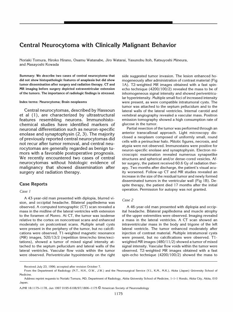
Central Neurocytoma with Clinically Malignant Behavior Noriaki Tomura, Hiroko Hirano, Osamu Watanabe, Jiro Watarai, Yasunobu Itoh, Katsuyoshi Mineura, and Masayoshi Kowada Summary: We describe two cases of central neurocytoma that did not show histopathologic features of anaplasia but did show tumor dissemination after surgery and radiation therapy. CT and MR imaging before surgery depicted extraventricular extension of the tumors. The importance of radiologic findings is stressed. Index terms: Neurocytoma; Brain neoplasms Central neurocytomas, described by Hassoun et al (1), are characterized by ultrastructural features resembling neurons. Immunohisto- chemical studies have identified markers of neuronal differentiation such as neuron-specific enolase and synaptophysin (2, 3). The majority of previously reported central neurocytomas did not recur after tumor removal, and central neu- rocytomas are generally regarded as benign tu- mors with a favorable postoperative prognosis. We recently encountered two cases of central neurocytomas without histologic evidence of malignancy that showed dissemination after surgery and radiation therapy. Case Reports Case 1 A 43-year-old man presented with diplopia, blurred vi- sion, and occipital headache. Bilateral papilledema was observed. A computed tomographic (CT) scan revealed a mass in the midline of the lateral ventricles with extension to the foramen of Monro. At CT, the tumor was isodense relative to the cortex on noncontrast scans and enhanced moderately on postcontrast scans. Multiple small cysts were present in the periphery of the tumor, but no calcifi- cations were observed. T1-weighted magnetic resonance (MR) images, 520/13/2 (repetition time/echo time/exci- tations), showed a tumor of mixed signal intensity at- tached to the septum pellucidum and lateral walls of the lateral ventricles. Vascular flow voids within the tumor were observed. Periventricular hypointensity on the right side suggested tumor invasion. The lesion enhanced ho- mogeneously after administration of contrast material (Fig 1A). T2-weighted MR images obtained with a fast spin- echo technique (4200/100/2) revealed the mass to be of inhomogeneous signal intensity and showed periventricu- lar hyperintensity. Multiple small foci of increased intensity were present, as were compatible intratumoral cysts. The tumor was attached to the septum pellucidum and to the lateral walls of the lateral ventricles. Internal carotid and vertebral angiography revealed a vascular mass. Positron emission tomography showed a high consumption rate of glucose in the tumor. Partial resection of the tumor was performed through an anterior transcallosal approach. Light microscopy dis- closed a neoplasm composed of uniformly small, round cells with a perinuclear halo. Mitotic figures, necrosis, and atypia were not observed. Immunostains were positive for neuron-specific enolase and synaptophysin. Electron mi- croscopic examination revealed numerous synapselike structures and spherical and/or dense-cored vesicles. Af- ter surgery, the patient received 60.6 Gy of radiation ther- apy. Two months after discharge, the patient’s visual acu- ity worsened. Follow-up CT and MR studies revealed an increase in the size of the residual tumor and newly formed disseminated tumors in the ventricular wall (Fig 1B). De- spite therapy, the patient died 17 months after the initial operation. Permission for autopsy was not granted. Case 2 A 46-year-old man presented with diplopia and occip- ital headache. Bilateral papilledema and muscle atrophy of the upper extremities were observed. Imaging revealed a mass in the lateral ventricles. A CT scan showed an intraventricular mass in the body and trigone of the left lateral ventricle. The tumor enhanced moderately after injection of contrast material. Multiple intratumoral cysts were present, but no calcifications were observed. T1- weighted MR images (480/11/2) showed a tumor of mixed signal intensity. Vascular flow voids within the tumor were observed. T2-weighted MR images obtained with a fast spin-echo technique (4200/100/2) showed the mass to Received July 23, 1996; accepted after revision October 7. From the Department of Radiology (N.T., H.H., O.W., J.W.) and the Neurosurgical Service (Y.I., K.M., M.K.), Akita (Japan) University School of Medicine. Address reprint requests to Noriaki Tomura, MD, Department of Radiology, Akita University School of Medicine, 1–1–1 Hondo, Akita City, Akita, 010 Japan. AJNR 18:1175–1178, Jun 1997 0195-6108/97/1806 –1175 © American Society of Neuroradiology 1175
-
Upload
truongtruc -
Category
Documents
-
view
219 -
download
0
Transcript of Central Neurocytoma with Clinically Malignant Behavior · Central Neurocytoma with Clinically...
Central Neurocytoma with Clinically Malignant Behavior
Noriaki Tomura, Hiroko Hirano, Osamu Watanabe, Jiro Watarai, Yasunobu Itoh, Katsuyoshi Mineura,and Masayoshi Kowada
Summary: We describe two cases of central neurocytoma thatdid not show histopathologic features of anaplasia but did showtumor dissemination after surgery and radiation therapy. CT andMR imaging before surgery depicted extraventricular extensionof the tumors. The importance of radiologic findings is stressed.
Index terms: Neurocytoma; Brain neoplasms
Central neurocytomas, described by Hassounet al (1), are characterized by ultrastructuralfeatures resembling neurons. Immunohisto-chemical studies have identified markers ofneuronal differentiation such as neuron-specificenolase and synaptophysin (2, 3). The majorityof previously reported central neurocytomas didnot recur after tumor removal, and central neu-rocytomas are generally regarded as benign tu-mors with a favorable postoperative prognosis.We recently encountered two cases of centralneurocytomas without histologic evidence ofmalignancy that showed dissemination aftersurgery and radiation therapy.
Case Reports
Case 1
A 43-year-old man presented with diplopia, blurred vi-sion, and occipital headache. Bilateral papilledema wasobserved. A computed tomographic (CT) scan revealed amass in the midline of the lateral ventricles with extensionto the foramen of Monro. At CT, the tumor was isodenserelative to the cortex on noncontrast scans and enhancedmoderately on postcontrast scans. Multiple small cystswere present in the periphery of the tumor, but no calcifi-cations were observed. T1-weighted magnetic resonance(MR) images, 520/13/2 (repetition time/echo time/exci-tations), showed a tumor of mixed signal intensity at-tached to the septum pellucidum and lateral walls of thelateral ventricles. Vascular flow voids within the tumorwere observed. Periventricular hypointensity on the right
side suggested tumor invasion. The lesion enhanced ho-mogeneously after administration of contrast material (Fig1A). T2-weighted MR images obtained with a fast spin-echo technique (4200/100/2) revealed the mass to be ofinhomogeneous signal intensity and showed periventricu-lar hyperintensity. Multiple small foci of increased intensitywere present, as were compatible intratumoral cysts. Thetumor was attached to the septum pellucidum and to thelateral walls of the lateral ventricles. Internal carotid andvertebral angiography revealed a vascular mass. Positronemission tomography showed a high consumption rate ofglucose in the tumor.
Partial resection of the tumor was performed through ananterior transcallosal approach. Light microscopy dis-closed a neoplasm composed of uniformly small, roundcells with a perinuclear halo. Mitotic figures, necrosis, andatypia were not observed. Immunostains were positive forneuron-specific enolase and synaptophysin. Electron mi-croscopic examination revealed numerous synapselikestructures and spherical and/or dense-cored vesicles. Af-ter surgery, the patient received 60.6 Gy of radiation ther-apy. Two months after discharge, the patient’s visual acu-ity worsened. Follow-up CT and MR studies revealed anincrease in the size of the residual tumor and newly formeddisseminated tumors in the ventricular wall (Fig 1B). De-spite therapy, the patient died 17 months after the initialoperation. Permission for autopsy was not granted.
Case 2
A 46-year-old man presented with diplopia and occip-ital headache. Bilateral papilledema and muscle atrophyof the upper extremities were observed. Imaging revealeda mass in the lateral ventricles. A CT scan showed anintraventricular mass in the body and trigone of the leftlateral ventricle. The tumor enhanced moderately afterinjection of contrast material. Multiple intratumoral cystswere present, but no calcifications were observed. T1-weighted MR images (480/11/2) showed a tumor of mixedsignal intensity. Vascular flow voids within the tumor wereobserved. T2-weighted MR images obtained with a fastspin-echo technique (4200/100/2) showed the mass to
Received July 23, 1996; accepted after revision October 7.From the Department of Radiology (N.T., H.H., O.W., J.W.) and the Neurosurgical Service (Y.I., K.M., M.K.), Akita (Japan) University School of
Medicine.Address reprint requests to Noriaki Tomura, MD, Department of Radiology, Akita University School of Medicine, 1–1–1 Hondo, Akita City, Akita, 010
Japan.
AJNR 18:1175–1178, Jun 1997 0195-6108/97/1806–1175 © American Society of Neuroradiology
1175

Fig 1. Case 1: 43-year-old man withdiplopia, blurred vision, and occipitalheadache.
A, Contrast-enhanced coronal T1-weighted MR image (520/13/2) showsmass (thick arrows) in the right lateralventricle extending into the left lateral ven-tricle. Contrast enhancement of the solidportion is observed. Note hypointensity inperiventricular white matter on right side(thin arrows), suggesting edema.
B, Contrast-enhanced axial T1-weighted MR image (500/19/2) 15months after surgery reveals disseminatedtumor in all cerebrospinal fluid–containingspaces.
1176 TOMURA AJNR: 18, June 1997
have heterogeneous signal intensity (Fig 2A). PostcontrastT1-weighted MR images depicted extension of the tumorto the thalamus and the cistern of the velum interpositum(Fig 2B and C). Angiography revealed a hypervasculartumor.
At surgery, the tumor occupied the body and trigone ofthe left lateral ventricle. The tumor appeared to arise fromthe superior aspect of the thalamus and to extended intothe cistern of the velum interpositum. Light microscopicpathologic examination revealed a neoplasm composed ofuniformly small, round cells with perinuclear halos. Nei-ther cell pleomorphism nor mitosis was observed. Immu-nostains were positive for neuron-specific enolase andsynaptophysin. Synapselike structures and sphericaland/or dense-cored vesicles were detected by electronmicroscopy.
After partial resection of the tumor, the patient received60 Gy of local brain irradiation. MR imaging 5 months aftersurgery revealed tumor growth in the third ventricle and inthe frontal horn of the lateral ventricle. An additional 56 Gyof local brain irradiation was administered. MR imaging 7months after surgery showed disseminated tumor (Fig 2Dand E).
Discussion
Since the report by Hassoun et al in 1982 (1),more than 80 central neurocytomas have beenreported. Central neurocytoma and cerebralneuroblastoma have often been confused in theliterature. Cerebral neuroblastoma may be dif-ferentiated from central neurocytomas bymeans of light microscopy, because cerebralneuroblastomas are composed of immaturecells (4). Radiologically, the typical appearanceof a central neurocytoma as reported in theliterature (5–12) is that of a well-circumscribed
mass confined to the anterior portion of thelateral ventricles. Punctate or coarse calcifica-tions and multiple small cysts within the tumorare often observed. Mild to moderate contrastenhancement is common. Intraventricular oli-godendroglioma or ependymoma may be indis-tinguishable from central neurocytoma withoutultrastructural and immunohistochemical stud-ies. A number of published cases of intraven-tricular oligodendroglioma may actually be ex-amples of central neurocytoma that weremisdiagnosed pathologically.
Previously reported central neurocytomashave usually been confined to the ventricularsystem. Our literature review found six cases ofextraventricular extension (13, 14). Three ofthese six were histologically proved to be ana-plastic or malignant central neurocytomas,showing increased mitotic activity, presence ofnecrosis, and vascular proliferation. Althoughour cases did not show histopathologic featuresof malignancy, Wichmann et al (9) reportedthat extraventricular extension of central neuro-cytoma most probably indicates malignanttransformation of the tumor.
The benefits of radiation therapy and/or che-motherapy after surgery for central neurocyto-mas are controversial and may depend on thepresence or absence of a residual tumor. Theresponse of central neurocytomas to radiationor chemotherapy have yet to be determined.More than 50% of patients described in the lit-erature received radiation therapy after subtotalresection or biopsy (3, 4, 6, 7, 13–19). Twocases of central neurocytoma with evidence of

Fig 2. Case 2: 46-year-old man withdiplopia and occipital headache.
A, Axial T2-weighted MR image (4200/100/2) shows hyperintense but inhomoge-neous mass (arrows) containing cysts.
B and C, Contrast-enhanced axial(480/11/1) (B) and coronal (440/14/2)(C) T1-weighted MR images show a sug-gestion of tumor invasion (arrowheads)into the thalamus and cistern of the veluminterpositum.
D and E, Postcontrast axial T1-weighted MR images (480/13/2) 7 monthsafter surgery reveal disseminated tumorsin the right frontal lobe and near the fora-men of Magendie.
AJNR: 18, June 1997 CENTRAL NEUROCYTOMA 1177
response to radiation therapy have been re-ported. Even when patients did not receive ra-diation therapy, the majority had no recurrence(5, 16, 20). We found recurrence of central neu-rocytoma reported in four cases. Three of eightcases reported by Yasargil et al (13) and one ofseven cases reported by Kim et al (14) (none ofwhich showed histopathologic features ofanaplasia) had recurrence after removal. Theperiod of recurrence after surgery ranged from 2to 72 months.
We found three cases of central neurocytomawith potentially malignant central neurocyto-mas reported previously (9, 13). Two patientsreceived postoperative radiation therapy andhad no recurrence after 5 and 12 months, re-spectively (13). Therefore, standard his-topathologic evaluation may not always corre-late with the potential for regrowth of centralneurocytomas. Evaluation of glucose con-sumption of the tumor with positron emissiontomography can be useful for predicting the
potential of regrowth (21). We suggest thatpostoperative radiation therapy be consideredin cases of tumors with radiologic evidence ofextraventricular extension. Radiation therapy isalso useful for patients in whom there is his-topathologic evidence of malignancy.
References1. Hassoun J, Gambarelli D, Grisoli F, et al. Central neurocytoma: an
electron-microscopic study of two cases. Acta Neuropathol (Berl)1982;56:151–156
2. von Deimling A, Janzer R, Kleihues P, Wiestler OD. Patterns ofdifferentiation in central neurocytoma: an immunohistochemicalstudy of eleven biopsies. Acta Neuropathol 1990;79:473–479
3. Kubota T, Hayashi M, Kawano H, et al. Central neurocytoma:immunohistochemical and ultrastructural study. Acta Neuro-pathol 1991;81:418–427
4. Louis DN, Swearingen B, Linggood RM, et al. Central nervoussystem neurocytoma and neuroblastoma in adults: report of eightcases. J Neurooncol 1990;9:231–238
5. Smoker WRK, Townsend JJ, Reichman MV. Neurocytoma ac-companied by intraventricular hemorrhage: case report and liter-ature review. AJNR Am J Neuroradiol 1991;12:765–770
6. Bolen JW Jr, Lipper MH, Caccamo D. Case report. Intraventricular
central neurocytoma: CT and MR findings. J Comput Assist To-mogr 1989;13:495–497
7. Patil AA, McComb RD, Gelber B, McConnell J, Sasse S. Intraven-tricular neurocytoma: a report of two cases. Neurosurgery 1990;26:140–144
8. Porter-Grenn LM, Silbergleit R, Stern HJ, Patel SC, Mehta B, Sand-ers WP. Intraventricular primary neuronal neoplasms: CT, MR, andangiographic findings. J Comput Assist Tomogr 1991;15:365–368
9. Wichmann W, Schubiger O, von Deimling A, Schenker Ch, Vala-vanis A. Neuroradiology of central neurocytoma. Neuroradiology1991;33:143–148
10. Goergen SK, Gonzales MF, McLean CA. Intraventricular neurocy-toma: radiologic features and review of the literature. Radiology1992;182:787–792
11. Chang KH, Han MH, Kim DG, et al. MR appearance of centralneurocytoma. Acta Radiol 1993;34:520–526
12. McConachie NS, Worthington BS, Cornford EJ, Balsitis M, Ker-slake RW, Jaspan T. Review article: computed tomography andmagnetic resonance in the diagnosis of intraventricular cerebralmasses. Br J Radiol 1994;67:223–243
13. Yasargil MG, von Ammon K, von Deimling A, Valavanis A, Wich-mann W, Wiestler OD. Central neurocytoma: histopathological
1178 TOMURA
variants and therapeutic approaches. J Neurosurg1992;76:32–37
14. Kim DG, Chi JG, Park SH, et al. Intraventricular neurocytoma:clinicopathological analysis of seven cases. J Neurosurg 1992;76:759–765
15. Laidlaw JD, McLean A, Siu K, Gonzales MF. Intraventricular neu-rocytoma, a recently recognized pathological entity: report of twocases and review of the literature. Br J Neurosurg 1991;5:371–378
16. Nishio S, Tashima T, Takeshita I, Fukui M. Intraventricular neu-rocytoma: clinicopathological features of six cases. J Neurosurg1988;68:665–670
17. Tsujita Y, Nagashima K, Takakura K. A clinicopathological studyof central neurocytoma. Brain Nerve 1989;41:547–558
18. Barbosa MD, Balsitis M, Jaspan T, Lowe J, Path MRC. Intraventric-ular neurocytoma: a clinical and pathological study of three casesand review of the literature. Neurosurgery 1990;26:1045–1054
19. Harada M, Morioka T, Nishio S, Fukui M. Neurocytoma in leftfrontal lobe. Neurol Surg 1991;19:89–92
20. Ferreol E, Sawaya R, de Courten-Myers GM. Primary cerebralneuroblastoma (neurocytoma) in adults. J Neurooncol 1989;7:121–128
21. Mineura K, Sasajima T, Itoh Y, et al. Blood flow and metabolism ofcentral neurocytoma. Cancer 1995;76:1224–1232
AJNR: 18, June 1997