Center f Replication Origins in Bacterial Genomes - … · k DTU Replication Origins in Bacterial...
11
Comparative Microbial Genomics group Center for Biological Sequence Analysis The Technical University of Denmark DTU Replication Origins in Bacterial Genomes Dave Ussery Advanced Bioinformatics 24 November, 2006
Transcript of Center f Replication Origins in Bacterial Genomes - … · k DTU Replication Origins in Bacterial...

Comparative Microbial Genomics groupC
en
ter fo
r Bio
log
ica
l Se
qu
en
ce
An
aly
sis
Th
e T
ech
nic
al U
niv
ers
ity o
f De
nm
ark
DT
U
Replication Origins in Bacterial Genomes
Dave UsseryAdvanced Bioinformatics24 November, 2006

Comparative Microbial Genomics groupC
en
ter fo
r Bio
log
ica
l Se
qu
en
ce
An
aly
sis
Th
e T
ech
nic
al U
niv
ers
ity o
f De
nm
ark
DT
U
(Nielsen et al., 2006). The rearrangement mechanismitself may bias the relative nucleoid configurations to thetandem arrangement. Alternatively, the rearrangementmechanism might be indifferent to which arm is to bemoved, but the DNA segregation mechanism biases theplacement of newly replicated DNA in the central cellquarters in such a way that a tandem configurationbecomes likely.
In any case, it is clear that a previously unsuspectedmechanism exists for DNA arrangement at the cellquarters. The nature of this mechanism, the informationpresent on the chromosome arms to direct it, and the
proteins present at the quarters that we presume to carryit out, remain to be identified.
Experimental procedures
Bacterial strains and plasmids
The strains used in this study are all derivatives of strainFH2926, a DlacI-lacA derivative of MG1655, carrying thedifferent P1parS sequence and the pMT1parS sequence atvarious chromosome positions. The strains were constructedas described in Supplementary material. The construction ofplasmid pFHC2973, which carries both the P1-parB gene andthe pMT1-parB gene fused to the genes for the fluorescentproteins CFP and yGFP, respectively, is also described inSupplementary material.
Growth conditions, microscopy and measurements
The general methods employed were as previouslydescribed (Nielsen et al., 2006).
References
Bates, D., and Kleckner, N. (2005) Chromosome and repli-some dynamics in E. coli: loss of sister cohesion triggersglobal chromosome movement and mediates chromosomesegregation. Cell 121: 899–911.
Brendler, T., Sawitzke, J., Sergueev, K., and Austin, S.(2000) A case for sliding SeqA tracts at anchored replica-tion forks during Escherichia coli chromosome replicationand segregation. EMBO J 19: 6249–6258.
Dabrazhynetskaya, A., Sergueev, K., and Austin, S. (2005)Species and incompatibility determination within the P1parfamily of plasmid partition elements. J Bacteriol 187: 5977–5983.
Dingman, C.W. (1974) Bidirectional chromosome replication:some topological considerations. J Theor Biol 43: 187–195.
Lau, I.F., Filipe, S.R., Soballe, B., Okstad, O.A., Barre, F.X.,and Sherratt, D.J. (2003) Spatial and temporal organizationof replicating Escherichia coli chromosomes. Mol Microbiol49: 731–743.
Li, Y., Sergueev, K., and Austin, S. (2002) The segregation ofthe Escherichia coli origin and terminus of replication. MolMicrobiol 46: 985–996.
Nielsen, H.J., Li, Y., Youngren, B., Hansen, F.G., and Austin,S. (2006) Progressive segregation of the Escherichia colichromosome. Mol Microbiol 61: 383–393.
Niki, H., and Hiraga, S. (1998) Polar localization of the repli-cation origin and terminus in Escherichia coli nucleoidsduring chromosome partitioning. Genes Dev 12: 1036–1045.
Sunako, Y., Onogi, T., and Hiraga, S. (2001) Sister chromo-some cohesion of Escherichia coli. Mol Microbiol 42:1233–1241.
Wang, X., Possoz, C., and Sherratt, D.J. (2005) Dancingaround the divisome: asymmetric chromosome segrega-tion in Escherichia coli. Genes Dev 19: 2367–2377.
Yamazoe, M., Adachi, S., Kanaya, S., Ohsumi, K., andHiraga, S. (2005) Sequential binding of SeqA protein to
Fig. 6. Chromosome rearrangement model.A. At initiation, the origin region of the chromosome (black circles)is at the cell centre, and the replication forks (yellow triangles) formthere. The two arms (light blue and pink lines) of the chromosomeare arranged either side of the cell centre. The terminus (greensquare) is near the new cell pole.B. At mid-replication, the forks have dissociated from the cell centreand are following the path of the DNA template. The newlyreplicated DNA products (dark blue and red) stay together for awhile (sister loci cohesion, grey hatching) and then segregate toopposite sides of the cell centre. This DNA is relativelydisorganized and resides in the central half of the cell.C. As replication progresses, the new origins attach to the cellquarter positions and the two arms of each nascent chromosomeare sorted out so that the arms lie on either side of the cellquarters.D. At termination, the origins are at the quarter positions, and theterminus and forks are at the cell centre. The arms (replichores) ofthe two new chromosomes are arranged with respect to the cellquarters. After the following post-replicational period (D-period), celldivision will restore the starting state of the cycle.
E. coli chromosome segregation 337
© 2006 The AuthorsJournal compilation © 2006 Blackwell Publishing Ltd, Molecular Microbiology, 62, 331–338
Nielsen et al., Mol. Micro., 62:331-338 (Okt. 2006).
Location, location, location...

Comparative Microbial Genomics groupC
en
ter fo
r Bio
log
ica
l Se
qu
en
ce
An
aly
sis
Th
e T
ech
nic
al U
niv
ers
ity o
f De
nm
ark
DT
U
Transcriptio
n
Perc
enta
ge o
f gen
ome
Num
ber o
f gen
omes
Replication,
recombination
and repair Signal
transductio
n
mechanismsCell w
all or
cell membrane
biogenesis
V. choleraeV. fischeriV. parahaemolyticusV. vulnificus YJ016
P. profundumAquaticHost-associatedTerrestrial
Cell motili
ty
Post-translatio
nal
modification and
protein turnover
Energy production
and conversion
0
01 2 3 4 5 6 7 8 9 10 11 12 13 14 15 0 20 40 60 80 100 120 140 160
20
40
60
Num
ber o
f gen
omes
0
20
40
60
2
4
6
8
10a
b c
Number of 16S rRNAs Number of tRNAs
COGs
!"#$%&'()*$+(,()$-).$/"(%()$/&))#/%(&)
The two main virulence factors of V. cholerae, cholera toxin (CT), which is encoded on the filamentous cholera toxin phage (CTX!) and causes a profuse watery diarrhoea, and the toxin co-regulated pilus (TCP), which is an essential intestinal coloniza tion factor and the host receptor for CT, were both acquired by a subset of isolates by horizontal gene transfer (HGT)44,45. In the aquatic environment, V. cholerae has been found in association with zooplankton and phytoplankton, on the chitinous exoskeletons and moults of copepods (crustaceans) and in the mucilaginous sheaths of blue-green algae46–49. The mannose-sensitive haemag-glutinin (MSHA), which is encoded by all the sequenced Vibrionaceae genomes, is involved in V. cholerae adherence to zooplankton50. Recent studies have described a role for a
chitin-binding protein, ORF VCA0811, from V. cholerae in attachment to both the chitinous exoskeleton of zooplankton and human epithelial cells, by binding to the sugars present on both surfaces51,52. This protein is found in the genomes of all sequenced Vibrionaceae and might also be involved in the environmental persistence of these bacteria.
V. fischeri exists naturally either in a free-living planktonic state or as a sym-biont of the luminescent bobtail squid, E. scolopes53. V. fischeri provides the source of luminescence, which the squid can use for camouflage, protection against preda-tors, and which might also be involved in mating8. The bacteria only luminesce in this symbiotic state in the light organ and not when they are free living. The interaction of V. fischeri with the light organ of squid
involves several genes, including mot, fla, flr and rpoN8. Colonization is aided by the secretion of a mucoid substance above the pores of the light organ, which traps the V. fischeri, and MSHA is required for subsequent colonization54,55.
Although, at first glance, the human pathogen V. cholerae and the squid symbiont V. fischeri have markedly different lifestyles, there are suggestive similarities between both species. In particular, the abundance of chitinase genes in their genomes reflects the high degradative ability attributed to vibrios56. Chitin is a biopolymer of N-acetylglucosamine and, after cellulose, is the most abundant carbohydrate polymer; it is present in large quantities in the sea, being a constituent of the exoskeletons of crus-taceans and zooplankton. Chitin induces TCP production, which, along with MSHA, allows V. cholerae to colonize the exoskel-etons of crustaceans and zooplankton57,58. V. cholerae, in addition to V. fischeri, can use chitin as a carbon and nitrogen source. Homologues of TCP were identified in the V. fischeri genome sequence5. Chitin might therefore have a similar role in V. fischeri, stimulating TCP-mediated biofilm forma-tion or perhaps even facilitating colonization of the squid.
The diversity of TCP-mediated bacteria –host interactions in the aquatic niche has led to speculation on the role of CT in the environment59,60. CT might function as an osmoregulator for crustaceans by the removal of salts from the cell by its intrinsic function. Briefly, CT increases Cl– secretion and reduces Na+ absorption, which could be advantageous to the crustacean as it moves into environments of increasing salinity60. As V. cholerae is associated with copepods, V. cholerae might establish a symbiotic relationship with these crustaceans, obtain-ing a suitable place (chitinous exoskeleton) to attach and feed, and providing the host with a powerful osmoregulator (CT). Homologues of the CTX prophage were identified in the sequenced genome of V. fischeri, however this prophage did not carry the ctxAB genes5. CTX prophage lacking the ctxAB genes were also observed in V. cholerae, indicating the presence of a precursor CTX phage that acquired the CT genes from a still-unknown source61.
Another pathogenicity factor in V. cholerae is neuraminidase, which is encoded by the nanH gene on Vibrio pathogenicity island-2 (VPI-2)62. Neuraminidase cleaves mucin from intestinal cells, unmasking GM1 gangliosides, the receptors for CT, and releasing sialic acid, a carbon source63,64.
!"#$%&'(')'!"#$%"&%'#'#(&)$&*"+'#"&,'-.'$#/0"/"&01/./0)".'2)'023&/')'*"+,%"-$,"./'.0'12$+,&%+'
.0'.%,3.2.#.$+'#%.$4+'5678+9':;./#',3&'<"-%"./:1&:&'"/',3&'=">&%'1./,&?,'.0':@$:,"1A'3.+,B
:++.1":,&>':/>',&%%&+,%":2'+4&1"&+C'D3&'>:,:':E:"2:-2&':,'F6GH'=&%&'$+&>',.'#&/&%:,&':'>"+,%"-$B
,"./'13:%,'"/'=3"13',3&'#&/&'1./,&/,'0.%':/I'4:%,"1$2:%'0$/1,"./'1:/'-&'1.;4:%&>':1%.++',3&'
#&/$+':/>'"/',3&'1./,&?,'.0',3&'J%.,&.-:1,&%":':/>'-:1,&%":':+':'=3.2&C'-A'0')'*"+,%"-$,"./'.0'
%KFL':/>',KFL'#&/&+':;./#',3&'+&@$&/1&>'-:1,&%":2'#&/.;&+C'D3&'+&@$&/1&>'E"-%".+':%&'
"/>"1:,&>'"/'I&22.=C'D3&'IB:?"+'%&4%&+&/,+',3&'/$;-&%'.0'+&@$&/1&>'#&/.;&+'1./,:"/"/#':'
4:%,"1$2:%'/$;-&%'.0'%KFL'.%',KFL'#&/&+'5?B:?"+9C'
!"#$!"%& '("$
0$| ADVANCE ONLINE PUBLICATION &4443#/)5."30$%6."7'"426%'0.$
©!2006!Nature Publishing Group!
!
Nature Rev. Micro, 4:697-704, (Sept. 2006).

Comparative Microbial Genomics groupC
en
ter fo
r Bio
log
ica
l Se
qu
en
ce
An
aly
sis
Th
e T
ech
nic
al U
niv
ers
ity o
f De
nm
ark
DT
U
rRN
ArR
NA
rRN
A
rRNArR
NA
rRN
ArR
NA
rRN
A
rRNArR
NA
Ori
gin
0M
0.5M1M
1.5M2
M2.5
M
BASE ATLAS
Center for Biological Sequence Analysishttp://www.cbs.dtu.dk/
G Contentfixavg
0.00 0.23
A Contentfixavg
0.00 0.45
T Contentfixavg
0.00 0.45
C Contentfixavg
0.00 0.23
Annotations:
CDS +
CDS -
rRNA
tRNA
AT Skewfixavg
-0.12 0.12
GC Skewfixavg
-0.10 0.10
Percent ATdevavg
0.67 0.75
Resolution: 1120
C. tetani E88 2,799,251 bp
Comparative Microbial Genomics groupC
en
ter fo
r Bio
log
ica
l Se
qu
en
ce
An
aly
sis
Th
e T
ech
nic
al U
niv
ers
ity o
f De
nm
ark
DT
U
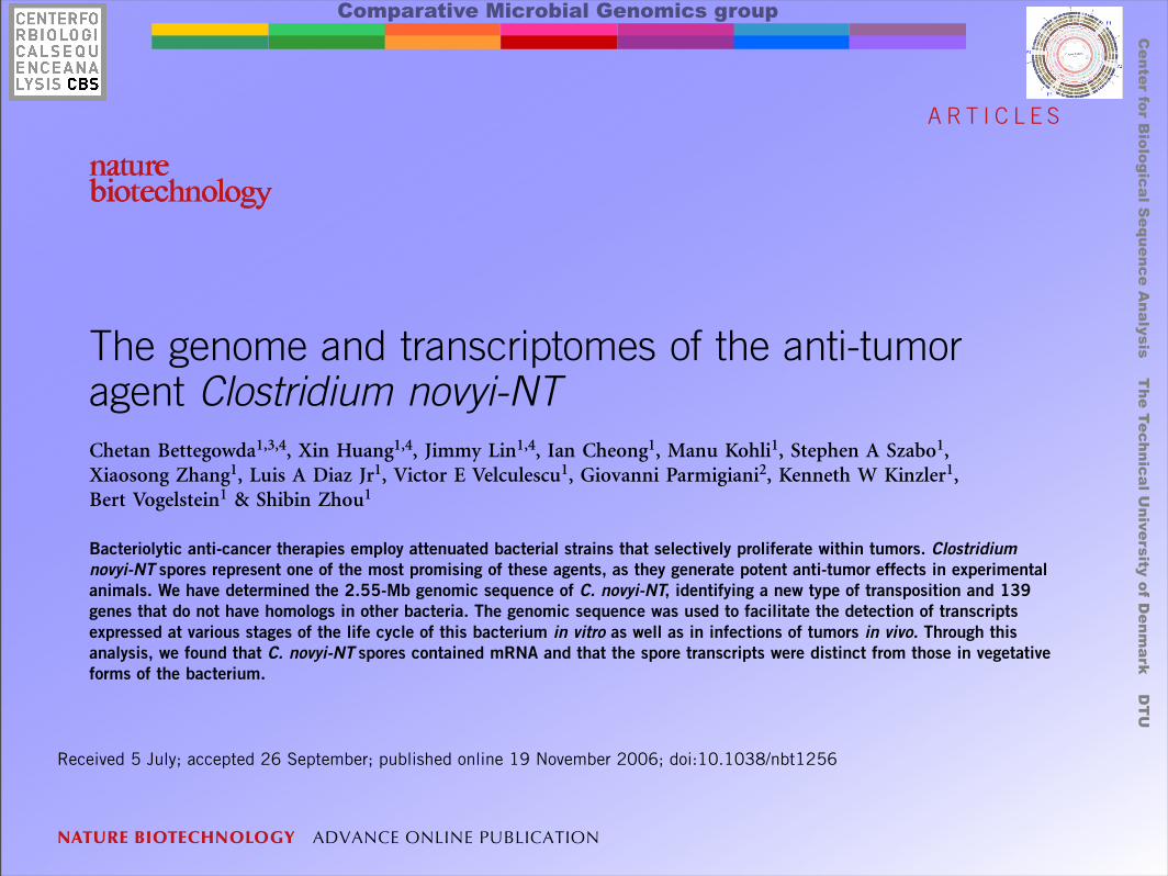
The genome and transcriptomes of the anti-tumoragent Clostridium novyi-NTChetan Bettegowda1,3,4, Xin Huang1,4, Jimmy Lin1,4, Ian Cheong1, Manu Kohli1, Stephen A Szabo1,Xiaosong Zhang1, Luis A Diaz Jr1, Victor E Velculescu1, Giovanni Parmigiani2, Kenneth W Kinzler1,Bert Vogelstein1 & Shibin Zhou1
Bacteriolytic anti-cancer therapies employ attenuated bacterial strains that selectively proliferate within tumors. Clostridiumnovyi-NT spores represent one of the most promising of these agents, as they generate potent anti-tumor effects in experimentalanimals. We have determined the 2.55-Mb genomic sequence of C. novyi-NT, identifying a new type of transposition and 139genes that do not have homologs in other bacteria. The genomic sequence was used to facilitate the detection of transcriptsexpressed at various stages of the life cycle of this bacterium in vitro as well as in infections of tumors in vivo. Through thisanalysis, we found that C. novyi-NT spores contained mRNA and that the spore transcripts were distinct from those in vegetativeforms of the bacterium.
It has been known for over a century that severe bacterial infections incancer patients occasionally result in the eradication of malignancy1.These clinical observations spawned attempts to use bacteria to treattumors in laboratory animals and in cancer patients2–4. Anaerobicbacteria are particularly intriguing agents for this purpose, as the onlytissues allowing the growth of such bacteria in otherwise healthymammals are within tumors5. Indeed, a majority of human tumorscontain large hypoxic regions that make them relatively insensitive toradiation or chemotherapy but provide an ideal environment for thegrowth of anaerobic bacteria6,7. Most previous attempts to useanaerobic bacteria for tumor therapeutics employed Clostridiumsporogenes, a nonpathogenic species often used as a control forsterilization in the food industry. We developed an attenuated strainof the pathogenic clostridial species C. novyi, called C. novyi-NT, inwhich the phage episome containing the major systemic toxin genewas deleted8. When injected intravenously, C. novyi-NT spores pro-duced substantial anti-tumor effects in experimental animals withoutexcessive toxicity8–11. The spores of C. novyi-NT are very stable but thevegetative form is exquisitely sensitive to oxygen11.C. novyi-NT lies on a distinct branch of the genus Clostridium
and no previous sequencing studies of it have been published.Although a considerable number of clostridial species are of medicaland biotechnological importance, only four (C. acetobutylicum12,C. difficile13, C. perfringens14 and C. tetani15) have been analyzedby complete genomic sequencing. Additionally, the genome ofC. botulinum is currently under annotation (http://www.sanger.ac.uk/Projects/C_botulinum/). As judged by 16S RNA sequences, C. novyi isnot highly related to any of these sequenced organisms. To better
understand how clostridia function as anti-neoplastic agents, as well asto gain insight into the pathology of human clostridial diseases, wehave determined the sequence of the C. novyi-NT genome andanalyzed the transcriptomes of its vegetative and spore forms.
RESULTSC. novyi-NT genomeThe C. novyi-NT genome consists of a single circular chromosome,2,547,720 base pairs (bp) in length with a G+C content of 28.9%(Fig. 1 and Table 1). Deviant G+C content and trinucleotide compo-sition were almost completely confined to the regions that harboredthe rRNA operons (Fig. 1). No extrachromosomal sequences, such asthose that would be found in plasmids or phages, were identified. Thegeneral features of clostridial genomes are summarized in Table 1. TheC. novyi-NT genome is smaller and contains fewer coding sequences(CDS) than the other genomes. Although atypical G+C content istightly associated with the rRNA operons, several putative mobileelements were identified in the C. novyi-NT genome, includingtransposons, clustered regularly interspaced short palindromic repeats(CRISPR) and prophage elements (Table 2 and SupplementaryTables 1 and 2 online). A putative replication origin of theC. novyi-NT genome was indicated by a distinct inflection point inthe coding strand, which was localized around the DnaA gene (Fig. 1,at the top of the genome circle). Closer inspection of this regionrevealed multiple putative DnaA boxes (TTATCCACA) on bothstrands in intergenic regions between the dnaA (NT01CX0867) andrpmH (NT01CX0868) genes as well as between the dnaA and dnaN(NT01CX0866) genes. This distribution pattern of the DnaA boxes
©20
06 N
atur
e P
ublis
hing
Gro
up h
ttp:
//ww
w.n
atur
e.co
m/n
atur
ebiotechnology
Received 5 July; accepted 26 September; published online 19 November 2006; doi:10.1038/nbt1256
1The Howard Hughes Medical Institute, The Ludwig Center for Cancer Genetics & Therapeutics at The Sidney Kimmel Comprehensive Cancer Center, and Departmentof Pharmacology and Molecular Sciences, Johns Hopkins Medical Institutions, 1650 Orleans Street, Baltimore, Maryland 21231, USA. 2Departments of Oncology,Biostatistics and Pathology, Johns Hopkins Medical Institutions, 550 North Broadway, Baltimore, Maryland 21205, USA. 3Present address: Department of Neurologyand Neurosurgery, Johns Hopkins Medical Institutions, 600 N. Wolfe Street, Baltimore, Maryland 21287, USA. 4These authors contributed equally to this work.Correspondence should be addressed to S.Z. ([email protected]).
NATURE BIOTECHNOLOGY ADVANCE ONLINE PUBLICATION 1
A R T I C L E S

Comparative Microbial Genomics groupC
en
ter fo
r Bio
log
ica
l Se
qu
en
ce
An
aly
sis
Th
e T
ech
nic
al U
niv
ers
ity o
f De
nm
ark
DT
U
gene expression can be rescued separately bydistinct spatial expression patterns of Bmal1.Genome-wide profiling experiments suggestthat ~10% of the transcriptome is under cir-cadian regulation; however, the majority ofthese cycling transcripts are tissue-specific(18–22). Our results are consistent with thistissue-specific diversity of circadian expres-sion and further suggest that core circadianclock components may play distinct roles indifferent tissues, perhaps in addition to theirfunction in regulating circadian rhythms. Therestoration of circadian activity rhythms inbrain-rescued Bmal1!/! mice is consistent withprevious SCN transplant studies in rodents(23, 24). However, the transgenic approachused here has the advantages of preserving theanatomical integrity of the brain as well asallowing the conditional manipulation of therescue via Dox treatment. The use of tissue-specific and conditional regulation of circadianclock gene expression should be a valuablemethod for understanding the molecular-,
cellular-, and systems-level regulation of cir-cadian rhythms in mammals.
References and Notes1. P. L. Lowrey, J. S. Takahashi, Annu. Rev. Genomics Hum.
Genet. 5, 407 (2004).2. M. Ikeda, M. Nomura, Biochem. Biophys. Res. Commun.
233, 258 (1997).3. J. B. Hogenesch et al., J. Biol. Chem. 272, 8581 (1997).4. N. Gekakis et al., Science 280, 1564 (1998).5. J. B. Hogenesch, Y. Z. Gu, S. Jain, C. A. Bradfield,
Proc. Natl. Acad. Sci. U.S.A. 95, 5474 (1998).6. M. K. Bunger et al., Cell 103, 1009 (2000).7. R. D. Rudic et al., PLoS Biol. 2, e377 (2004).8. M. K. Bunger et al., Genesis 41, 122 (2005).9. A. Laposky et al., Sleep 28, 395 (2005).10. S. Shimba et al., Proc. Natl. Acad. Sci. U.S.A. 102, 12071
(2005).11. R. V. Kondratov, A. A. Kondratova, V. Y. Gorbacheva,
O. V. Vykhovanets, M. P. Antoch, Genes Dev. 20, 1868(2006).
12. Y. Sun et al., J. Biomed. Sci. 10.1007/s11373-006-9108-4(2006).
13. See supporting material on Science Online.14. M. Gossen, H. Bujard, Proc. Natl. Acad. Sci. U.S.A. 89,
5547 (1992).15. P. A. Furth et al., Proc. Natl. Acad. Sci. U.S.A. 91, 9302
(1994).
16. H. Hong et al., paper presented at the biennial meetingof the Society for Research on Biological Rhythms,Sandestin, FL, 21 to 25 May 2006 (www.conferences.uiuc.edu/conferences/conference.asp?ID=292).
17. N. Preitner et al., Cell 110, 251 (2002).18. B. Kornmann, N. Preitner, D. Rifat, F. Fleury-Olela,
U. Schibler, Nucleic Acids Res. 29, E51 (2001).19. R. A. Akhtar et al., Curr. Biol. 12, 540 (2002).20. S. Panda et al., Cell 109, 307 (2002).21. H. R. Ueda et al., Nature 418, 534 (2002).22. K. F. Storch et al., Nature 417, 78 (2002).23. M. R. Ralph, R. G. Foster, F. C. Davis, M. Menaker, Science
247, 975 (1990).24. M. Sujino et al., Curr. Biol. 13, 664 (2003).25. We thank N. Lampl, A. Jyawook, and A. Falk for technical
assistance; members of the Takahashi laboratory andF. Davis for expert advice; and K. Esser, E. Hardeman,and M. Mayford for plasmids. Supported by NIH grantsR01 ES005703 (C.A.B.) and P50 MH074924 (J.S.T.).
Supporting Online Materialwww.sciencemag.org/cgi/content/full/314/5803/1304/DC1Materials and MethodsSOM TextFigs. S1 to S8References
13 July 2006; accepted 11 October 200610.1126/science.1132430
A Bacterial Protein Enhances theRelease and Efficacy of LiposomalCancer DrugsIan Cheong, Xin Huang, Chetan Bettegowda, Luis A. Diaz Jr., Kenneth W. Kinzler,Shibin Zhou,* Bert Vogelstein*
Clostridium novyi-NT is an anaerobic bacterium that can infect hypoxic regions within experimentaltumors. Because C. novyi-NT lyses red blood cells, we hypothesized that its membrane-disruptingproperties could be exploited to enhance the release of liposome-encapsulated drugs withintumors. Here, we show that treatment of mice bearing large, established tumors with C. novyi-NTplus a single dose of liposomal doxorubicin often led to eradication of the tumors. The bacterialfactor responsible for the enhanced drug release was identified as a previously unrecognizedprotein termed liposomase. This protein could potentially be incorporated into diverse experimentalapproaches for the specific delivery of chemotherapeutic agents to tumors.
There is no dearth of drugs that can killcancer cells. The challenge is killing thecancer cells selectively while sparing the
normal cells. Three basic strategies are currentlyused to achieve this specificity. The first (se-lective toxicity) uses drugs that have more po-tent growth-inhibitory effects on tumor cellsthan on normal cells (1, 2). This strategy un-derlies the success of conventional chemothera-peutic agents as well as those of newer targetedtherapies such as imatinib (Gleevec). Thesecond strategy (delivery) uses agents such asantibodies that specifically react with molecules
that are predominantly expressed in tumor cells(3, 4). The third strategy (angiogenic) exploitsabnormal aspects of the tumor vasculature withagents such as bevacizumab (Avastin) (5, 6) ordrugs incorporated into liposomes (7–9). Lipo-somes are relatively large particles that canpenetrate through the fenestrated endotheliumpresent in tumors and a limited number of otherorgans (8, 9). Once they gain access to tumors,they persist and eventually release their contentsand raise local drug concentrations through theenhanced permeabilization and retention effect(10). Although each of these strategies hasmerit, the specificity achieved with any one ofthem is imperfect, limiting the amount of drugthat can be safely administered without causingsystemic toxicity.
Here, we describe our efforts to combine allthree strategies. We investigated C. novyi-NT,
an attenuated strain of the obligate anaerobeC. novyi. Similar to other bacteriolytic thera-pies, C. novyi-NT can selectively infect andpartially destroy experimental cancers becauseof the hypoxic nature of the tumor environment(11, 12). C. novyi-NT is also hemolytic (lyseserythrocytes). Because enzymes that ruptureerythrocytes can disrupt lipid bilayers (13), wehypothesized that the bacterium’s hemolyticproperties could be exploited to enhance the re-lease of liposome-encapsulated drugs withintumors. This approach would theoretically in-crease specificity by combining the selectivetumor toxicity of chemotherapeutic agents, theselective delivery of C. novyi-NT to tumors, andthe selective uptake of liposomes mediated bythe abnormal tumor vasculature.
To test this hypothesis, we first treated syn-geneic CT26 colorectal tumors in BALB/c mice.C. novyi-NT spores were injected intravenously,and once germination had begun in the tumors(~16 hours after injection), we administered asingle intravenous dose of liposomal doxoru-bicin (Doxil). Doxil is a liposomal formula-tion that encapsulates doxorubicin, a widelyused DNA-damaging chemotherapeutic agent.Liposome-encapsulated doxorubicin has beenshown to result in improved outcomes comparedwith unencapsulated doxorubicin (14). As previ-ously documented (15, 16), treatment with C.novyi-NT spores alone resulted in germinationand necrosis within the centrally hypoxic re-gion of tumors but left a well-oxygenatedviable rim that eventually regrew (Fig. 1A).Neither doxorubicin nor Doxil alone resultedin prolonged therapeutic effects in these mice.The combination of Doxil and C. novyi-NTspores, however, resulted in complete regressionof tumors in 100% of mice (Fig. 1A), and 65%of the mice were still alive at 90 days (Fig. 1B).
The Howard Hughes Medical Institute and the LudwigCenter for Cancer Genetics and Therapeutics, JohnsHopkins Kimmel Comprehensive Cancer Center, Baltimore,MD 21231, USA.
*To whom correspondence should be addressed. E-mail:[email protected] (S.Z.); [email protected] (B.V.)
24 NOVEMBER 2006 VOL 314 SCIENCE www.sciencemag.org1308
REPORTS

Comparative Microbial Genomics groupC
en
ter fo
r Bio
log
ica
l Se
qu
en
ce
An
aly
sis
Th
e T
ech
nic
al U
niv
ers
ity o
f De
nm
ark
DT
U
rplD
dnaJ
0M
0.5M1M
1.5M
2M C. novyi NT 2,547,720 bp
BASE ATLAS
Center for Biological Sequence Analysishttp://www.cbs.dtu.dk/
G Contentfixavg
0.00 0.25
A Contentfixavg
0.00 0.50
T Contentfixavg
0.00 0.50
C Contentfixavg
0.00 0.25
Annotations:
CDS +
CDS -
rRNA
tRNA
AT Skewfixavg
-0.15 0.15
GC Skewfixavg
-0.15 0.15
Percent ATfixavg
0.25 0.75
Resolution: 1020

Comparative Microbial Genomics groupC
en
ter fo
r Bio
log
ica
l Se
qu
en
ce
An
aly
sis
Th
e T
ech
nic
al U
niv
ers
ity o
f De
nm
ark
DT
U
rpoC
etfA
etfA
hgdC
0M
0.5M1M
1.5M
2M C. novyi NT 2,547,720 bp
GENOME ATLAS
Center for Biological Sequence Analysishttp://www.cbs.dtu.dk/
Intrinsic Curvaturedevavg
0.20 0.26
Stacking Energydevavg
-7.39 -6.45
Position Preferencedevavg
0.14 0.17
Annotations:
CDS +
CDS -
rRNA
tRNA
Global Direct Repeatsfixavg
5.00 7.50
Global Inverted Repeatsfixavg
5.00 7.50
GC Skewfixavg
-0.15 0.15
Percent ATfixavg
0.25 0.75
Resolution: 1020

Comparative Microbial Genomics groupC
en
ter fo
r Bio
log
ica
l Se
qu
en
ce
An
aly
sis
Th
e T
ech
nic
al U
niv
ers
ity o
f De
nm
ark
DT
U
??????? 3
© 2005 Society for Applied Microbiology and Blackwell Publishing Ltd, Environmental Microbiology
emi_917.fm
genomes, including Proteobacteria and Cyanobacteria wefound only a good match to the dnaE sequence. We thusobserved perfect agreement between the direction of theA/T skew and the presence/absence of a polC homologue(see Figs 1 and 2).
Skew strength and growth rate
The T. maritima origin of replication has proven difficult tofind from skews due to the low signal-to-noise ratio. How-ever, our method predicts an origin at position 164 kb (seeFig. 2D), close to where a consensus sequence for bac-terial origins has been found (Lopez et al., 2000). In con-trast, the origin is easily located within the F. nucleatumchromosome as the signal-to-noise ratio is high (seeFig. 2C). Nonetheless, the origin replication appears tohave been misplaced in the published sequence (Kapatralet al., 2002). Bacteriodes thetaiotaomicron is anotherrecent example where our origin prediction method pro-vides a reliable prediction, which differs considerably from
position zero in the published sequence (see Fig. 2B) (Xuet al., 2003).
The five sequenced Clostridia genomes stand out byhaving an extraordinarily strong strand bias. The signal-to-noise ratios are about 40 for all five genomes, for whichreason it is surprising that the origin was misplaced by50 kb in the recent publication of the Clostridium tetanigenome (Brüggemann et al., 2003). The Clostridiumgroup contains the fastest replicating organisms known,e.g. C. perfringens that has a minimal generation time ofeight minutes (Shimizu et al., 2002), which is likely to putsevere strains on the genome architecture. In contrast, theworst signal-to-noise ratios among the Firmicutes areobserved for Mollicutes, i.e. Mycoplasma and Urea-plasma. These organisms have long doubling time despitetheir small genomes, e.g. 94 min for Mycoplasma capri-colum where replication takes place at a rate of only~100 bp s-1 (Seto and Miyata, 1998). From these obser-vations we speculate that high growth rate may in generalresult a strong strand bias, makes sense given that the
1
Proteobacteria(101)
Chlamydiae(9)
Planctomycetes(1)
Cyanobacteria(8)
Actinobacteria(17)
Aquificae(1)
Thermotogae(1)
Deinococcus(3)
Firmicutes(55)
Fusobacteria(1)
Bacteriodetes(3)
Spirochaetes(8)
Bacteriodes thetaiotaomicronPorphyromonas gingivalis
Fusobacterium nucleatum
Bacillus subtilisClostridium perfringensMycoplasma genitalium
Deinococcus radiodurans Chr. 1Deinococcus radiodurans Chr. 2Thermus thermophilus
Thermotoga maritima
Aquifex aeolicus
Mycobacterium tubercolosisStreptomyces coelicolor
Gloeobacter violaceusSynechococcus sp.Synechocystis sp.
Rhodopirellula baltica
Chlamydia trachomatis
Rickettsia prowazekiiNeisseria meningitidisEscherichia coli K-12Desulfovibrio vulgarisCampylobacter jejuni
Leptospira interrogans Chr. 1Leptospira interrogans Chr. 2Treponema pallidum
------
-
-
-
---
-----
PolCG/C skewA/T skewPhyla
-10% 10% 20%0%
Example speciesSignal-to-noise ratio
0 10 20 30
Fig. 1. Phylogenetic overview of skews. For each phyla, the distribution of signal-to-noise ratios is represented by a box-and-whiskers plot based on the 10, 25, 50, 75 and 90 per-centiles. Within each phyla the presence/absence of a PolC homologue is shown for selected species along with the G/C and A/T skews, shown as blue and green bars respectively. Note that the A/T skew is positive for all species with a PolC homologue and vice versa.
CO
LOR
FIG
UR
E
environmental m
icrobiologyV
OLU
ME
8N
UM
BE
R 2
PAG
ES
185–370F
EB
RU
AR
Y2006
1462-2912(200602)8:2;1-2
Environmental Microbiology, Volume 8, Issue 2, February 2006
Genomics update185 Protecting sausages with bacteria instead of salt
M. Y. Galperin
Minireview
193 Environmental tuning of mutation rates C. Saint-Ruf & I. Matic
Research articles
200 daime, a novel image analysis program for microbial ecology and biofilmresearchH. Daims, S. Lücker & M. Wagner
214 Nitrosospira spp. can produce nitrous oxide via a nitrifier denitrification pathway L. J. Shaw, G. W. Nicol, Z. Smith, J. Fear, J. I. Prosser & E. M. Baggs
223 Microbial community structure in polluted Baltic Sea sediments A. Edlund, T. Soule, S. Sjöling & J. K. Jansson
233 Effect of above-ground plant species on soil microbial community structure andits impact on suppression of Rhizoctonia solani AG3 P. Garbeva, J. Postma, J. A. van Veen & J. D. van Elsas
247 Alteration and resilience of the soil microbial community following compostamendment: effects of compost level and compost-borne microbial community C. Saison, V. Degrange, R. Oliver, P. Millard, C. Commeaux, D. Montange & X. Le Roux
258 Diversity and phylotype consistency of bacteria in the guts of three bee species(Apoidea) at an oilseed rape field K. I. Mohr & C. C. Tebbe
273 Genomic comparisons among g-proteobacteria J. Mrázek, A. M. Spormann & S. Karlin
289 Development and validation of a prototype 16S rRNA-based taxonomicmicroarray for AlphaproteobacteriaH. Sanguin, A. Herrera, C. Oger-Desfeux, A. Dechesne, P. Simonet, E. Navarro, T. M. Vogel, Y. Moënne-Loccoz, X. Nesme & G. L. Grundmann
This journal is available online at Blackwell Synergy. Visit www.blackwell-synergy.com to search the articles and register for table of contents e-mail alerts.
Environmental Microbiology is covered by Index Medicus, SciSearch, ISI Alerting Services, Biotechnology Citation Index, Current Contents/Life Sciences,
and Current Contents/Agriculture, Biology and Environmental Sciences.
Typeset by SNP Best-set Typesetter Ltd., Hong Kong.
Published jointly by the Society for Applied Microbiology and Blackwell Publishing.
Forthcoming papers in Environmental Microbiology
MinireviewExocellular electron transfer in anaerobic microbial communitiesA. J. M. Stams, F. A. M. de Bok, C. M. Plugge, M. H. A. van Eekert, J. Dofling & G. Schraa
Research articlesStructural diversity of bacterial communities in a heavy metal mineralized graniteoutcropD. Gleeson, F. McDermott & N. Clipson
Diversity and ubiquity of thermophilic methanogenic archaea in temperate anoxicsoilsX.-L. Wu, M. W. Friedrich & R. Conrad
Selective enrichment and molecular characterization of a previously unculturedNitrospira-like bacterium from activated sludgeE. Spieck, C. Hartwig, I. McCormack, F. Maixner, M. Wagner, A. Lipski & H. Daims
The ‘pH optimum anomaly’ of intracellular enzymes of Ferroplasma acidiphilumO. V. Golyshina, P. N. Golyshin, K. N. Timmis & M. Ferrer
Design of Shewanella-specific 16S rRNA primers and application to analysis of Shewanella in a minerotrophic wetlandS. G. Todorova & A. M. Costello
Identifying pioneer bacterial species responsible for biofouling membranebioreactorsK. Zhang, H. Choi, D. D. Dionysiou, G. A. Sorial & D. B. Oerther
Measurement of Prochlorococcus ecotypes using real-time polymerase chainreaction reveals different abundances of genotypes with similar light physiologiesN. A. Ahlgren, G. Rocap & S. W. Chisholm
Pseudomonas, the dominant polycyclic aromatic hydrocarbon-degrading bacteriaisolated from Antarctic soils and the role of large plasmids in horizontal genetransferY. Ma, L. Wang & Z. Shao
Brief reportsAnalysis of the transmission of Salmonella spp. through generations of pet snakesM. Schröter, A. Speicher, J. Hofmann & P. Roggentin
Utilization of capsaicin and vanillylamine as growth substrates by Capsicum (hotpepper)-associated bacteriaS. F. Flagan & J. R. Leadbetter
VOLUME 8 NUMBER 2 FEBRUARY 2006 www.env-micro.com ISSN 1462-2912
environmentalmicrobiology
Contents
Microarray for Alphaproteobacteria
Diversity of bacteria in three bee species
Environmental tuning of mutation rates
Methylation of naphthalene by sulfate reducers
308 DNA extraction procedure: a critical issue for bacterial diversity assessment inmarine sediments G. M. Luna, A. Dell’Anno & R. Danovaro
321 The active methanotrophic community in hydromorphic soils changes inresponse to changing methane concentration C. Knief, S. Kolb, P. L. E. Bodelier, A. Lipski & P. F. Dunfield
334 Sequence analysis of three plasmids harboured in Rhodococcus erythropolisstrain PR4 M. Sekine, S. Tanikawa, S. Omata, M. Saito, T. Fujisawa, N. Tsukatani, T. Tajima, T. Sekigawa, H. Kosugi, Y. Matsuo, R. Nishiko, K. Imamura, M. Ito,H. Narita, S. Tago, N. Fujita & S. Harayama
347 Methylation is the initial reaction in anaerobic naphthalene degradation by asulfate-reducing enrichment culture M. Safinowski & R. U. Meckenstock
353 Origin of replication in circular prokaryotic chromosomes P. Worning, L. J. Jensen, P. F. Hallin, H.-H. Stærfeldt & D. W. Ussery
Brief report
362 Reduction of Prussian Blue by the two iron-reducing microorganisms Geobactermetallireducens and Shewanella algaM. K. Jahn, S. B. Haderlein & R. U. Meckenstock
Web alert
368 Environmental microbiological microarraysL. P. Wackett
Corrigendum
370 Exploring the diversity of myxobacteria in a soil niche by myxobacteria-specificprimers and probes Z.-H. Wu, D.-M. Jiang, P. Li & Y.-Z. Li
Peder Worning and Lars Juhl Jensen et al., Environmental Microbiology, 8:353-361 (Feb. 2006)

Comparative Microbial Genomics groupC
en
ter fo
r Bio
log
ica
l Se
qu
en
ce
An
aly
sis
Th
e T
ech
nic
al U
niv
ers
ity o
f De
nm
ark
DT
U
# 1
6S
rRN
As
0
3
6
9
12
15
1010101111
121212131313
1414
0
30
60
90
120
150
1129686
108
7395959896
10895105
150
# t
RN
As
50
60
70
80
90
Photo
bac
teri
um
pro
fundum
Bac
illus
thuri
ngi
ensi
s 9727
Bac
illus
anth
raci
s St
erne
Bac
illus
cere
us
AT
CC
14579
Bac
illus
cere
us
ZK
Bac
illus
cere
us
AT
CC
10987
Bac
illus
anth
raci
s A
mes
Bac
illus
anth
raci
s A
mes
A2084
Clo
stri
diu
m a
ceto
buty
licum
Vib
rio fis
cher
i ES1
14
Bac
illus
subtilis
168
Clo
stri
diu
m p
erfr
inge
ns
13
Vib
rio p
arah
aem
oly
ticu
s
60
83
74
60
7975757375757575
57
Gen
e Sk
ew (
%)
0
9
18
27
36
45
4
44
115
40
303029303230288
Sign
al /
Nois
e ra
tio
A.
B.
C.
D.
Nature Rev. Micro, 4:697-704, (2006).

Comparative Microbial Genomics groupC
en
ter fo
r Bio
log
ica
l Se
qu
en
ce
An
aly
sis
Th
e T
ech
nic
al U
niv
ers
ity o
f De
nm
ark
DT
U
Location, location, location...
Fig. 2. Comparison of AT content within different parts of prokaryotic chromosomes. (a) For each of the 158 sequencedbacterial chromosomes, the AT content of 200 bp upstream and 200 bp downstream of translation start sites for all geneswas calculated. The mean value for each genome was used to generate the ‘box and whiskers’ plot shown. The notch in themiddle of each box represents the 95% confidence interval for the median. Thus, the two medians differ significantly. Notethat the values plotted are the relative differences in AT content, to allow for comparison of genomes with different mean ATcontent. (b) For each chromosome, the mean AT content was calculated for a region on either side of the replication originand terminus (representing 8% of the length of the chromosome). The difference of the medians for the replication terminusand origin regions is significant. [Note that the 158 chromosomes come from only 152 genomes, since some bacterialgenomes contain multiple chromosomes.]
http://mic.sgmjournals.org 751
Microbiology Comment
Microbiology, 150:749-752, (2004).
Origin region is more GC rich, more stable....
Terminus region is more AT rich, more variable (and more viruses...)