CELLULITIS FOLLOWING DENTAL EXTRACTION* - … andFleishman and deatlh, severe loss ofvision,...
23
ORBITAL CELLULITIS FOLLOWING DENTAL EXTRACTION* BY John D. Bullock, MD, MS AND (BY INVITATION)John A. Fleishman, MD INTRODUCTION THE DEVELOPMENT OF ORBITAL CELLULITIS FOLLOWING EXTRACTION OF TEETH has been recognized clinically in a number of studies. 1-7 Because of the large number of dental extractions performed in the United States each year (estimated at 50 million in the American Dental Association Bureau of Economic and Behavioral Research 1979 Survey of Services Ren- dered), it is important to recognize the process by which this may occur. Organisms from an odontogenic source may gain entrance to the orbit through local tissue planes, by hematogenous spread, or by involvement of the paranasal sinuses. 7-9 With the present widespread use of antibiotics the clinician rarely observes the contiguous spread of dental infection to the orbit. When this process does occur the use of antibiotics may slow the spread of infection so that the underlying disease process may not be recognized. In some cases, however, such secondary factors as the viru- lence of the organism, the general health of the patient, or a poor choice in initial antimicrobial therapy may dispose certain patients to a rapid spread of infection. In four cases presented in this paper, all patients demonstrated ele- vated white blood cell counts and radiologic evidence of acute ipsilateral paranasal sinus infection. Fever was present in three patients. Meningitis developed in one. Possible predisposing factors were pregnancy with an upper respiratory tract infection in one patient, heroin addiction in an- other, and nephrotic syndrome with chronic antral infection in a third. The interval between dental extraction and development of orbital symp- toms ranged from 2 hours to 13 days. The sequelae-subdural empyema *From the Departments of Ophthalmology, and Microbiology and Immunology, Wright State University School of Medicine, Dayton, Ohio (Dr Bullock) and the Kellogg Eye Center of the University of Michigan School of Medicine, Ann Arbor (Dr Fleishman). TR. AM. OPHTH. Soc. vol. LXXXII, 1984
-
Upload
truongcong -
Category
Documents
-
view
216 -
download
1
Transcript of CELLULITIS FOLLOWING DENTAL EXTRACTION* - … andFleishman and deatlh, severe loss ofvision,...
ORBITAL CELLULITIS FOLLOWINGDENTAL EXTRACTION*
BY John D. Bullock, MD, MS AND
(BY INVITATION)John A. Fleishman, MD
INTRODUCTION
THE DEVELOPMENT OF ORBITAL CELLULITIS FOLLOWING EXTRACTION OF TEETH
has been recognized clinically in a number of studies. 1-7 Because of thelarge number of dental extractions performed in the United States eachyear (estimated at 50 million in the American Dental Association Bureauof Economic and Behavioral Research 1979 Survey of Services Ren-dered), it is important to recognize the process by which this may occur.Organisms from an odontogenic source may gain entrance to the orbitthrough local tissue planes, by hematogenous spread, or by involvementof the paranasal sinuses. 7-9 With the present widespread use of antibioticsthe clinician rarely observes the contiguous spread of dental infection tothe orbit. When this process does occur the use of antibiotics may slowthe spread of infection so that the underlying disease process may not berecognized. In some cases, however, such secondary factors as the viru-lence of the organism, the general health of the patient, or a poor choicein initial antimicrobial therapy may dispose certain patients to a rapidspread of infection.
In four cases presented in this paper, all patients demonstrated ele-vated white blood cell counts and radiologic evidence of acute ipsilateralparanasal sinus infection. Fever was present in three patients. Meningitisdeveloped in one. Possible predisposing factors were pregnancy with anupper respiratory tract infection in one patient, heroin addiction in an-other, and nephrotic syndrome with chronic antral infection in a third.The interval between dental extraction and development of orbital symp-toms ranged from 2 hours to 13 days. The sequelae-subdural empyema
*From the Departments of Ophthalmology, and Microbiology and Immunology, WrightState University School of Medicine, Dayton, Ohio (Dr Bullock) and the Kellogg EyeCenter of the University of Michigan School of Medicine, Ann Arbor (Dr Fleishman).
TR. AM. OPHTH. Soc. vol. LXXXII, 1984
Bullock and Fleishman
and deatlh, severe loss of vision, blindness with ptosis and exotropia-demonstrate the need for early diagnosis and the immediate institution ofappropriate antimicrobial therapy and surgical drainage when indicated.
CASE REPORTSCASE 1
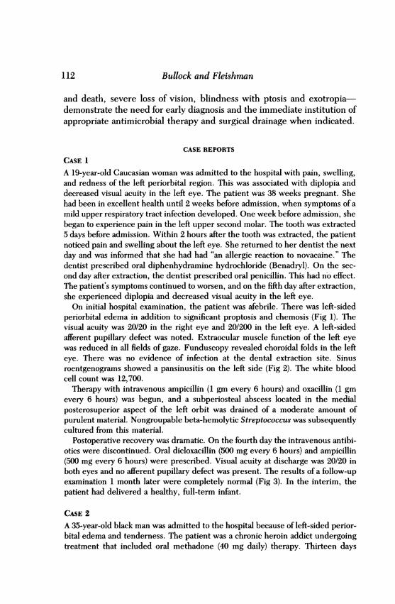
A 19-year-old Caucasian woman was admitted to the hospital with pain, swelling,and redness of the left periorbital region. This was associated with diplopia anddecreased visual acuity in the left eye. The patient was 38 weeks pregnant. Shehad been in excellent health until 2 weeks before admission, when symptoms of amild upper respiratory tract infection developed. One week before admission, shebegan to experience pain in the left upper second molar. The tooth was extracted5 days before admission. Within 2 hours after the tooth was extracted, the patientnoticed pain and swelling about the left eye. She returned to her dentist the nextday and was informed that she had had "an allergic reaction to novacaine." Thedentist prescribed oral diphenhydramine hydrochloride (Benadryl). On the sec-ond day after extraction, the dentist prescribed oral penicillin. This had no effect.The patient's symptoms continued to worsen, and on the fifth day after extraction,she experienced diplopia and decreased visual acuity in the left eye.On initial hospital examination, the patient was afebrile. There was left-sided
periorbital edema in addition to significant proptosis and chemosis (Fig 1). Thevisual acuity was 20/20 in the right eye and 20/200 in the left eye. A left-sidedafferent pupillary defect was noted. Extraocular muscle function of the left eyewas reduced in all fields of gaze. Funduscopy revealed choroidal folds in the lefteye. There was no evidence of infection at the dental extraction site. Sinusroentgenograms showed a pansinusitis on the left side (Fig 2). The white bloodcell count was 12,700.Therapy with intravenous ampicillin (1 gm every 6 hours) and oxacillin (1 gm
every 6 hours) was begun, and a subperiosteal abscess located in the medialposterosuperior aspect of the left orbit was drained of a moderate amount ofpurulent material. Nongroupable beta-hemolytic Streptococcus was subsequentlycultured from this material.
Postoperative recovery was dramatic. On the fourth day the intravenous antibi-otics were discontinued. Oral dicloxacillin (500 mg every 6 hours) and ampicillin(500 mg every 6 hours) were prescribed. Visual acuity at discharge was 20/20 inboth eyes and no afferent pupillary defect was present. The results of a follow-upexamination 1 month later were completely normal (Fig 3). In the interim, thepatient had delivered a healthy, full-term infant.
CASE 2A 35-year-old black man was admitted to the hospital because of left-sided perior-bital edema and tenderness. The patient was a chronic heroin addict undergoingtreatment that included oral methadone (40 mg daily) therapy. Thirteen days
112
Orbital Cellulitis
FIGURE 1Case 1 with left-sided periorbital edema, proptosis, and chemosis.
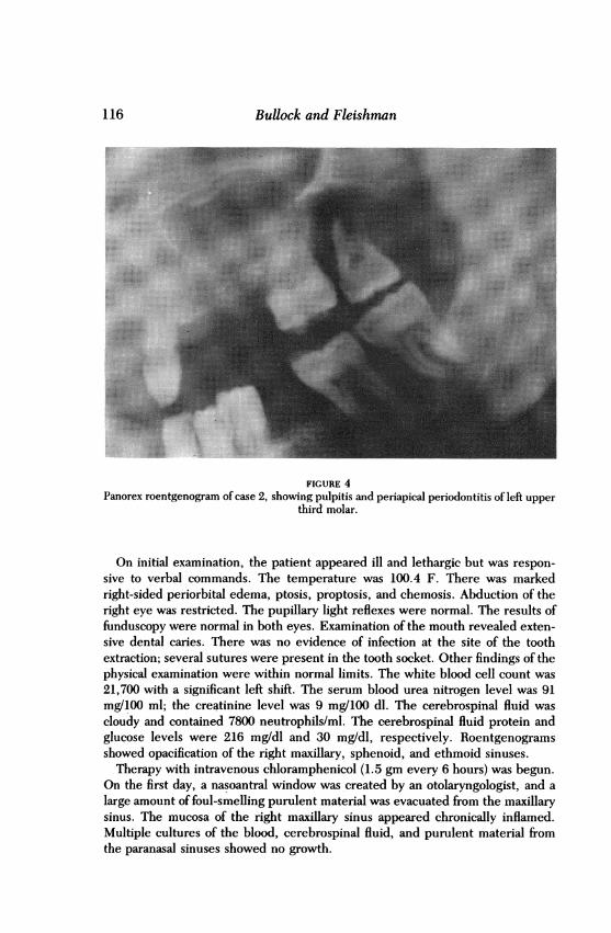
before admission he was seen in the dental clinical for pain of 1 month's durationin the left upper third molar. A panorex roentgenogram showed irreversiblepulpitis and periapical periodontitis (Fig 4). The tooth was extracted, and thepatient was given oral penicillin (250 mg every 6 hours). Two days after theextraction the patient returned to his dentist complaining of a severe headacheand purulent discharge from the left nostril. There was no evidence of infection atthe extraction site. He was given propoxyphene hydrochloride (Darvon) for painand dismissed. Thirteen days after the extraction he again returned to his dentistcomplaining of severe headaches, nasal drainage, and pain at the extraction site.No facial or periorbital edema was noted and the extraction site was unchanged.He was again dismissed. Later in the day, left-sided periorbital edema andtenderness developed; the patient was then admitted to the hospital.On initial examination, the patient was alert and febrile (T = 100.7 F). There
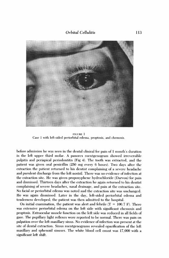
was extensive periorbital edema on the left side with significant chemosis andproptosis. Extraocular muscle function on the left side was reduced in all fields ofgaze. The pupillary light reflexes were reported to be normal. There was pain onpalpation over the left maxillary sinus. No evidence of infection was present at thesite of dental extraction. Sinus roentgenograms revealed opacification of the leftmaxillary and sphenoid sinuses. The white blood cell count was 17,000 with asignificant left shift.
113
Bullock and Fleishman
FIGURE 2Sinus roentgenogram of same patient, showing opacification of left ethmoid and maxillary
sinuses. Arrow indicates site of tooth extraction.
Therapy was begun with intramuscular ampicillin (500 mg every 6 hours) andwarm compresses to the left orbit. The patient remained febrile, and the periorbi-tal edema and chemosis increased. On the second day the left pupil reactedsluggishly. No change in the therapeutic regimen was undertaken. On the thirdday, there was no light perception in the left eye.An otolaryngologist attempted to drain the orbital abscess through a small stab
incision made in the supranasal aspect of the left orbit. A minimal amount ofpurulent material was recovered. In addition, a left nasoantral window was cre-ated, and 20 ml of purulent material was drained from the maxillary sinus.Cultures of this material subsequently grew penicillinase-producing Staphylococ-cus aureus and alpha-hemolytic Streptococcus. Postoperatively, the antibioticregimen was changed to intravenous methicillin (1 gm every 4 hours) and oralerythromycin (500 mg every 6 hours). The patient's condition deteriorated overthe next 4 days, with increasing periorbital edema and proptosis.On the seventh day, he was seen in consultation. There was massive left-sided
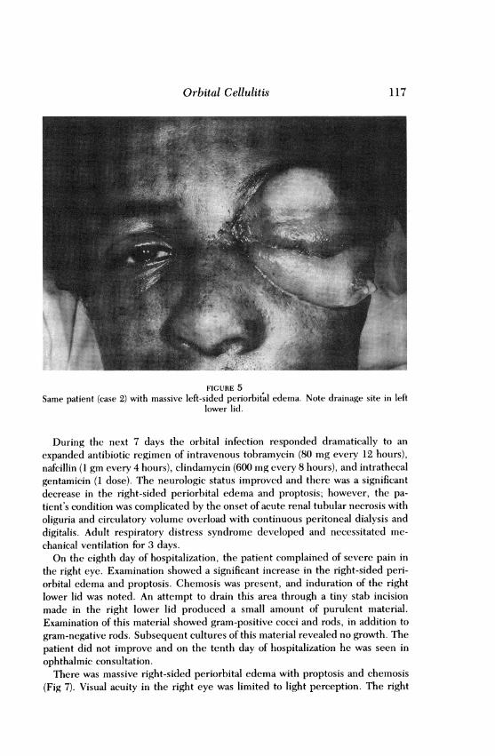
periorbital edema and proptosis (Fig 5). A large amount of foul-smelling purulentmaterial was expelled through a drainage site that opened spontaneously in theleft lower lid during examination.
In surgery, copious amounts of purulent material were evacuated from the leftorbit through an incision made in the drainage site of the lower lid. Additional
114
Orbital Cellulitis
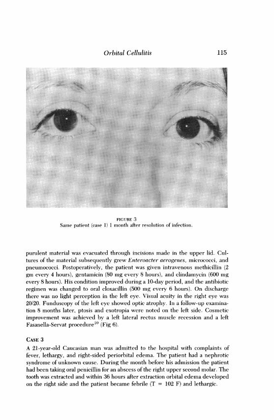
FIGURE 3Same patient (case 1) 1 month after resoluition of infection.
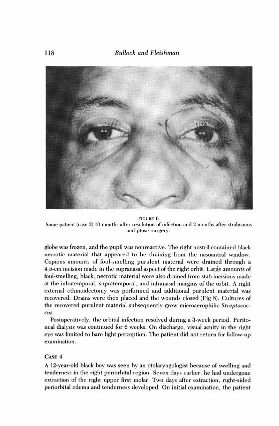
purulent material was evacuated through incisions made in the upper lid. Cul-tures of the material subsequently grew Enteroacter aerogenes, micrococci, andpneumococci. Postoperatively, the patient was given intravenous methicillin (2gm every 4 hours), gentamicin (80 mg every 8 hours), and clindamycin (600 mgevery 8 hours). His condition improved during a 10-day period, and the antibioticregimen was changed to oral cloxacillin (500 mg every 6 hours). On dischargethere was no light perception in the left eye. Visual acuity in the right eye was20/20. Funduscopy of the left eye showed optic atrophy. In a follow-up examina-tion 8 months later, ptosis and exotropia were noted on the left side. Cosmeticimprovement was achieved by a left lateral rectus muscle recession and a leftFasanella-Servat procedure'0 (Fig 6).
CASE 3A 21-year-old Caucasian man was admitted to the hospital with complaints offever, lethargy, and right-sided periorbital edema. The patient had a nephroticsyndrome of unknown cause. During the month before his admission the patienthad been taking oral penicillin for an abscess of the right upper second molar. Thetooth was extracted and within 36 hours after extraction orbital edema developedon the right side and the patient became febrile (T = 102 F) and lethargic.
115
Bullock and Fleishman
FIGURE 4Panorex roentgenogram of case 2, showing pulpitis and periapical periodontitis of left upper
third molar.
On initial examination, the patient appeared ill and lethargic but was respon-sive to verbal commands. The temperature was 100.4 F. There was markedright-sided periorbital edema, ptosis, proptosis, and chemosis. Abduction of theright eye was restricted. The pupillary light reflexes were normal. The results offunduscopy were normal in both eyes. Examination of the mouth revealed exten-sive dental caries. There was no evidence of infection at the site of the toothextraction; several sutures were present in the tooth socket. Other findings of thephysical examination were within normal limits. The white blood cell count was21,700 with a significant left shift. The serum blood urea nitrogen level was 91mg/100 ml; the creatinine level was 9 mg/100 dl. The cerebrospinal fluid wascloudy and contained 7800 neutrophils/ml. The cerebrospinal fluid protein andglucose levels were 216 mg/dl and 30 mg/dl, respectively. Roentgenogramsshowed opacification of the right maxillary, sphenoid, and ethmoid sinuses.Therapy with intravenous chloramphenicol (1.5 gm every 6 hours) was begun.
On the first day, a nasoantral window was created by an otolaryngologist, and alarge amount of foul-smelling purulent material was evacuated from the maxillarysinus. The mucosa of the right maxillary sinus appeared chronically inflamed.Multiple cultures of the blood, cerebrospinal fluid, and purulent material fromthe paranasal sinuses showed no growth.
116
Orbital Cellulitis
FIGURE 5Same patient (case 2) with massive left-sided periorbital edema. Note drainage site in left
lower lid.
During the next 7 days the orbital infection responded dramatically to anexpanded antibiotic regimen of intravenous tobramycin (80 mg every 12 hours),nafcillin (1 gm every 4 hours), clindamycin (600 mg every 8 hours), and intrathecalgentamicin (1 dose). The neurologic status improved and there was a significantdecrease in the right-sided periorbital edema and proptosis; however, the pa-tient's condition was complicated by the onset of acute renal tubular necrosis witholiguria and circulatory volume overload with continuous peritoneal dialysis anddigitalis. Adult respiratory distress syndrome developed and necessitated me-chanical ventilation for 3 days.On the eighth day of hospitalization, the patient complained of severe pain in
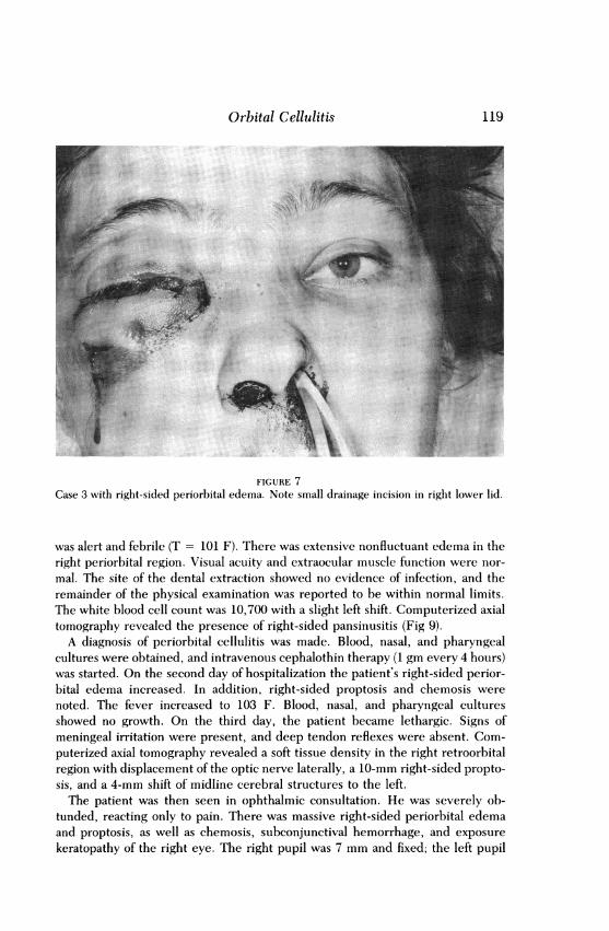
the right eye. Examination showed a significant increase in the right-sided peri-orbital edema and proptosis. Chemosis was present, and induration of the rightlower lid was noted. An attempt to drain this area through a tiny stab incisionmade in the right lower lid produced a small amount of purulent material.Examination of this material showed gram-positive cocci and rods, in addition togram-negative rods. Subsequent cultures of this material revealed no growth. Thepatient did not improve and on the tenth day of hospitalization he was seen inophthalmic consultation.There was massive right-sided periorbital edema with proptosis and chemosis
(Fig 7). Visual acuity in the right eye was limited to light perception. The right
117
Bullock and Fleishnan
FIGURE 6Same patient (case 2) 10 months after resolution of infection and 2 months after strabismus
and ptosis surgery.
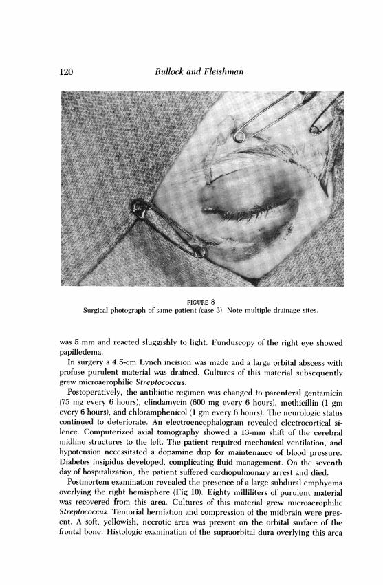
globe was frozen, and the pupil was nonreactive. The right nostril contained blacknecrotic material that appeared to be draining from the nasoantral window.Copious amounts of foul-smelling purulent material were drained through a4.5-cm incision made in the supranasal aspect of the right orbit. Large amounts offoul-smelling, black, necrotic material were also drained from stab incisions madeat the infratemporal, supratemporal, and infranasal margins of the orbit. A rightexternal ethmoidectomy was performed and additional purulent material wasrecovered. Drains were then placed and the wounds closed (Fig 8). Cultures ofthe recovered purulent material subsequently grew microaerophilic Streptococ-cus.
Postoperatively, the orbital infection resolved during a 3-week period. Perito-neal dialysis was continued for 6 weeks. On discharge, visual acuity in the righteye was limited to bare light perception. The patient did not return for follow-upexamination.
CASE 4A 12-year-old black boy was seen by an otolaryngologist because of swelling andtenderness in the right periorbital region. Seven days earlier, he had undergoneextraction of the right upper first molar. Two days after extraction, right-sidedperiorbital edema and tenderness developed. On initial examination, the patient
118
Orbital Cellulitis
FIGURE 7Case 3 with right-sided periorbital edema. Note small drainage incision in right lower lid.
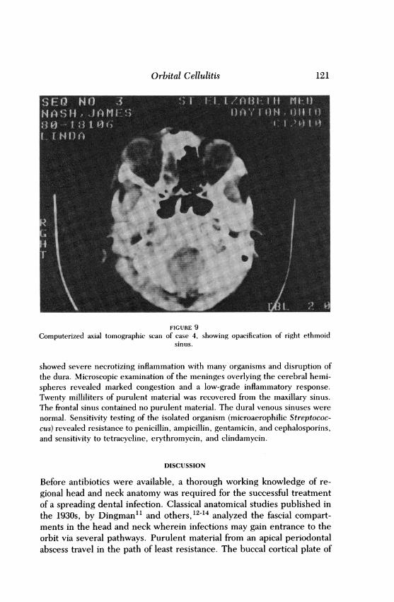
was alert and febrile (T = 101 F). There was extensive nonfluctuant edema in theright periorbital region. Visual acuity and extraocular muscle function were nor-mal. The site of the dental extraction showed no evidence of infection, and theremainder of the physical examination was reported to be within normal limits.The white blood cell count was 10,700 with a slight left shift. Computerized axialtomography revealed the presence of right-sided pansinusitis (Fig 9).A diagnosis of periorbital cellulitis was made. Blood, nasal, and pharyngeal
cultures were obtained, and intravenous cephalothin therapy (1 gm every 4 hours)was started. On the second day of hospitalization the patient's right-sided perior-bital edema increased. In addition, right-sided proptosis and chemosis werenoted. The fever increased to 103 F. Blood, nasal, and pharyngeal culturesshowed no growth. On the third day, the patient became lethargic. Signs ofmeningeal irritation were present, and deep tendon reflexes were absent. Com-puterized axial tomography revealed a soft tissue density in the right retroorbitalregion with displacement of the optic nerve laterally, a 10-mm right-sided propto-sis, and a 4-mm shift of midline cerebral structures to the left.The patient was then seen in ophthalmic consultation. He was severely ob-
tunded, reacting only to pain. There was massive right-sided periorbital edemaand proptosis, as well as chemosis, subconjunctival hemorrhage, and exposurekeratopathy of the right eye. The right pupil was 7 mm and fixed; the left pupil
119
Bullock and Fleishman
FIGURE 8Surgical photograph of same patient (case 3). Note multiple drainage sites.
was 5 mm and reacted sluggishly to light. Funduscopy of the right eye showedpapilledema.
In surgery a 4.5-cm Lynch incision was made and a large orbital abscess withprofuse purulent material was drained. Cultures of this material subsequentlygrew microaerophilic Streptococcus.
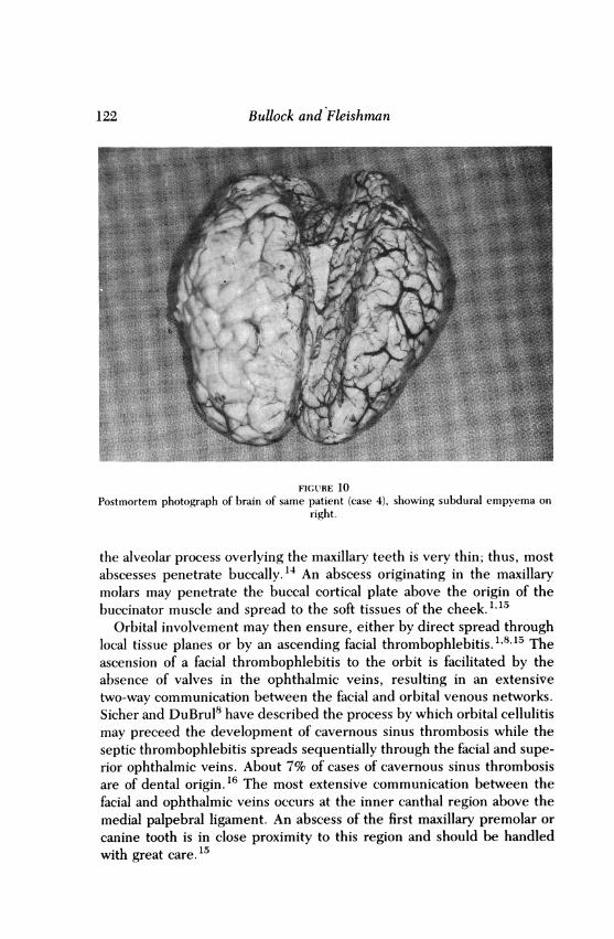
Postoperatively, the antibiotic regimen was changed to parenteral gentamicin(75 mg every 6 hours), clindamycin (600 mg every 6 hours), methicillin (1 gmevery 6 hours), and chloramphenicol (1 gm every 6 hours). The neurologic statuscontinued to deteriorate. An electroencephalogram revealed electrocortical si-lence. Computerized axial tomography showed a 13-mm shift of the cerebralmidline structures to the left. The patient required mechanical ventilation, andhypotension necessitated a dopamine drip for maintenance of blood pressure.Diabetes insipidus developed, complicating fluid management. On the seventhday of hospitalization, the patient suffered cardiopulmonary arrest and died.Postmortem examination revealed the presence of a large subdural emphyema
overlying the right hemisphere (Fig 10). Eighty milliliters of purulent materialwas recovered from this area. Cultures of this material grew microaerophilicStreptococcus. Tentorial herniation and compression of the midbrain were pres-ent. A soft, yellowish, necrotic area was present on the orbital surface of thefrontal bone. Histologic examination of the supraorbital dura overlying this area
120
Orbital Cellulitis
FIGURE 9Computerized axial tomographic scan of case 4, showing opacification of right ethmoid
sinus.
showed severe necrotizing inflammation with many organisms and disruption ofthe dura. Microscopic examination of the meninges overlying the cerebral hemi-spheres revealed marked congestion and a low-grade inflammatory response.Twenty milliliters of purulent material was recovered from the maxillary sinus.The frontal sinus contained no purulent material. The dural venous sinuses werenormal. Sensitivity testing of the isolated organism (microaerophilic Streptococ-cus) revealed resistance to penicillin, ampicillin, gentamicin, and cephalosporins,and sensitivity to tetracycline, erythromycin, and clindamycin.
DISCUSSION
Before antibiotics were available, a thorough working knowledge of re-gional head and neck anatomy was required for the successful treatmentof a spreading dental infection. Classical anatomical studies published inthe 1930s, by Dingman"l and others,12-14 analyzed the fascial compart-ments in the head and neck wherein infections may gain entrance to theorbit via several pathways. Purulent material from an apical periodontalabscess travel in the path of least resistance. The buccal cortical plate of
121
Bullock and Fleishman
FIGURE 10Postmortem photograph of brain of same patient (case 4), showing subdural empyema on
right.
the alveolar process overlying the maxillary teeth is very thin; thus, mostabscesses penetrate buccally.'4 An abscess originating in the maxillarymolars may penetrate the buccal cortical plate above the origin of thebuccinator muscle and spread to the soft tissues of the cheek."'5
Orbital involvement may then ensure, either by direct spread throughlocal tissue planes or by an ascending facial thrombophlebitis." 8"15 Theascension of a facial thrombophlebitis to the orbit is facilitated by theabsence of valves in the ophthalmic veins, resulting in an extensivetwo-way communication between the facial and orbital venous networks.Sicher and DuBrul8 have described the process by which orbital cellulitismay preceed the development of cavernous sinus thrombosis while theseptic thrombophlebitis spreads sequentially through the facial and supe-rior ophthalmic veins. About 7% of cases of cavernous sinus thrombosisare of dental origin. 16 The most extensive communication between thefacial and ophthalmic veins occurs at the inner canthal region above themedial palpebral ligament. An abscess of the first maxillary premolar orcanine tooth is in close proximity to this region and should be handledwith great care. 15
122
Orbital Cellulitis
Infections of the maxillary molars may also spread posteriorly into theinfratemporal and pterygopalatine fossae. 1,2,7,15 Purulent material maythen extend along the tuberosity of the maxilla and thus gain access to theorbit through the inferior orbital fissure."',5 This fissure is closed by astrong fascia and smooth muscle fibers; however, an opening is oftenfound at its lateral aspect. 7
Because of the intimate anatomic relationship between the paranasalsinuses and the orbit, paranasal sinusitis is the most common cause oforbital cellulitis. 18-21 In the adult, more than 50% of the orbital circumfer-ence is surrounded by sinus cavities. Venous blood flows freely betweenthe ophthalmic and ethmoid veins.22 An extensive plexus of veins nearthe nasolacrimal duct communicates freely with venous plexuses of theturbinates, the linings of the sinuses, and the ophthalmic veins.23 Inaddition, there can be congenital dehiscences in the medial and superiorwalls of the orbit.24
It is, therefore, not surprising that odontogenic infections most com-monly reach the orbit through the paranasal sinuses.1 As many as 20% ofall cases of sinusitis have been attributed to dental origin.2'5 The apices ofthe maxillary molars and premolars are in close apposition with the floorof the maxillary sinus. Occasionally the apices of these teeth are in directcontact with the mucous membrane of the sinus. During extraction of anabscessed tooth, the floor of the sinus may be fractured, resulting ininnoculation of the antrum with purulent material. In 1927, Hempstead26reviewed 385 cases of maxillary sinusitis. In 22.5% of these cases, theinfection was attributed to extraction of abscessed molars. In 16% of thecases, a fistula extended through the alveolar process into the maxillaryantrum.
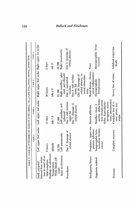
In all of the cases detailed in the present study, development of orbitalcellulitis followed extraction of an ipsilateral maxillary molar (Table I).Three of the four patients were febrile on admission. All had elevatedwhite blood cell counts. Roentgenographic evidence of paranasal sinusinfection was present on admission in all cases. In each patient, it isprobable that infection reached the orbit via the paranasal sinuses.The time interval between extraction of the tooth and onset of orbital
symptoms ranged from 2 hours (patient 1) to 13 days (patient 2). Severeheadaches and purulent nasal discharge developed within 2 days in thesecond patient, indicating that infection had spread to the paranasalsinuses. In cases 1 and 3, the spread of the infection to the orbit wasparticularly rapid. Case 3 had evidence of chronically inflamed mucosa inthe maxillary sinus. In addition, his debilitated state of health was a likelycontributing factor in the accelerated spread of the infection not only to
123
124 Bullock and Fleishmnn
-0
0~~~ ~ U 0~~~~~ ~ ~ m0
~~ C~~~~ - Z ~u .) -0 CCsef2H ~~~~~~~~~~ Q~~rC
.0.o 04
Ze~~~~~ ~~~~ ~ ~ ~~~~ ~~~CU.~~c USm CtQ~~~~ ~~~~~~~~~~to 0~~~~~~~~CbC -O o~ &2
o --~~~~~~~~~~~~~~~~
H~~~~~~~Q~~~~~~CU.~t0.-50
"0
0~~~~~~~~~~~b
~~~ U~~~~~~~ 0L C1 0X
U."4.s.
H 0 ~~~~~~~~~~~~~~~~~~~~ ~~~~~~~C
0biC~~~~~~.
0U4 004
Orbital Cellulitis
the orbit, but also to the meninges. In case 1, orbital symptoms devel-oped only 2 hours after extraction. Her 2-week history of coryza pre-ceding the dental extraction may have contributed to a rapid spread ofinfection through an already inflamed antral mucosa. The pregnancy itselfmay have predisposed her to the original odontogenic infection. The rateof occurrence of gingivitis during pregnancy has been reported at from30% to 100%, reaching a peak during the 8th month of gestation.9
In cases 3 and 4, infection spread to involve the central nervous system.In case 4, a subdural empyema developed approximately 10 days afterdental extraction; in case 3, meningitis developed in the patient 36 hoursafter extraction. In 1945, Haymaker7 analyzed 28 cases of fatal centralnervous system infections following dental extraction. Spread ofthe odon-togenic infection to the intracranial cavity was attributed to direct exten-sion in 17 cases and to hematogenous spread in 11 cases. His seriesincluded six cases of intraorbital abscess. In three of these cases, theorbital abscess was directly involved in the subsequent intracranial spreadof infection. In two cases, cavernous sinus thrombosis occurred. In theremaining case, purulent material from the orbital infection penetratedthe frontal bone and frontal sinus, ultimately gaining entrance to thesubdural space. A similar process appears to have occurred in case 4. Theorbital infection spread upward to involve the orbital roof. Subsequentdisruption of the dura overlying the orbital portion of the frontal boneallowed organisms to invade the subdural space.These cases demonstrate the importance of appropriate antimicrobial
therapy and the implementation of adequate and timely surgical drainagewhen indicated. In case 4, the infecting organism was resistant to theinitial antibiotic employed and drainage of the orbital abscess was de-layed, allowing the infection to penetrate the orbital roof and invade thesubdural space. A similar situation occurred in case 2, in which theinfecting organism also was resistant to the initial antibiotic employed. Ingeneral, when Gram staining and cultures of the infecting organisms areunavailable, multiple-drug regimens should be used. Agents should beadministered intravenously and in high doses. Initial therapy shouldinclude a penicillinase-resistant penicillin. Ampicillin should be includedin patients less than 4 years old to provide coverage against Hemophilisinfluenze.27 The clinician should be aware that a patient with orbitalcellulitis may respond dramatically to initial antimicrobial therapy, onlyto relapse several days later after an orbital abscess has formed, as oc-curred in case 3. After 7 days of antimicrobial therapy, the patient'sorbital signs had almost returned to normal; however, within 24 hours theperiorbital edema, proptosis, and chemosis returned, and the visual acu-ity in the eye was severely impaired.
125
Bullock and Fleishman
Deciding when intensive antimicrobial therapy must be augmentedwith surgical drainage requires astute clinical judgment. Duke-Elder28and Dingman" have emphasized the difficulties inherent in making thisdecision. If drainage is attempted prematurely, a localized infection maybe disseminated throughout the orbital tissues. Conversely, if left un-drained, a true orbital abscess that has not responded to medical therapymay result in loss of vision, cavernous sinus thrombosis, meningitis,subdural empyema, or brain abscess. If the clinical situation is ambiguousand a decision is made to delay surgery, the ophthalmological findingsmust be followed meticulously. B-scan ultrasonography and computer-ized axial tomography of the orbit may aid in the detection of an ab-scess2930; however, careful observation of the eye should take precedencein influencing the decision to attempt surgical drainage. The presence ofchoroidal folds may represent traction on the optic nerve.31-33 Increasingproptosis and chemosis associated with extraocular muscle impairmentare general indications for surgery. Surgical drainage should be employedbefore impairment of vision develops.
It is essential that any surgical drainage be established adequately:small stab incisions proved inadequate in cases 2 and 3. In general, a largeincision should be used. This can be made over the region of a suspectedabscess or in the supranasal quadrant of the orbit (Lynch incision). Addi-tional incisions can be made if needed. Drains should be placed in thewounds and advanced a small distance each day.The high incidence of dental extraction makes it important to recognize
the possibility of infection that may spread to the orbit, and the potentialconsequences of such an infection must not be underestimated. The onefatality occurred in a previously healthy 12-year-old boy. No apparentpredisposing factors were uncovered that should have rendered himsusceptible to such a rapid spread of infection. This demonstrates theimportance of careful choice in initial antimicrobial therapy and closemonitoring of physical signs once therapy has been instituted.
ACKNOWLEDGMENTS
Reed 0. Dingman, MD, DDS, and William Coyne, DDS, providedresearch consultation.
REFERENCES
1. Kaban LB, McGill T: Orbital cellulitis of dental origin: Differential diagnosis and theuse of the computed tomography as a diagnostic aid. J Oral Surg 1980; 38:682-685.
2. Gold RS, Sager E: Pansinusitis, orbital cellulitis, and blindness as sequelae of delayedtreatment of dental abscess. J Oral Surg 1974; 32:40-43.
126
Orbital Cellulitis 127
3. Yates C, Monks A: Orbital cellulitis complicating the extraction of teeth. J Dent 1978;6:229-232.
4. Pellegrino SV: Extension of dental abscess to the orbit. J Am Dent Assoc 1980;100:873-875.
5. Dener CB, Sazima HJ, Schaberg SJ: Life-threatening infection after extraction of thirdmolars. J Am Dent Assoc 1980; 101:649-650.
6. Limongelli WA, Clark MS, Williams AC: Panfacial cellulitis with contralateral orbitalcellulitis and blindness after tooth extraction. J Oral Surg 1977; 35:38-43.
7. Haymaker W: Fatal infections of the central nervous system and meninges after toothextraction with analysis of 28 cases. Am J Orthod 1945; 31:117-188.
8. Sicher H, DuBrul EL: Oral Anatomy, ed 7. St Louis, CV Mosby Co, 1980, pp 498-518.9. Loe H: Endocrinologic influences on periodontal disease, pregnancy and diabetes
mellitus. Ala J Med Sci 1968; 5:336-348.10. Fasanella RM, Servat J: Levator resection for minimal ptosis: Another simplified opera-
tion. Arch Ophthalmol 1961; 65:493-496.11. Dingman RO: The management of acute infections of the face and jaws. Am J Orthod
1939; 25:780-794.12. Singer E: Fasciae ofthe Human Body and Their Relations to the Organs They Envelop.
Baltimore, Williams & Wilkins, 1935, pp 1-105.13. Grodinsky M, Holyoke EA: Fasciae and fascial spaces of the head, neck, and adjacent
regions. Am J Anat 1938; 63:367-408.14. Coller FA, Yglesias L: Infections of the lip and face. Surg Gynecol Obstet 1935;
60:277-288.15. Birn H: Spread of dental infections. Dent Pract Dent Rec 1972; 22:347-356.16. Shaw RE: Cavernous sinus thrombophlebitis: A review. BrJ Surg 1952; 40:40-48.17. Wunderer S: Die Ausbreitung der retromaxillaren Abszesse im Lichte neuerer anatom-
ischer Forschung. Ost Z Stomat 1955; 52:651-660.18. Chandler JR, Langenbrunner DJ, Stevens ER: The pathogenesis of orbital complica-
tions in acute sinusitis. Laryngoscope 1970; 80:1414-1428.19. Haynes RE, Cramblett HG: Acute ethmoiditis: Its relationship to orbital cellulitis. AmJ
Dis Child 1967; 114:261-267.20. Jarrett WH, Gutman FA: Ocular complications of infection in the paranasal sinuses.
Arch Ophthalmol 1969; 81:683-688.21. Smith AT, Spencer JT: Orbital complications resulting from lesions of the sinuses. Ann
Otol Rhinol Laryngol 1948; 57:5-27.22. Gamble RC: Acute inflammations of the orbit in children. Arch Ophthalmol 1933;
10:483-497.23. Batson OV: Relationship of the eye to the paranasal sinuses. Arch Ophthalmol 1936;
16:322-323.24. Williamson-Noble FA: Diseases of the orbit and its contents, secondary to pathological
conditions of the nose and paranasal sinuses. Ann R Coll Surg Engl 1954; 15:46-64.25. Knight JS, Stacy GC: Antral infection of dental origin with a report of a case via an
unusual dental route. Aust Dent J 1963; 8:483-491.26. Hempstead BE: Intranasal surgical treatment of chronic maxillary sinusitis. Arch Oto-
laryngol 1927; 6:426-430.27. Watters EC, Waller PH, Hiles DA, Michaels RH: Acute orbital cellulitis. Arch Oph-
thalmol 1976; 94:785-788.28. Duke-Elder S: The ocular adnexa, in System of Ophthalmology. St Louis, CV Mosby
Co, 1974, vol 13, part 2, pp 859-866.29. Goldberg F, Berne AS, Oski FA: Differentiation of orbital cellulitis from preseptal
cellulitis by computed tomography. Pediatrics 1978; 62:1000-1005.30. Schramm VL, Myers EN, Kennerdell JS: Orbital complications of acute sinusitis:
Evaluation, management, and outcome. Otolaryngology 1978; 86:ORL 221-20.31. Bullock JD, Egbert PR: The origin of choroidal folds: A clinical, histopathological, and
experimental study. Doc Ophthalmol 1974; 37:261-293.
Bullock and Fleishman
32. Experimental choroidal folds. Am J Ophthalmol 1974; 78:618-623.33. Bullock JD, Waller RR: Choroidal folding in orbital disease, in Proceedings of the Third
International Symposium on Orbital Disorders. The Hague, Dr W. Junk bv Publishers,1977, pp 483-488.
DISCUSSION
DR R. R. WALLER. I appreciate very much the opportunity to discuss this informa-tive and beautifully presented paper. There are a variety of mishaps affecting thevisual system which occur while in the dental office. Most injuries, fortunately,are not serious, only transiently affect vision and can be prevented by wearingprotective eyewear. Doctor Robert Hales, in 1970, reported several cases ofocular injuries sustained in the dental office, including corneal abrasions from anexplosion of an ampule of local anesthetic; chemical keratitis from pumice, em-bedded corneal foreign bodies of alloy materials, and the resultant recurrentcorneal erosions which often follow. Corneal perforation was also reported in hispaper when, during the course of filling a tooth of a 10-year-old girl, a double-ended excavation instrument which was caught in a gauze, flipped into the air,and impaled in the child's eye. Blaxter and Britten in 1961 and Cooper in 1962reported transient amaurosis, diplopia, and pupil dilation following mandibularnerve block with procaine. The probable explanation was that an intra-arterialinjection had taken place and because of anastomosis between the internal andexternal carotid system, the anesthetic agent reached the eye. This latter explana-tion makes sense, especially in those situations where the middle meningealartery is anomalously the major blood supply to the orbit.
Doctor Bullock's paper reminds us of yet another complication of dental sur-gery which in contrast is not an immediate complication, but rather a delayedevent of days or weeks and it can be devastating. The entrance of organismsthrough local tissue planes is probably the least common route of entry for thepathogen in these cases in the 1980s. On the other hand, entry of organisms byhematogenous route appears more plausible. Doctor Bullock pointed out thepresence of rich anastomoses between facial and ophthalmic veins and mentionsthe more than chance relationship between dental disease and cavernous sinusthrombosis. That rich vascular anastomoses between the blood supply to thesinuses and the orbit also exist is evidenced by reports in the literature detectingimmediately within the retinal vessels, choroidal circulation, and conjunctivalvessels, the presence of depo-steroid, following injection into the nasal cavity fortreatment of allergic polyps. The common relationship between paranasal sinus-itis and orbital cellulitis is beautifully documented in this paper, in that all fourpatients presented with radiologic evidence of paranasal sinusitis on the side ofdental extraction. It is also striking that all patients had upper teeth extracted, nolower teech, and we are reminded that the apices of the maxillary premolars andmolars can be in contact with the maxillary sinus mucosa.The principles of management of orbital cellulitis have been emphasized and
include hospitalization, close monitoring, and vigorous (as well as appropriate)
128
Orbital Cellulitis
antibiotic therapy. This is not the time for outpatient care. Orbital cellulitisshould be a true ophthalmic emergency. We prefer to evaluate separately thesigns of orbital inflammation (chemosis, lid swelling, erythema, proptosis, dis-placement, strabismus, and visual disturbance) recording trends in the medicalrecord at least every 12 hours, and more frequently if necessary. If the clinicalcondition deteriorates, one must reevaluate medical therapy, but deteriorationshould mean abscess until proven otherwise, and this means emergency evalua-tion with computed tomography (CT) or ultrasound. CT examination appears tomore precisely identify the smooth, dome-shaped, more readily drained subperi-osteal abscess confined by the periorbita adjacent to the affected sinus. PerhapsDoctor Bullock would comment on efficacy of CT vs ultrasound in this situation.Abscesses should be drained externally through adequate incisions, not small stabwounds. We believe the establishment of excellent drainage from the contiguoussinus into the nasopharynx if the abscess is in the peripheral orbital space.
Doctor Bullock has reminded us of the importance of early detection andaggressive management of orbital cellulitis. This can avoid the serious ocularcomplications of ophthalmoplegia and visual loss, and the tragic complications ofmeningitis and death. I have enjoyed very much having the opportunity to discussthis excellent paper.
DR LEONARD APr. Several years ago we experienced a small epidemic of eye andcentral nervous system complications following dental surgery in the colony ofmonkeys at the UCLA Medical Center. The complications included orbital cellu-litis, retrobulbar abscess, orbital apex syndrome, superior orbital fissure syn-drome, and brain abscess followed by death.
After a monkey escaped from his cage and mutilated the leg of a caretaker, thestaffveterinarians decided to remove or saw offthe canine teeth of the monkeys toprevent further mishaps. Most often the tooth was cut off at the gum level, thenerve destroyed, and the canal packed in the manner used by dentists to preservea tooth.
Ocular infections followed these procedures. The baboon shown on the pro-jected slide developed orbital cellulitis a few days after dental surgery. Orbitalswelling was followed by proptosis, a hyperemic fixed globe, and systemic signsand symptoms of infection and toxicity. The dental consultant ordered systemicchloramphenicol therapy. Little improvement was seen after 1 week. Orbitalincision and drainage was attempted. No frank pus was obtained. Routine culturesof the scant aspirate were negative. Cultures for anaerobic bacteria were not takenby the attending veterinarian. A change of antibiotic therapy to methicillin andgentamicin was recommended. The baboon's condition improved temporarily,but later in the week he was found dead in his cage. A brain abscess was found atautopsy.
In recent years at the Jules Stein Eye Institute we have seen two adults whodeveloped severe orbital cellulitis following dental extraction. Both patients werediabetic. One patient, a dentist, responded well to antibiotic therapy. The otherpatient died when the markedly disturbed metabolic state, complicated by the
129
Bullock and Fleishman
infection, could not be controlled. These events suggest that diabetes may beanother predisposing factor contributing to the occurrence of orbital infectionafter dental procedurs. I suspect that the spread of infection to adjacent structures(soft tissue, sinuses, etc) and the eye occurs more frequently than surmised but isnot reported because improvement occurs with prompt antibiotic therapy.One intriguing aspect of this subject is why eye complications and infections
elsewhere in the body do not occur more frequently after dental procedures sincetransient bacteremia is not uncommon. In reported studies the incidence ofbacteremia following extractions has ranged from 15% to 85%. The higher inci-dences are found more often in the studies that included culture procedures foranaerobic (normal in the mouth) as well as aerobic bacteria. Perhaps the reason fornegative culture reports in cases of eye and other body infections following dentalextraction is the failure to use appropriate culture methods for both groups ofbacteria. Failure to identify the causative microorganisms deters the choice ofproper antibiotic therapy.From dental studies we learn that transient bacteremia sometimes occurs after
seemingly innocuous manipulation around teech, even in patients with healthy-appearing gingiva. For example, it may occur after tooth brushing, vigorousflossing, and the use of Stimudents and the Water Pik. The incidence of bactere-mia increasese if periodontal disease exists.
Complications from transient bacteremia after dental extraction is averted inthe healthy individual mainly by the body's immune system. Certain conditions,however, predispose a person to infection, and therefore antibiotic prophylaxisshould be considered before dental treatment. This group would include patientswho are chemically ill, debilitated, or immunosuppressed, patients with meta-bolic disorders such as the nephrotic syndrome (described by Doctor Bullock thismorning), and patients and diabetes as mentioned by me earlier. For years,cardiologists have given antibiotics prophylactically to patients with congenital oracquired heart disease to avoid bacterial endocarditis. Recently orthopedic sur-geons have recommended that patients with prosthetic hips receive prophylacticantibiotic therapy because there have been some cases of infection in quiet,healed joints after dental procedures. Similarly, I would seriously consider givingantibiotics prophylactically to a patient who had an intraocular lens implant. Theartificial lens could act as a nidus for circulating microorganisms released into thebloodstream during dental treatment.
I have enjoyed Doctor Bullock's presentation and Doctor Waller's remarks. Iappreciate the opportunity to share my thoughts and experience on this importantsubject.
DR F. C. BLODI. I think we are all indebted to Doctor Bullock for calling ourattention again to the pathogenesis of orbital cellulitis. I would like to emphasizethree points:
1) It is interesting that most of these patients are quite young and the same isborne out in Doctor Bullock's series. I am not sure why because you would expect
130
Orbital Cellulitis
that many more older patients get a tooth extraction and yet most of these patientsare children.
2) It is not necessarily a pansinusitis that produces the orbital cellulitis. DoctorBullock has mentioned that. I vividly remember a case which is still in the courts.A young child was managed, again poorly, by a pediatrician but no ophthalmicconsultant had been called in. An otolaryngologist on repeated examinations didnot find a pansinusitis.
3) For the third point, I would like to stress what Doctor Waller has alreadymentioned: management of this condition is obviously an emergency. There is noquestion about that, but the great question remains when and where to drain. Thelist of indications for drainage was shown in Doctor Bullock's paper and is a littlebit vague. When is the motility somewhat reduced and when is the conditiongetting worse? This reminds me of medieval mysticism. We can follow the chang-es in the orbit nowadays with computerized tomography scans and ultrasound(preferably with both) very well. These patients have to be examined daily and theminute an abscess forms, it has to be drained. These wild stab incisions, notknowning where and when to drain, is 19th century medicine.
DR WILLIAM JARRErr. I think Doctor Bullock has done us a service by callingattention to this now rare disease. Although rare today, it was something that wasquite common in the past, and it illustrates the changes in the disease patternsthat come about with advances in medicine, in this case antibiotics. The wide-spread use of antibiotics, for example, completely changed the nature of otolaryn-gology as a specialty; acute mastoiditis with resultant intracranial abscess was acommon and serious complication in the past but these are now rarely seen, as arecases of orbital cellulitis. Most residents will go through their training withoutever seeing such a case, and a presentation such as Doctor Bullock's is needed toraise the level of our consciousness in order to make the correct diagnosis.
I have seen several such cases in my own practice; none of mine were the resultof dental extraction, but all were due to pansinusitis. I think the important lessonto be learned is to think about this disease when you see such a patient. When adisease becomes uncommon, the diagnosis may be missed initially, with poten-tially disastrous results.
I cannot stress too much the importance of draining an orbital abscess, asDoctor Blodi pointed out. It is hard to know exactly when to intervene surgically.Ifjust a cellulitis is present, drainage doesn't do any good and may indeed spreadthe infection. But if a subperiosteal orbital abscess develops, drainage becomesmanditory. When drainage is done, an adequate incision down through theperiosteum is needed, or the case won't be cured.
Again, I think it is important for us to be exposed to these cases and to knowthat they do occur even in this day of widespread antibiotic usage.
DR MELVIN ALPER. To carry this group of complications further, Doctor Apt hasreferred to the bacteremia that occurs following dental intervention. I recentlyhad a physician patient who came in with an occlusion of the central retinal artery.
131
Bullock and Fleishman
He was running a slight fever. We had him evaluated. He had gone to a dentistabout 2 weeks previous to the occluded vessel. He thought he had had a viralsyndrome. We studied him and found that he had prolapse of the mitral valve(Barlow's) syndrome. He had developed vegetative endocarditis and had thrownan embolism which had occluded the central retinal artery. This is anotheretiology to add to your long list of ocular complications from visits to the dentists'office.
DR RONALD BURDE. I think the only point I would like to make is that the list thatDr Dan Jones gives for treating is for bugs that are assumed to be anaerobic forthe most part and that what we are dealing with here are bugs that may bemicroaerobic, microaerophilic, or may be totally aerobic. I would ask DoctorBullock to address the fact that in fact when one is dealing with a source that mayhave this type of organism, might one better use a different group of antibiotics oradd at least one that is more specific for anaerobic organisms? I believe if we useonly penicillin and gentamicin in these cases, we will miss treating the offendingorganism. That, of course, does not obviate the need for drainage of the abscess,but it may help us with the prevention of hematogenous spread or contiguousspread to the central nervous system.
DR WILLIAM H. SPENCER. Nobody has mentioned anything about phycomycoticinfection. Phycomycotic orbital cellulitis may follow dental extraction, particularlyin individuals who are acidotic and diabetic. I notice that in your recommenda-tions for detecting possible causative organisms you included various forms ofculture but do not suggest biopsy. I would like to emphasize that phycomycoticinfection is rarely detectable by culture alone. A biopsy of the infected tissue hasto be taken before these organisms can be shown. I wonder if you would consideradding biopsy to your list of things that might be done to diagnose the infectiousagent?
DR JOHN BULLOCK. I really appreciate everyone's comments. Doctor Waller men-tioned ultrasound vs computed tomography (CT). I have had no experience withultrasound in these cases, I have used only CT. He also mentioned drainageexternally or internally into the nasopharynx, and I have no experience withinternal drainage. I've approached them all externally as I have shown. DoctorApt questions: "Why does this occur; there is so much infection in dental extrac-tions, so why don't more patients get this." In my four cases I think that thenumber one answer is delay in diagnosis and treatment. All these patients wereneglected both by the dentist and by the originally treating pediatricians and ear,nose, and throat doctors, and I think that the infections rapidly got out ot hand. Ithink it emphasizes the importance of early, proper management. Doctor Blodimentioned, "how does one decide when to do surgical drainage." I see a lot ofcases of orbital cellulitis and my feeling is that most of them do not need surgicaldrainage. Most of them just need antibiotic therapy.; I think it is wrong to rush tosurgery and I think most of these patients should be managed for at least 24 hours
132
Orbital Cellulitis 133
with massive doses of appropriate antibiotics and then if the patient is worsening,in spite of antibiotic therapy, I think drainage should then be done. I appreciateDoctor Jarrett's comments and in terms of Doctor Burde's comment about antibi-otics, the list I showed included both aerobic and anaerobic organisms. In DoctorDan Jones' chapter in the Duane series, he mentioned that the treatment ofchoice for anaerobic infections is massive doses of penicillin G.

![ptosis [emedicine]](https://static.fdocuments.us/doc/165x107/577cdd4a1a28ab9e78acb3ee/ptosis-emedicine.jpg)
![Index [link.springer.com]978-0-387-92855-5/1.pdf · Bell’s palsy acquired myogenic ptosis, 95 congenital myogenic ptosis, 79 involutional ptosis, 74 patient selection, 156 prior](https://static.fdocuments.us/doc/165x107/5e0ec41cf5a39e518c0f1033/index-link-978-0-387-92855-51pdf-bellas-palsy-acquired-myogenic-ptosis.jpg)