When is a cell not a cell? A theory relating coenocytic structure

− 151 −
Cellular Morphogenesis in Valonia sp.: with Emphasis on the Formation of Lenticular and Rhizoid Cells
Alex P. Camaya* and Kazuo OkudaGraduate School of Kuroshio Science, Kochi University(2-5-1, Akebono-cho, Kochi 780-8520, Japan)
AbstractThe unspecified macrophyte Valonia sp. obtained from Bicol Region, the Philippines portrays two distinct types of cell divisions. Higher light intensity induced lenticular cells, whereas lower light intensity pro-duced rhizoid cells, which varied in time and structure. These initiated at stage I where chloroplasts assem-bled roughly at the indefinite surface of a cell. Microtubules (MTs) at the protoplasmic assembly appeared to curve and overlap to each other. At stage II a disc-shaped assembly of protoplasm was transformed where radial MT arrays extended from the periphery to the central area of the protoplasmic assembly. In stage III the aggregation of protoplasm stopped, and the average diameter of lenticular cells was ca. 1 mm whereas rhizoid cells were ca. 0.1 mm in diameter. Short but dense cortical MTs were arranged randomly all over the protoplasmic aggregations, followed by the depolymerization of MTs. In stage IV septum walls formed to divide lenticular cells or rhizoid cells from the mother cells. Densely crammed MTs were evenly dispersed all over lenticlular and rhizoid cells. Then in stage V juvenile cells started to puff out in lenticular cells and likewise elongated for rhizoid cells. New parallel arrays of MTs were formed from the base towards the apex portion of these new cells. Furthermore, the maximum attainable height measured about 2 mm in lenticular cells, while it was about 1.4 mm for rhizoid cells. Lenticular and rhizoid cells tended to develop near the zones of both ends of mother cells.
keywords: cytomorphogenesis, cytoskeleton, immunofluorescent microscopy, lenticular cell, microtubule, protoplasmic aggregation, rhizoid cell, Valonia
Introduction
Thalli of Valonia sp. samples were obtained from Lagonoy Gulf, Bicol Region, the Philippines, a site in the country where a prominent "Kuroshio Current" is relatively adjacent. Efforts have been made by the group of faculties from Kochi University, Japan assisted by researchers from Bicol University, the Philippines pres-ently investigating "Kuroshio current" effect in those named countries including Taiwan. This type of green algae occur scarcely in the seagrass and seaweeds beds in the said area, although other species like Caulerpa sp. and Halimeda sp. seem to be relatively more (David et al., unpubl. data 2004). The species of Valonia used in the present study has not been identified specifically, but it seems to be relatively congruent in morphology to V. utricularis, which has been studied by Okuda et al. (1997a), Okuda et al. (2000) and Satoh et al. (2000).
Valonia species is a coenocytic green alga belonging to the order Siphonocladales, and the thallus consists of a few very large cells or a single giant cell (Satoh et al., 2000). The thallus cells are obovoid to clavate in shape,
each of which has a huge central vacuole and thin parietal layer of protoplasm containing many nuclei and chlo-roplasts. Cellular morphogenesis involves local growth and differentiation, which are regulated by time and space (Okuda et al., 1997a). During cell division a local accumulation of nuclei and chloroplasts is cut off by a domed cell wall, forming a small lens-shaped daughter cell. If it is formed from the apical part of the mother cell, it will develop into a lateral cell, whereas if it arises basally, it will develop into a rhizoid. (Bold and Wynne, 1985). This suggests that wild thalli of Valonia growing on the substrate may produce lateral cells (lenticular cells), which are directly exposed to light penetration whereas the basal portion of the thalli may be hidden at the dark sides and presumably produce elongated rhizoid arms. Cell division occurs during cleavage of protoplasm by a septum wall, which is produced inwardly from the mother cell wall at the periphery of a lenticular cell (Okuda et al., 1997a). According to Leliaert et al. (2003), this type of cell division can be seen as a modification of CI (centripetal invagination) in taxa with inflated cells, where it is impossible to bridge the large diameter of the
黒潮圏科学(Kuroshio Science),2,151−159,2009
Received 26 January 2009; accepted 5 March 2009* JASSO exchange scholar from Bicol University, Philippines during a school year of 2008. Present address: Coastal Resources Management Unit, Bicol University Tabaco Campus, Tabaco City, Philippines. (In memory of my dear brother Al who flourished 25 pleasant years of his life on land and joined GOD in pursue to his sacred covenant: by AP Camaya)

− 152 −
Alex P. Camaya and Kazuo Okuda
cells by invagination of cell walls. Lenticular cell forma-tion is compared with segregative cell division (SCD) as designated for Siphonocladalean algae. Okuda et al. (1997b) have demonstrated SCD in Dictyospheria caver-nosa, where contracted granular segments of protoplasm lead to many cleavages of protoplasts, which eventually become daughter cells forming a hollow globular thallus.
Cell division and mitosis are associated with cellular reproduction (Sze, 1993), where during these stages cel-lular components are constantly moving from one place to another. These cellular movements are involved in the cytoskeleton "cell skeleton" composed of microtubules, microfilaments and intermediate filaments, which may maintain and regulate the dynamic activities and the shape of cells. Microtubule threads are made of spherical proteins called alpha and beta tubulin that frequently serve as tracks to direct movement within the cells (Murray, 2004). In the advent of the powerful techniques to investigate the role of various microtubular cytoskel-eton (Gunnings and Steer, 1996), indirect immunofluores-cence microscopy can visibly show its three-dimensional, over-all distribution of microtubules in Valonia species (Okuda et al., 1990). In siphonocladalean algae, cortical MTs seem to be unique in appearance and regulation where they are extremely long and parallel to each other and persist without depolymerization during mitosis, while perinuclear MTs radiate from the peripheries of each interphase nucleus (La Claire, 1987). Sugimoto et al. (2000) have studied the role of cortical MTs in the land plant Arabidopsis and compared microfibrils (MFs) with cortical MTs alignment pattern at equivalent stages of cell expansion. According to Sugimoto et al. (2000), both cortical MTs and MFs have similar orientation only during initial phase of elongation growth, but MTs change in orientation even when cells are still expanding, whereas MFs remain transverse until cell expansion has finished. In the siphonocladalean species Boodlea coacta and Chaetomorpha moniligera, cortical MTs are always arranged longitudinally to the cells, whereas MFs are arranged longitudinally, obliquely and transversely to the cells (Mizuta and Okuda, 1987; Okuda and Mizuta, 1987; Okuda et al., 1990; Nishioka et al., 1990). These results indicate that cortical MTs are not always related to MF arrangement. In addition, MTs can be disassembled and reassembled in different conformations when they play the same or different function (Gunnings and Steer, 1996).
Objectives of the study
Intended to provide significant ideas, this research gives consistent data which relates the siphonocladalean Valonia sp. with V. utricularis that has been studied previously by Okuda et al. (1997a). Hence, this study to
some extent might share pertinent information for further identification of this unspecified tropical macrophyte. Moreover due to currently deliberate studies on Kuroshio current effect, this report was subjected to crucial phy-cological knowledge linking green algae growing in the Philippines to those in subtropical Japan. Specifically as revealed herein, the sequential cytological morphogenesis of the two distinct cell types, lenticular and rhizoid cells of Valonia sp. are discussed later. Such as its congruency or disparity from each other; spatio-temporal determina-tion as well as reproductive distribution analysis; and indirect immunofluorescent microscopy and examination of cytoskeletal's microtubules filaments (MTs) behaviors/arrangements which are known to play significant roles (re: controlling over cell movement and shape) during cellular morphogenesis in this alga were studied in the present study.
Materials and Methods
Culture and observationUsing 500 ml pyrex glass petri containers containing
filtered seawater with PES medium at 1/4 strength (Okuda et al., 1990), Valonia sp. strain no. 8 cells were cultured in a incubator set at 25 C under light conditions with a 14h:10h photo-regime and the light intensity of 27.5 µmol m-2 s-1 using cool white fluorescent lamps (FL 40 SW, Toshiba Ltd. Tokyo). Some were cultured under a dim light condition (0.0083 µmol m-2 s-1). Valonia cells cultured under these two different light conditions were observed under a light microscope (SMZ800 Nikon, Japan with built-in digital camera Olympus Camedia C-5060 Wide Zoom). When cells began with an initial protoplasmic aggregation, they were settled on the bottom of a glass petri dish using vaseline wax to fix positioning in culture medium. Regular monitoring during cellular development was done by taking photos with corre-sponding scale and magnification in time. Cells were monitored at intervals of 5-6 hours for 7 or more days. Culture media were replaced after 4 to 5 days. Images of photographs were transferred into the computer (using multi card reader) for documentation and analysis. Data obtained were computed to determine the basic param-eters for cellular development analysis.
Indirect immunofluoresence microscopyThe methodology for indirect immunofluorescent
microscopy for observing MTs and nuclei was the same as the procedures described by Okuda et al. (1990) and Okuda et al. (1997a), except for the concentration of fixa-tive. Cells were immersed for 10-15 min in a small cup containing 5 ml of fixative. The cells were cut into pieces with improvised fine razor attached to wooden stick

− 153 −
Cellular morphogenesis in Valonia sp.: with emphasis on the formation of lenticular and rhizoid cells
and blown with glass pipette in order for protoplasm to slightly disintegrate from cell walls. Thin protoplasmic layers thus removed from cell walls were mounted on poly-L-lysine-coated coverslips. Coverslips with samples were incubated in PBS containing 5% Nonidet P40 for 30 min. After rinsing with PBS, the samples were ready for antibody staining processes. For microtubules staining, 50 µl of anti-β-tubulin, rat monoclonal to yeast tubulin (clone YL 1/2 from Sera Labs, Crawley Down, England diluted 1:240 with PBS) was applied to the samples for 6-12 hrs. This was followed by incubation with 50 µl of FITC-labeled rabbit anti-rat immunoglobulin G diluted 1:480 with PBS (from Miles Scientific, Naperville, Illinois, USA) . Finally, the samples were mounted on glass slides with a drop of VECTASHIELD mounting medium with DAPI (4,6-diamidino-2-phenyllindole from Vector Lab.) then sealed with clear nail polish around the entire cover slips. The specimens were observed under an epi-fluorescence microscope (Olympus BX51 Fluorescence Microscope together with Keyence VB-7010 for digi-tally- computerized fluorescence image provider).
Results and Discussion
Lenticular cell formationAccording to Okuda et al. (1997a), vital events in
the formation of lenticular cells in V. utricularis can be divided into five stages depending on chloroplast distri-butions concomitant with the significant changes in the arrangement of cortical MTs. In this study, the findings of the experiments done for lenticular cell induction in Valonia sp. strain no. 8 were likewise in fact from the report of the said citation, hence the ideas of analysis herein were mainly based upon it, except further on details for rhizoid cell formation. Moreover, this study characterized both types of cell formation in terms of some basic reproductive parameters.
In Fig.1, views were taken constantly at the same portion on the surface of a cell. Figure 1A shows an assembly of chloroplasts in a certain portion of a mother cell, which is the initial phase or stage I of lenticular cell formation. The average diameter of the aggregations measured half to less than a millimeter. After more than 6 hours (Fig. 1B) the size of aggregation became larger than the previous one where stage II happened at this time. The initial group of protoplasm in the central area (white arrow) remained its size but tended to darken due to the aggregation of chloroplasts. Then after 12 hours, stage III shown in (Fig. 1C) came where the protoplasm spot turned into a disk-shaped aggregate with clear boundaries around it. After about 24 hours, the aggrega-tion of protoplasm begun to stop stage IV (Fig. 1D). Then eventually stage V occurred after 30 hours (Fig. 1E),
where a bowl-like juvenile lenticular cell was developing in the direction of an arrow. The lenticular cell continu-ously enlarged as time passed (Figs. I-F).
Rhizoid cell formationReferring to Figs. 2A-L, arrows were shown a posi-
tion of the same portion on a mature cell. A localized small spot of a chloroplast assembly (Fig. 2A) was the first sign for stage I in rhizoid cell formation. After 6 hrs (Fig. 2B), the spot slightly darkened and increased in diameter (more than 0.1 mm). As shown in Fig. 2C, stage II happened where a chloroplast aggregation turned to a small disk-shaped spot. The boundary around the aggregation was not obvious at this stage. Twenty-four hours after the initiation, stage III came (Fig. 2D), where the aggregation showed a clear boundary around it. In this stage the aggregation attained to about 0.17 mm in diameter. After that, no significant change was observed (Fig. 2E) for about 12 hrs. Stage IV came 35 hrs after the initiation, where a flat disk-shaped cell bulged to become a round button-like cell (Figs. 2F, G). The new rhizoid cell was completely formed and independently divided from its mother cell by a septum wall, while the size in diameter remains as well as its dark pigmentations except at the top surface. This rhizoidal cell development was distinctive cellular formation from lenticular cell formation. Then, final stage V occurred 53 hrs after the initiation (Fig. 2H). This juvenile rhizoid cell performed another typical behavior at this stage. The dark apex portion of the cell bulged and formed a pyramid-shaped configuration (see inlet in Fig. 2H). This reformation in a rhizoid cell was never observed in lenticular cell forma-tion. Consequently, Figs. 2I-L showed the continuous
Fig. 1. Chronological sequence of a developing len-ticular cell in Valonia sp.Japanese standard time is shown in the right bottom side of each photograph, A-C taken on the first day; D-F on the second day; G-I on the third day.

− 154 −
Alex P. Camaya and Kazuo Okuda
elongation of a slender rhizoid filament extending from the rhizoid cell. There was no change in girth length of the basal part of the cell.
Lenticular cell formation and rhizoid cell formation were schematically illustrated in Fig. 3. It is important to note that although both types of cell divisions underwent similar developmental processes, they varied convention-ally in size and time matter.
Figure 4 shows that the average growth in height (mm) of Valonia sp. in both types of cellular forma-tion has significant relationship in time. We called this spatio-temporal parameter here, where spatio or space refers to height with temporal or time referring to hours. Monitoring was conducted for almost a week for both
types of cell formation. In 150 hours, lenticular cells developed much bigger and higher than rhizoid cells. Furthermore, it was also found out that lenticular cells started to outgrow earlier within 24 hours, while rhizoid cells elongated 40 hrs after the initiation. Further, lentic-ular cells at 24-35 hrs after the initiation depicted a steep growth curve, suggesting abrupt increase of lenticular cells in height during this stage (immediately after stage V). The height of lenticular cells reached more than 2.19 mm within 150 hrs, while rhizoid cells elongated 1.33 mm in length.
Figure 5 shows Valonia thalli with mature lenticular cells (A-C) and with rhizoid cells (D-F). These lenticular and rhizoid cells had been divided from single mother cells. Using a cross-sectioning method, mother cells were partitioned as shown in Fig. 6 to determine where daughter cells were formed on mother cells.
It seems that lenticular and rhizoid cells may form at indefinite portions of mother cells, but they often appeared at the lateral sides of obovoid and cylindrically-shaped mother cells in the present study. Bold and Wynne (1983) describe that in Valonia lens-shaped cells are formed around the periphery of the mother cell, where
Fig. 2. Chronological succession of a developing rhizoid cell in Valonia sp.Arrows indicate the same sites on a cell. Japanese standard time is shown in the right bottom side of each photograph, A-C taken on the first day; D-F on the second day; G-I on the third day; J-L on the fourth day.
Fig. 3. Schematic diagram showing the development of lenticular and rhizoid cells in time.
Fig. 4. Growth of lenticular and rhizoid cells in Valonia sp.Showing the length of cells.
Fig. 5. Nine zones in each of mother cells.Lenticular cells (A-C) and rhizoid cells (D-F) induced under high intensity of light and under low intensity of light, respectively.

− 155 −
Cellular morphogenesis in Valonia sp.: with emphasis on the formation of lenticular and rhizoid cells
towards the top of the plants these develop into side branches, while towards the base they develop rhizoids. This statement means that either side branches (lenticular cells) or rhizoid cells develop dependently on the por-tions of mother cells.
To prove this fact, single mother cells, obovoid in shape, were randomly selected and cultured for 15 days, with their lateral sides being settled on the bottom of a petri dish. Then the mother cells were allowed to produce lenticular and rhizoid cells. Upon reaching due day, samples were taken photos under a microscope, and then placed with cross-sectioned scale (Fig. 6) to exactly determine the zones where daughter cells were produced. The computed relative frequency (quotient of the no. of individuals per zone in the total individual present multi-plied to 100%) was used to satisfy such a parameter.
Figure 7 shows the rate of frequency in appearance of lenticular and rhizoid cells at each zone of a mother cell. As the whole, both lenticular and rhizoid cells were produced at right and left side zones more frequently than at a middle zone. This indicates that these cells tend to form at the both ends of the longitudinal axis of the
mother cells. Among each of right and left side zones, the rate of L1 or R2 was higher than that of other zones, sug-gesting the formation of lenticular and rhizoid cells may occur most frequently near the apices of the mother cells.
Daughter lenticular cells produced the third genera-tions (arrows in Fig. 5C). As the third generation cells matured, they developed into a clavate, cylindrical cells, which resembled their mother daughter cells.
Indirect immunofluorescense microscopy for MTs As one of the components of cytoskeleton, MTs
perform dynamic activities towards the regulations of cellular developments in plants. According to Gunning and Steer (1996), MTs in land plants are organized into four discrete arrays in association with those dynamic cellular events namely as interphase cortical MTs, the preprophase band of MTs; mitotic spindle MTs; and the phragmoplast. In some siphonocladalean algal species, cortical MTs persist during cell development and are always arranged longitudinally to the axis of a cell (Okuda et al., 1993). Okuda et al. (1997a) reported that parallel, radial and random systems of cortical MTs occur during lenticular cell formation in Valonia utricularis. In the present study, in Valonia sp. the behaviors of cortical MTs during rhizoid cell formation were demonstrated first and compared with those during lenticular cell for-mation.
In stage I, cortical MTs appeared to curve and overlap only at the locus where chloroplasts aggregated, whereas the other MTs surrounding the locus retained their parallel arrangement. Since the diameter of a chlo-roplast aggregation shown in Fig. 8F was relatively large, this was regarded as the initial phase of lenticular cell formation. Figure 8A shows the broad image of disori-ented cortical MTs at the central portion of a chloroplast aggregation, and the enlarged views were shown in Figs. 8B-D. Cortical MTs surrounding the central portion (Fig.
Fig. 7. Relative frequency of the occurrence of len-ticular and rhizoid cells.
Fig. 8. Fluorescence images of MTs (A-D) and nuclei (E) in a cell at stage I (F) in lenticular cell for-mation.
Fig. 6. A modeling scale for determining zones where lenticular and rhizoid cells form on a mother cell.L1-3, left zones; M1-3, middle zones; R1-3, right zones.

− 156 −
Alex P. Camaya and Kazuo Okuda
8B) and perinuclear MTs (Fig. 8D) were undisturbed as they had stood before. Nuclei were quite crowded in the protoplasm where chloroplasts aggregated (Fig. 8E), probably as the result of nuclear divisions.
Changes in the arrangement of cortical MTs and the distribution of nuclei at a protoplasmic assembly at stage I in lenticular cell formation also occurred in rhizoid cell formation (Fig. 9). Cortical MTs were disoriented (Fig. 9A) at the central portion of a chloroplast aggregation (Fig. 9F), where nuclei were distributed densely (Fig. 8E). In the other area of a cell, the arrangement or distri-bution of cortical MTs (Fig. 9B), perinuclear MTs (Fig. 9C) and nuclei (Fig. 9D) was unchanged.
In stage II in lenticular cell formation (Fig. 10F), cortical MTs formed a radial array extending from areas around the central portion of a protoplasmic assembly (Fig. 10A, see also inlet in it). However, random MTs emerged at the center of the protoplasmic assembly (Fig. 10D). Cortical MTs located along the boundary of the protoplasmic assembly tended to curve and overlap to each other (Figs. 10B, C). Nuclei increased in density
more than those in Stage I (Fig. 10E). The features of cortical MTs at stage II in rhizoid
cell formation were clearly shown in Fig. 11. Random MTs (Figs. 11A, B) were located around the center of a protoplasmic assembly (Fig. 11F), whereas MTs extended radially out from the periphery of the center of the protoplasmic assembly (Figs. 11C, D). Nuclei accu-mulated within the protoplasmic assembly (Fig. 11E).
In stage III, as previously stated, the accumulation of protoplasm for new lenticular and rhizoid cells stopped. The arrangement pattern of cortical MTs in stage III was similar to that in stage II. However, random MTs became shorter (Figs. 12A, D; Figs. 13A, D), and MTs around the peripheries of the protoplasmic assemblies either divided into several pieces (Figs. 12A, C) or undulated (Figs. 13A-C). These were probably caused by the depo-lymerization of cortical MTs, as described by Okuda et al. (1997a). No nucleus was observed in the periphery of the protoplasmic assembly (Fig. 12 E), but protoplasm in the outside of the periphery contained nuclei and parallel arranged MTs (Fig. 12B). This was not observed at stage III in rhizoid cell formation (Fig. 13E) because the cell
Fig. 9. Fluorescence images of MTs (A-C) and nucli (D-E) in a cell at stage I (F) in rhizoid cell formation.
Fig. 11. Fluorescence images of MTs (A-D) and nuclei (E) in a cell at stage II (F) in rhizoid cell formation.
Fig. 12. Fluorescence images of MTs (A-D) and nuclei (E) in a cell at stage III (F) in lenticular cell formation.
Fig. 10. Fluorescence images of MTs (A-D) and nuclei (E) in a cell at stage II (F) in lenticular cell formation.
− 157 −
Cellular morphogenesis in Valonia sp.: with emphasis on the formation of lenticular and rhizoid cells
used might have been early stage III.
Septum formation occurred in stage IV. According to Okuda et al. (1997a), in Valonia utricularis a septum wall is produced inwardly from the original cell wall at the periphery of accumulated protoplasm and developed centripetally towards a vacuole to divide the accumulated protoplasm from the other outside protoplasm, eventually forming a lenticular cell. Since a septum wall was a trans-lucent ring-shaped material surrounding protoplasm, it was difficult to obtain protoplasm at the side of a septum wall in the present study.
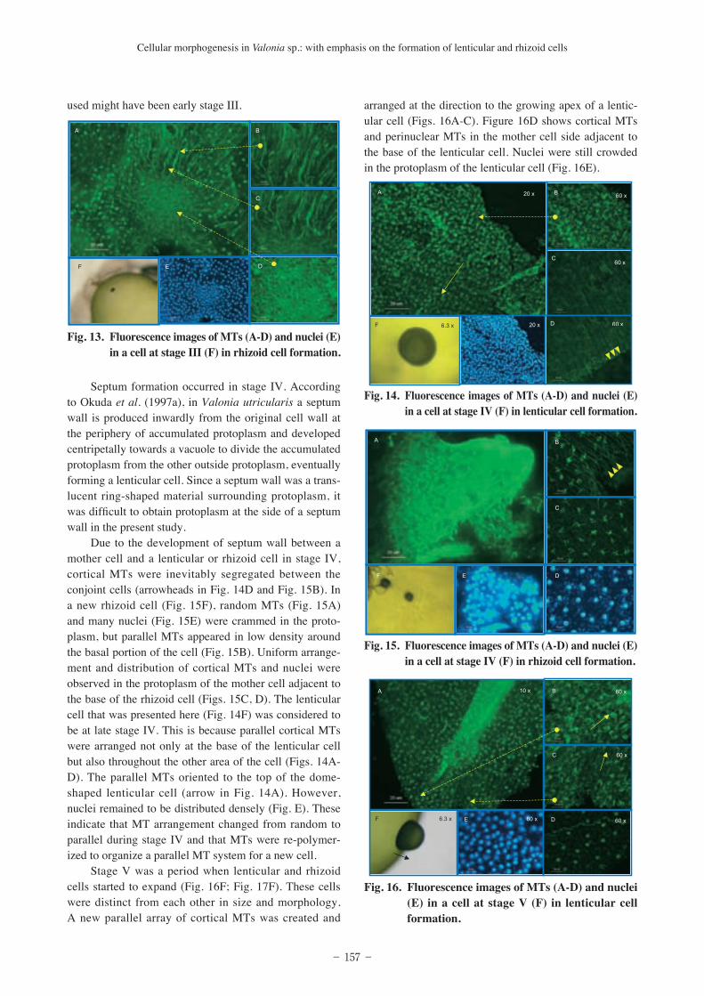
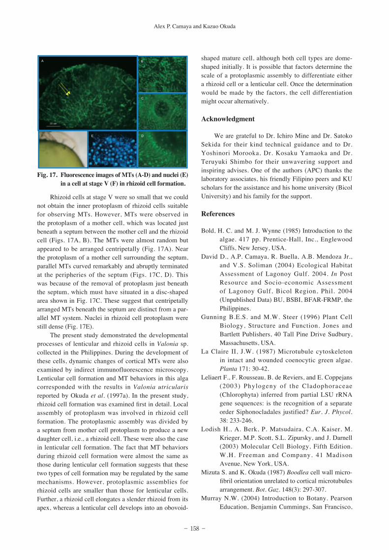
Due to the development of septum wall between a mother cell and a lenticular or rhizoid cell in stage IV, cortical MTs were inevitably segregated between the conjoint cells (arrowheads in Fig. 14D and Fig. 15B). In a new rhizoid cell (Fig. 15F), random MTs (Fig. 15A) and many nuclei (Fig. 15E) were crammed in the proto-plasm, but parallel MTs appeared in low density around the basal portion of the cell (Fig. 15B). Uniform arrange-ment and distribution of cortical MTs and nuclei were observed in the protoplasm of the mother cell adjacent to the base of the rhizoid cell (Figs. 15C, D). The lenticular cell that was presented here (Fig. 14F) was considered to be at late stage IV. This is because parallel cortical MTs were arranged not only at the base of the lenticular cell but also throughout the other area of the cell (Figs. 14A-D). The parallel MTs oriented to the top of the dome-shaped lenticular cell (arrow in Fig. 14A). However, nuclei remained to be distributed densely (Fig. E). These indicate that MT arrangement changed from random to parallel during stage IV and that MTs were re-polymer-ized to organize a parallel MT system for a new cell.
Stage V was a period when lenticular and rhizoid cells started to expand (Fig. 16F; Fig. 17F). These cells were distinct from each other in size and morphology. A new parallel array of cortical MTs was created and
arranged at the direction to the growing apex of a lentic-ular cell (Figs. 16A-C). Figure 16D shows cortical MTs and perinuclear MTs in the mother cell side adjacent to the base of the lenticular cell. Nuclei were still crowded in the protoplasm of the lenticular cell (Fig. 16E).
Fig. 13. Fluorescence images of MTs (A-D) and nuclei (E) in a cell at stage III (F) in rhizoid cell formation.
Fig. 14. Fluorescence images of MTs (A-D) and nuclei (E) in a cell at stage IV (F) in lenticular cell formation.
Fig. 15. Fluorescence images of MTs (A-D) and nuclei (E) in a cell at stage IV (F) in rhizoid cell formation.
Fig. 16. Fluorescence images of MTs (A-D) and nuclei (E) in a cell at stage V (F) in lenticular cell formation.
− 158 −
Alex P. Camaya and Kazuo Okuda
Rhizoid cells at stage V were so small that we could not obtain the inner protoplasm of rhizoid cells suitable for observing MTs. However, MTs were observed in the protoplasm of a mother cell, which was located just beneath a septum between the mother cell and the rhizoid cell (Figs. 17A, B). The MTs were almost random but appeared to be arranged centripetally (Fig. 17A). Near the protoplasm of a mother cell surrounding the septum, parallel MTs curved remarkably and abruptly terminated at the peripheries of the septum (Figs. 17C, D). This was because of the removal of protoplasm just beneath the septum, which must have situated in a disc-shaped area shown in Fig. 17C. These suggest that centripetally arranged MTs beneath the septum are distinct from a par-allel MT system. Nuclei in rhizoid cell protoplasm were still dense (Fig. 17E).
The present study demonstrated the developmental processes of lenticular and rhizoid cells in Valonia sp. collected in the Philippines. During the development of these cells, dynamic changes of cortical MTs were also examined by indirect immunofluorescence microscopy. Lenticular cell formation and MT behaviors in this alga corresponded with the results in Valonia utricularis reported by Okuda et al. (1997a). In the present study, rhizoid cell formation was examined first in detail. Local assembly of protoplasm was involved in rhizoid cell formation. The protoplasmic assembly was divided by a septum from mother cell protoplasm to produce a new daughter cell, i.e., a rhizoid cell. These were also the case in lenticular cell formation. The fact that MT behaviors during rhizoid cell formation were almost the same as those during lenticular cell formation suggests that these two types of cell formation may be regulated by the same mechanisms. However, protoplasmic assemblies for rhizoid cells are smaller than those for lenticular cells. Further, a rhizoid cell elongates a slender rhizoid from its apex, whereas a lenticular cell develops into an obovoid-
shaped mature cell, although both cell types are dome-shaped initially. It is possible that factors determine the scale of a protoplasmic assembly to differentiate either a rhizoid cell or a lenticular cell. Once the determination would be made by the factors, the cell differentiation might occur alternatively.
Acknowledgment
We are grateful to Dr. Ichiro Mine and Dr. Satoko Sekida for their kind technical guidance and to Dr. Yoshinori Morooka, Dr. Kosaku Yamaoka and Dr. Teruyuki Shimbo for their unwavering support and inspiring advises. One of the authors (APC) thanks the laboratory associates, his friendly Filipino peers and KU scholars for the assistance and his home university (Bicol University) and his family for the support.
References
Bold, H. C. and M. J. Wynne (1985) Introduction to the algae. 417 pp. Prentice-Hall, Inc., Englewood Cliffs, New Jersey, USA.
David D., A.P. Camaya, R. Buella, A.B. Mendoza Jr., and V.S. Soliman (2004) Ecological Habitat Assessment of Lagonoy Gulf. 2004. In Post Resource and Socio-economic Assessment of Lagonoy Gulf, Bicol Region, Phil. 2004 (Unpublished Data) BU, BSBI, BFAR-FRMP, the Philippines.
Gunning B.E.S. and M.W. Steer (1996) Plant Cell Biology, Structure and Function. Jones and Bartlett Publishers, 40 Tall Pine Drive Sudbury, Massachusetts, USA.
La Claire II, J.W. (1987) Microtubule cytoskeleton in intact and wounded coenocytic green algae. Planta 171: 30-42.
Leliaert F., F. Rousseau, B. de Reviers, and E. Coppejans (2003) Phylogeny of the Cladophoraceae (Chlorophyta) inferred from partial LSU rRNA gene sequences: is the recognition of a separate order Siphonocladales justified? Eur. J. Phycol. 38: 233-246.
Lodish H., A. Berk, P. Matsudaira, C.A. Kaiser, M. Krieger, M.P. Scott, S.L. Zipursky, and J. Darnell (2003) Molecular Cell Biology, Fifth Edition. W.H. Freeman and Company. 41 Madison Avenue, New York, USA.
Mizuta S. and K. Okuda (1987) Boodlea cell wall micro-fibril orientation unrelated to cortical microtubules arrangement. Bot. Gaz. 148(3): 297-307.
Murray N.W. (2004) Introduction to Botany. Pearson Education, Benjamin Cummings, San Francisco,
Fig. 17. Fluorescence images of MTs (A-D) and nuclei (E) in a cell at stage V (F) in rhizoid cell formation.

− 159 −
Cellular morphogenesis in Valonia sp.: with emphasis on the formation of lenticular and rhizoid cells
USA. Nishioka H., K. Okuda and S. Mizuta (1990) Growth and
cell-shape control in the marine coenocytic green alga, Chaetomorpha moniligera. Bot. Mar. 33: 289-297.
Okuda K. and S. Mizuta (1987) Modification in Cell shape unrelated to cellulose microfibrils orienta-tion in growing thallus cells of Chaetomorpha moniligera. Plant Cell Physiol. 28: 461-473
Okuda K., I. Mine and S. Ueno (1997a) Cytomorpho-genesis in coenocytic green algae. IV. The con-struction of cortical microtubules during lenticular cell formation in Valonia utricularis. Mem. Fac. Sci. Kochi Univ. Ser. D (Biol.) 18: 17-25.
Okuda K., I. Mine, T. Morinaga and N. Kuwaki (1997b) Cytomorphogenesis in coenocytic green algae. V. Segregative cell division and cortical microtubules in Dictyosphaeria cavernosa (Siphonocladales, Chlorophyceae). Phycol. Res. 45: 189-196.
Okuda K., K. Matsuo, S. Mizuta (1990) Preparation for immunofluorescence microscopy causes change in arrangements of cortical microtubules in some coenocytic green algae. Mem. Fac. Sci. Kochi Univ. Ser. D (Biol.) 11: 5-10.
Okuda K., K. Matsuo, S. Mizuta (1993) The meridional arrangement of cortical microtubules defines the site of tip growth in the coenocytic green alga, Chamaedoris orientalis. Bot. Mar. 36: 53-62.
Okuda K., N. Sakurai, K. Yuasa, I. Mine and T. Matsui (2000) Indirect immunofluorescence microscopy for observing cytoskeletons in giant celled green algae. Mem. Fac. Sci. Kochi Univ. Ser. D (Biol.) 21: 49-57.
S a t o h T . , N . S a k u r a i a n d K . O k u d a ( 2 0 0 0 ) Cytomorphogenesis in coenocytic green algae. VI. Dynamic changes in the actin cytoskeleton during wound-induced contraction in Valonia utricularis. Hikobia 13:153-161.
Sze, P. (1993) A biology of the algae, Second Edition. Wm. C. Brown Publishers, Wm. C. Brown Communications, Inc., 2460 Kerper Blvd. Dubuque, IA 52001, USA.
Sugimoto K., R.E. Williamson and G.O. Wasteneys (2000) New techniques enable comparative anal-ysis of microtubule orientation, wall texture, and growth rate in intact roots of Arabidopsis. Plant Physiol. 124: 1493-1506