Cellular Levels of Class 1 and Class 3 Aldehyde...
15
Vol. 3, 1901-1914, November 1997 Clinical Cancer Research 1901 Advances in Brief Cellular Levels of Class 1 and Class 3 Aldehyde Dehydrogenases and Certain Other Drug-metabolizing Enzymes in Human Breast Malignancies’ Lakshmaiah Sreerama and Norman E. Sladek2 Department of Pharmacology, University of Minnesota Medical School, Minneapolis, Minnesota 55455 Abstract Molecular determinants of cellular sensitivity to cyclo- phosphamide, long the mainstay of chemotherapeutic regi- mens used to treat metastatic breast cancer, include class 1 and class 3 aldehyde dehydrogenases (ALDH.1 and ALDH-3, respectively), which catalyze the detoxification of this agent. Thus, mterindividual variation in the activity of either of these enzymes in breast cancers could contribute to the wide variation in clinical responses that are obtained when such regimens are used to treat these malignancies. Consistent with this notion, ALDH-1 levels in primary and metastatic breast malignancies were found to range from 1-276 and 8-160 mIU/g tissue, respectively, and those of ALDH-3 range from 1-242 and 6-97 mIU/g tissue, respec- tively. ALDH-1 and ALDH-3 levels in normal breast tissue predicted the levels of these enzymes in primary and meta- static breast malignancies present in the same individuals. Confirming and extending the observations of others, levels of glutathione, a molecular determinant of cellular sensitiv- ity to various DNA cross-linking agents including cyclophos- phamide, and of DT-diaphorase, glutathione S-transferases, and cytochrome P450 1A1, each of which is known to cata- lyze the detoxification/toxification of one or more anticancer agents (although not of cyclophosphamide), also varied widely in primary and metastatic breast malignancies. Given the wide range of ALDH-1, ALDH-3, and glutathione levels that were observed in malignant breast tissues, meas- urement of their levels in normal breast tissue and/or pri- mary breast malignancies prior to the initiation of chemo- therapy is likely to be of value in predicting the therapeutic potential, or lack thereof, of cyclophosphamide in the treat- ment of metastatic breast cancer, thus providing a rational Received 3/13/97; revised 7/1 1/97; accepted 7/1 1/97. The costs of publication of this article were defrayed in part by the payment of page charges. This article must therefore be hereby marked advertisement in accordance with 18 U.S.C. Section 1734 solely to indicate this fact. I Supported by USPHS Grant CA 21737 and DOA DAMD 17-94-J- 4057. Descriptions of parts of this investigation have appeared in abstract form (Sladek, N. E., and Sreerama, L., Proc. Am. Assoc. Cancer Res., 36: 325, 1995). 2 To whom requests for reprints should be addressed, at Department of Pharmacology, University of Minnesota, 3-249 Millard Hall, 435 Del- aware Street SE, Minneapolis, MN 55455. Phone: (612) 625-0691; Fax: (612) 625-8408; E-mail: sladeO0l @maroon.tc.umn.edu. basis for the design of individualized therapeutic regimens when treating this disease. Introduction Cyclophosphamide is perhaps the most widely used che- motherapeutic drug in the conventional treatment, as well as the high-dose treatment (followed by autologous multipotent/pluri- potent hematopoietic cell reinfusion to ameliorate the severe myelosuppression that accompanies it), of metastatic breast cancer (reviewed in Refs. 1-4). Unfortunately, its use, even in combination with other agents as is usual, rarely results in cures. Most often underlying the failure of cyclophosphamide to rid the patient of all malignant cells are the facts that drug-resistant mutant clones appear early in the natural history of tumor progression, i.e., even before drug treatment (intrinsic resist- ance), and that new drug-resistant clones may develop quite rapidly after the initiation of therapy (acquired resistance). Until resistant subpopulations become the dominant popu- lation, cyclophosphamide and related compounds are clinically effective in the treatment of metastatic breast cancer and, in- deed, play a lead role in that regard, even when combined with other agents; therefore, an understanding of how resistance to these agents is effected would likely be of value because meas- ures may then become apparent as to how to prevent and/or negate it. Molecular determinants of cellular sensitivity to cy- clophosphamide and other oxazaphosphorines, e.g., 4-hydroper- oxycyclophosphamide, mafosfamide, and ifosfamide, include class 1 and class 3 ALDHs3 (ALDH-l and ALDH-3, respec- tively). Specifically, cellular sensitivity to the oxazaphospho- rines is inversely related to the cellular content of these enzymes because they each catalyze the detoxification of these agents (reviewed in Refs. S and 6; Refs. 7-18). Interindividual varia- tion in ALDH-1 and/or ALDH-3 levels has been observed in colon, ovarian, and salivary gland malignancies (19-21). Not known is the extent of interindividual variation, if any, in the activity of either of these enzymes in breast malignancies. Thus, the investigation reported herein sought to ascertain to what extent ALDH-l and ALDH-3 levels varied in malignant (and normal) breast tissues, i.e., whether clinical resistance to the oxazaphosphorines could be accounted for, at least in some cases, by relatively elevated levels of these enzymes. Surgically removed malignant (and normal) breast tissue samples were used for this purpose. A second objective was to ascertain whether ALDH- 1 and ALDH-3 levels in normal and malignant (primary and meta- 3 The abbreviations used are: ALDH, aldehyde dehydrogenase; GST, glutathione S-transferase; DT-D, DT-diaphorase; CYP 1A 1 , cytochrome P450 lAl ; GSH, glutathione; ER, estrogen receptor. Research. on August 25, 2018. © 1997 American Association for Cancer clincancerres.aacrjournals.org Downloaded from
-
Upload
truongminh -
Category
Documents
-
view
216 -
download
0
Transcript of Cellular Levels of Class 1 and Class 3 Aldehyde...

Vol. 3, 1901-1914, November 1997 Clinical Cancer Research 1901
Advances in Brief
Cellular Levels of Class 1 and Class 3 Aldehyde Dehydrogenases
and Certain Other Drug-metabolizing Enzymes in Human
Breast Malignancies’
Lakshmaiah Sreerama and Norman E. Sladek2
Department of Pharmacology, University of Minnesota Medical
School, Minneapolis, Minnesota 55455
Abstract
Molecular determinants of cellular sensitivity to cyclo-
phosphamide, long the mainstay of chemotherapeutic regi-
mens used to treat metastatic breast cancer, include class 1
and class 3 aldehyde dehydrogenases (ALDH.1 and
ALDH-3, respectively), which catalyze the detoxification of
this agent. Thus, mterindividual variation in the activity of
either of these enzymes in breast cancers could contribute to
the wide variation in clinical responses that are obtained
when such regimens are used to treat these malignancies.
Consistent with this notion, ALDH-1 levels in primary and
metastatic breast malignancies were found to range from
1-276 and 8-160 mIU/g tissue, respectively, and those of
ALDH-3 range from 1-242 and 6-97 mIU/g tissue, respec-
tively. ALDH-1 and ALDH-3 levels in normal breast tissue
predicted the levels of these enzymes in primary and meta-
static breast malignancies present in the same individuals.
Confirming and extending the observations of others, levels
of glutathione, a molecular determinant of cellular sensitiv-
ity to various DNA cross-linking agents including cyclophos-
phamide, and of DT-diaphorase, glutathione S-transferases,
and cytochrome P450 1A1, each of which is known to cata-
lyze the detoxification/toxification of one or more anticancer
agents (although not of cyclophosphamide), also varied
widely in primary and metastatic breast malignancies.
Given the wide range of ALDH-1, ALDH-3, and glutathione
levels that were observed in malignant breast tissues, meas-
urement of their levels in normal breast tissue and/or pri-
mary breast malignancies prior to the initiation of chemo-
therapy is likely to be of value in predicting the therapeutic
potential, or lack thereof, of cyclophosphamide in the treat-
ment of metastatic breast cancer, thus providing a rational
Received 3/13/97; revised 7/1 1/97; accepted 7/1 1/97.
The costs of publication of this article were defrayed in part by the
payment of page charges. This article must therefore be hereby marked
advertisement in accordance with 18 U.S.C. Section 1734 solely to
indicate this fact.
I Supported by USPHS Grant CA 21737 and DOA DAMD 17-94-J-
4057. Descriptions of parts of this investigation have appeared in
abstract form (Sladek, N. E., and Sreerama, L., Proc. Am. Assoc. Cancer
Res., 36: 325, 1995).2 To whom requests for reprints should be addressed, at Department ofPharmacology, University of Minnesota, 3-249 Millard Hall, 435 Del-
aware Street SE, Minneapolis, MN 55455. Phone: (612) 625-0691; Fax:(612) 625-8408; E-mail: sladeO0l @maroon.tc.umn.edu.
basis for the design of individualized therapeutic regimens
when treating this disease.
Introduction
Cyclophosphamide is perhaps the most widely used che-
motherapeutic drug in the conventional treatment, as well as the
high-dose treatment (followed by autologous multipotent/pluri-
potent hematopoietic cell reinfusion to ameliorate the severe
myelosuppression that accompanies it), of metastatic breast
cancer (reviewed in Refs. 1-4). Unfortunately, its use, even in
combination with other agents as is usual, rarely results in cures.
Most often underlying the failure of cyclophosphamide to rid
the patient of all malignant cells are the facts that drug-resistant
mutant clones appear early in the natural history of tumor
progression, i.e., even before drug treatment (intrinsic resist-
ance), and that new drug-resistant clones may develop quite
rapidly after the initiation of therapy (acquired resistance).
Until resistant subpopulations become the dominant popu-
lation, cyclophosphamide and related compounds are clinically
effective in the treatment of metastatic breast cancer and, in-
deed, play a lead role in that regard, even when combined with
other agents; therefore, an understanding of how resistance to
these agents is effected would likely be of value because meas-
ures may then become apparent as to how to prevent and/or
negate it. Molecular determinants of cellular sensitivity to cy-
clophosphamide and other oxazaphosphorines, e.g., 4-hydroper-
oxycyclophosphamide, mafosfamide, and ifosfamide, include
class 1 and class 3 ALDHs3 (ALDH-l and ALDH-3, respec-
tively). Specifically, cellular sensitivity to the oxazaphospho-
rines is inversely related to the cellular content of these enzymes
because they each catalyze the detoxification of these agents
(reviewed in Refs. S and 6; Refs. 7-18). Interindividual varia-
tion in ALDH-1 and/or ALDH-3 levels has been observed in
colon, ovarian, and salivary gland malignancies (19-21). Not
known is the extent of interindividual variation, if any, in the
activity of either of these enzymes in breast malignancies.
Thus, the investigation reported herein sought to ascertain
to what extent ALDH-l and ALDH-3 levels varied in malignant
(and normal) breast tissues, i.e., whether clinical resistance to
the oxazaphosphorines could be accounted for, at least in some
cases, by relatively elevated levels of these enzymes. Surgically
removed malignant (and normal) breast tissue samples were
used for this purpose.
A second objective was to ascertain whether ALDH- 1 and
ALDH-3 levels in normal and malignant (primary and meta-
3 The abbreviations used are: ALDH, aldehyde dehydrogenase; GST,
glutathione S-transferase; DT-D, DT-diaphorase; CYP 1A 1 , cytochrome
P450 lAl ; GSH, glutathione; ER, estrogen receptor.
Research. on August 25, 2018. © 1997 American Association for Cancerclincancerres.aacrjournals.org Downloaded from
1902 ALDH-l and ALDH-3 Levels in Human Breast Malignancies
static) breast tissue samples taken from the same patients were
quantitatively related. This was because, in the event that such
a relationship did exist, determination of these enzyme levels in
easily obtainable normal or malignant primary breast tissue
samples would be of prognostic value with regard to the success,
or lack thereof, that might be anticipated upon the subsequent
use of an oxazaphosphorine to eliminate microscopic malignant
metastatic nodules.
Xenobiotics that are abundantly present in the diet/
environment, e.g. , 3-methylcholanthrene and catechol, rap-
idly, coordinately, and reversibly induce ALDH-3, DT-D,
GSTs, UDP-glucuronosyl transferase and, in some cases,
CYP lAl in cultured human breast cancer cell models. Con-
sequently, reversible multienzyme-mediated multidrug resist-
ance/collateral sensitivity to cyclophosphamide and certain
other anticancer drugs is rapidly effected (22). Some of the
latter are also already used, e.g. , mitoxantrone (reviewed in
Ref. 23), or show promise, e.g., E09 (24), in the treatment of
metastatic breast cancer. Ingestion of certain dietary sub-
stances, namely, coffee and broccoli, has been shown to
result in the coordinated elevation of ALDH-3, DT-D, and
the GSTs in human saliva (25). Stable (irreversible) intrinsic
as well as acquired phenotypes of this sort have also been
observed in cultured human cancer models (12, 13, 22). Not
known is whether coordinated elevation of these enzymes
ever occurs in normal and/or malignant breast tissue. Thus, in
a first attempt to address this question, DT-D, pan-GST,
GSTa, GSTp., GST’rr, and CYP lAl levels in the malignant
(and normal) breast tissue samples were also quantified.
The sulthydryl, glutathione, appears to be yet another mo-
lecular determinant of cellular sensitivity to the oxazaphospho-
rines (reviewed in Ref. 5). Thus, its levels in malignant (and
normal) breast tissues were determined as well.
Materials and Methods
Normal (n = 26) and malignant (n = 1 12) female breast
tissue samples obtained from 1 10 donors were procured from
the Cooperative Human Tissue Network, Midwestern Divi-
sion, Columbus, OH. Surgically removed normal and malig-
nant breast tissue samples were snap-frozen in liquid nitrogen
(within 6 h after removal), stored at -70#{176}C (5 to 60 days),
and shipped to us in dry ice. Patient characteristics, diag-
noses, and cellular characteristics (Table 1) were provided by
the pathology reports that accompanied the tissue specimens.
Purified human GSTs a, p., and ‘i� and affinity-purified poly-
clonal antibodies specific for each of these isozymes, i.e.,
anti-GSTa IgG, anti-GSTp. IgG, and anti-GST-n� IgG, respec-
tively (26), were generously provided by Dr. A. J. Townsend
(Department of Biochemistry, Bowman Gray School of Mcd-
icine, Wake Forest University, Winston-Salem, NC). Micro-
somes, isolated from a cell line (hlAl v2) transfected with
human CYP lAl cDNA and constitutively expressing the
enzyme (15 ng CYP lAl/mg microsomal protein), were
purchased from Gentest Corporation (Woburn, MA), as was
polyclonal anti-CYP lAl IgG (obtained from goats immu-
nized with rat CYP lAl). Anti-goat IgG-alkaline phosphatase
conjugate was purchased from Sigma Chemical Co. (St.
Louis, MO). Enhanced protein binding 96-well ELISA plates
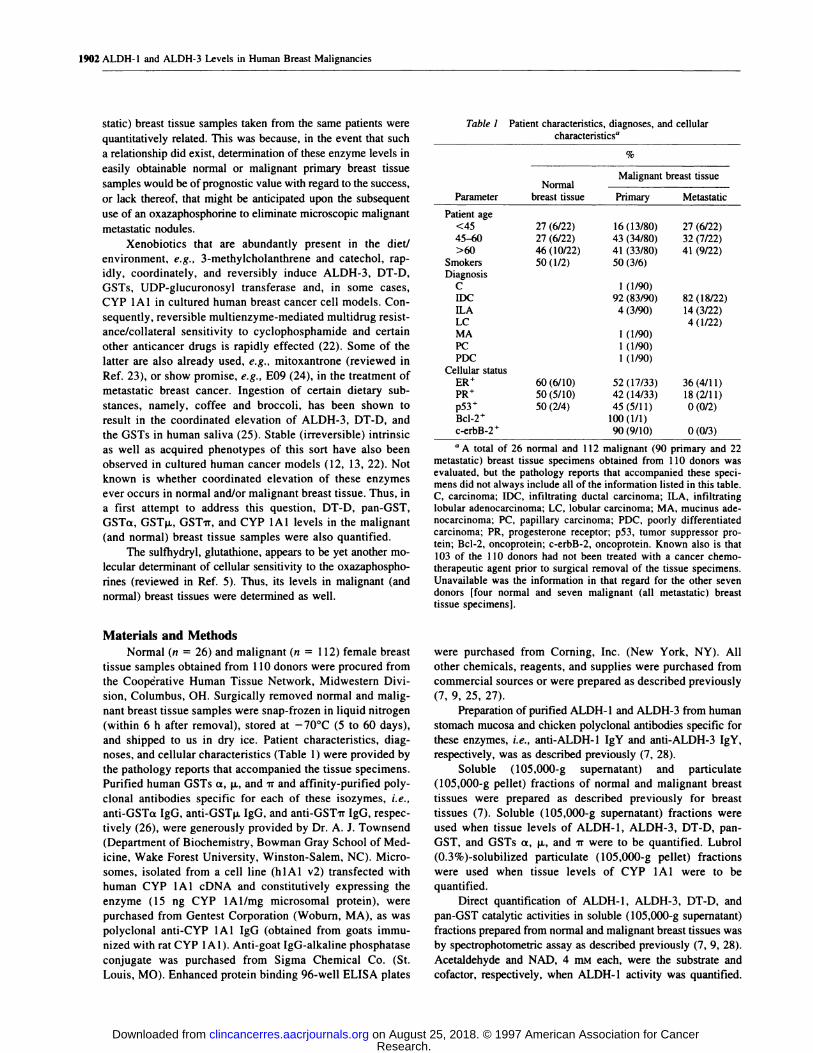
Table 1 Patient characteristics, diagnoses, and cellular
characteristicsa
%
Malignant breast tissueNormal
Parameter breast tissue Primary Metastatic
Patient age
<45 27(6/22) 16(13/80) 27 (6/22)
45-60 27 (6/22) 43 (34/80) 32 (7/22)
>60 46 (10/22) 41 (33/80) 41 (9/22)Smokers 50(1/2) 50(3/6)Diagnosis
C 1(1/90)
IDC 92(83/90) 82 (18/22)
ThA 4(3/90) 14 (3/22)LC 4(1/22)
MA 1(1/90)PC 1 (1/90)PDC 1 (1/90)
Cellular statusER� 60(6/10) 52(17/33) 36(4/11)
PR� 50(5/10) 42(14/33) 18(2/11)p53+ 50(2/4) 45(5/11) 0(0/2)
Bcl-2� 100(1/1)
c-erbB-2� 90(9/10) 0(0/3)
a A total of 26 normal and 112 malignant (90 primary and 22
metastatic) breast tissue specimens obtained from 1 10 donors was
evaluated, but the pathology reports that accompanied these speci-mens did not always include all of the information listed in this table.C, carcinoma; IDC, infiltrating ductal carcinoma; ILA, infiltrating
lobular adenocarcinoma; LC, lobular carcinoma; MA, mucinus ade-nocarcinoma; PC, papillary carcinoma; PDC, poorly differentiated
carcinoma; PR, progesterone receptor; p53, tumor suppressor pro-tein; Bcl-2, oncoprotein; c-erbB-2, oncoprotein. Known also is that
103 of the I 10 donors had not been treated with a cancer chemo-
therapeutic agent prior to surgical removal of the tissue specimens.
Unavailable was the information in that regard for the other seven
donors [four normal and seven malignant (all metastatic) breasttissue specimens].
were purchased from Corning, Inc. (New York, NY). All
other chemicals, reagents, and supplies were purchased from
commercial sources or were prepared as described previously
(7, 9, 25, 27).
Preparation of purified ALDH-l and ALDH-3 from human
stomach mucosa and chicken polyclonal antibodies specific for
these enzymes, i.e., anti-ALDH-l IgY and anti-ALDH-3 IgY,
respectively, was as described previously (7, 28).
Soluble (105,000-g supernatant) and particulate
(105,000-g pellet) fractions of normal and malignant breast
tissues were prepared as described previously for breast
tissues (7). Soluble (l05,000-g supernatant) fractions were
used when tissue levels of ALDH-1, ALDH-3, DT-D, pan-
GST, and GSTs a, p., and ir were to be quantified. Lubrol
(0.3%)-solubilized particulate ( 105,000-g pellet) fractions
were used when tissue levels of CYP 1A1 were to be
quantified.
Direct quantification of ALDH-1, ALDH-3, DT-D, and
pan-GST catalytic activities in soluble (105,000-g supernatant)
fractions prepared from normal and malignant breast tissues was
by spectrophotometric assay as described previously (7, 9, 28).
Acetaldehyde and NAD, 4 mM each, were the substrate and
cofactor, respectively, when ALDH-l activity was quantified.
Research. on August 25, 2018. © 1997 American Association for Cancerclincancerres.aacrjournals.org Downloaded from

0
0
0
0)
E
I
-J
200
100
0
0
0
0)
EC,)I
-I
200
100
.#{149} A
#{149}
AAA
I.
A#{149}
AA0 #{149}I#{149}#{149}
.#{149} A#{149}#{149}S.
0055
0 5.555 AAS... AS5SS A0 #{149}�
00 � AAA000 SSSSSSSSSSSS AA000 SSSSSSS$5SSsssss �000000 SSSSSSSSSS#{149} A000000 SSSSSSS
.0�-
Normal Metastatic
SS
A
A
S AAA
0 5
550 55
A0 #{149}su..s.0 _______ LA�o ::::ss::.0 #{149}#{149}ssuuu:uc�cxjoo ussssp$IpuuuuuI AL0� ssssuusu::::ssuusssuus AL
-0
Normal Metastatlc
Benzaldehyde and NADP, 4 m�i each, were the substrate and
cofactor, respectively, when ALDH-3 activity was quantified.
Substrate, cofactor, and inhibitor were 2,6-dichlorophenol-indo-
phenol (40 �.LM), NADH (160 p.M), and dicumarol (10 tiM),
respectively, when DT-diaphorase activity was quantified. Co-
substrates were l-chloro-2,4-dinitrobenzene and GSH, 1 mr�i
each, when pan-GST activity was quantified.
Spectrophotometric quantification of GSH levels in normal
and malignant breast tissue was as described by Anderson (29).
Spectrophotometric quantification of protein levels in soluble
(105,000-g supernatant) and Lubrol-solubilized particulate
(105,000-g pellet) fractions of normal and malignant breast
tissues was as described previously (7).
Indirect quantification of ALDH-l, ALDH-3, and GSTs
a, ji., and IT catalytic activities in soluble (105,000-g super-
natant) fractions was by ELISAs, as described previously (22,
27). Dilution with blocking solution of primary antibodies
was 1:1000 in the cases ofALDH-l and ALDH-3 and 1:2000
in the cases of GSTs a, p�, and ‘it. Normal and malignant
breast tissue levels (catalytic activities/g of tissue) of
ALDH-l, ALDH-3, and GSTs a, p., and i� were estimated
from standard curves generated with purified enzymes; spe-
cific activities of the latter were 2,850, 60,500, 44,600,
24,100, and 56,800 mIU/mg protein, respectively, when sub-
strates and cofactors were as in the direct assays.
Quantification of CYP lAl levels in Lubrol-solubilized
particulate (l05,000-g pellet) fractions was by an ELISA as
described immediately above, except that the: (a) primary
antibody was anti-CYP lAl IgG diluted 1:1000 with block-
ing solution; and (b) secondary antibody was anti-goat
IgG-alkaline phosphatase conjugate diluted I : 1000 with
blocking solution. Normal and malignant breast tissue levels
(pg/g tissue) of CYP lAl were estimated from standard
curves generated with Lubrol-solubilized CYP I A 1-contain-
ing microsomes (15 ng of CYP lAl/mg of microsomal
protein).
The Macintosh-based STATView II (Brainpower, Inc.,
Calabas, CA) computer program was used to generate Ps
(one- and two-tailed, unpaired, Student’s t-tests), and linear
regression lines, r� (regression coefficients), and Ps thereof.
Clinical Cancer Research 1903
Primary
Fig. 1 ALDH-l levels in human normal breast (n = 26) and primary
(n = 90) and metastatic (n = 22) breast tumor tissue samples. ALDH- 1
catalytic activity [NAD (4 mM)-linked oxidation of acetaldehyde (4
mM)] was quantified both directly by spectrophotometric assay and
indirectly by an ELISA as described in “Materials and Methods.” Only
catalytic activity quantified indirectly is shown because the values
obtained were independent of the method used to obtain them (r2 =
0.987, P � 0.0001). Points are means (rounded off for clarity of
presentation to zero if they were <2.5 mIU/g, and to 5 mIU/g or the
nearest multiple thereof if they were �2.5 mIU/g) of duplicate deter-minations made on single normal and malignant, or just malignant,tissue samples taken from each of 110 patients.
Primary
Fig. 2 ALDH-3 levels in human normal breast (n = 26) and primary
(n 90) and metastatic (n = 22) breast tumor tissue samples. ALDH-3
catalytic activity [NADP (4 mM)-hnked oxidation of benzaldehyde (4
mM)] was quantified both directly by spectrophotometric assay and
indirectly by an ELISA as described in “Materials and Methods.” Onlycatalytic activity quantified indirectly is shown because the values
obtained were independent of the method used to obtain them (r2 =
0.976, P � 0.0001). Points are means (rounded off for clarity ofpresentation to zero if they were <2.5 mIU/g, and to 5 mIU/g or thenearest multiple thereof if they were �2.5 mIU/g) of duplicate deter-minations made on single normal and malignant, or just malignant,tissue samples taken from each of 110 patients.
Research. on August 25, 2018. © 1997 American Association for Cancerclincancerres.aacrjournals.org Downloaded from

PRI MARY
40
20
NORMAL
Dp�0o��jg:i�
D
METASTATIC
S
200�
S
100��;:
S � #{149}�#{149}1100 200
ALLC I
20O�
S
S � � � � I
100 200
ALDH-1, mlU/g tissue
Fig. 3 ALDH-1 and ALDH-3 levels in normal(n 26; 0) and malignant [primary (n = 90; #{149})and metastatic (n = 22; A)] human breast tissues.
ALDH-1 [NAD (4 mrsi)-linked oxidation of acet-
aldehyde (4 mM)] and ALDH-3 [NADP (4 mid)-
linked oxidation of benzaldehyde (4 mM)] cata-lytic activities were indirectly quantified by
ELISAs as described in “Materials and Methods.”Points are means of duplicate determinations
made on single, normal and malignant, or just
malignant, breast tissue samples taken from each
of 110 patients.
50 100 150
I mlU/g tissue
A x2 test (2 X 2 table) was used to ascertain whether there
were any statistically significant differences in frequencies of
expression (positive or negative) of GSTa, GSTp., and CYP
lAl as a function of tissue type and of CYP 1A1 as a
to ascertain whether the observed simultaneously elevated
levels of ALDH-3, DT-D, GST (pan-GST and GST’rr), and
CYP lAl, and/or simultaneously elevated levels of ALDH-3,
DT-D, and GST (pan-GST and GSTi�), were the consequence
1904 ALDH-1 and ALDH-3 Levels in Human Breast Malignancies
function of estrogen receptor status. This test was also used of a common or of independent event(s).
Table 2 ALDH-l, ALDH-3, DT-D, pan-GST, GSTs a, �i, and ir, CYP 1A1, and GSH levels in human normal and malignant (primary and
metastatic) breast tissue samples: summary’�
Normal Primary Metastatic All
Enzyme n Mean ± SD Range n Mean ± SD Range n Mean ± SD Range n Mean ± SD Range
ALDH-l 26 19 ± 19 3-75 90 37 ± 43 1-276 22 54 ± 41 8-160 138 36 ± 41 1-276
ALDH-3 26 14 ± 12 2-54 90 24 ± 41 1-242 22 34 ± 27 6-97 138 24 ± 36 1-242
DT-D 26 438 ± 690 23-2700 90 1190 ± 1520 10-6250 22 1110 ± 810 84-2720 138 1030 ± 1330 10-6250
pan-GST 26 920 ± 1050 56-4550 90 2400 ± 2030 123-8880 22 3330 ± 2380 464-8130 138 2270 ± 2070 56-8880
GSTa 26 125 ± 171 0-900 90 293 ± 427 0-2500 22 501 ± 605 0-1700 138 294 ± 438 0-2500
GSTp. 26 96 ± 129 0-522 90 301 ± 560 0-3400 22 295 ± 654 0-3050 138 261 ± 529 0-3400GSTir 26 694 ± 829 100-3800 90 1730 ± 1410 120-6200 22 2360 ± 1800 400-6500 138 1630 ± 1470 100-6500
CYP lAl 21 1 ± 5 0-24 80 36 ± 92 0-570 19 52 ± 120 0-485 120 33 ± 90 0-570
GSH 25 264 ± 681 2-3470 82 1130 ± 1990 16-10400 18 2120 ± 3360 16-1 1 100 125 1 100 ± 2130 2-11100
a Values are summaries of the data presented in Figs. 1, 2, and 5-11. Zero values obtained for GSTa, GSTp., and CYP lAl are included in the
calculation of mean values for these enzymes. Units are mIU/g tissue except in the cases ofCYP 1A1 and GSH, where they are pg/g and nmol/g tissue,
respectively. Statistically, mean enzyme and GSH levels in primary and metastatic breast tumor tissue are significantly higher (P < 0.05; one-tailed,
unpaired, Student’s t-test) than those in normal breast tissue in all cases except in that of metastatic breast tumor tissue GSTp. (P = 0.084). Mean
enzyme and GSH levels in metastatic breast tumor tissues are significantly higher (P S 0.05) than those in primary breast tumor tissue only in the
cases of ALDH-l and pan-GST.
0
SISI
0)
EC.)
I0-J4
03SISI
0)
E
C.)
I0-I4
Research. on August 25, 2018. © 1997 American Association for Cancerclincancerres.aacrjournals.org Downloaded from

ALDH-1 ALDH-3
80’
Fig. 4 ALDH-1 and ALDH-3 levels in pairedhuman normal and primary tumor (n = 21; 0),
and normal and metastatic tumor (n = 7; S)breast tissues. ALDH-l [NAD (4 mM)-linked
oxidation of acetaldehyde (4 mM)] and ALDH-3[NADP (4 nmi)-linked oxidation of benzalde-hyde (4 mM)] catalytic activities were indirectlyquantified by ELISAs as described in “Materialsand Methods.” Points are means of duplicatedeterminations made on single normal and ma-lignant breast tissue samples taken from each of26 patients.
0)
E
SS#{149}1
I-
0E
I-
S
E
a.
0)
D
E
S3SS
I-
0E3
I-.
U
S
SS
S
0)
E
53SS
0E3
I-
U
S
4,SS
.5
S
Ea.
>�‘ 0.78p � 0.0001
40
80
40
200
0
20 40 60
I J I
S
0 � I
, �=0.58
� 0 p�0.000180 � � I
Normal Tissue, mlU/g
100
�v’_� �p � 0.0001
Normal Tissue, mlU/g
20 40 60
Clinical Cancer Research 1905
Results
Shown in Figs. 1 and 2 are scatter plots of, respectively,
ALDH-l and ALDH-3 levels in normal and malignant (primary
as well as metastatic) breast tissue samples. Mean values and
SDs are presented in Table 2. Immediately apparent is that the
level of each enzyme varies widely in all three tissues. For
example, highest levels of ALDH-1 and ALDH-3 in primary
breast malignancies were �-250-fold greater than the lowest
levels of these enzymes in these tissues. Statistical analysis of
this data revealed that mean levels of each enzyme in the
malignant (primary or metastatic) breast tissue samples were
significantly higher (P � 0.05) than those in the normal breast
tissue samples. The mean ALDH-1 level in the metastatic breast
tumor samples was significantly higher (P = 0.029) than that in
the primary breast tumor samples. The mean ALDH-3 level in
the metastatic breast tumor samples was not significantly higher
than that in the primary breast tumor samples at a P level of 0.05
but was very nearly so as the P value was 0.05 1.
Unexpectedly, cellular levels of ALDH-l and ALDH-3
appeared to be directly related in the normal, as well as in the
malignant (primary as well as metastatic), breast tissue samples
(Fig. 3).
ALDH-l and ALDH-3 levels in the normal breast tissue
samples predicted the respective levels of these enzymes in
paired primary, as well as metastatic, breast tumor tissue sam-
ples (Fig. 4). We did not have enough paired samples to ascer-
tam whether cellular levels of ALDH-l or ALDH-3 in primary
breast malignancies predicted cellular levels of these enzymes in
metastatic breast malignancies.
Confirming and extending the observations of others (Refs.
Research. on August 25, 2018. © 1997 American Association for Cancerclincancerres.aacrjournals.org Downloaded from

6000
4000
2000
4,3In4)
a)
E
0I-0
S
S
S
S
S
S
S
SSS
S
.5
0 A
0 55 A
S AS
SS
AAAAA
S. A55555 AS. AL
00 5555.5.
0 555 A555 AS
0 555 LA88� ::::u::: AL00 555555555555 A0000000000 55555555555 A
8000
6000
4000
2000
4,34)In
a)
EI-Cl)
C4,a.
S
S
A
S
AS
S
S
A
A
S
A
.5
$0 i:
I0
IS A
CS A
88o _0
SSSSSSS A
$1, A
0 �$... LA
$55
1906 ALDH-l and ALDH-3 Levels in Human Breast Malignancies
Normal Primary Metastatic
Fig. 5 DT-D levels in human normal breast (n = 26) and primary (n =
90) and metastatic (n = 22) breast tumor tissue samples. A spectropho-
tometric assay was used as described in “Materials and Methods” to
directly quantify DT-D catalytic activity; substrate, cofactor, and inhib-itor were 2,6-dichlorophenol-indophenol (40 p.M), NADH (160 jiM),
and dicumarol (10 jiM), respectively. Points are means (rounded off for
clarity of presentation to zero if they were <50 mlU/g, and to 100
mIU/g or the nearest multiple thereof if they were �50 mIU/g) ofduplicate determinations made on single normal and malignant, or justmalignant, tissue samples taken from each of 1 10 patients.
30 and 3 1 ; reviewed in Ref. 32), DT-D, pan-GST, GSTa,
GSTp., GSTi�, and CYP lAl levels also varied widely in
normal and malignant (primary and metastatic) breast tissue
(Figs. 5-10 and Table 2). As in the cases of ALDH-l and
ALDH-3, statistical analysis of the data revealed that mean
levels of each enzyme in the malignant (primary or metastatic)
breast tissue samples were significantly higher (P � 0.05) than
those in the normal breast tissue samples with one exception,
metastatic breast tumor tissue GSTp. (P = 0.084). The mean
pan-GST level in the metastatic breast tumor samples was
significantly higher (P = 0.041) than that in the primary breast
tumor samples. Mean DT-D, GSTa, GSTp., GST’rr, and CYP
I A 1 levels in the metastatic breast tumor samples were not
significantly higher at a P level of 0.05 than those in the primary
breast tumor samples, although those of GSTa (P = 0.061) and
GSTir (P = 0.058) were very nearly so.
Statistically, lack of detectable GSTa and GSTp. (Figs. 7
and 8, respectively) was independent of tissue type (P > 0.1), as
was the lack of detectable CYP lAl (Fig. 10; P > 0.05);
however, a more frequent lack of detectable CYP 1A I in normal
Normal Primary Metastatic
Fig. 6 Pan-GST levels in human normal breast (n 26), and primary(n 90) and metastatic (n = 22) breast tumor tissue samples. A
spectrophotometric assay was used as described in “Materials and
Methods” to directly quantify pan-GST catalytic activity; cosubstrates
were l-chloro-2,4-dinitrobenzene and GSH, I mM each. Points aremeans (rounded off for clarity of presentation to zero if they were <50
mlU/g, and to 100 mIU/g or the nearest multiple thereof if they were�50 mIU/g) of duplicate determinations made on single normal and
malignant, or just malignant, tissue samples taken from each of 1 10
patients.
breast tissue was very nearly statistically significant (P =
0.0523).
As expected, tissue levels of GSTa, GSTp., GST’ir, and
pan-GST were directly related to each other when all of the data
(n 138) was grouped (P � 0.0001 in each case; linear
regression analyses of data not shown). Similarly, there was a
direct relationship between pan-GST and ALDH-l, ALDH-3,
and DT-D levels (P = 0.01, 0.02, and 0.003, respectively; linear
regression analyses of data not shown). However, DT-D levels
were not related to those of ALDH-l or ALDH-3 (P 0.33 and
0.42, respectively), and CYP lAl levels were unrelated (P >
0. 1) to those of the other enzymes (linear regression analyses of
data not shown).
Evidence (levels that are each more than 1 SD above
normal breast tissue mean levels) for the coordinated induction
of ALDH-3, DT-D, pan-GST, and CYP lAl (induced gene
expression effected by transactivation of a cis-acting DNA ele-
ment, xenobiotic responsive element, present in the 5’-upstream
regions of the genes coding for these enzymes; Refs. 1 3 and
Research. on August 25, 2018. © 1997 American Association for Cancerclincancerres.aacrjournals.org Downloaded from

4,3SIS
0)
E
I-Cl)C,
2000
1000
3000
2000
1000
S
A
S LAS A
S
SS A
S
S
0
SS A55
S
A55S5555 A55555 LA
0 55555555 LA00 5555555 A000 SSS55.5SSS5.SS A00000000 55555 LA000000 i!ff!!#{149}� A
4,3In4)
a)
E
I-Cl)
C,
S
A
S
S
55
SS
S
S
S
S
A
S A55
0 555 A55 A55
0 55 AA00 5555500 55555 AA000 SSSS#{149}SS
55555 A22900 55SSS#{149}#{149} -
Normal Metastatic Normal Metastatlc
33-41) was observed in only three samples, two primary and
one metastatic breast tumors (Table 3). As judged by the same
criteria, coordinated induction of ALDH-3, DT-D, and pan-
GST, but not of CYP lAl (induced gene expression effected by
transactivation of a cis-acting DNA element, antioxidant respon-
sive element, present in the 5’-upstream regions of the genes
coding for these enzymes; Refs. 13 and 34-41) was observed in
only seven additional samples, four primary and three metastatic
breast tumors (Table 3). In one case, sample 6, a normal breast
sample obtained from the same patient was available. ALDH-3
and DT-D levels in this sample were each more than 2 SDs, and
pan-GST and GST’n� levels were nearly (-=0.86) 1 SD, above
corresponding normal breast tissue mean values; CYP lAl was
not detected in this sample (data not shown).
DT-D, pan-GST, GSTa, GSTji, GSTi�, and CYP 1A1
levels in normal breast tissue did not predict (P > 0.1) for
corresponding enzyme levels in paired, primary, or metastatic
breast tumor tissue (linear regression analyses of data not
shown). However, detectable GSTp. was always absent in ma-
lignant breast tissue samples when it was not found in normal
breast tissue samples obtained from the same patient (n = 1 1),
as would be expected if the absence of this enzyme was due to
a GSTji null genotype (reviewed in Ref. 41 ; data not shown). In
contrast, GSTa was detected in two of three malignant breast
tissue samples obtained from patients from which normal breast
tissue samples lacked detectable levels of this enzyme; more-
over, in five cases, GSTa was found in normal, but not malig-
nant, breast tissue (data not shown). In the case of CYP lAl, 15
of 19 malignant breast tissue samples tested negative when the
paired normal breast tissue samples tested negative, and one
malignant breast tissue sample tested negative when the paired
normal breast tissue sample tested positive (data not shown). We
did not have enough paired samples to ascertain whether cellular
levels of these enzymes in primary breast malignancies pre-
Clinical Cancer Research 1907
Primary
Fig. 7 GSTa levels in human normal breast (n = 26) and primary (n =
90) and metastatic (n = 22) breast tumor tissue samples. GSTa catalytic
activity [conjugation of GSH to l-chloro-2,4-dinitrobenzene (1 m�seach)] was indirectly quantified by an ELISA as described in “Materialsand Methods.” Points are means (rounded off for clarity of presentationto zero if they were >0 and <25 mJU/g, and to 50 mIU/g or the nearestmultiple thereof if they were �25 mIU/g) of duplicate determinationsmade on single normal and malignant, or just malignant, tissue samples
taken from each of 1 10 patients. GSTa was not detected in 4 of 26(15%) normal breast tissue samples, 14 of 90 (16%) primary breasttumor samples, and 5 of 22 (23%) metastatic breast tumor samples.These zero values are not shown in this figure.
Primary
Fig. 8 GSTja levels in human normal breast (n = 26) and primary (n =
90) and metastatic (n = 22) breast tumor tissue samples. GSTp. catalytic
activity [conjugation of GSH to l-chloro-2,4-diaitrobenzene (1 ms�each)] was indirectly quantified by an ELISA as described in “Materials
and Methods.” Points are means (rounded off for clarity of presentationto zero if they were >0 and <25 mIU/g, and to 50 mIU/g or the nearest
multiple thereof if they were �25 mIU/g) of duplicate determinations
made on single normal and malignant, or just malignant, tissue samples
taken from each of 110 patients. GSTp. was not detected in 9 of 26(35%) normal breast tissue samples, 38 of 90 (42%) primary breasttumor samples, and 1 1 of 22 (50%) metastatic breast tumors samples.
These zero values are not shown in this figure.
Research. on August 25, 2018. © 1997 American Association for Cancerclincancerres.aacrjournals.org Downloaded from

6000
4000
2000
a)3InIn
0)
E
I-Cl)
C,
A
S
A55S
S
55S
A0 5 LA
SS A
AA
AA
555S
0 555555555
55555555555S A555S. A
0 5 AS A555555 A5555.55
000 555 A0 555555 A00 5555 A
5.500 5555500 555 ALLA00000 555000000 555500 � 555
a)3InIn
a)
a.
4
a.>.
(I
400
200
A
S
S
S
S
AS55
A
S A
S
555S55S
A0 55
555555 A
Normal Metastatic
1908 ALDH-l and ALDH-3 Levels in Human Breast Malignancies
Normal Primary Metastatic
Fig. 9 GST’i� levels in human normal breast (n = 26) and primary (n =
90) and metastatic (n = 22) breast tumor tissue samples. GST’i� catalytic
activity [conjugation of GSH to l-chloro-2,4-dinitrobenzene (1 mM
each)] was indirectly quantified by an ELISA as described in “Materialsand Methods.” Points are means (rounded off for clarity of presentationto zero if they were <50 mIU/g, and to 100 mIU/g or the nearestmultiple thereof if they were �50 mIU/g) of duplicate determinations
made on single normal and malignant, or just malignant, tissue samples
taken from each of 1 10 patients.
dicted cellular levels of the corresponding enzyme in metastatic
breast malignancies.
Again confirming and extending the observations of others
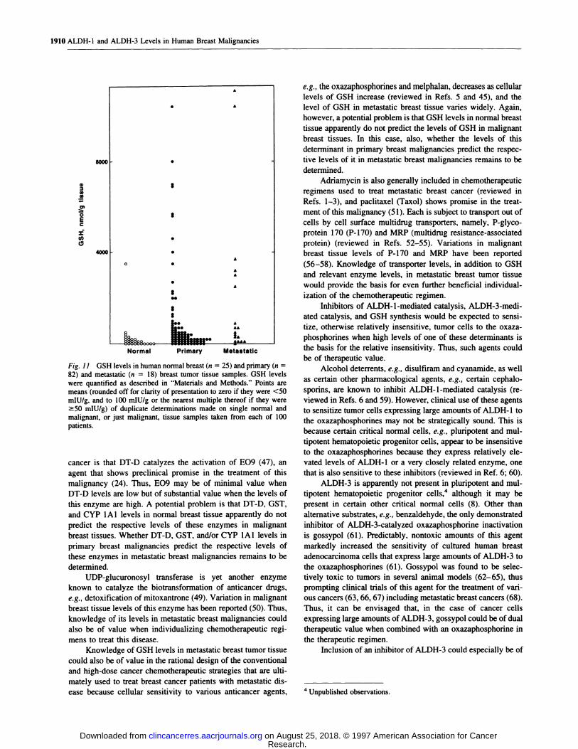
(42, 43), GSH levels, also, varied widely in normal and malig-
nant (primary and malignant) breast tissue (Fig. 1 1 and Table 2).
As in the cases of the enzymes, statistical analysis of the data
revealed that mean levels of GSH in the malignant (primary or
metastatic) breast tissue samples were significantly higher (P �
0.05) than that in the normal breast tissue samples. The mean
GSH level obtained for metastatic breast tumor samples was not
significantly higher (P > 0. 1) than that obtained for the primary
breast tumor samples.
GSH levels were not related to any of the enzyme levels
when all of the data (n = 1 1 5-1 25) were grouped (P > 0.1;
linear regression analyses of data not shown).
GSH levels in normal breast tissue samples did not predict
(P > 0. 1) for the corresponding GSH levels in paired, primary,
or metastatic breast tumor tissue samples (linear regression
analyses of data not shown). We did not have enough paired
samples to ascertain whether cellular levels of GSH in primary
Primary
Fig. 10 CYP lAl levels in human normal breast (n = 21) and primary(n 80) and metastatic (n = 19) breast tumor tissue samples. CYP lAllevels were quantified by an ELISA as described in “Materials and
Methods.” Points are means (rounded off for clarity of presentation tozero if they were >0 and <5 pg/g, and to 10 pg/g or the nearest multiplethereof if they were �5 mIU/g) of duplicate determinations made onsingle normal and malignant, or just malignant, tissue samples takenfrom each of 99 patients. CYP lAl was not detected in 19 of 21 (90%)normal breast tissue samples, 53 of 80 (66%) primary breast tumor
samples, and 13 of 19 (68%) metastatic breast tumors samples. These
zero values are not shown in this figure.
breast malignancies predicted cellular levels of GSH in meta-
static breast malignancies.
Except for those of ALDH-l and ALDH-3, enzyme and
GSH levels in the four normal and seven metastatic specimens
obtained from the seven donors for whom treatment histories (if
any) prior to specimen removal were not known were not
significantly different (P > 0.1) from those found in the 22
normal and 15 metastatic specimens, respectively, obtained
from donors known not to have been treated with antitumor
agents prior to specimen removal. Statistically, ALDH-l and
ALDH-3 levels were significantly greater (P < 0. 1 ) in the four
normal, as well as the seven metastatic, specimens obtained
from the seven donors for whom treatment histories were not
available.
Statistically, enzyme and GSH levels in normal and
malignant (primary as well as metastatic) breast tissue sam-
ples were not always independent (P � 0.05) of patient age
or of ER, progesterone receptor, or p53 status (Table 4).
Perhaps meaningful, high levels of CYP lAl (>50 pg/g
tissue) may have occurred more frequently in ER� than in
ER tissue samples [4 of 26 (15%) versus 1 of 21 (5%),
Research. on August 25, 2018. © 1997 American Association for Cancerclincancerres.aacrjournals.org Downloaded from

Clinical Cancer Research 1909
Table 3 Malignant (primary and metastatic) breast tissue samples exhibiting markedly elevated levels of ALDH-3, DT-D,(each > 1 SD above their respective normal breast tissue mean values)’�
pan-GST, and GSTi�
Sample Malignancy ALDH-3 DT-D pan-GST GSTi� CYP IA1
1 Primary 28 5520 2630 2100 0
2 Primary 35 4310 7280 6200 177”
3 Primary 39 6250 4870 1900 5
4 Primary 45 1410 3000 2900 206”Sc Primary 171 1890 4450 3600 5
6” Primary 231 2950 5180 4150 0
7 Metastatic 28 1730 5650 3850 ND�
8 Metastatic 53 1510 6430 3800 09 Metastatic 64 1600 4630 3900 485”
l0’� Metastatic 97 2540 7600 6500 0
a Included in this table are the 10 malignant samples, of 112, in which ALDH-3, DT-D, pan-GST, and GST’rr levels were each >1 SD above
their respective normal breast tissue mean values (Table 2). ALDH-3, GSTir, and CYP lAl levels were quantified by ELISAs, and those of DT-Dand pan-GST were quantified by spectrophotometric assays, as described in “Materials and Methods.” Values are means of duplicate determinationsmade on single malignant tissue samples taken from each of 10 patients. Units are mlU/g tissue except in the case of CYP lAl, where they are pg/g.There were no normal breast tissue samples (n = 26) in which all four levels were > 1 SD above their respective normal breast tissue mean values.There were no samples excluded from this listing because only pan-GST or only GSTi� failed to meet the criteria of levels > 1 SD above theirrespective normal tissue mean values.
b Value is > 1 SD above the normal breast tissue mean value (Table 2).
C Samples in which ALDH-3, DT-D, pan-GST, and GSTir levels were each >2 SD above their respective normal breast tissue mean values
(Table 2).d Samples in which ALDH-3, DT-D, pan-GST, and GSTi� levels were each > 1 SD above their respective primary breast tumor tissue mean
values (Table 2).C ND, not determined.
I Samples in which ALDH-3, DT-D, pan-GST, and GST’rr levels were each > 1 SD above their respective metastatic breast tumor tissue meanvalues (Table 2).
respectively; data not shown] as might be expected if xeno-
biotic induction of this enzyme cannot be effected in the
absence of ER (reviewed in Refs. 13 and 44), although the
putative more frequent appearance in ER� tissue samples
was not statistically significant (P = 0.1073).
Discussion
Given that: (a) metastatic breast cancer is usually treated
with a combination of chemotherapeutic agents (reviewed in
Refs. 1-4); (b) one of these agents is virtually invariably cy-
clophosphamide (reviewed in Refs. 1-4); (c) established mo-
lecular determinants of cellular sensitivity to cyclophosphamide
and other oxazaphosphorines include ALDH-l and ALDH-3
(reviewed in Refs. 5 and 6; Refs. 7-18); (d) ALDH-l and
ALDH-3 each catalyze the detoxification of cyclophosphamide
and other oxazaphosphorines (reviewed in Refs. 5, 6, and 13;
Ref. 16); and (e) ALDH-l and ALDH-3 levels vary widely in
primary and metastatic breast tumors as reported herein, it
follows that the wide range of clinical responses to cyclophos-
phamide (oxazaphosphorine)-based combination chemotherapy
of metastatic breast cancer must be, at least in part, due to the
substantial variability of ALDH-l and ALDH-3 levels in these
malignancies. Furthermore, given that: (a) GSH is yet another
established molecular determinant of cellular sensitivity to cy-
clophosphamide and other oxazaphosphonnes (as well as to
other DNA cross-linking agents, e.g., melphalan, chlorambucil,
and cisplatin; reviewed in Refs. 5, 45, and 46); (b) cellular
sensitivity to cyclophosphamide and other oxazaphosphorines
(as well as to other DNA cross-linking agents) decreases as
cellular concentrations of GSH increase (reviewed in Refs. 5,
45, and 46); and (c) GSH levels vary widely in primary and
metastatic breast tumors as reported herein and by others (42,
43), it follows that the wide range of clinical responses to
cyclophosphamide (oxazaphosphorine)-based combination
chemotherapy of metastatic breast cancer must be, at least in
part, due to the substantial variability of GSH levels in these
malignancies.
Knowledge of ALDH-l and ALDH-3 levels in metastatic
breast tissue would be of value in the rational design of the
conventional and high-dose cancer chemotherapeutic strategies
that are ultimately used to treat breast cancer patients with
metastatic disease. Thus, cyclophosphamide and other oxaza-
phosphorines may well be the drugs of choice when ALDH-l
and ALDH-3 levels are low, but they likely would not be when
the level of one or both of these enzymes is high in metastatic
breast tumors. Metastatic breast tumor samples of sufficient size
or, indeed, any size, may only infrequently be obtainable for
testing of this type, but that would not be a problem because
ALDH-l and ALDH-3 levels in normal breast tissues predict
corresponding malignant metastatic, as well as primary, breast
tissue levels of these enzymes. Whether cellular levels of
ALDH-l or ALDH-3 in primary breast malignancies predict
cellular levels of these enzymes, respectively, in corresponding
metastatic breast malignancies remains to be determined, but
that is likely to be the case.
Cancer chemotherapeutic strategies could be beneficially
individualized even further if cellular levels of DT-D, the GSTs,
and CYP 1A1 in metastatic breast tumors were also taken into
account because these enzymes are known to catalyze the bio-
transformation of various anticancer agents, e.g., mitomycin C
and E09; melphalan and chlorambucil; and ellipticine, respec-
lively (reviewed in Refs. 45, 47, and 48), and the levels of these
enzymes in metastatic breast tissue vary widely. Perhaps most
relevant with regard to the chemotherapeutic treatment of breast
Research. on August 25, 2018. © 1997 American Association for Cancerclincancerres.aacrjournals.org Downloaded from
a
S #{163}
S
I
I
S
a0 I
aa
I
a
IIS
II
�88oo.� Iiiiii; IaaaaftII aaaa
1910 ALDH-l and ALDH-3 Levels in Human Breast Malignancies
4 Unpublished observations.
a)3SS
a)
0
EC
xCl)C,
8000
4000
Normal Primary Metastatic
Fig. 11 GSH levels in human normal breast (n = 25) and primary (n =
82) and metastatic (n = 18) breast tumor tissue samples. GSH levelswere quantified as described in “Materials and Methods.” Points aremeans (rounded off for clarity of presentation to zero if they were <50mIU/g, and to 100 mIU/g or the nearest multiple thereof if they were�50 mIU/g) of duplicate determinations made on single normal and
malignant, or just malignant, tissue samples taken from each of 100
patients.
cancer is that DT-D catalyzes the activation of EO9 (47), an
agent that shows preclinical promise in the treatment of this
malignancy (24). Thus, E09 may be of minimal value when
DT-D levels are low but of substantial value when the levels of
this enzyme are high. A potential problem is that DT-D, GST,
and CYP lAl levels in normal breast tissue apparently do not
predict the respective levels of these enzymes in malignant
breast tissues. Whether DT-D, GST, and/or CYP lAl levels in
primary breast malignancies predict the respective levels of
these enzymes in metastatic breast malignancies remains to be
determined.
UDP-glucuronosyl transferase is yet another enzyme
known to catalyze the biotransformation of anticancer drugs,
e.g., detoxification of mitoxantrone (49). Variation in malignant
breast tissue levels of this enzyme has been reported (50). Thus,
knowledge of its levels in metastatic breast malignancies could
also be of value when individualizing chemotherapeutic regi-
mens to treat this disease.
Knowledge of GSH levels in metastatic breast tumor tissue
could also be of value in the rational design of the conventional
and high-dose cancer chemotherapeutic strategies that are ulti-
mately used to treat breast cancer patients with metastatic dis-
ease because cellular sensitivity to various anticancer agents,
e.g., the oxazaphosphorines and melphalan, decreases as cellular
levels of GSH increase (reviewed in Refs. 5 and 45), and the
level of GSH in metastatic breast tissue varies widely. Again,
however, a potential problem is that GSH levels in normal breast
tissue apparently do not predict the levels of GSH in malignant
breast tissues. In this case, also, whether the levels of this
determinant in primary breast malignancies predict the respec-
tive levels of it in metastatic breast malignancies remains to be
determined.
Adriamycin is also generally included in chemotherapeutic
regimens used to treat metastatic breast cancer (reviewed in
Refs. 1-3), and paclitaxel (Taxol) shows promise in the treat-
ment of this malignancy (51). Each is subject to transport out of
cells by cell surface multidrug transporters, namely, P-glyco-
protein 170 (P-l70) and MRP (multidrug resistance-associated
protein) (reviewed in Refs. 52-55). Variations in malignant
breast tissue levels of P-l70 and MRP have been reported
(56-58). Knowledge of transporter levels, in addition to GSH
and relevant enzyme levels, in metastatic breast tumor tissue
would provide the basis for even further beneficial individual-
ization of the chemotherapeutic regimen.
Inhibitors of ALDH-l-mediated catalysis, ALDH-3-medi-
ated catalysis, and GSH synthesis would be expected to sensi-
tize, otherwise relatively insensitive, tumor cells to the oxaza-
phosphorines when high levels of one of these determinants is
the basis for the relative insensitivity. Thus, such agents could
be of therapeutic value.
Alcohol deterrents, e.g., disulfiram and cyanamide, as well
as certain other pharmacological agents, e.g., certain cephalo-
sporins, are known to inhibit ALDH-l-mediated catalysis (re-
viewed in Refs. 6 and 59). However, clinical use of these agents
to sensitize tumor cells expressing large amounts of ALDH-l to
the oxazaphosphorines may not be strategically sound. This is
because certain critical normal cells, e.g., pluripotent and mul-
tipotent hematopoietic progenitor cells, appear to be insensitive
to the oxazaphosphorines because they express relatively dc-
vated levels of ALDH-l or a very closely related enzyme, one
that is also sensitive to these inhibitors (reviewed in Ref. 6; 60).
ALDH-3 is apparently not present in pluripotent and mul-
tipotent hematopoietic progenitor cells,4 although it may be
present in certain other critical normal cells (8). Other than
alternative substrates, e.g., benzaldehyde, the only demonstrated
inhibitor of ALDH-3-catalyzed oxazaphosphorine inactivation
is gossypol (61). Predictably, nontoxic amounts of this agent
markedly increased the sensitivity of cultured human breast
adenocarcinoma cells that express large amounts of ALDH-3 to
the oxazaphosphonnes (61). Gossypol was found to be selec-
tively toxic to tumors in several animal models (62-65), thus
prompting clinical trials of this agent for the treatment of van-
ous cancers (63, 66, 67) including metastatic breast cancers (68).
Thus, it can be envisaged that, in the case of cancer cells
expressing large amounts of ALDH-3, gossypol could be of dual
therapeutic value when combined with an oxazaphosphorine in
the therapeutic regimen.
Inclusion of an inhibitor of ALDH-3 could especially be of
Research. on August 25, 2018. © 1997 American Association for Cancerclincancerres.aacrjournals.org Downloaded from

Clinical Cancer Research 1911
Table 4 Enzyme levels in normal and malig nant (primary and metastatic) breast tissues as a function of patient
receptor status, and p53 status: statistical analysis”
age, ER status, progesterone
Tissue and group
P (unpaired, two-tailed, Students’s t test)
ALDH-l ALDH-3 DT-D pan-GST GSTa GSTji GSTir
Normal<45 y vs 45-60 y 0.134 0.136 0.378 0.362 0.460 0.466 0.323
<45 y vs >60 y 0.973 0.370 0.320 0.713 0.967 0.054 0.94145-60 y vs >60 y 0.023 �0.000l 0.719 0.045 0.002 0.290 0.030
ER� vs ER 0.716 0.651 0.092 0.822 0.010 0.303 0.326
PR� vs PR 0.750 0.171 0.223 0.186 0.001 0.520 0.062Primary
<45 y vs 45-60 y 0.931 0.481 0.843 0.350 0.926 0.001 0.912
<45 y vs >60 y 0.250 0.688 0.008 0.962 0.196 0.071 0.616
45-60 y vs >60 y 0.185 0.346 0.004 0.327 0.152 0.056 0.706ER� vs ER 0.799 0.635 0.281 0.188 0.590 0.136 0.096PR� vs PR 0.885 0.777 0.335 0.665 0.886 0.022 0.283p53+ vs pS3�
0.120 0.005 0.509 0.571 0.706 0.500 0.097Metastatic
<45 y vs 45-60 y 0.204 0.407 0.136 0.538 0.918 0.214 0.980
<45 y vs >60 y 0.937 0.214 0.049 0.144 �0.000l 0.055 0.32445-60 y vs >60 y 0.019 0.031 0.549 0.016 �0.000l 0.001 0.343
ER� vs ER 0.1 16 0.751 0.303 0.006 0.059 0.265 0.008PR� vs PR �0.000l 0.002 0.632 0.037 0.027 0.930 0.057
a Primary data is among that presented in Figs. 1, 2, and 5-9; ns are as listed in Table 1. CYP lAl and GSH levels were analyzed in a similar
fashion, but the results of that analysis are not given in the table because P > 0.1 in all cases except GSH, <45 y vs 45-60 y (P � 0.0001). PR,
progesterone receptor.
value when autologous pluripotent and multipotent hematopoi-
etic progenitor cells are used to repopulate bone marrow and
other tissues that have been depleted of these cells and their
progeny as a consequence of very high-dose chemotherapy
and/or radiation. This is because breast cancer cells frequently
metastasize to the bone marrow, and they have been found in
peripheral blood (69, 70); bone marrow and peripheral blood are
the two most commonly used sources of multipotent/plunipotent
hematopoietic progenitor cells. Analogues of cyclophospha-
mide, mafosfamide, and 4-hydroperoxycyclophosphamide are
used to “purge” bone marrow and peripheral blood of these
cells, thus allowing the use of such marrow and peripheral blood
in autologous transplantation (reviewed in Ref. 2; Refs. 71 and
72). In some cases, however, purging is not complete, and tumor
cells are reinfused into the patient with predictable conse-
quences (72). Why purging is sometimes incomplete is not
known. High levels of ALDH-3 are a possibility. It is in this
scenario that an inhibitor of ALDH-3 could be especially useful.
Buthionine sulfoximine is a demonstrated inhibitor of GSH
synthesis (73). Clinical trials designed to evaluate its efficacy in
negating resistance to alkylating agents have been initiated (74,
75).
Uncertain is whether elevated levels of ALDH-3, DT-D,
and the GSTs, and/or elevated levels of these enzymes and of
CYP lAl were ever the consequence of coordinated induction
effected either by a relevant mutation (enzyme levels are stably
elevated) or by the introduction of certain dietary or pharmaco-
logical agents that transiently induce the expression of these
enzymes (enzyme levels return to basal levels within days upon
cessation of inducer intake). Malignant breast tissue levels of
ALDH-3, DT-D, GST, and CYP lAl were each more than 1 SD
above normal breast tissue mean levels in 3 of 99 samples
(observed frequency of 0.030), and malignant breast tissue
levels of ALDH-3, DT-D, and GST, but not those of CYP 1A 1,
were greater than 1 SD above normal breast tissue mean levels
in 10 of 1 10 samples (observed frequency of 0.091). Expected
frequencies, based on the assumption that elevated expression
(level greater than 1 SD above normal breast tissue mean level)
of these enzymes is the consequence of independent events,
were 0.0ll� and 0.048,6 respectively. As judged by x2 analysis,
observed frequencies did not differ significantly (P = 0.09 and
0. 13, respectively) from expected frequencies. The reader is
advised that the choice of 1 SD above normal breast tissue mean
values as indication of coordinated induction was entirely arbi-
trary. The clinical ramifications of coordinated enzyme induc-
tion by pharmacological and/or dietary/environmental agents
5 ALDH-3, DT-D, GST (pan-GST and GSTir), and CYP lAl levels
were > 1 SD above their respective normal breast tissue mean values in30, 28, 43, and 29 of 99 samples, respectively. Therefore, the expected
frequency = (0.303) (0.283) (0.434) (0.293) = 0.011.
6 ALDH-3, DT-D, and GST (pan-GST and GSTir) levels were > I SD
above their respective normal breast tissue mean values in 32, 38, and 53
of 110 samples, respectively. Therefore, the expected frequency =
(0.291) (0.345) (0.482) = 0.048. A denominator of 1 10, rather than 112,was used to calculate observed and expected frequencies because pri-
mary and metastatic tumor samples were obtained from the same patient
in each of two instances; levels of any given enzyme, or groups of
enzymes, in primary and metastatic tumor samples obtained from the
same donor are likely to parallel each other. In none of the four samples
were ALDH-3, DT-D, pan-GST, and GSTir levels each >1 SD above
their respective normal breast tissue mean values. Pan-GST and GSTir
levels were each > 1 SD above their respective normal breast tissue
mean values in each of one set of paired samples but not in either of theother set of paired samples. ALDH-3 and DT-D levels were not > 1 SD
above their respective normal breast tissue mean values in any of the
paired samples.
Research. on August 25, 2018. © 1997 American Association for Cancerclincancerres.aacrjournals.org Downloaded from
1912 ALDH-l and ALDH-3 Levels in Human Breast Malignancies
27. Sreerama, L., and Sladek, N. E. Class 1 and class 3 aldehyde
dehydrogenase levels in the human tumor cell lines currently used by the
are potentially substantial, especially with regard to chemother-
apeutic strategies. These have been detailed elsewhere (22, 25).
Unexpectedly, cellular levels of ALDH-l and ALDH-3
appeared to be directly related in normal and malignant (primary
as well as metastatic) breast tissue. This finding was unexpected
because these enzymes are not known to have anything in
common with regard to regulation of their expression. At this
time, then, this observation can only be viewed as a curiosity.
In an effort to substantiate the contentions made herein
with direct evidence, attempts are currently being made to
collect information as to how the specimen donors were subse-
quently treated and, in those cases where anticancer agents were
given, the clinical responses thereto. Clinical responses to anti-
cancer drugs as a function of the enzyme and GSH levels
reported herein will be the subject of a future publication if this
effort proves sufficiently fruitful. In addition, an independent,
also retrospective, study addressing this issue as it concerns
cyclophosphamide and relevant aldehyde dehydrogenases is al-
ready in progress and will be the subject of a future communi-
cation.
Acknowledgments
The authors gratefully acknowledge the technical assistance pro-vided by Susan M. Johnson and Dr. Ganaganur K. Rekha.
References
1 . Henderson, I. C. Adjuvant systemic therapy of early breast cancer.in: J. R. Harris, S. Heilman, I. C. Henderson, and D. W. Kinne (eds.),
Breast Diseases, Ed. 2, pp. 427-486. Philadelphia: J. B. Lippincou,1991.
2. Henderson, I. C. Chemotherapy for metastatic disease. in: J. R.Harris, S. Hellman, I. C. Henderson, and D. W. Kinne (eds.), BreastDiseases, Ed. 2, pp. 604-665. Philadelphia: J. B. Lippincott, 1991.
3. Davidson, N. E., and Abeloff, M. D. Adjuvant systemic therapy in
women with early-stage breast cancer at high risk for relapse. J. Natl.
Cancer Inst., 84: 301-305, 1992.
4. Triozzi, P. L., Rhoades, C., and Thornton, D. E. High-dose chemo-
therapy for breast cancer. Cancer Treat. Rev., 21: 185-198, 1995.
5. Sladek, N. E. Oxazaphosphorine-specific acquired cellular resist-
ance. In: B. A. Teicher (ed), Drug Resistance in Oncology, pp. 375-
41 1 . New York: Marcel Dekker, 1993.
6. Sladek, N. E. Metabolism and pharmacokinetic behavior of cyclo-
phosphamide and related oxazaphosphonnes. in: G. Powis (ed), Anti-cancer Drugs: Reactive Metabolism and Drug Interactions, pp. 79-156.
United Kingdom: Pergamon Press, 1994.
7. Sreerama, L., and Sladek, N. E. Identification and characterization of
a novel class 3 aldehyde dehydrogenase overexpressed in a human
breast adenocarcinoma cell line exhibiting oxazaphosphorine-specific
acquired resistance. Biochem. Pharmacol., 45: 2487-2505, 1993.
8. Sreerama, L., and Sladek, N. E. Overexpression or polycyclic aro-
matic hydrocarbon-mediated induction of an apparently novel class 3
aldehyde dehydrogenase in human breast adenocarcinoma cells and its
relationship to oxazaphosphorine-specific acquired resistance. Adv.
Exp. Med. Biol., 328: 99-113, 1993.
9. Sreerama, L., and Sladek, N. E. Identification of a methylcholan-
threne-induced aldehyde dehydrogenase in a human breast adenocarci-
noma cell line exhibiting oxazaphosphorine-specific acquired resist-
ance. Cancer Res., 54: 2176-2185, 1994.
10. Sreerama, L., and Sladek, N. E. Human breast adenocarcinoma
MCF-7/0 cells electroporated with cytosolic class 3 aldehyde dehydro-
genases obtained from tumor cells and normal tissue exhibit differential
sensitivity to mafosfamide. Drug Metab. Dispos., 23: 1080-1084, 1995.
11. Bunting, K. D., Lindahl, R., and Townsend, A. J. Oxazaphospho-
rime-specific resistance in human MCF-7 breast carcinoma cell linesexpressing transfected rat class 3 aldehyde dehydrogenase. J. Biol.
Chem., 269: 23197-23203, 1994.
12. Rekha, G. K., Sreerama, L., and Sladek, N. E. Intrinsic cellular
resistance to oxazaphosphorines exhibited by a human colon carcinoma
cell line expressing relatively large amounts of a class-3 aldehyde
dehydrogenase. Biochem. Pharmacol., 48: 1943-1952, 1994.
13. Sladek, N. E., Sreerama, L., and Rekha, G. K. Constitutive and
overexpressed human cytosolic class-3 aldehyde dehydrogenases in
normal and neoplastic cells/secretions. Adv. Exp. Med. Biol., 372:
103-114, 1995.
14. Sreerama, L., Rekha, G. K., and Sladek, N. E. Phenolic antioxidant-
induced overexpression of class-3 aldehyde dehydrogenase and oxaza-phosphonne-specific resistance. Biochem. Pharmacol., 49: 669-675,
1995.
15. Bunting, K. D., and Townsend, A. J. De novo expression of trans-
fected human class 1 aldehyde dehydrogenase (ALDH) causes resist-
ance to oxazaphosphorine anti-cancer alkylating agents in hamster V79
cell lines. Elevated class 1 ALDH activity is closely correlated withreduction in DNA interstrand cross-linking and lethality. J. Biol. Chem.,271: 11884-11890, 1996.
16. Bunting, K. D., and Townsend, A. J. Protection by transfected rat or
human class 3 aldehyde dehydrogenases against the cytotoxic effects ofoxazaphosphorine alkylating agents in hamster V79 cell lines. Demon-
stration of aldophosphamide metabolism by the human cytosolic class 3isozyme. J. Biol. Chem., 271: 11891-11896, 1996.
17. Magni, M., Shammah, S., Schir#{243},R., Mellado, W., Dalla-Favera,
R., and Gianni, A. M. Induction of cyclophosphamide-resistance byaldehyde-dehydrogenase gene transfer. Blood, 87: 1097-1 103, 1996.
18. Moreb, J., Schweder, M., Suresh, A., and Zucali, J. R. Overexpres-sion of the human aldehyde dehydrogenase class I results in increased
resistance to 4-hydroperoxycyclophosphamide. Cancer Gene Ther., 3:
24-30, 1996.
19. Marselos, M., and Michalopoulos, G. Changes in the patterns of
aldehyde dehydrogenase activity in primary and metastatic adenocarci-nomas of the human colon. Cancer Lett., 34: 27-37, 1987.
20. Djunc, Z., Malviya, V. K., Deppe, G., Malone, J. M., Jr.,McGunagle, D. L., Heilbrun, L. K., Reading, B. A., and Lawrence,
w. D. Detoxifying enzymes in human ovarian tissues: comparison ofnormal and tumor tissues and effects of chemotherapy. J. Cancer Res.
Clin. Oncol., 116: 379-383, 1990.
21 . Sreerama, L., and Sladek, N. E. Over-expression of glutathione
S-transferases, DT-diaphorase and an apparently tumor-specific cyto-
solic class-3 aldehyde dehydrogenase by Warthin tumors and mucoepi-
dermoid carcinomas of the human parotid gland. Arch. Oral Biol., 41:
597-605, 1996.
22. Rekha, G. K., and Sladek, N. E. Multienzyme-mediated stable and
transient multidrug resistance and collateral sensitivity induced by xc-
nobiotics. Cancer Chemother. Pharmacol., 40: 215-224, 1997.
23. Hainsworth, J. D. The use of mitoxantrone in the treatment of breast
cancer. Semin. Oncol., 22 (Suppl. 1): 17-20, 1995.
24. Smitskamp-Wilms, E., Hendniks, H. R., and Peters, G. J. Develop-
ment, pharmacology, role of DT-diaphorase and prospects of the in-doloquinone E09. Gen. Pharmacol., 27: 421-429, 1996.
25. Sreerama, L., Hedge, M. W., and Sladek, N. E. Identification of a
class 3 aldehyde dehydrogenase in human saliva and increased levels of
this enzyme, glutathione S-transferases, and DT-diaphorase in the saliva
of subjects who continually ingest large quantities of coffee or broccoli.Clin. Cancer Res., 1: 1 153-1 163, 1995.
26. Townsend, A. J., Goldsmith, M. E., Pickett, C. B., and Cowan,
K. H. Isolation, characterization, and expression in Escherichia coli of
two murine mu class glutathione S-transferase cDNAs homologous to
the rat subunits 3 (Yb!) and 4 (Yb2). J. Biol. Chem., 264: 21582-21590,
1989.
Research. on August 25, 2018. © 1997 American Association for Cancerclincancerres.aacrjournals.org Downloaded from
Clinical Cancer Research 1913
National Cancer Institute to screen for potentially useful antitumor
agents. Adv. Exp. Med. Biol., 414: 81-94, 1997.
28. Dockham, P. A., Lee, M-O., and Sladek, N. E. Identification ofhuman liver aldehyde dehydrogenases that catalyze the oxidation ofaldophosphamide and retina.ldehyde. Biochem. Pharmacol., 43: 2453-
2469, 1992.
29. Anderson, M. E. Determination of glutathione and glutathione
disulfide in biological samples. Methods Enzymol., 113: 548-555,
1985.
30. Schlager, J. J., and Powis, G. Cytosolic NAD(P)H:(quinone-accep-
tor)oxidoreductase in human normal and tumor tissue: effects of ciga-rette smoking and alcohol. mt. J. Cancer, 45: 403-409 1990.
3!. Murray, G. I., Weaver, R. J., Paterson, P. J., Ewen, S. W. B.,
Melvin, W. T., and Burke, M. D. Expression of xenobiotic metabolizing
enzymes in breast cancer. J. Pathol., 169: 347-353, 1993.
32. Mulders, T. M. T., Keizer, H. J., Breimer, D. D., and Mulder, G. J.in vivo characterization and modulation of the glutathione/glutathione
S-transferase system in cancer patients. Drug Metab. Rev., 27: 19 1-229,
1995.
33. Nebert, D. W., and Jones, J. E. Regulation of the mammaliancytochrome P1-450 (CYPJAI) gene. mt. J. Biochem., 21: 243-252,
1989.
34. Rushmore, T. H., and Pickett, C. B. Transcriptional regulation of
the rat glutathione S-transferase Ya subunit gene. Characterization of a
xenobiotic-responsive element controlling inducible expression by phe-
nolic antioxidants. J. Biol. Chem., 265: 14648-14653, 1990.
35. Rushmore, 1. H., King, R. G., Paulson, K. E., and Pickett, C. B.Regulation of glutathione S-transferase Ya subunit gene expression:identification of a unique xenobiotic-responsive element controlling
inducible expression by planar aromatic compounds. Proc. NatI. Acad.
Sci. USA, 87: 3826-3830, 1990.
36. Rushmore, T. H., Morton, M. R., and Pickett, C. B. The antioxidant
responsive element. Activation by oxidative stress and identification of
the DNA consensus sequence required for functional activity. J. Biol.Chem., 266: 11632-11639, 1991.
37. Favreau, L. V., and Pickett, C. B. Transcriptional regulation of the
rat NAD(P)H:quinone reductase gene. Identification of regulatory dc-
ments controlling basal level expression and inducible expression byplanar aromatic compounds and phenolic antioxidants. J. Biol. Chem.,
266: 4556-4561, 1991.
38. Li, Y., and Jaiswal, A. K. Regulation of human NAD(P)H:quinoneoxidoreductase gene. Role of AP1 binding site contained within human
antioxidant response element. J. Biol. Chem., 267: 15097-15104, 1992.
39. Asman, D. C., Takimoto, K., Pitot, H. C., Dunn, 1. J., and Lindahl,
R. Organization and characterization of the rat class 3 aldehyde dehy-drogenase gene. J. Biol. Chem., 268: 12530-12536, 1993.
40. Belinsky, M., and Jaiswal, A. K. NAD(P)H:quinone oxidoreduc-tase1 (DT-diaphorase) expression in normal and tumor tissues. Cancer
Metastasis Rev., 12: 103-117, 1993.
41. Hayes, J. D., and Pulford, D. J. The glutathione S-transferase
supergene family: regulation of GST and the contribution of the isoen-
zymes to cancer chemoprotection and drug resistance. Crit. Rev. Bio-
chem. Mol. Biol., 30: 445-600, 1995.
42. El-Sharabasy, M. M. H., El-Dosoky, I., Horria, H., and Khalaf,
A. H. Elevation of glutathione, glutathione-reductase and nucleic acidsin both normal tissues and tumour of breast cancer patients. CancerLeu., 72: 11-15, 1993.
43. Perry, R. R., Mazetta, J., Levin, M., and Barranco, S. C. Glutathione
levels and variability in breast tumors and normal tissue. Cancer
(Phila.), 72: 783-787, 1993.
44. Safe, S. H. Modulation of gene expression and endocrine response
pathways by 2,3,7,8-tetrachlorodibenzo-p-dioxin and related corn-
pounds. Pharmacol. Ther., 67: 247-281, 1995.
45. O’Brien, M. L., and Tew, K. D. Glutathione and related enzymes in
multidrug resistance. Eur. J. Cancer, 32A: 967-978, 1996.
46. Gosland, M., Lurn, B., Schimrnelpfennig, J., Baker, J., and Doukas,
M. Insights into mechanisms of cisplatin resistance and potential for itsclinical reversal. Pharmacotherapy, 16: 16-39, 1996.
47. Workman, P. Enzyme-directed bioreductive drug development re-
visited: a commentary on recent progress and future prospects with
emphasis on quinone anticancer agents and quinone metabolizing en-zymes, particularly DT-diaphorase. Oncol. Res.. 6: 461-475, 1994.
48. Paoletti, C., Lecointe, P., Lesca, P., Cros, S., Mansuy, D., and DatXuong, N. Metabolism of ellipticine and derivatives and its involvement
in the antitumor action of these drugs. Biochimie, 60: 1003-1009, 1978.
49. Wolf, C. R., Macpherson, J. S., and Smyth, J. F. Evidence for the
metabolism of mitozantrone by microsomal glutathione transferases and3-methylcholanthrene-inducible glucuronosyl transferases. Biochem.Pharmacol., 35: 1577-1581, 1986.
50. Albin, N., Massaad, L., Toussaint, C., Mathieu, M-C., Morizet, J.,Parise, 0., Gouyette, A., and Chabot, G. G. Main drug-metabolizingenzyme systems in human breast tumors and peritumoral tissues. Cancer
Res., 53: 3541-3546, 1993.
5! . McVie, J. G., and Bonadonna, G. Paclitaxel (taxol): current prac-tices and future directions in breast cancer management. Semin. Oncol.,
23 (Suppl. 1): 1-68, 1996.
52. Rowinsky, E. K., Cazenave, L. A., and Donehower, R. C. Taxol: anovel investigational antimicrotubule agent. J. Nail. Cancer Inst., 82:
1247-1259, 1990.
53. Horwitz, S. B., Cohen, D., Rao, S., Ringel. I., Shen, H-i., and Yang,C-P. H. Taxol: mechanisms of action and resistance. J. Nail Cancer Inst.Monogr., 15: 55-61, 1993.
54. Childs, S., and Ling, V. The MDR superfamily of genes and itsbiological implications. in: V. 1. DeVita, S. HeIlman, and S. A.Rosenberg (eds.), Important Advances in Oncology, pp. 21-36. Phila-
delphia: J. B. Lippincott, 1994.
55. Goldstein, L. J. Clinical reversal of drug resistance. in: R. F. Ozols(ed), Current Problems in Cancer, pp. 67-123. St. Louis: Mosby-Year
Book, 1995.
56. Sanfilippo, 0., Ronchi, E., Dc Marco, C., Di Fronzo, G., and
Silvestrini, R. Expression of P-glycoprotein in breast cancer tissue andin vitro resistance to doxorubicin and vincristine. Eur. J. Cancer, 27:
155-158, 1991.
57. Charpin, C., Vielh, P., Duffaud, F., Devictor, B., Andrac, L.,Lavaut, M. N., Allasia, C., Horschowski, N., and Piana, L. Quanti-tative immunocytochemical assays of P-glycoprotein in breast car-cinomas: correlation to messenger RNA expression and to immuno-
histochemical prognostic indicators. J. NatI. Cancer Inst., 86: 1539-
1545, 1994.
58. Filipits, M., Suchomel, R. W., Dekan, G., Haider, K., Valdima-rsson G., Depisch, D., and Pirker, R. MRP and MDRJ gene expres-
sion in primary breast carcinomas. Clin. Cancer Res., 2: 123 1-1237,
1996.
59. Sladek, N. E., Manthey, C. L., Maki, P. A., Zhang, Z., andLandkamer, G. J. Xenobiotic oxidation catalyzed by aldehyde dehydro-genases. Drug Metab. Rev., 20: 697-720, 1989.
60. Moreb, J. S., Turner, C., Sreerama, L., Zucali, I. R., Sladek, N. E..and Schweder, M. Interleukin-1 and tumor necrosis factor alpha induce
class 1 aldehyde dehydrogenase mRNA and protein in bone marrow
cells. Leukemia Lymphoma, 20: 77-84, 1995.
61. Rekha, G. K., and Sladek, N. E. Inhibition of human class 3
aldehyde dehydrogenase, and sensitization of tumor cells that expresssignificant amounts of this enzyme to oxazaphosphorines, by the natu-
rally occurring compound gossypol. Adv. Exp. Med. Biol., 414: 133-
146, 1997.
62. Tso, W-W. Gossypol inhibits Ehrlich ascites tumor cell prolifera-
tion. Cancer Lett., 24: 257-261, 1984.
63. Wu, Y-W., Chik, C. L., and Knazek, R. A. An in vitro and in vivo
study of antitumor effects of gossypol on human SW-l3 adrenocorticalcarcinoma. Cancer Res., 49: 3754-3758, 1989.
64. Chang, C. J. G., Ghosh, P. K., Hu, Y. F., Brueggemeier, R. W., andLin, Y. C. Antiproliferative and antimetastatic effects of gossypol on
Research. on August 25, 2018. © 1997 American Association for Cancerclincancerres.aacrjournals.org Downloaded from

1914 ALDH-l and ALDH-3 Levels in Human Breast Malignancies
Dunning prostate cell-bearing Copenhagen rats. Res. Commun. Chem.
Pathol. Pharmacol., 79: 293-312, 1993.
65. Naik, H. R., Petrylak, D.. Yagoda. A., Lehr, J. E., Akhtar, A., andPienta, K. J. Preclinical studies of gossypol in prostate carcinoma. Int. J.
Oncol., 6: 209-213, 1995.
66. Stein, R. C., Joseph, A. E. A., Matlin, S. A., Cunningham, D. C.,
Ford, H. T., and Coombes, R. C. A preliminary clinical study ofgossypol in advanced human cancer. Cancer Chemother. Pharmacol.,
30: 480-482, 1992.
67. Flack, M. R., Pyle, R. G., Mullen, N. M., Lorenzo, B., Wu, Y. W.,
Knazek, R. A., Nisula, B. C., and Reidenberg, M. M. Oral gossypol inthe treatment of metastatic adrenal cancer. J. Clin. Endocrinol. Metab.,
76: 1019-1024. 1993.
68. Seidman, A. D. Chemotherapy for advanced breast cancer: a currentperspective. Semin. Oncol., 23: 55-59, 1996.
69. Ross, A. A., Cooper, B. W., Lazarus, H. M., Mackay, W., Moss,
1. J.. Ciobanu, N., Tallman, M. S., Kennedy, M. J., Davidson, N. E.,Sweet, D., Winter, C., Akard, L., Jansen, J., Copelan, E., Meagher,
R. C., Herzig. R. H., Klumpp, T. R., Kahn, D. G., and Warner, N. E.Detection and viability of tumor cells in peripheral blood stem cell
collections from breast cancer patients using immunocytochemical andclonogenic assay techniques. Blood, 82: 2605-2610, 1993.
70. Datta, Y. H., Adams, P. T., Drobyski, W. R., Ethier, S. P., Terry,
V. H., and Roth, M. S. Sensitive detection of occult breast cancer by the
reverse-transcriptase polymerase chain reaction. J. Clin. Oncol., 12:
475-482, 1994.
71. Kennedy, M. J., Beveridge, R. A., Rowley, S. D., Gordon, G. B.,
Abeloff, M. D., and Davidson, N. E. High-dose chemotherapy with
reinfusion of purged autologous bone marrow following dose-intenseinduction as initial therapy for rnetastatic breast cancer. I. Natl. CancerInst., 83: 920-926, 1991.
72. Shpall, E. J., Jones, R. B., Bast, R. C., Jr., Rosner, G. L., Vandermark,R., Ross, M., Affronti, M. L., Johnston, C., Eggleston, S., Tepperburg, M.,Coniglio, D., and Peters, W. P. 4-Hydropemxycyclophosphamide purgingof breast cancer from the mononuclear cell fraction of bone marrow inpatients receiving high-dose chemotherapy and autologous marrow support:a Phase I trial. J. Clin. Oncol., 9: 85-93, 1991.
73. Griffith, 0. W., and Meister, A. Potent and specific inhibition of
glutathione synthesis by buthionine sulfoximine (S-n-butyl homocys-
teine sulfoximine). J. Biol. Chem., 254: 7558-7560, 1979.
74. O’Dwyer, P. J., Hamilton, T. C., Yao, K-S., Tew, K. D., and Owls,R. F. Modulation of glutathione and related enzymes in reversal of resist-ance to anticancer drugs. Hematol. Oncol. Clin. N. Am., 9: 383-396, 1995.
75. O’Dwyer, P. J., Hamilton, T. C., LaCreta, F. P., Gallo, J. M.,Kilpatrick, D., Halbherr, T., Brennan, J., Bookman, M. A., Hoffman, J.,Young, R. C., Comis, R. L., and Ozols, R. F. Phase I trial of buthioninesulfoximine in combination with melphalan in patients with cancerJ. Clin. Oncol., 14: 249-256, 1996.
Research. on August 25, 2018. © 1997 American Association for Cancerclincancerres.aacrjournals.org Downloaded from
1997;3:1901-1914. Clin Cancer Res L Sreerama and N E Sladek malignancies.and certain other drug-metabolizing enzymes in human breast Cellular levels of class 1 and class 3 aldehyde dehydrogenases
Updated version
http://clincancerres.aacrjournals.org/content/3/11/1901
Access the most recent version of this article at:
E-mail alerts related to this article or journal.Sign up to receive free email-alerts
Subscriptions
Reprints and
To order reprints of this article or to subscribe to the journal, contact the AACR Publications
Permissions
Rightslink site. Click on "Request Permissions" which will take you to the Copyright Clearance Center's (CCC)
.http://clincancerres.aacrjournals.org/content/3/11/1901To request permission to re-use all or part of this article, use this link
Research. on August 25, 2018. © 1997 American Association for Cancerclincancerres.aacrjournals.org Downloaded from


![Aldehyde dehydrogenase (ALDH) in Alzheimer’s and Parkinson’s … · 2016. 10. 13. · and supplementary data in (Gru¨nblatt et al. 2010; Molo-chnikov et al. 2012)], as well as](https://static.fdocuments.us/doc/165x107/610846aa0addfd0c045b1e73/aldehyde-dehydrogenase-aldh-in-alzheimeras-and-parkinsonas-2016-10-13.jpg)