Cellular and Molecular Immunology Module1:...
32
NPTEL – Biotechnology – Cellular and Molecular Immunology Joint initiative of IITs and IISc – Funded by MHRD Page 1 of 32 Cellular and Molecular Immunology Module1: Introduction Lecture 1: Introduction The term immunity comes from the Latin word immunitas, means protection from legal prosecution. Immunity refers to protection from disease and other pathogens. The cells and molecules responsible for immunity are called immune system and their efforts in regards to any etiological agent are called immune responses. Normally the immune responses are elicited against the foreign substances but occasionally to the self molecules and are referred as autoimmune responses. Immunology is a branch of life- science which deals with the cellular and molecular events occurring in the body after encounters of micro-organisms and other foreign substances. The history of immunology is quite old. In ancient China, people often used skin lesions of patients recovered from small pox to cure small pox in young children. The first successful record of vaccination came from the work of Edward Jenner’s efficacious vaccination against smallpox. Jenner observed that milkmaid who had recovered from cowpox never showed any symptom of smallpox. Following this observation he inoculated the cowpox pustules into the arm of a young boy who later did not show full progressive smallpox symptoms. Small pox was the first disease that was eradicated worldwide by vaccination. Recently the science of immunology has grown up by the advent of new molecular biology tools. Our current understanding of the human and animal immune system and its functions has remarkably improved. Advances such as recombinant DNA technology, immunohistochemistry, monoclonal antibody production and x-ray crystallography have changed the immunology to a broader area. The development of techniques to produce transgenic and knockout mice has also played a great role to understand many complex immunological pathways.
Transcript of Cellular and Molecular Immunology Module1:...

NPTEL – Biotechnology – Cellular and Molecular Immunology
Joint initiative of IITs and IISc – Funded by MHRD Page 1 of 32
Cellular and Molecular Immunology
Module1: Introduction
Lecture 1: Introduction
The term immunity comes from the Latin word immunitas, means protection from legal
prosecution. Immunity refers to protection from disease and other pathogens. The cells
and molecules responsible for immunity are called immune system and their efforts in
regards to any etiological agent are called immune responses. Normally the immune
responses are elicited against the foreign substances but occasionally to the self
molecules and are referred as autoimmune responses. Immunology is a branch of life-
science which deals with the cellular and molecular events occurring in the body after
encounters of micro-organisms and other foreign substances.
The history of immunology is quite old. In ancient China, people often used skin lesions
of patients recovered from small pox to cure small pox in young children. The first
successful record of vaccination came from the work of Edward Jenner’s efficacious
vaccination against smallpox. Jenner observed that milkmaid who had recovered from
cowpox never showed any symptom of smallpox. Following this observation he
inoculated the cowpox pustules into the arm of a young boy who later did not show full
progressive smallpox symptoms. Small pox was the first disease that was eradicated
worldwide by vaccination.
Recently the science of immunology has grown up by the advent of new molecular
biology tools. Our current understanding of the human and animal immune system and its
functions has remarkably improved. Advances such as recombinant DNA technology,
immunohistochemistry, monoclonal antibody production and x-ray crystallography have
changed the immunology to a broader area. The development of techniques to produce
transgenic and knockout mice has also played a great role to understand many complex
immunological pathways.

NPTEL – Biotechnology – Cellular and Molecular Immunology
Joint initiative of IITs and IISc – Funded by MHRD Page 2 of 32
1.1 Innate and adaptive immunity
Defense against microbes includes an early response action called innate immunity and
a later response called as adaptive immunity. Innate immunity is also called natural or
native immunity and provides first line of defense against any microbial infection in
human body. It usually involves many cellular and biochemical events that react to
microbes and their products in order to clear them from the body. The main components
of innate immune system are
1) Barriers – skin and outer epithelial surface.
2) Scavenger cells – neutrophils, macrophages, dendritic cell and natural killer cells.
3) Complement system
4) Cytokines
5) Chemical mediators of inflammation
Microbial agents and pathogens contain some molecules over their surface that act as
foreign substance for the body and are collectively called as pathogen associated
molecular pattern (PAMP). PAMP’s are recognized by specific proteins and
biochemical molecules produced by cells of innate immunity and these recognition
molecules are called as pattern recognition receptors. The innate immune responses are
produced against the specific structures present over the microbes and are common to
many of them. Thus, they cannot distinguish the minute differences among microbes.
In contrast, adaptive immunity is stimulated by constant exposure of infectious agents.
The most characteristic feature of adaptive immunity is memory against the repetitive
exposure of same pathogen. Furthermore, it has a capacity to distinguish between fine
differences among microbes and hence also called as specific immunity. As specific
immunity is gathered by constant exposure to the foreign agent, it is better termed as
acquired immunity. The central components of adaptive immunity are
1) Lymphocytes and their secreted products e.g. antibodies
2) Foreign substances that trigger specific immune responses and are identified by
lymphocytes or antibodies are called as antigens.

NPTEL – Biotechnology – Cellular and Molecular Immunology
Joint initiative of IITs and IISc – Funded by MHRD Page 3 of 32
Figure1.1 Graph showing the relation between innate and acquired immunity:
Almost all the higher organisms have well developed mechanisms for defending against
the microorganisms. Innate and adaptive system work together as they are the
components of host integrated system. However there are many microbes that have
developed and adapted to resist the innate immunity and hence more robust mechanisms
are required for their expulsion. Innate and adaptive immune systems are interlinked;
stimulation of anyone against the foreign substances instigates the other and hence
functions cooperatively.

NPTEL – Biotechnology – Cellular and Molecular Immunology
Joint initiative of IITs and IISc – Funded by MHRD Page 4 of 32
Table 1.1 common features of innate and adaptive immunity:
The mechanism of innate immunity provides an initial defense against the infection.
Adaptive immune responses develop later and consist of lymphocytes.

NPTEL – Biotechnology – Cellular and Molecular Immunology
Joint initiative of IITs and IISc – Funded by MHRD Page 5 of 32
Figure 1.2 Innate and adaptive immunity:

NPTEL – Biotechnology – Cellular and Molecular Immunology
Joint initiative of IITs and IISc – Funded by MHRD Page 6 of 32
Lecture 2: Properties of Immune system
2.1 Cells of the immune system
Cells of the immune system are present as circulating cells in the blood and lymph. They
are distributed to almost every organ and tissue of the animal body. Their distributions
upon exposure to an external agent or pathogens are utmost important in the generation of
effective immune response. At beginning, the immune system must respond to the
entering pathogen followed by an adaptive immune response by specific lymphocyte.
Finally, the cells of adaptive immune response destroy the pathogens by the effector
cells.
Followings are the major cells of immune system
2.1.1 Macrophages and Phagocytes- They are present in virtually every tissue
and organ of the body and respond instantaneously to the entering pathogens. The job of
phagocytic cells involves recruitment of cells at the site of infection, ingestion, and
destruction of the pathogens.
2.1.2 Neutrophils- These are the granulocytes present in the blood stream and are the
first line of the defense in the body. They are the most abundant cells present in the blood
stream. They are about 12-15 µm in diameter with projection on their surface. The
nucleus of neutrophils contains 3-5 lobes (polymorphonuclear cells). The cytoplasm of
the neutrophils contains the granules that are filled with enzymes like lysozyme,
collagenase, and elastase. They are stained with neutral dyes and produced in the bone
marrow. The production of neutrophils is stimulated by granulocyte colony stimulating
factors (G-CSF). On an average about 1011
cells/day are produced in a normal human
individual. Usually neutrophils are recruited at the site of infection immediately
following the invasion of the foreign substance, if not they undergo apoptosis and get
cleared from the circulation. The other two granulocytes present in the blood are
basophils and eosinophils which are stained by basic (hematoxylin) and acidic (eosin)
dye, respectively.

NPTEL – Biotechnology – Cellular and Molecular Immunology
Joint initiative of IITs and IISc – Funded by MHRD Page 7 of 32
2.1.3 Mononuclear phagocytes- They play a central role in the innate and adaptive
immune system. They are formed by precursor hematopoietic cells and are called
monocytes. They are about 10-15 µm in diameter and have bean shaped nuclei. Once
enter into the circulation they are called macrophages. The major function of macrophage
includes following
1. To ingest and kill the microbes.
2. To ingest and clear dead cells and unused cells.
3. They secrete cytokines upon activation.
4. They serve as antigen presenting cells to display the antigens to the T lymphocyte.
5. They also help in angiogenesis (formation of blood vessels).
2.1.4 Mast cells- These are derived from bone marrow cells and contain histamine and
other chemical mediators of allergic diseases. Mast cells express the receptors for IgE and
IgG antibodies. They also provides defense against helminth infection.
2.1.5 Basophils- They are structurally and functionally similar to mast cells and
mediate allergic conditions. The granules of basophils contain acidic proteins which bind
to basic dyes (hematoxylin)
2.1.6 Eosinophils- They are granulocytes present in the blood and contains the
enzyme required to damage the cell wall of the parasite. The granules of the eosinophils
contain the basic proteins which bind to acidic dye (eosin).
2.1.7 Dendritic cells- They are the specialized antigen presenting cells which
captures the microbes and microbial antigens, and transport them to lymphoid tissues to
be recognized by lymphocytes. They activate the naive T cells and form a bridge between
innate and adaptive immune response. They are widely distributed into many organs and
epithelial surface. Plasmacytoid dendritic cells are the subpopulation of dendritic cells
involved in the recognition of the virus infected cells.
2.1.8 Naïve lymphocytes- The lymphocytes that are not previously encountered with
antigens are called as Naïve lymphocytes. They trigger the adaptive immune response
after encountering with the antigen.
NPTEL – Biotechnology – Cellular and Molecular Immunology
Joint initiative of IITs and IISc – Funded by MHRD Page 8 of 32
2.1.9 Lymphocytes- These are the cells of the adaptive immune system. There are
two subsets of the lymphocytes.
a) B lymphocyte- Involved in the production of the antibodies (bursa of Fabricius
derived lymphocyte). The two major subsets of the B lymphocytes are follicular B
cells and marginal B cells.
b) T lymphocyte- Involved in the production of cellular immune response (Thymus
derived lymphocyte). The two major subsets of the T lymphocytes are CD4+ and
CD8+ cells.
2.1.10 Effector and memory lymphocytes- They circulate through the normal
blood stream and are responsible for systemic immunity against a particular pathogen.
The memory cells are important for providing protection against second exposure of the
antigens. They are produced in the body during their first encounter with the antigen and
expand following their repeated exposure.

NPTEL – Biotechnology – Cellular and Molecular Immunology
Joint initiative of IITs and IISc – Funded by MHRD Page 9 of 32
Figure 2.1 Overview of hematopoiesis:
NPTEL – Biotechnology – Cellular and Molecular Immunology
Joint initiative of IITs and IISc – Funded by MHRD Page 10 of 32
2.2 Anatomy of lymphoid tissues and organs
In order to properly activate the immune system following antigen-antibody interaction,
the immune cells need to be localized to a specific area where they properly express the
receptors for Ag recognition and attain maturity. Bone marrow and thymus are the
central or primary lymphoid organs which produce B and T lymphocytes, respectively.
B and T lymphocytes are produced in the primary lymphoid organs and complete its
functional maturation in the peripheral or secondary lymphoid organs such as spleen
and lymph node. Two important functions shared by the primary lymphoid organs are to
i) Provide growth factors required for maturation of lymphocytes.
ii) Present self antigens for recognition and selection of maturing lymphocytes.
2.2.1 Bone marrow
Bone marrow is the major site for the generation of circulating RBC, granulocyte,
monocytes and B cells. All the cells are formed in the bone marrow by the process of
hematopoiesis by hematopoietic stem cells (HSC) during fetal stage. HSCs give rise to
the common lymphoid and common myeloid progenitor cells under the influence of
interleukin-6 (IL-6), stem cell factor (SCF), and fms-like tyrosine kinase receptor-3
ligand (Flt3L). The common lymphoid progenitor is the source of T cells, B cells, and
natural killer (NK) cells. Majority of the B cell maturation takes place in the bone
marrow, but the final maturation completes in the secondary lymphoid organs (spleen). T
cell maturation occurs entirely in the thymus while NK cell maturation occurs entirely in
the bone marrow. The common myeloid progenitors give rise to the lineages of erythroid,
granulocytic, megakaryocytic, and monocytic cells, which give rise to mature red blood
cells, granulocytes (neutrophils, eosinophils, and basophils), platelets, and monocytes,
respectively. Monocytic lineage give arise to dendritic cells.
2.2.2 Thymus
The thymus gland is the site for the maturation of T lymphocytes. It is situated in the
anterior side of mediastinum and bilobed in shape. The thymus is divided into outer
cortex which is densely filled with T lymphocyte and inner medulla which are sparse in
lymphocyte population. Interleukin- 7, secreted by the cortical cells is responsible for the

NPTEL – Biotechnology – Cellular and Molecular Immunology
Joint initiative of IITs and IISc – Funded by MHRD Page 11 of 32
development of T lymphocytes. Medulla of thymus contains Hassall’s corpuscles which
are supposed to be the remnants of the degenerating epithelial cells.
2.2.3 Lymphatic system
This consists of specialized vessels which drain fluids from tissues and lymph node into
the blood circulation. The skin, epithelial cells, and lymphatic capillaries absorb the fluid
called lymph, present in the tissue spaces and drain it into the blood vessels. The
lymphatic system collects the microbial antigens from the entry point and delivers it to
the lymph node to activate the adaptive immune response. The antigens are captured and
transported to the lymphoid organs during their initial encounter. Antigens are displayed
by the antigen presenting cells in the lymphoid tissue and presented to the lymphocytes.
a) Lymph nodes are the organs that carry the lymph and help in the activation of
adaptive immune response. The segregation of B and T lymphocyte depends on
the cytokines secreted by the lymph node.
b) Spleen
Spleen is also called as grave yard of red blood cells. It is made up of red pulp
which is full of blood cells and white pulp rich in lymphocyte. The white pulp
helps to stimulate adaptive immune response against blood borne antigens. The
white pulp area is divided into T cell and B cell zone. The T cell zone is also the
resident area for mature dendritic cells which activates the naïve T cells upon
antigen stimulation. Follicular dendritic cells reside in the B cell zone and activate
the humoral immune response.
2.2.4 Other lymphoid tissues
Skin, gastrointestinal mucosa, and respiratory epithelium mucosa have their own lymph
nodes. The lymphoid tissues associated with the gastrointestinal tract are called gut
associated lymphoid tissues (GALT) while bronchial mucosa associated lymphatic
tissues are called mucosa associated lymphoid tissues (MALT).

NPTEL – Biotechnology – Cellular and Molecular Immunology
Joint initiative of IITs and IISc – Funded by MHRD Page 12 of 32
2.3 Cytokines and chemical mediators of immune system
Cytokines are the proteins secreted by different cell types and are the regulators of cell
cycle and various aspects of innate and adaptive immunity. On an average human
genome contains more than 180 genes that encode different kinds of cytokines. The
nomenclature of cytokines is mostly based on their biological activity (e.g. interferon).
By analogy many cytokines which were thought to be made by leucocytes are called as
interleukins. The productions of cytokines are transient and are not stored for long
period of time. Once needed, they are synthesized and secreted out for their biological
effect and degraded rapidly upon completion of their assigned job. Cytokines are
pleiotropic in nature which means one cytokine can do multiple biological actions.
Cytokines may be redundant which means many cytokines can do similar kind of
biological activity.
Many cytokines act close of their production, either on the same cell called autocrine or
to the nearby cells called paracrine action. Cytokines may enter into the circulation from
their site of production to act on distant organ; the property is called as endocrine action.

NPTEL – Biotechnology – Cellular and Molecular Immunology
Joint initiative of IITs and IISc – Funded by MHRD Page 13 of 32
Lecture 3: Innate immune system (Part I)
Innate immunity is the first line of defense against any invading pathogen. Innate immune
system is remarkably conserved among animals, plants and insects, suggesting common
source of origin and their diversion during the course of evolution. Families of receptors
called Toll like receptors are found in every form of life from insects up to mammals.
Major signal transduction pathway that activates the Toll-like receptors in mammals are
called NF-κB pathway.
3.1 Important functions of innate immunity
Controls and eliminates the infection at the entry point itself.
Eliminate the infected cells and correct the damage by tissue repair.
Stimulates adaptive immune response
3.2 Immune response to microbes
The early innate immune response is the first check point for any microbe that enters the
body through different portals (skin, blood, aerosol and mucous membrane). If pathogen
enters successfully inside the body the innate immune response counter attacks the
pathogens. The major way of innate immunity interventions are inflammation and
antiviral defense.
3.2.1 Inflammation
Inflammation is the migration of leukocytes, plasma proteins, and blood to the area of
breach. They are recruited to the site of injury and destroy the evading pathogens by the
help of cytokines and phagocytic cells (neutrophils, macrophages, monocytes). The
mechanism of killing may involve formation of free oxygen and nitrogen radical by the
phagocytes. The effect of inflammation in the body has some cardinal features which are
described as rubor, calor, dolor, tumor, and functio laesa.
Rubor- Redness (because of increased blood supply).
Calor-Heat ((because of increased blood supply).
Dolor- Pain (because of the P substance produced following the secretion of cytokines).
Tumor- swelling (due to accumulation of fluid).
Functio laesa - Loss of function.

NPTEL – Biotechnology – Cellular and Molecular Immunology
Joint initiative of IITs and IISc – Funded by MHRD Page 14 of 32
3.2.2 Antiviral defense
These are the responses against viral infection and are mediated by cytokines and natural
killer cells. Pathogens which are able to survive against inflammation and antiviral
defense in turns enter into blood circulation. Blood contains another important
component of innate immunity called complements. The pathogens are destroyed by
typical classical and alternate pathways of the complement system (lecture 4). The innate
immune responses many time fails to eradicate the pathogens. In those cases the immune
system is evolved with more robust and powerful cells and antibodies of adaptive
immune system.
3.3 Recognition system of innate immunity
Innate immune system recognizes the structures present on the microbial pathogens and
are collectively called pathogen associated molecular pattern (PAMP). Similarly
innate immune system also recognizes the molecules produced by the damaged cell and
are collectively called damage associated molecular pattern (DAMP). The PAMP and
DAMP are collectively called pattern recognition receptors. The receptors for innate
immune system are developed at the level of germline (adaptive are generated by somatic
recombination). The innate immunity does not react with the normal and healthy cells.
3.4 Toll like receptors
Toll like receptors (TLRs) are the families of PAMP expressed in many cell types and are
involved in the recognition of various kind of antigens. The gene for TLR was first
discovered in Drosophila. There are nine different kinds of TLRs (TLR1-9) each
recognizing different antigenic molecules. TLR contains leucine repeats and cysteine rich
motif at their extracellular domain while intracellular domain contains toll IL-1 receptor
(TIR). The extracellular part of TLR is involved in ligand binding while the intracellular
domains are involved in the downstream signaling cascade.

NPTEL – Biotechnology – Cellular and Molecular Immunology
Joint initiative of IITs and IISc – Funded by MHRD Page 15 of 32
Figure 3.1 Structure of Toll like receptor:
TLRs are found in the cell surface as well as inside the cells and hence are able to
recognize a wide variety of antigens. TLR-1, -2, -4, -5 and -6 are expressed over the
surface of plasma membrane while TLR-3, -7, -8, and -9 are expressed inside endosomal
membrane (Figure 3.2). Different TLRs can recognize different antigens as listed below.
TLR-1 Bacterial lipoprotein
TLR-2 Bacterial peptidoglycans
TLR-3 Double stranded RNA
TLR-4 Lipopolysaccharides
TLR-5 Bacterial flagella
TLR-6 Bacterial lipoprotein
TLR-7 Single stranded RNA
TLR-8 Single stranded RNA
TLR-9 CpG DNA

NPTEL – Biotechnology – Cellular and Molecular Immunology
Joint initiative of IITs and IISc – Funded by MHRD Page 16 of 32
Figure 3.2 Location of Toll like receptor:
Signaling pathway of TLR activation begins with the ligand binding either on the cell surface or
in the endosome. The binding of ligand to TLR leads to dimerization of the TLR which further
recruits the adaptor proteins such as MyD88. The adaptor proteins then activate the transcription
factor such as NF-κβ, activation protein-1, interferon response factor-3 (IRF-3) and IRF-7. NF-κβ
and activation protein-1stimulates the production of inflammatory cytokines (TNF and IL-1)
while IRF-3 and -7 promote the production of type-I interferon.

NPTEL – Biotechnology – Cellular and Molecular Immunology
Joint initiative of IITs and IISc – Funded by MHRD Page 17 of 32
Lecture 4: Innate immune system (Part II)
4.1 Receptors for PAMPs and DAMPs present in cytosol
In addition to membrane bound TLR many receptors are present in the cytosol which can
sense the invading antigenic structures.
Nucleotide oligomerization domain (NOD) like receptors present in the cytosol is
specialized to sense PAMP and DAMP and recruit the chemical mediators of
inflammation. NOD1 can sense invading gram negative bacteria while NOD2 can
recognize the muramyl dipeptide from both gram positive and negative bacteria.
Retinoic acid-inducible gene-I (RIG-I) like receptors are another sensors present in the
cytosol which are specific against viral RNA and induce the production of type I
interferon.
Carbohydrates present on the surface of microbes are recognized by C-type lectin
receptors and facilitate their phagocytosis. Similarly, mannose receptors are also playing
an important role in sensing the carbohydrate entity present over the surface of many
microorganisms. Another group of receptor called dendritic cell-associated C type
lectin-1(Dectin-1) and dectin-2 are important sensors of fungal antigens.
In addition, many cell types express different receptors involved in the phagocytosis of
the antigens. Scavenger receptors present over the surface of macrophages senses
specifically the oxidized lipoproteins from bacterial cell. N-formyl met-leu-phe
receptors are expressed over the surface of neutrophils and macrophages and are
involved in the recognition of the N formylmethionyl residues of bacterial origin.
4.2 Cellular components of innate immune system
Many different cellular components are involved in the proper functioning of the body
innate immune system.
4.2.1 Epithelial cells
Epithelial cells over the skin surface are the physical barriers for the invading pathogens.
Epithelial cells produce antimicrobial substances such as defensins and cathelicidins
which also hinder the entry of pathogens. Epithelial barriers include skin, gastrointestinal
and respiratory mucosa. Intraepithelial T lymphocytes present in the skin and
gastrointestinal tract can respond to the encountering pathogens.
NPTEL – Biotechnology – Cellular and Molecular Immunology
Joint initiative of IITs and IISc – Funded by MHRD Page 18 of 32
4.2.2 Phagocytes
Macrophages and neutrophils are the first line of defense against the pathogens and are
specialized in phagocytic function. Usually phagocytic cells are involved in killing of the
microbes and secretion of cytokines that mediate the inflammatory response.
4.2.3 Dendritic cells
They are one of the most important components of the innate immune system. Their role
of presenting the antigens to the cells of adaptive immune system makes it unique among
the others. They express variety of TLRs, PAMPs and DAMPs for the recognition of the
pathogens and present it to the naïve T lymphocytes to trigger the adaptive immune
response.
4.2.4 Natural killer cells
Many natural killer (NK) cells express the inhibitory receptors that recognizes the MHC
class I molecules. Inhibitory receptors contain a unique structure in their cytoplasmic tail
called immunoreceptor tyrosine-based inhibition motif (ITIM), which blocks the
signaling pathways of the activating receptors. Activating receptor of NK cells contains
immunoreceptor tyrosine-based activation motif (ITAM), which promotes the infected
cell killing and cytokine secretion. NK cells contains a unique CD molecule called CD16
over their surface that has an affinity towards microbial bound IgG molecules, the
phenomenon is called antibody-dependent cell-mediated cytotoxicity.
NK cells recognize the ligands of infected cells or cells undergoing stress and kill the
host cells. This helps in the elimination of infection and also unwanted cell population in
the human body. IL-12 is produced by the macrophages that phagocytize the microbial
antigens, the NK cells secretes interferon-γ in response to IL-12 and kill the phagocytized
pathogen.
NPTEL – Biotechnology – Cellular and Molecular Immunology
Joint initiative of IITs and IISc – Funded by MHRD Page 19 of 32
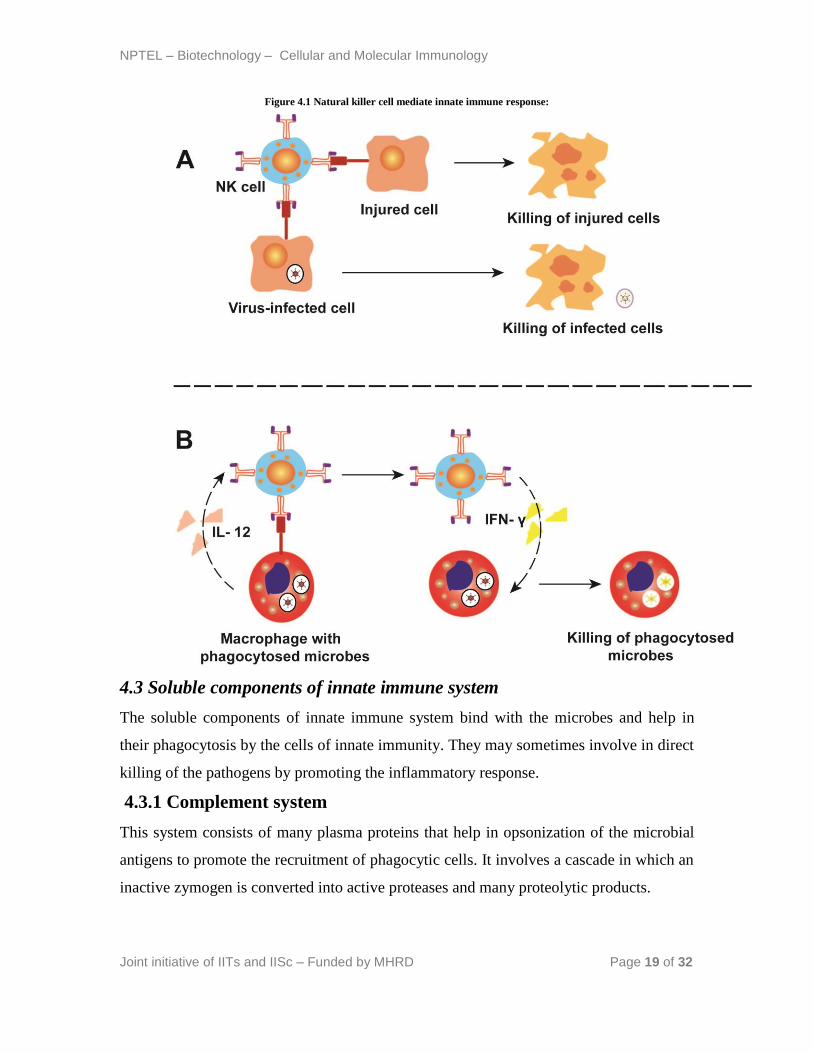
Figure 4.1 Natural killer cell mediate innate immune response:
4.3 Soluble components of innate immune system
The soluble components of innate immune system bind with the microbes and help in
their phagocytosis by the cells of innate immunity. They may sometimes involve in direct
killing of the pathogens by promoting the inflammatory response.
4.3.1 Complement system
This system consists of many plasma proteins that help in opsonization of the microbial
antigens to promote the recruitment of phagocytic cells. It involves a cascade in which an
inactive zymogen is converted into active proteases and many proteolytic products.

NPTEL – Biotechnology – Cellular and Molecular Immunology
Joint initiative of IITs and IISc – Funded by MHRD Page 20 of 32
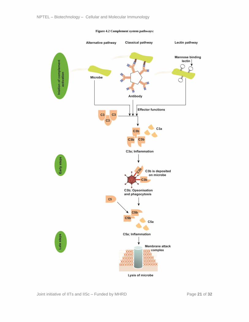
First step in the activation of complement system is the recognition of the foreign
substance and that occurs by three different pathways
1. Classical pathway
2. Alternate pathway
3. Lectin pathway
Classical pathway was named as it was discovered first and uses the plasma protein C1q
to detect antibody bound over the surface of microbes. Following the binding, C1q starts
the cascade (figure 4.2) which leads to lysis of the microbes. Alternate pathway is
triggered with a protein called C3 which recognizes the lipopolysaccharides present in the
bacterial cell. Lectin pathway is triggered by the mannose binding lectin that recognizes
the mannose residues present in the microbes.
Recognition of antigens by any of these pathways converts C3 into C3a and C3b with the
help of C3 convertase. C3b binds with the microbial antigen while C3a stimulates the
release of inflammatory cytokines. C3b activates the C5 convertase to convert C5 into
C5a and C5b. C5a is a chemoattractant while C5b initiates the formation of complex with
other complement proteins C6, C7, C8, and C9. This sequential cascade leads to
formation of membrane attack complex which causes lysis of the cell.
NPTEL – Biotechnology – Cellular and Molecular Immunology
Joint initiative of IITs and IISc – Funded by MHRD Page 21 of 32
Figure 4.2 Complement system pathways:

NPTEL – Biotechnology – Cellular and Molecular Immunology
Joint initiative of IITs and IISc – Funded by MHRD Page 22 of 32
Lecture 5: Adaptive immune system (Part I)
Adaptive immune responses are of two types
Humoral immune response
Cell mediated immune response
5.1 Humoral immune response
Humoral immune responses are mediated by the antibodies which are produced by
activated B cells. Antibodies recognize the microbial antigen, neutralize the infectivity,
and target the microbes to other effector system for degradation. Humoral immunity is
the major type of immune response against extracellular microbes and toxins because the
secreted form of the antibody can easily bind and eliminate the microbes and toxins.
Occasionally antibodies may bind to the microbes to promote their phagocytosis in order
to eliminate the infection.
5.2 Cell mediated immune response
This is also called cellular immunity, and is mediated by T lymphocytes. Cell mediated
immunity plays an important role against intracellular microbes, viruses, and some
intracellular bacteria. The cellular immunity promotes the destruction of microbes by
direct killing or phagocytosis of the infected cells.

NPTEL – Biotechnology – Cellular and Molecular Immunology
Joint initiative of IITs and IISc – Funded by MHRD Page 23 of 32
Figure 5.1 Schematic representation of humoral and cell mediated immunity:
Immunity against a pathogen is usually induced by the exposure of microbial antigen to
the host and is called active immunity. Immunity can also be transferred by serum or
lymphocyte from an immunized individual to a diseased individual and is called passive
immunity. Passive immunization is a rapid way to transfer the immunity in the absence
of active immunity. Passive immunization against toxin and venoms is a life saving
treatment in many lethal conditions (Tetanus toxoid, snake antivenom).

NPTEL – Biotechnology – Cellular and Molecular Immunology
Joint initiative of IITs and IISc – Funded by MHRD Page 24 of 32
Figure 5.2 Schematic representation of active and passive immunity:
The first concept of humoral immunity was given by Emil von Behring and Shibasabro
Kitasato; they showed for the first time that serum transferred from a recovered
diphtheria patient protected the recipient from active diphtheria infection. The active
ingredients are called antitoxins because they nullify the effect of toxins. They won the
noble prize for their landmark discovery. Paul Ehrlich coined the term antibodies for the
proteins present in the serum and showed that it is capable to bind and neutralize the
toxins. The substances that induce the production of antibodies are called antigens. The
definition of the antigen changed in due course of time with the modern discoveries. The
antigens are defined as substances that bind to a specific lymphocyte with or without
further production of antibody. However the substances that induce the production of
antibodies are called immunogens in modern immunology world.
The cellular theory of immunity started with the work of Elie Metchnikoff, who first
demonstrated the phenomenon of phagocytosis. Another remarkable finding was put

NPTEL – Biotechnology – Cellular and Molecular Immunology
Joint initiative of IITs and IISc – Funded by MHRD Page 25 of 32
forth by Almroth Wright, who showed that the factors present in the serum can coat the
bacteria and help them to phagocytize by the cells of the immune system, the process
known as opsonization.
5.3 Feature of adaptive immunity
The adaptive immunity has some fundamental properties.
5.3.1 Specificity
The adaptive immunity is specific to a particular antigen, which means specific
antibodies are produced against a particular antigen. The structures present over the
antigen that stimulate the production of antibodies are called antigenic determinants or
epitopes. Minute differences exist among lymphocytes that express membrane receptors
which are able to distinguish fine differences present on the epitopes. The specificity of
immune system leads to a huge population of lymphocytes that are antigenically specific
and are called lymphocyte repertoire.
5.3.2 Diversity
The ability of lymphocyte repertoire to recognize a wide variety of antigens is called
diversity. In fact lymphocyte repertoire that contains receptors for different antigen
contributes to a large population of extremely diverse lymphocyte clones.
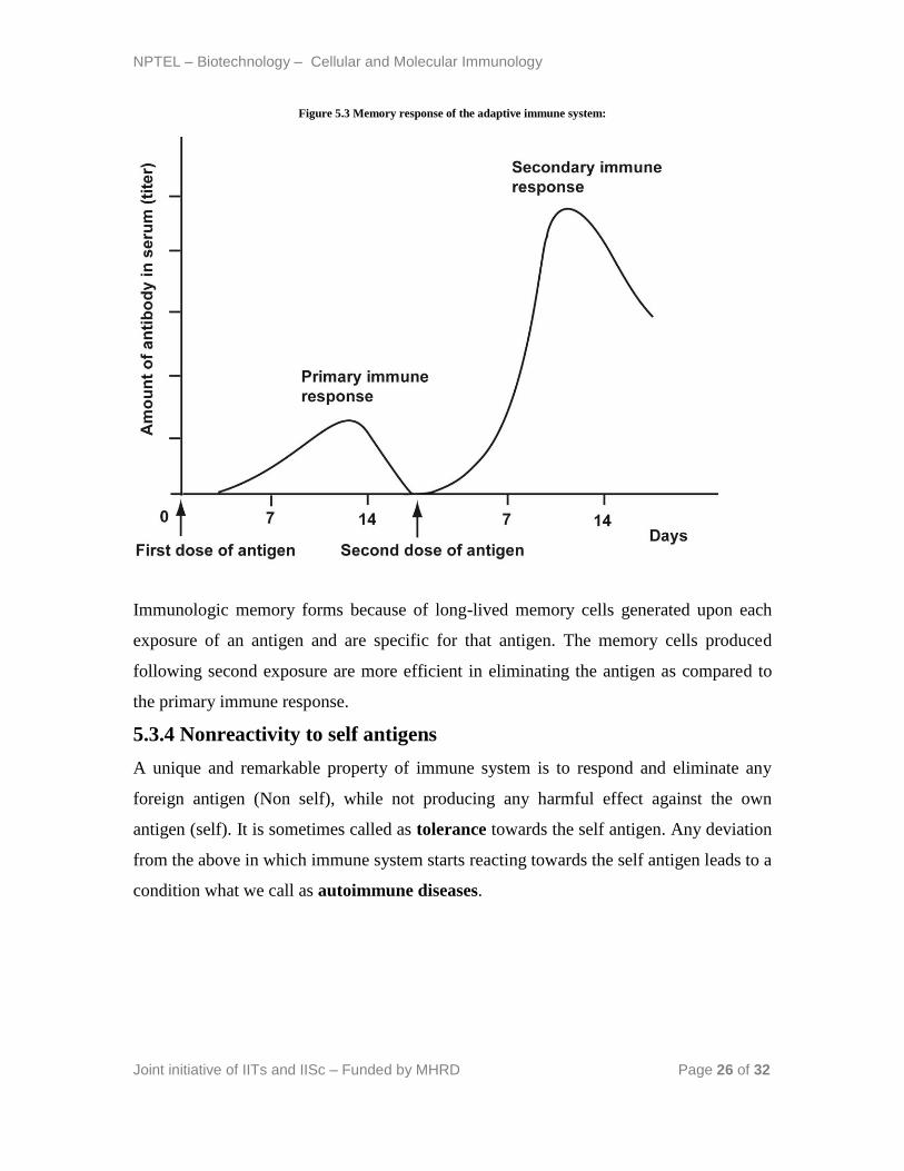
5.3.3 Memory
The ability of the immune system to remember the antigens and respond again to the
same upon exposure is called immunological memory. The immune response against the
second exposure of the same antigen or subsequent exposure is usually rapid and larger
than the primary immune response (figure 5.2).
NPTEL – Biotechnology – Cellular and Molecular Immunology
Joint initiative of IITs and IISc – Funded by MHRD Page 26 of 32
Figure 5.3 Memory response of the adaptive immune system:
Immunologic memory forms because of long-lived memory cells generated upon each
exposure of an antigen and are specific for that antigen. The memory cells produced
following second exposure are more efficient in eliminating the antigen as compared to
the primary immune response.
5.3.4 Nonreactivity to self antigens
A unique and remarkable property of immune system is to respond and eliminate any
foreign antigen (Non self), while not producing any harmful effect against the own
antigen (self). It is sometimes called as tolerance towards the self antigen. Any deviation
from the above in which immune system starts reacting towards the self antigen leads to a
condition what we call as autoimmune diseases.

NPTEL – Biotechnology – Cellular and Molecular Immunology
Joint initiative of IITs and IISc – Funded by MHRD Page 27 of 32
5.3.5 Specialization
Both humoral and cellular immune system responds to the foreign pathogens in a
different way. The responses are different against extracellular and intracellular
pathogens. Each type of response is unique for a particular type of pathogen and is
specialized to perform specific functions.
5.3.6 Clonal expansion
It is another unique property of immune system in which the lymphocyte starts producing
similar kind of cells upon exposure to an antigen in order to eliminate the pathogen more
effectively and more rapidly. The cells formed after clonal expansion has similar surface
markers and responds to similar kind of antigen.
5.3.7 Homeostasis
Every process in the body has a regulatory mechanism. The immune responses are
produced against an antigen or pathogen upon entry inside the body and wane following
the clearance of antigen. This is done in order to maintain homeostasis mechanism inside
the body. Any deviation from the homeostasis leads to an immunological disease
condition.

NPTEL – Biotechnology – Cellular and Molecular Immunology
Joint initiative of IITs and IISc – Funded by MHRD Page 28 of 32
Lecture 6: Adaptive immune system (Part II)
6.1 Cells of the adaptive immune system
Major cells of the adaptive immune system includes following.
Lymphocytes
Antigen presenting cells
Effector cells
Lymphocytes are the main cells which can recognize the antigen and produce an antibody
in order to eliminate the antigens. They are considered as a mediator of both humoral and
cellular immunity. B lymphocytes are the only cells in the body capable of producing
antibodies. B lymphocytes recognize the extracellular antigens and are differentiated into
the plasma cells. T lymphocytes recognize the intracellular antigens and they either help
in their phagocytosis or direct killing of the infected cells. T lymphocytes are activated by
the antigen loaded over the major histocompatibility complex (MHC) molecules, which
are expressed on the surfaces of other cells. T lymphocytes are divided into different
subsets which are specific for certain specialized functions. Helper T cells are the subsets
of T lymphocyte that secretes the cytokines and help in the proliferation and activation of
T lymphocyte. Helper T cells also activate the B cells (to secret the antibody),
macrophages and other leucocytes by the virtue of the cytokines. Cytotoxic T
lymphocytes kill the cells infected by viruses or other intracellular pathogens.
Regulatory T cells help to reduce or inhibit the immune response and are negative
regulators of immune system. Natural killer cells are involved in the innate immunity
against viruses or other intracellular pathogens. Different classes of lymphocytes can be
identified and differentiated by the expression of their surface receptors called cluster of
differentiation (CD).
Antigen-presenting cells are mostly the dendritic cells which capture the antigens,
transport it to the lymphoid organs and present the antigens to naïve lymphocytes in order
to activate the immune response.
Effector cells of the immune system mainly include activated T lymphocytes,
mononuclear phagocytes, and other leukocytes. Effector cells are required to complete
the immune cascade, i.e. to eliminate the microbes.

NPTEL – Biotechnology – Cellular and Molecular Immunology
Joint initiative of IITs and IISc – Funded by MHRD Page 29 of 32
Figure 6.1 Lymphocyte classes:

NPTEL – Biotechnology – Cellular and Molecular Immunology
Joint initiative of IITs and IISc – Funded by MHRD Page 30 of 32
6.2 Different stages of adaptive immune response
Steps of adaptive immune response follow a cascade orchestrated by the antibodies and
the cells of the adaptive immune system.
Step 1 Capture and display of antigens
↓
Step 2 Recognition of antigen by lymphocytes
↓
Step 3 Activation of T lymphocytes
↓
Step 4 Activation of B lymphocytes
↓
Step 5 Production of memory cells
6.2.1 Capture and display of antigens
Dendritic cells present in the epithelial body surfaces and connective tissues are the major
antigen presenting cells. They display the antigens to the CD4+ T cells to activate the
antibody mediated (humoral) immune response and CD8+ T cells to activate cell
mediated immune response. The antigen presenting cells contain a specialized structure
over their surface called the MHC molecules that helps in the display of antigenic
peptides to the cells of immune system (discussed in later chapters). Pathogens entering
into lymph nodes and spleen are displayed by the antigen presenting cells to the B and T
lymphocytes.

NPTEL – Biotechnology – Cellular and Molecular Immunology
Joint initiative of IITs and IISc – Funded by MHRD Page 31 of 32
6.2.2 Recognition of antigen by lymphocytes
Lymphocyte specific for an antigen is activated upon encountering with antigen
presenting cells loaded with an antigenic peptide. The concept of activation of
lymphocyte is called clonal selection theory. The theory was put forth first by Neils
Jerne and explained further by Burnet. This theory says that antigen specific clones of
lymphocytes exist even before the exposure of antigens and a large number of clones are
generated during lymphocytic maturation to diversify the recognition of microbial
antigens.
6.2.3 Activation of T lymphocytes
Activated T helper cells proliferate and differentiate into effector cells with the help of
cytokines. Interleukin-2, secreted by T-helper cells modulate the clonal expansion of
activated T lymphocytes. The effector cells help in killing the pathogen by phagocytosis.
Activated cytotoxic T cells kill the intracellular pathogens in the cytoplasm of infected
cells. Alternatively, cytotoxic T cells eliminate the infection by phagocytosis of the
infected cells.
6.2.4 Activation of B lymphocytes
B cells are activated with the help of CD4+ T lymphocyte and differentiated into
antibody secreting plasma cells. Usually lipids and carbohydrate antigens stimulate the
production of IgM class of antibody while protein antigens can induce IgG, IgA, IgE type
of immunoglobulins. The production of different form of antibodies requires class
switching of the surface immunoglobulin present over the B cells. Antibodies bind and
prevent the pathogens, thus “neutralizing” the pathogens and block their ability to infect
host cells. IgA antibodies specifically act on the mucosal surface of the gastrointestinal
and respiratory tract and neutralize the invading pathogens. IgG antibodies coat
pathogens and target them for phagocytosis while IgM can activate the complement
pathway.

NPTEL – Biotechnology – Cellular and Molecular Immunology
Joint initiative of IITs and IISc – Funded by MHRD Page 32 of 32
6.2.5 Production of memory cells
Initial activation of T lymphocyte produces the long-lived memory cells that survive for
many days following infection. Memory cells respond much faster than the naïve
lymphocytes. Generations of long lasting memory cells are the major target for vaccine
design against microbial pathogens.