Cellular Adaptation and Cell Injury CLDavis Foundation On the
32
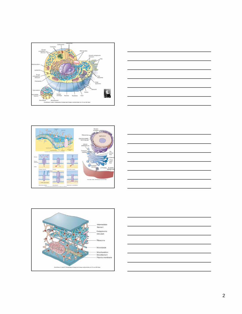
1 Cellular Adaptation and Cell Injury CLDavis Foundation On the Beach Lec 1 Cell Injury Causes R K Myers 2008 Cell Injury, Causes, Responses, Reversible, Necrosis Ron Myers, DVM PhD DACVP Professor Veterinary Pathology Iowa State University Rudolf Virchow (1821-1902) The “elemental patient”
Transcript of Cellular Adaptation and Cell Injury CLDavis Foundation On the

1
Cellular Adaptation and Cell InjuryCLDavis Foundation
On the Beach
Lec 1
Cell Injury Causes
R K Myers 2008
Cell Injury, Causes, Responses, Reversible, Necrosis
Ron Myers, DVM PhDDACVP
Professor Veterinary PathologyIowa State University
Rudolf Virchow (1821-1902)
The “elemental patient”
2

3
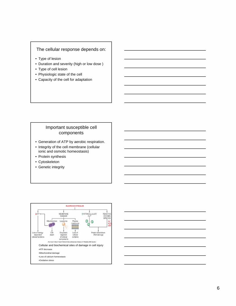
Cellular responses to stress
• Many causes of cell injury• Homeostasis, a more or less narrow range of
steady state, can be altered.• Cellular adaptations can result in an alteredCellular adaptations can result in an altered
steady state with changes in morphology.• Limited ways to respond to injury.• The body is a fiendishly complicated and clever
aggregation of cells and the lesions resulting from cell injury can be complex.
Cells can and must adapt to stimulus or stress
• In response to stress, cells may:– Adapt
– Be reversibly injured
– Die
atrophy dysplasia
hyperplasia
metaplasia
hypertrophy
4
Responses to Cell stress
Normal Cell
Cell Death
Injurious agent
Ad i
Reversible LesionAdaptation Lesion
Residual LesionLipofuscin, etc.
HypertrophyHyperplasia
AtrophyMetaplasiaDysplasia
Inadequate adaptation
The normal cell, adapted cell, injured cell, irreversibly injured cell, and dead cell are inexact states along a continuum of function and structure.
Causes of Cell Injury
• Oxygen deficiency—common and important– Hypoxia reduces aerobic oxidative respiration– Results from l) cardiorespiratory failure, 2) loss of
blood supply, 3) reduced transport of O2 in blood (i.e. i CO t i it ) d 4) bl k f ll
• Extrinsic or Intrinsic.
anemia, or CO toxicity), and 4) blockage of cell enzymes (cyanide toxicosis).
– Compare to ischemia: loss of blood supply.• Physical agents
– Trauma, extremes of heat and cold, radiation, and electrical energy may seriously injure cells

5
Causes of Cell Injury• Infectious agents
– Viruses– Bacteria– Mycotic agents– Protozoa– Metazoan parasites
• Nutritional deficiency and imbalances– Protein-calorie deficiencies – Protein-calorie excess – Vitamin and mineral imbalances
Causes of Cell Injury• Genetic derangement
Mutations, whatever their origin, may cause no disease, deprive a cell of a critical protein (enzyme), or may be incompatible with cell survival.
• Workload imbalance– Overworked or under worked cells must adapt
Causes of Cell Injury• Chemicals, drugs, toxins
– Influence cells by a multitude of mechanisms – block or stimulate cell membrane receptors, alter specific
enzyme systems, produce toxic free radicals, alter cell permeability, damage chromosomes, modify metabolic pathways, and damage structural components of cells. p y , g p
• Immune dysfunction– Failure to respond due to congenital or acquired defects– Autoimmunity -- Immune response directed against host
antigens– Hypersensitivity -- inappropriate or exaggerated response to
certain antigens
6
The cellular response depends on:
• Type of lesion • Duration and severity (high or low dose )• Type of cell lesion • Physiologic state of the cell • Capacity of the cell for adaptation
Important susceptible cell components
• Generation of ATP by aerobic respiration.• Integrity of the cell membrane (cellular
ionic and osmotic homeostasis))• Protein synthesis • Cytoskeleton• Genetic integrity
Cellular and biochemical sites of damage in cell injury•ATP decrease
•Mitochondrial damage
•Loss of calcium homeostasis
•Oxidative stress
7
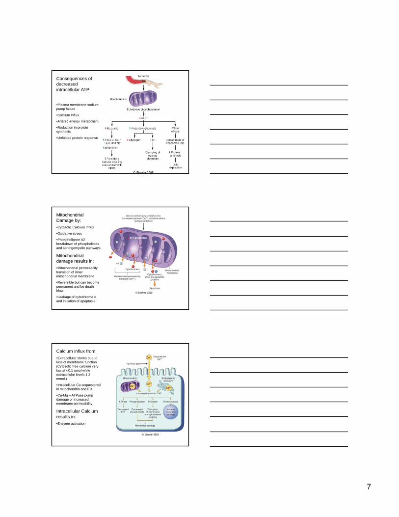
Consequences of decreased intracellular ATP:
•Plasma membrane sodium pump failure
•Calcium influx
•Altered energy metabolismAltered energy metabolism
•Reduction in protein synthesis
•Unfolded protein response
Mitochondrial Damage by:•Cytosolic Calcium influx
•Oxidative stress
•Phospholipase A2 breakdown of phospholipids and sphingomyelin pathways
Mitochondrial damage results in:•Mitochondrial permeability transition of inner mitochondrial membrane
•Reversible but can become permanent and be death blow
•Leakage of cytochrome c and initiation of apoptosis
Calcium influx from:•Extracellular stores due to loss of membrane function. (Cytosolic free calcium very low at <0.1 umol while extracellular levels 1.3 mmol.)
•Intracellular Ca sequestered in mitochondria and ER.
Ca Mg ATPase pump•Ca-Mg – ATPase pump damage or increased membrane permeability
Intracellular Calcium results in:•Enzyme activation

8
Oxygen-derived free radicals – Reactive oxygen species leading to oxidative stress
Free radicals initiated by:Radiant energy: UV, X ray
Inflammation: e.g. neutrophil killing
Chemicals and drugs and their metabolism – eg chloroform
Redox reactions of normal metabolism
Transition metals: especially iron and copper, which donate or accept free electrons
Nitric oxide (just say NO!)
Lipid peroxidation of membranes:ROS attack on unsaturated Fas of membrane lipids yield peroxides and autocatalyze chain reactions (propagation)
Oxidative modification of proteins:A i id id h i id iAmino acid side chain oxidation, cross linkages (disulfide bonds), and protein backbone oxidation result in fragmentation and increased degradation by proteasome complex
DNA Lesions:Thymine reaction in nuclear and mitochondrial DNA single stranded breaks (aging?)
9
Reactive oxygen species: brief summary
• Hydrogen peroxide (H2O2)– Forms free rads via Fe2+ catalyzed Fenton Reaction– Diffuses widely in cell
• Superoxide anion (O2-)– Generated by leaks in electron transport chain and
some cytosolic reactions– Produces other ROS– Does not diffuse far from origin
Reactive oxygen species: brief summary continued
• Hydroxyl radical (.OH)– Generated from H2O2 by Fe2+ catalyzed Fenton
reaction– Intracellular radical most responsible for
l l tt kmacromolecule attack
• Peroxynitrite (ONOO.)– From the reaction of nitric oxide (NO) with O2-– Damages macromolecules
Reactive oxygen species: brief summary continued
• Lipid peroxide radicals (RCOO.)– Organic radicals produced during lipid
peroxidation
• Hypochlorous acid (HOCl)– Produced by macrophages and pmns during
respiratory burst accompanying phagocytosis– Dissociates to yield hypochlorite radical (OCl-)

10
Protection from ROS:Inherently unstable
Antioxidants block or scavenge
Vit. E and A
Binding of catalyzing iron and copper to transport proteins (e.g. transferrin, ceruloplasmin, lactoferrin, ferritin)
Enzymes:Enzymes:
•Catalase (in peroxisomes) decomposes hydrogen peroxide
•Superoxide dismutase converts superoxide to water and oxygen
•Glutathione peroxidase catalyzes free radical breakdown
Membrane permeability defects:•Many causes
•Mitochondrial dysfunction
•Loss of membrane phospholipids
•Cytoskeletal abnormalities
•ROS (reactive oxygen species)
•Lipid breakdown products
Lysosomal membrane injury:•Enzyme leakage and activation in cytoplasm cell digestion: loss of glycogen, ribonucleoprotein, deoxyribonucleoprotein and eventually necrosis
Cell Injury: reversible and irreversible
11
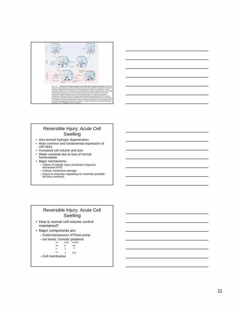
Fig. 1-13 Normal cell and the changes in reversible and irreversible cell injury. Reversible injury is characterized by generalized swelling of the cell and its organelles, blebbing of the plasma membrane, detachment of ribosomes from the endoplasmic reticulum, and clumping of nuclear chromatin. Transition to irreversible injury is characterized by increasing swelling of the cell, swelling and disruption of lysosomes, presence of large amorphous densities in swollen mitochondria, disruption of cellular membranes, and profound nuclear changes. The latter include nuclear condensation (pyknosis), followed by fragmentation (karyorrhexis) and dissolution of the nucleus (karyolysis). Laminated structures (myelin figures) derived from damaged membranes of organelles and the plasma membrane first appear during the reversible stage and become more pronounced in irreversibly damaged cells. (From Kumar V, Abbas A, Fausto N: Robbins & Cotran pathologic basis of disease, ed 7, Philadelphia, 2005, Saunders.)
Reversible Injury: Acute Cell Swelling
• Also termed hydropic degeneration• Most common and fundamental expression of
cell injury• Increased cell volume and size• Water overload due to loss of normal• Water overload due to loss of normal
homeostasis• Major mechanisms:
– Failure of cellular injury production (hypoxia, decreased ATP)
– Cellular membrane damage– (injury to enzymes regulating ion channels possible
but less common)
Reversible Injury: Acute Cell Swelling
• How is normal cell volume control maintained?
• Major components are:– Sodium/potassium ATPase pumpSodium/potassium ATPase pump– Ion levels: Osmotic gradients
– Cell membranes
Ion Inside Outside
Na 12 146
K
Ca

12
Sodium/Potassium Pump
Na
Ca
Na
K
Normal
Ca
H2O
H2O
MitochondriaATP synthesis O2
KATP
Ion Inside Outside
Na 12 146
K
Ca
Normal volume control
Sodium/Potassium Pump
Na
KCa
Na
K
Without O2 or ATP
Ca
H2O
X
H2O
MitochondriaATP synthesis O2
K
ATP
X
Cell swellling
Hypoxic injury resulting in cell swelling
13
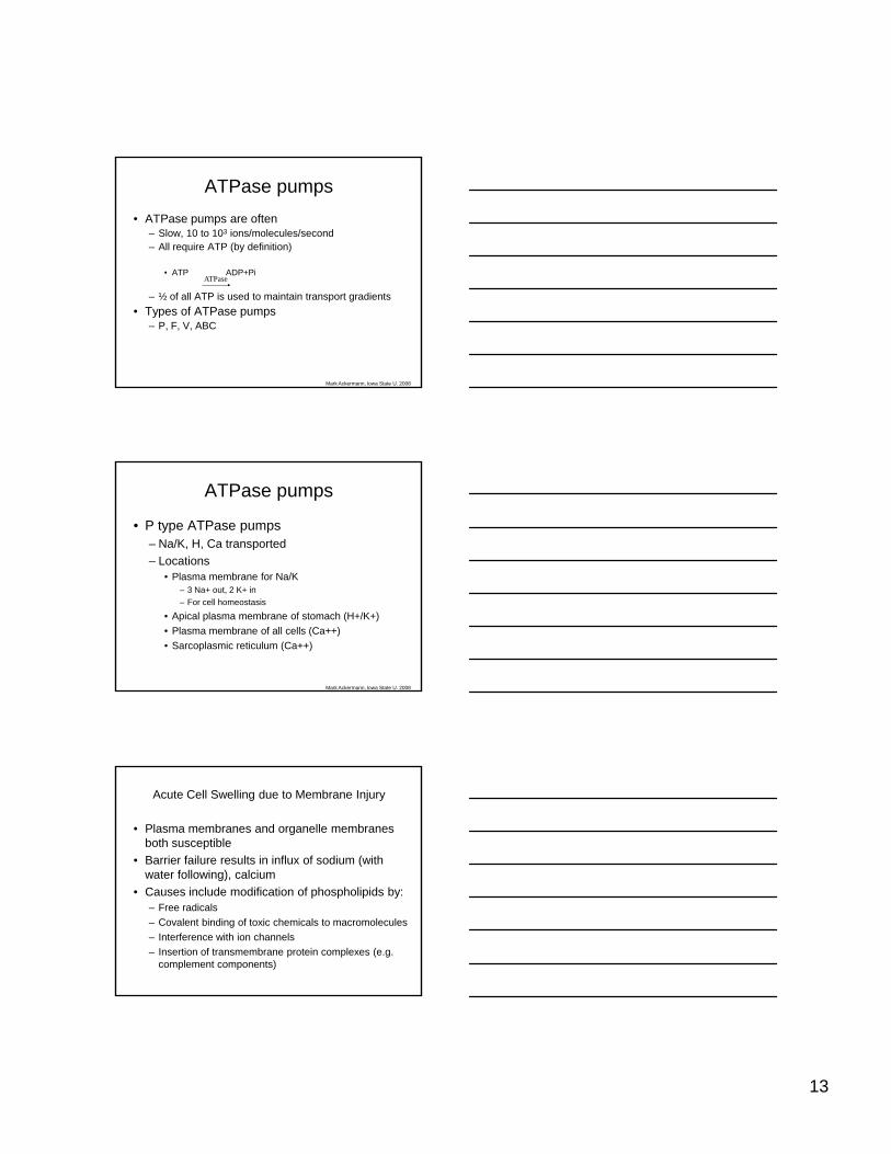
ATPase pumps• ATPase pumps are often
– Slow, 10 to 103 ions/molecules/second– All require ATP (by definition)
• ATP ADP+Pi• ATP ADP+Pi
– ½ of all ATP is used to maintain transport gradients• Types of ATPase pumps
– P, F, V, ABC
ATPase
Mark Ackermann, Iowa State U. 2008
ATPase pumps
• P type ATPase pumps– Na/K, H, Ca transported– Locations
• Plasma membrane for Na/K• Plasma membrane for Na/K– 3 Na+ out, 2 K+ in– For cell homeostasis
• Apical plasma membrane of stomach (H+/K+)• Plasma membrane of all cells (Ca++)• Sarcoplasmic reticulum (Ca++)
Mark Ackermann, Iowa State U. 2008
• Plasma membranes and organelle membranes both susceptible
• Barrier failure results in influx of sodium (with water following), calcium
Acute Cell Swelling due to Membrane Injury
g)• Causes include modification of phospholipids by:
– Free radicals– Covalent binding of toxic chemicals to macromolecules– Interference with ion channels– Insertion of transmembrane protein complexes (e.g.
complement components)

14
Acute Cell Swelling due to Membrane Injury:
Carbon tetrachloride
Cell Swelling
Progression to necrosis
Organelle membrane injury (e.g. mitochondrial membranes) can lead to cell swelling also by decreased ATP production route.
Membrane Injury:Carbon tetrachloride
Can also result in another type of reversible injury—Fatty Change
15
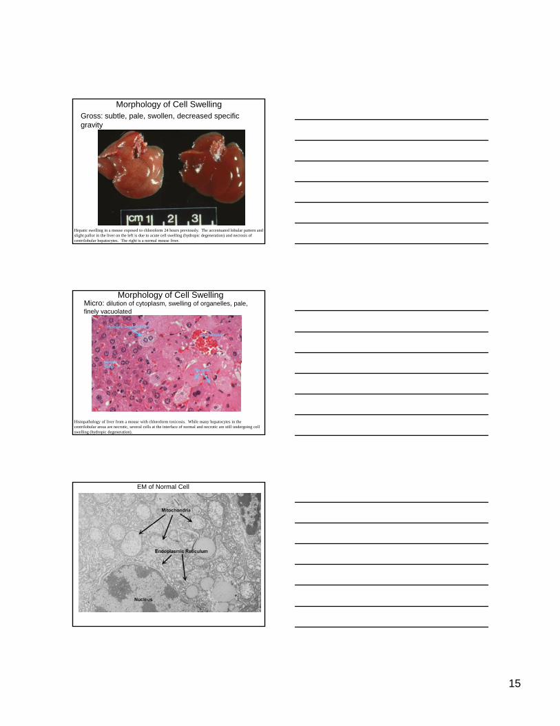
Morphology of Cell SwellingGross: subtle, pale, swollen, decreased specific gravity
Hepatic swelling in a mouse exposed to chloroform 24 hours previously. The accentuated lobular pattern and slight pallor in the liver on the left is due to acute cell swelling (hydropic degeneration) and necrosis of centrilobular hepatocytes. The right is a normal mouse liver.
Morphology of Cell SwellingMicro: dilution of cytoplasm, swelling of organelles, pale, finely vacuolated
Histopathology of liver from a mouse with chloroform toxicosis. While many hepatocytes in the centrilobular areas are necrotic, several cells at the interface of normal and necrotic are still undergoing cell swelling (hydropic degeneration).
EM of Normal Cell

16
Morphology of Cell SwellingEM: dilated ER, distended nuclear membrane, swollen mitochondria, others – loss of microvilli, blebs
Morphology of Cell Swelling
EM: dilated ER, swollen mitochondria, others – myelin figure
Significance and Fate of Acute Cell Swelling
• Volume regulation and other functions lost• Depends on number and type of cell affected• Vulnerable cells include: cardiac myocytes,
proximal cortical tubules, hepatocytes, endothelial cells, neurons and gliaendothelial cells, neurons and glia
• Changes are reversible if ATP/O2 restored and membrane injury repaired
• May return to normal or near normal and may have evidence of injury
• May reach “point of no return” and progress to cell death.

17
Cell Injury and Cell Death
Cell Injury and Cell Death
• Point of no return: not precise and likely no single pathway
• Two consistent characteristics of irreversibility
1. Inability to restore mitochondrial function2. Severe cell membrane damage
• Leak of lysosomal contents leading to cytoplasmic and nuclear degradation
• Massive leak of intracellular substances and influx of calcium
Cell Death:
2 main types• Necrosis: morphologic changes (gross or microscopic) indicative of
cell death in a living animal.
Cell death following swelling (oncosis)
• Apoptosis: a genetically determined process of cell self-destruction that is marked by the fragmentation of nuclear DNA is activatedthat is marked by the fragmentation of nuclear DNA, is activated either by the presence of a stimulus or by the removal of a stimulus or suppressing agent, is a normal physiological process eliminating DNA-damaged, superfluous, or unwanted cells (as immune cells targeted against the self in the development of self-tolerance or larval cells in amphibians undergoing metamorphosis), and when halted (as by genetic mutation) may result in uncontrolled cell growth and tumor formation called also programmed cell deathMerriam-Webster's Medical Dictionary, © 2002 Merriam-Webster, Inc
Cell death with shrinkage

18
Cell Death: Some new usage
• Cell death and necrosis are not synonyms• Necrosis by oncosis and apoptosis may be
seen together• Proposed: use necrosis for histologicProposed: use necrosis for histologic
changes following cell death by either mechanism– Use oncotic necrosis and apoptotic necrosis
when a distinction can be and needs to be made.
Cell death to Necrosis lag time:
Reversible changes may be visible within minutes
EM changes within 6 hrs.
LM changes occur 4-12 hrs following cell death in myocardium (irreversible injury within 20-60 minutes
Gross lesions may take 24-48 hrs unless vascular changes occur
Oncotic necrosis (oncosis): irreversible cell injury by hypoxia, ischemia, membrane damage.
• Can follow acute cell swellingFi l th• Final pathways involve calcium

19
Events in ischemia: reversible and irreversible injury
Morphologic appearance of necrotic cells and tissues (oncotic necrosis)
• Due to denaturation of intracellular proteins and enzymatic digestion of the cell.
• Enzymatic digestion within the dying cells can be termed autolysis (if after death postmortem autolysis)
• Denaturation (coagulation) and protein lysis (autolysis) are both occurring while the cells is still aliveare both occurring while the cells is still alive
• Denaturation begins in the live cells, contributes to cell death, and continues in the dead cell
• Denaturation stops autolysis by inactivating enzyme and removing their substrates, and by doing this decreases inflammation
• It takes several hours for changes to develop
Ultrastructural changes
• Early stages have swelling of mitochondria with ER dilation and fragmentation, nuclear membrane folding, pale structureless cytoplasm, poorly visible organelles.
• Later stage has cell collapse, shrinking, cytoplasmic and organelle homogeneity, nuclear density or lysis. Plasma membrane structures (desmosomes, microvilli, cilia) distorted, lost
20
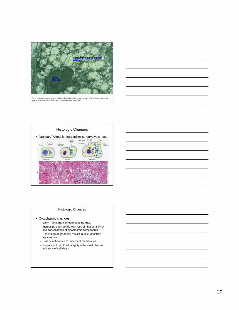
Electron microscopy of a renal epithelial cell that is in early oncotic necrosis. The nucleus is condensed (pyknosis) and the mitochondria are very swollen (high amplitude).
Histologic Changes
• Nuclear. Pyknosis, karyorrhexis, karyolysis, loss.
Histologic Changes
• Cytoplasmic changes– Early – pink and homogeneous by H&E– Increasing eosinophilia with loss of ribosomal RNA
and consolidation of cytoplasmic components– Continuing degradation results in pale, ghostlike
appearance– Loss of adherence to basement membranes– Rupture of loss of cell integrity – the most obvious
evidence of cell death

21
Renal cortex of a mouse with chloroform toxicosis. Notice that some of the epithelial cells are necrotic. Some exhibit pyknosis while others have lost the nucleus or have a very pale nucleus (karyolysis.) Necrosis is irreversible, but replacement cells may be regenerated depending on the tissue.
Spleen of a dog with parvovirus infection has fragmentation of lymphocyte nuclei (karyorrhexis) because of the infection. Many of these nuclear changes may be due to apoptosis.
Centrilobular hepatocytes of a mouse with chloroform toxicosis are pale and necrotic. Note that in the necrotic areas many of the nuclei are lost. This is karyolysis. Some of the dark nuclei that are remaining are actually endothelial cell nuclei lining the sinusoids. Note the "pre-necrotic" cells that are only at the stage of cell swelling?

22
Macroscopic Changes
• Pale, unless blood is abundant• Soft and friable• Often sharply demarcated• May be surrounded by a red zone of hyperemia
or hemorrhageor hemorrhage• Because of the lag time, gross evidence of
necrosis may be inapparent in peracute of acute death unless there are vascular changes.
Mammary gland from a cow with "peracute mastitis" due to coliform bacterial infection has large areas of necrosis. These areas are slightly pink to gray. In the liver the necrotic areas would have been paler than normal parenchyma but mammary gland parenchyma is very pale normally and necrotic areas are not paler. The key to the necrosis is the line of hyperemia present here denoted as a line of demarcation.
Coagulation necrosis, infarcts, kidney, cow. A, Note the pale regions of acute coagulation necrosis surrounded by a red rim of active hyperemia and inflammation (far left). B, Acute coagulation necrosis of renal tubular epithelial cells. Necrotic cells have homogeneous eosinophilic cytoplasm, more or less retained cell outlines, and nuclear changes such as pyknosis and nuclear absence. H&E stain. (A, Courtesy Dr. D.E. Tyler, College of Veterinary Medicine, The University of Georgia; and Noah's Arkive, College of Veterinary Medicine, The University of Georgia. B, Courtesy Dr. S. Newman, College of Veterinary Medicine, University of Tennessee.) (McGavin, M. Donald. Pathologic Basis of Veterinary Disease, 4th Edition. C.V. Mosby,

23
Types of Oncotic Necrosis
• Appearance depends on tissue involved, cause, and time since injury has occurred as well as vascular changes.
• Patterns historically derived, maybe outmoded, but still used.– Coagulation (coagulative): denaturation develops and basic cell g ( g ) p
outlines are preserved– Caseation (caseous): implies degradation of cells into a granular
friable mass– Liquefactive: enzyme digestion dominates, usual in CNS– Fat necrosis: also termed enzymatic necrosis of fat– Gangrenous necrosis: coagulation necrosis plus liquefaction and
saprophytic bacteria or mummification
Coagulation necrosis
Liver from another mouse with chloroform toxicosis has centrilobular necrosis. In this case the necrotic cells retain their cell outlines and usually have a pyknotic nucleus. This is coagulation necrosis.
Coagulation necrosis
Kidney of a mouse with coagulation necrosis of some proximal tubules. The change is subtle but you can detect the pyknotic cells and the slightly brighter homogeneous cytoplasm especially in the lower middle part of the image.

24
Coagulation necrosis
Liver of a mouse with multifocal coagulation necrosis of hepatocytes. The change is easy to see here in contrast to the more viable hepatocytes. This was due to a viral infection in this mouse. (Mouse hepatitis virus.)
Coagulation necrosis
Heart of a dog with coagulation necrosis and mineralization. The dog had renal failure. The small blue punctate areas in the necrotic myocytes are mineral, perhaps associated with mitochondria.
Coagulation necrosis
Heart of a cow showing the wall of the left ventricle with a discrete pale yellow area. There is also a pale area associated with one of the papillary muscles. This lesion was due to embolism from endocarditis.
25
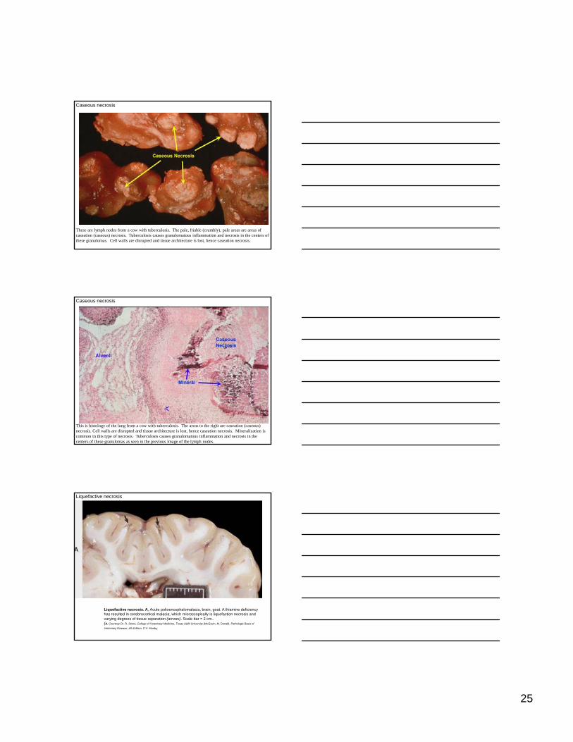
Caseous necrosis
These are lymph nodes from a cow with tuberculosis. The pale, friable (crumbly), pale areas are areas of caseation (caseous) necrosis. Tuberculosis causes granulomatous inflammation and necrosis in the centers of these granulomas. Cell walls are disrupted and tissue architecture is lost, hence caseation necrosis.
Caseous necrosis
This is histology of the lung from a cow with tuberculosis. The areas to the right are caseation (caseous) necrosis. Cell walls are disrupted and tissue architecture is lost, hence caseation necrosis. Mineralization is common in this type of necrosis. Tuberculosis causes granulomatous inflammation and necrosis in the centers of these granulomas as seen in the previous image of the lymph nodes.
Liquefactive necrosis
Liquefactive necrosis. A, Acute polioencephalomalacia, brain, goat. A thiamine deficiency has resulted in cerebrocortical malacia, which microscopically is liquefaction necrosis and varying degrees of tissue separation (arrows). Scale bar = 2 cm..(A, Courtesy Dr. R. Storts, College of Veterinary Medicine, Texas A&M University.(McGavin, M. Donald. Pathologic Basis of
Veterinary Disease, 4th Edition. C.V. Mosby,

26
Liquefactive necrosis
This is histology of the brain of a dog with distemper. The pale areas are zones of total cell and tissue loss, in the CNS termed liquefactive necrosis. Grossly the areas would be fluid consistency.
This is cross section of the brain of a sheep with brain abscesses. Although abscesses are by definition an inflammatory lesion, the neutrophils that make up the lesion lyse and also cause dissolution of parenchymal cells leading to a watery (liquefactive) to pasty (caseous) appearance of the tissue. Abscesses with liquefactive necrosis can occur in virtually any system, including CNS.
Gangrenous necrosis: dry
The blue line outlines the scrotal and inguinal area of a bull with gangrene of the skin. In this location it is often an ischemic lesion. The tissue feels dry and leathery and is usually very dark. This is termed dry gangrene.

27
Gangrenous necrosis: wet
This is udder of a sheep with a condition called "blue bag" due to Pasteurella multocida infection. The necrosis seen here is actually wet gangrene. The surrounding tissue is well-vascularized leading to the wet and bloody nature of it. Often saprophytic bacteria like Clostridial organisms contaminate areas of necrosis and cause wet gangrene.
Gangrenous necrosis: dry
These are the hooves of a bovine with "fescue toxicity" a disease in which the blood supply to the distal extremities is lost because of toxic effects on the vessels. The dry leathery appearance is termed dry gangrene. There is still some blood in the skin indicating at least partial blood supply being present or restored. Note that one of the claws has been lost due to the process.
Gangrenous necrosis: dry
Ring tail in a young rat

28
Fat necrosis
Three types in veterinary medicine:
•Enzymatic necrosis of fat: destruction of fat in the abdominal cavity especially around pancreas due to leakage of activated pancreatic lipases from fluid from injured pancreas. Lipases split the triglyceride esters of adipocytes. Fatty acids combine with calcium to form chalky white areas – white saponification.
T ti f t i h di ti i h d•Traumatic fat necrosis: seen where adipose tissue is crushed, such as in the pelvic canal of heifers following dystocia or sternum of recumbent cattle.
•Abdominal fat necrosis of cattle: large masses of retroperitoneal, mesenteric, and omental fat necrosis, cause undetermined
Enzymatic necrosis of fat.
This cat has had previous bouts of pancreatitis. Leakage of pancreatic enzymes, such as lipase, cause degradation and necrosis of the fat around the pancreas and in the adjacent mesentery. Necrotic fat often becomes mineralized and so grossly the lesion is chalky to gritty as well as being pale from the mineral in the necrotic fat.
Other lesions and terms traditionally used with cell death or necrosis
Zenker’s necrosis. A special antiquated term used for coagulation necrosis of muscle.
Fibrinoid necrosis. Accumulation of pink amorphous masses of fibrin, antibodies, and serum proteins in arterial walls due to immune injury. Neutrophils are also often present. Also termed fibrinoid degeneration.
Ul ti d i L l d f t ti f thUlceration and erosion. Local defects or excavations of the surface of an organ or mucous membrane resulting from sloughing or loss of necrotic tissue. An ulcer is full thickness epithelial loss (through basement membrane. An erosion is more superficial and does not penetrate basement membrane.

29
Zenker’s necrosis
Coagulation necrosis of muscle historically was termed Zenker's necrosis. You may see the term but the necrosis present is no different from coagulation necrosis in other organs. The pale areas here are necrotic and due to this cow being down for a long time. Pressure on the muscle prevents proper perfusion of the muscle thus causing necrosis.
Zenker’s necrosis
Histology of a skeletal muscle with coagulation necrosis, historically termed Zenker's necrosis. The necrosis present grossly or histologically is no different from coagulation necrosis in other organs. Hypereosinophilia, cell swelling, cell shrinkage, nuclear pyknosis, and karyolysis but with retention of cell borders are all present here and typical of coagulation necrosis.
Fibrinoid necrosis
Histology of a bovine with an inflammatory and necrotizing disease of the smooth muscle of the artery wall. This type of change is termed fibrinoid necrosis. The name comes from the apparent similarity in the appearance of the necrotic smooth muscle with fibrin. It turns out that electron microscopy has shown that much of the pink color is indeed due to fibrin likely also with immunoglobulin and complement.

30
Ulcer
Esophagus from a bovine that had a partial blockage for a few days. Pressure on the foreign body from esophageal contraction led to death of the mucosa and sloughing into the lumen leaving the raw surface. This is an ulcer rather than an erosion in that the basement membrane has been breached and has actually exposed underlying connective tissue and blood vessels.
Ulcers and erosions
This dog has an autoimmune disease called pemphigus vulgaris in which the cell adhesions of the epithelium are disrupted leaving erosions and eventually ulcerations. The difference between an erosion, in which the basement membrane is not breached, and an ulcer may be a matter of only one cell layer. Erosions often become ulcers, as in this case.
Sequelae of Necrosis
• Surrounding inflammatory reaction with hyperemia and phagocytes (necrotaxis).
• Digestion and liquefaction of necrotic debris by enzymes, autolytic and heterolytic. Absorption of liquid debris by lymphatics and removal of solid debris by macrophages
• Fibrosis and scarringFibrosis and scarring.• Regeneration of parenchyma if appropriate cell types
and stromal retention• Calcification of debris slow to be removed (dystrophic
calcification)• Formation of sequestrum

31
Sequestrum
The hoof and phalanges of this horse have been split down the middle. The horse had a penetrating nail wound that fractured P3 and also allowed bacteria to enter, causing osteomyelitis. A fragment of the bone became necrotic and isolated by the exudate and so was not resolved by normal processes. This persistent fragment is termed a sequestrum. Sequestra happen in other tissue also, but bone is most common.
1. Which is the best indicator of irreversible cell injury?
A. Clumping of chromatin B. Inactivation of sodium pump C. Severe mitochondrial damage D. Changes in nuclear structure and function E. Damage to the rough endoplasmic
reticulum
1. C. Slauson and Cooper, p. 51 and 60 2007
10. Which of the following is not a hallmark of reversible cell injury
1. Adenosine triphosphate depletion 2. Cell swelling 3. Loss of membrane permeability 4. Severe mitochondrial damage 5. Reduced oxidative phosphorylation
A. 1 B. 1, 2 C. 3 D. 3, 4 E. 4, 5
10. D. Robbins and Cotran, p. 11

32
25. All of the following cytomorphologic changes characterize irreversible cell injury EXCEPT:
1. Lysosomal swelling and disruption 2. Calcium entry into the cell 3. Acute cell swelling 4. Plasma membrane damage 5 Mitochondrial swelling with large amorphous densities5. Mitochondrial swelling with large amorphous densities
A. 1, 5 B. 2, 3 C. 3 D. 4, 5 E. 5
25. C. Pathologic Basis of Veterinary Disease, p. 10-16 2008
30. All of the following prevent free radical injury EXCEPT:
1. Vitamin C 2. Vitamin E 3. Superoxide dismutase 4. Sodium chloride 5. Glutathione peroxidase
A. 1, 4 B. 2, 3 C. 3 D. 4 E. 4, 5
30. D. Pathologic Basis of Veterinary Disease, p. 20 2008
35. The most common type of necrosis in the central nervous system is:
A. Liquefactive B. Coagulative C. Caseous D. Gangrene E. Apoptosis
35. A. Pathologic Basis of Veterinary Disease, p. 23 2008