Cell Wall Proteins 1998
51
MICROBIOLOGY AND MOLECULAR BIOLOGY REVIEWS, 1092-2172/98/$04.0010 Mar. 1998, p. 130–180 Vol. 62, No. 1 Copyright © 1998, American Society for Microbiology Cell Wall and Secreted Proteins of Candida albicans: Identification, Function, and Expression W. LAJEAN CHAFFIN, 1 * JOSE ´ LUIS LO ´ PEZ-RIBOT, 2 MANUEL CASANOVA, 3 DANIEL GOZALBO, 3 AND JOSE ´ P. MARTI ´ NEZ 3 Department of Microbiology and Immunology, Texas Tech University Health Sciences Center, Lubbock, 1 and Department of Medicine, University of Texas Health Science Center at San Antonio, San Antonio, 2 Texas, and Departamento de Microbiologı ´a y Ecologı ´a, Facultad de Farmacia, Universitat de Valencia, Valencia, Spain 3 INTRODUCTION .......................................................................................................................................................131 Cell Wall and Morphology ....................................................................................................................................131 Cell Wall and Interactions with the Host ...........................................................................................................132 CELL WALL COMPOSITION AND ORGANIZATION ......................................................................................132 Composition .............................................................................................................................................................132 Organization ............................................................................................................................................................133 Layering................................................................................................................................................................133 Fimbriae ...............................................................................................................................................................133 CELL WALL PROTEINS ..........................................................................................................................................135 Cell Wall versus Secreted Proteins ......................................................................................................................135 Extraction.................................................................................................................................................................135 Composition .............................................................................................................................................................136 Modification.............................................................................................................................................................137 Glycosylation........................................................................................................................................................137 Phosphorylation ..................................................................................................................................................137 Ubiquitination .....................................................................................................................................................138 Distribution and Expression .................................................................................................................................138 Enzymes with Cell Wall Function ........................................................................................................................140 Exo-b-(1,3)-glucanase.........................................................................................................................................140 b-1,3-Glucan transferase ...................................................................................................................................141 Chitinases.............................................................................................................................................................141 b-N-Acetylglucosaminidase ................................................................................................................................141 Glutaminyl-peptide-g-glutamylyl-transferase .................................................................................................142 Hydrolytic Enzymes and Proteins with Extracellular Targets .........................................................................142 Acid proteinase ....................................................................................................................................................142 Phospholipase ......................................................................................................................................................143 Esterase ................................................................................................................................................................144 Glucoamylase .......................................................................................................................................................144 Hemolytic factor ..................................................................................................................................................144 Acid phosphatase ................................................................................................................................................144 Miscellaneous ......................................................................................................................................................144 Morphology-Associated Proteins ..........................................................................................................................145 Epitope recognized by MAb 4C12 ....................................................................................................................145 Epitope recognized by MAb 3D9 ......................................................................................................................146 Hwp1p...................................................................................................................................................................147 Hyr1p ....................................................................................................................................................................147 Immunomodulatory 65-kDa Mannoprotein (MP65) ..........................................................................................147 Heat Shock Proteins ...............................................................................................................................................147 hsp90.....................................................................................................................................................................148 hsp70.....................................................................................................................................................................148 Heat shock or stress mannoproteins ...............................................................................................................149 Glycolytic Enzymes .................................................................................................................................................149 Enolase .................................................................................................................................................................149 Phosphoglycerate kinase ....................................................................................................................................150 Glyceraldehyde-3-phosphate dehydrogenase ...................................................................................................150 Alcohol dehydrogenase .......................................................................................................................................150 Binding Proteins (Receptors) for Host Ligands .................................................................................................150 * Corresponding author. Mailing address: Department of Microbi- ology and Immunology, Texas Tech University Health Sciences Cen- ter, Lubbock, TX 79430. Phone: (806) 743-2513. Fax: (806) 743-2334. E-mail: [email protected]. 130
-
Upload
isela-s-fujarte -
Category
Documents
-
view
223 -
download
1
description
Articulo de Chaffin et al. publicado en 1998 en el que aborda diferentes aspectos de las proteínas de pared celular de Candida.
Transcript of Cell Wall Proteins 1998
MICROBIOLOGY AND MOLECULAR BIOLOGY REVIEWS,1092-2172/98/$04.0010
Mar. 1998, p. 130–180 Vol. 62, No. 1
Copyright © 1998, American Society for Microbiology
Cell Wall and Secreted Proteins of Candida albicans:Identification, Function, and Expression
W. LAJEAN CHAFFIN,1* JOSE LUIS LOPEZ-RIBOT,2 MANUEL CASANOVA,3
DANIEL GOZALBO,3 AND JOSE P. MARTINEZ3
Department of Microbiology and Immunology, Texas Tech University Health Sciences Center, Lubbock,1 and Departmentof Medicine, University of Texas Health Science Center at San Antonio, San Antonio,2 Texas, and Departamento
de Microbiologıa y Ecologıa, Facultad de Farmacia, Universitat de Valencia, Valencia, Spain3
INTRODUCTION .......................................................................................................................................................131Cell Wall and Morphology ....................................................................................................................................131Cell Wall and Interactions with the Host ...........................................................................................................132
CELL WALL COMPOSITION AND ORGANIZATION ......................................................................................132Composition.............................................................................................................................................................132Organization ............................................................................................................................................................133
Layering................................................................................................................................................................133Fimbriae ...............................................................................................................................................................133
CELL WALL PROTEINS..........................................................................................................................................135Cell Wall versus Secreted Proteins ......................................................................................................................135Extraction.................................................................................................................................................................135Composition.............................................................................................................................................................136Modification.............................................................................................................................................................137
Glycosylation........................................................................................................................................................137Phosphorylation ..................................................................................................................................................137Ubiquitination .....................................................................................................................................................138
Distribution and Expression .................................................................................................................................138Enzymes with Cell Wall Function ........................................................................................................................140
Exo-b-(1,3)-glucanase.........................................................................................................................................140b-1,3-Glucan transferase ...................................................................................................................................141Chitinases.............................................................................................................................................................141b-N-Acetylglucosaminidase................................................................................................................................141Glutaminyl-peptide-g-glutamylyl-transferase .................................................................................................142
Hydrolytic Enzymes and Proteins with Extracellular Targets .........................................................................142Acid proteinase....................................................................................................................................................142Phospholipase......................................................................................................................................................143Esterase ................................................................................................................................................................144Glucoamylase.......................................................................................................................................................144Hemolytic factor..................................................................................................................................................144Acid phosphatase ................................................................................................................................................144Miscellaneous ......................................................................................................................................................144
Morphology-Associated Proteins ..........................................................................................................................145Epitope recognized by MAb 4C12 ....................................................................................................................145Epitope recognized by MAb 3D9 ......................................................................................................................146Hwp1p...................................................................................................................................................................147Hyr1p ....................................................................................................................................................................147
Immunomodulatory 65-kDa Mannoprotein (MP65)..........................................................................................147Heat Shock Proteins...............................................................................................................................................147
hsp90.....................................................................................................................................................................148hsp70.....................................................................................................................................................................148Heat shock or stress mannoproteins ...............................................................................................................149
Glycolytic Enzymes .................................................................................................................................................149Enolase .................................................................................................................................................................149Phosphoglycerate kinase ....................................................................................................................................150Glyceraldehyde-3-phosphate dehydrogenase ...................................................................................................150Alcohol dehydrogenase .......................................................................................................................................150
Binding Proteins (Receptors) for Host Ligands.................................................................................................150
* Corresponding author. Mailing address: Department of Microbi-ology and Immunology, Texas Tech University Health Sciences Cen-ter, Lubbock, TX 79430. Phone: (806) 743-2513. Fax: (806) 743-2334.E-mail: [email protected].
130
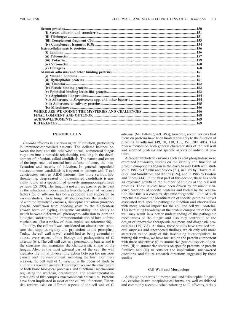
Serum proteins....................................................................................................................................................150(i) Serum albumin and transferrin ..............................................................................................................151(ii) Fibrinogen .................................................................................................................................................151(iii) Complement fragment C3d....................................................................................................................153(iv) Complement fragment iC3b...................................................................................................................154
Extracellular matrix proteins............................................................................................................................156(i) Laminin ......................................................................................................................................................156(ii) Fibronectin ................................................................................................................................................157(iii) Entactin ....................................................................................................................................................159(iv) Vitronectin ................................................................................................................................................160(v) Collagens....................................................................................................................................................160
Mannan adhesins and other binding proteins ...............................................................................................161(i) Mannan adhesins ......................................................................................................................................161(ii) Hydrophobic proteins ..............................................................................................................................161(iii) Fimbriae ...................................................................................................................................................162(iv) Plastic binding proteins..........................................................................................................................162(v) Epithelial binding lectin-like protein.....................................................................................................163(vi) Agglutinin-like proteins ..........................................................................................................................164(vii) Adherence to Streptococcus spp. and other bacteria ..........................................................................164(viii) Adherence to salivary proteins............................................................................................................165(ix) Miscellaneous...........................................................................................................................................166
WHERE ARE WE GOING? THE MYSTERIES AND CHALLENGES..............................................................166FINAL COMMENT AND OUTLOOK ....................................................................................................................168ACKNOWLEDGMENTS ...........................................................................................................................................169REFERENCES ............................................................................................................................................................169
INTRODUCTION
Candida albicans is a serious agent of infection, particularlyin immunocompromised patients. The delicate balance be-tween the host and this otherwise normal commensal fungusmay turn into a parasitic relationship, resulting in the devel-opment of infection, called candidiasis. The nature and extentof the impairment of normal host defense influence the man-ifestation and severity of infection. In general, superficialmucocutaneous candidiasis is frequent in patients with T-celldeficiencies, such as AIDS patients. The more serious, life-threatening, deep-seated or disseminated candidiasis is nor-mally found in a spectrum of severely immunocompromisedpatients (29, 390). The fungus is not a mere passive participantin the infectious process, and a hypothetical set of virulencefactors for C. albicans has been proposed and supported byvarious studies. These fungal attributes include the productionof secreted hydrolytic enzymes, dimorphic transition (morpho-genetic conversion from budding yeast to the filamentousgrowth form or hypha), antigenic variability, the ability toswitch between different cell phenotypes, adhesion to inert andbiological substrates, and immunomodulation of host defensemechanisms (for a review of these topics, see reference 96).
Initially, the cell wall was considered an almost inert struc-ture that supplies rigidity and protection to the protoplast.Today, the cell wall is well established as being essential toalmost every aspect of the biology and pathogenicity of C.albicans (64). The cell wall acts as a permeability barrier and isthe structure that maintains the characteristic shape of thefungus. Also, as the most external part of the cell, the wallmediates the initial physical interaction between the microor-ganism and the environment, including the host. For thesereasons, the cell wall of C. albicans is the focus of study bynumerous research groups. Their objectives are the elucidationof both basic biological processes and functional mechanismsregulating the synthesis, organization, and environmental in-teractions of this complex macromolecular structure. Proteinshave been implicated in most of the cell wall functions. Exten-sive reviews exist on different aspects of the cell wall of C.
albicans (64, 478–482, 491, 493); however, recent reviews thatfocus on proteins have been limited primarily to the function ofproteins as adhesins (49, 50, 110, 111, 151, 209, 406). Thisreview focuses on both general characteristics of the cell walland secreted proteins and specific aspects of individual pro-teins.
Although hydrolytic enzymes such as acid phosphatase wereexamined previously, studies on the identity and function ofprotein components began in the early to mid 1980s with stud-ies in 1983 by Chaffin and Stocco (71), in 1985 by Elorza et al.(125) and Sundstrom and Kenny (524), and in 1986 by Pontonand Jones (414). In the first part of this decade, there has beenan explosive growth in the number of studies of the cell wallproteins. These studies have been driven by presumed viru-lence functions of specific proteins and fueled by the realiza-tion that this is a complex, dynamic “organelle.” Out of suchimpetus has come the identification of specific proteins not yetassociated with specific pathogenic function and observationswith more general import for the cell and cell wall proteins.This increasing knowledge of the protein component of the cellwall may result in a better understanding of the pathogenicmechanisms of the fungus and also may contribute to thedesign of innovative therapeutic regimens and diagnostic pro-cedures (175, 333). At times, these studies have revealed sev-eral surprises and unexpected findings, which only add moreattraction to the study of this fascinating microorganism. Inwriting this review, we have focused on the protein componentwith three objectives: (i) to summarize general aspects of pro-teins; (ii) to summarize studies on specific proteins or proteinfamilies; and (iii) to consider the implications, unansweredquestions, and future research directions suggested by thesestudies.
Cell Wall and Morphology
Although the terms “dimorphism” and “dimorphic fungus”,i.e., existing in two morphological forms, are well establishedand commonly accepted when referring to C. albicans, strictly
VOL. 62, 1998 CELL WALL AND SECRETED PROTEINS OF C. ALBICANS 131
speaking this fungus has the ability to adopt a spectrum ofmorphologies, and thus C. albicans could be considered a“polymorphic” or “pleomorphic” organism (244, 390). Sincechanges in the cell wall determine the shape of the wholefungal cell, the cell wall is the structure ultimately responsiblefor a given morphology. C. albicans can reproduce by budding,giving rise to the formation of yeast cells (also designatedblastospores or blastoconidia). The production of germ tubesresults in the conversion to a filamentous growth phase orhypha, also called the mycelial form. The formation ofpseudohyphae occurs by polarized cell division when yeast cellsgrowing by budding have elongated without detaching fromadjacent cells. Under certain nonoptimal growing conditions,C. albicans can undergo the formation of chlamydospores,which are round, refractile spores with a thick cell wall. Thesemorphological transitions often represent a response of thefungus to changing environmental conditions and may permitthe fungus to adapt to different biological niches. The transi-tion from a commensal to a pathogenic lifestyle may also in-volve changes in environmental conditions and dispersionwithin the human host. The ultrastructure, composition, andbiological properties of the cell wall are affected by thesemorphological changes (64). Although progress has beenachieved in the recent years, the molecular mechanisms gov-erning these morphogenetic conversions are still not fully un-derstood, partly due to the difficulty of genetic manipulationsin this fungus (274, 275, 474). Recent reports that may heraldrapid advances in this area have identified transcriptional reg-ulatory genes, a general transcriptional repressor TUP1 (38), aputative transcriptional factor RBF1 (220), and a myc-like tran-scriptional factor EFG1 (516) that affect cellular morphologywhen their expression is altered. Most of the observations fromthese studies have been incorporated by Magee (310) into amodel for the regulation of pseudohyphal growth.
Cell Wall and Interactions with the Host
Two major aspects of the host-parasite interactions are theadhesion of C. albicans cells to host cells and tissues and theimmunomodulation of the host immune response.
Adhesion is a prerequisite for colonization and an essentialstep in the establishment of infection. C. albicans adheres toepithelial cells, endothelial cells, soluble factors, extracellularmatrix, and inert materials implanted in the body of the host.Multiple adherence mechanisms appear to be used by C. albi-cans cells (49, 50, 110, 111, 151, 209, 252, 406). Physical inter-actions of this fungus with the host are mediated at the cellsurface, and cell wall constituents implicated in binding havebeen designated adhesins (49). The large repertoire of ad-hesins displayed by this fungus may reflect the variety of hostsites that it can invade (49, 50, 110, 111, 209). Specific charac-teristics of individual cell wall moieties participating in adhe-sion events are discussed later in this review.
Another important aspect of interactions with the host, withdirect implications for pathogenesis, is the potential of thisfungus to modulate the immune response mounted by the host(64, 96, 107). The capacity of cell wall constituents, includingglucan, chitin, and mannoproteins, to modulate (activate ordepress) the immune response is well documented (11, 64).Mannans and mannoproteins display the most potent immu-nomodulatory activity, being able to regulate the action ofvirtually all arms of the immune system (natural killer cells,phagocytic cells, cell-mediated immunity, and humoral mech-anisms) (64, 107, 402, 412). Although individual cell wall moi-eties with immunomodulatory properties are described below,
readers are referred to excellent reviews on this topic (11, 64,107).
CELL WALL COMPOSITION AND ORGANIZATION
Composition
Approximately 80 to 90% of the cell wall of C. albicans iscarbohydrate. Three basic constituents represent the majorpolysaccharides of the cell wall: (i) branched polymers of glu-cose containing b-1,3 and b-1,6 linkages (b-glucans); (ii) un-branched polymers of N-acetyl-D-glucosamine (GlcNAc) con-taining b-1,4 bonds (chitin); and (iii) polymers of mannose(mannan) covalently associated with proteins (glyco[manno]-proteins). In addition, cell walls contain proteins (6 to 25%)and minor amounts of lipid (1 to 7%) (50, 64, 490, 493).
The microfibrillar polymers (b-glucans and chitin) representthe structural components of the wall. They form a rigid skel-eton that provides strong physical properties to the cell. Froma quantitative point of view, b-glucans are the main constitu-ent, accounting for 47 to 60% by weight of the cell wall. Chitinis a minor (0.6 to 9%) but important component of the C.albicans wall, particularly of the septa between independentcell compartments, budding scars, and the ring around theconstriction between mother cell and bud (126, 360).
On the other hand, mannose polymers (mannan), which donot exist as such but are found in covalent association withproteins (mannoproteins), represent about 40% of the totalcell wall polysaccharide and are the main material of the cellwall matrix (50, 64, 480, 490, 493). The term “mannan” hasbeen used also to refer to the main soluble immunodominantcomponent present in the outer cell wall layer of C. albicans,called phosphomannoprotein or phosphopeptidomannan com-plex. This cell wall fraction contains homopolymers of D-man-nose (as the main component), 3 to 5% protein, and 1 to 2%phosphate (436). The general features of cell wall mannopro-teins in C. albicans are basically identical to those found forSaccharomyces cerevisiae, one of the most thoroughly investi-gated yeasts in this regard. Several studies have resulted in adetailed knowledge of the structure of this cell wall constituentin C. albicans (12, 262–265, 494–498). Thus, mannose polymersare linked to the protein moiety through asparagine (by N-glycosidic bonds through two GlcNAc [di-N-acetylchitobiose]residues) and threonine or serine (by O-glycosidic, alkali-labilelinkages) residues. The N-glycosidically linked carbohydrate iscomposed of backbone chains of a-1,6-linked mannopyranosylresidues to which oligosaccharide side chains are attached. Theside chain mannopyranosyl residues contain a-1,2, a-1,3, b-1,2,b-1,6, and phosphodiester linkages as well as branches (a-1,6)that are oversynthesized under acidic growth conditions (150,152, 261–267, 494–498). The O-glycosidically-linked sugarcomponent consists of single mannose residues and short, un-branched mannose oligosaccharides (412). Several studiesraise the question of additional sugars present in cell wallconstituents. These observations include the following: (i) notall proteinaceous moieties present in cell wall extracts fromthis fungus react with concanavalin A, a lectin recognizinga-mannosylpyranose, or with polyclonal and monoclonal anti-bodies that recognize other mannan epitopes, such as factor 6,a mannooligosaccharide that confers serotype A specificity(57); (ii) differences in glycosylation and in sensitivity to neur-aminidase have been detected in candidal receptors for com-plement (4, 563); and (iii) treatment with neuraminidase af-fects the electrostatic surface properties of C. albicans asdetected with a fluorescent probe (227). As suggested in thesestudies, the observations raise the possibility that additional
132 CHAFFIN ET AL. MICROBIOL. MOL. BIOL. REV.

sugars are cell wall constituents. However, the observationscould reflect the existence of contaminating proteases in theglycosidase preparation. Sugar residues other than mannosemay define either additional functional or antigenic motifs orboth in cell wall glycoproteins.
The percent composition of walls from yeast cells and fila-mentous forms are similar, although the relative amounts ofb-glucans, chitin, and mannan vary according to the C. albicansgrowth form considered (50, 480, 491). Hyphal cells contain atleast three times as much chitin as yeast cells do (77, 127, 518).Chitin is the first polymer to appear in regenerating protoplasts(124, 375). Although the ratio of b-1,3- to b-1,6-glucan in theinsoluble fraction is similar in yeast and hyphal cells, the in-soluble glucan in the initial period of germ tube formationcontains considerably more b-1,3 linkages than that found inyeast and mature hyphal cells (518). The literature containsseveral reports on the identification of morphology-specificproteins and mannoproteins that are discussed later in thisreview.
Organization
The different cell wall components interact with each otherto give rise to the overall architecture of the cell wall. Besideshydrogen and hydrophobic bonds, there is also experimentalevidence for the presence of covalent linkages between differ-ent components (453, 482). Surarit et al. (527) reported thepresence of glycosidic linkages between glucan and chitin inthe nascent wall of C. albicans. Recent evidence indicates thatmannoproteins may also establish covalent associations withb-glucans (237, 238, 462, 463). It is suggested that b-1,3- andb-1,6-glucans are linked to proteins by phosphodiester link-ages, a process that may involve the participation of a GPI(glycosyl phosphatidylinositol) anchor (238) (see below). Pro-tein and mannoprotein species that are released only afterdigestion of the glucan cell wall network with b-glucanases mayplay a key role in configuring the final cell wall structure char-acteristic of each growth form (yeast and mycelium) of C.albicans (453, 479–482). Interactions between glyco(manno)-proteins and chitin also appear to exist in the wall of C. albi-cans cells as deduced from two lines of evidence: (i) chitinasetreatment of isolated cell walls solubilizes protein moieties,and (ii) the kinetics of incorporation of protein and manno-protein constituents into the walls of regenerating protoplastsis altered in the presence of nikkomycin, an antibiotic thatblocks chitin synthesis (124, 319).
Cell wall architecture has been studied most extensively in S.cerevisiae and is likely to be a model for C. albicans since thereare some similar observations, in particular sensitivity to enzy-matic digestion, glucan-mannoprotein linkages, and candidateproteins, that fit the same model (237, 238, 246, 247, 268, 462,463). In a very recent study, Kollar et al. (268) detected thepresence of material containing all four major cell wall com-ponents, b-1,3-glucan, b-1,6-glucan, chitin, and mannoprotein.Their analysis indicated that b-1,6-glucan has some b-1,3-glu-can branches that may be linked to the reducing end of chitin.The b-1,6-glucan and mannoprotein are attached through aremnant of the mannoprotein GPI anchor. Reducing ends ofb-1,6-glucan may also be attached to the nonreducing end ofb-1,3-glucan. The proportion of cell wall polysaccharide in-volved in this type of structure is not clear. The following cellwall building block, where the linkages are indicated by thelong dashes, is proposed (247, 268): Mannoprotein—GPI rem-nant—b-1,6-glucan—b-1,3-glucan—chitin. The authors pointout that these linkages are likely to be formed in the periplas-mic space as a common end of the individual biosynthetic
pathways. Chitin and b-1,3-glucan are synthesized at theplasma membrane and extruded into the periplasm, manno-protein is synthesized in the cytoplasm and transportedthrough the secretory pathway, and b-1,6-glucan synthesis mayoccur partially in the endoplasmic reticulum or Golgi complex(268). Not all components are necessarily present in a complex;therefore, the authors suggest that more chitin may be presentin inner cell wall layers and more mannoprotein may bepresent in the outer layers.
Layering. Since polysaccharides are poorly reactive to theordinary fixatives and stains used for transmission electronmicroscopy, only a few well-defined ultrastructural details areobtained by conventional protocols (Fig. 1A and B). However,transmission electron microscopy studies performed with morespecial techniques or with cytochemical stains and contrastingagents show several layers in the cell wall of C. albicans (Fig.1C to F). The appearance of these layers is variable and seemsto be related to the strain examined, growth conditions, mor-phology, and preparation of the specimens (50, 64, 422). Thus,there is no consensus about the number of layers present in thecell wall. Different authors have reported the presence of threeto eight different layers (28, 64, 186, 421, 438). The outer cellwall layer appears as a dense network with a fibrillar or floc-culent aspect (64, 480), whereas the inner wall layer appearscontiguous with the plasmalemma with extensive membraneinvaginations involved in anchoring of the cell wall to themembrane (192, 276). The microfibrillar polysaccharides glu-can and chitin, the components that supply rigidity to theoverall wall structure, appear to be more concentrated in theinner cell wall layer, adjacent to the plasma membrane. Incontrast, proteins and mannoproteins appear to be dominantin the outermost cell wall layer (Fig. 1B), although they arealso present through the entire wall and at the inner regions ofthe cell wall. Some of the latter proteins may be covalentlyassociated with glucans. Evidence from several cytochemicaland cytological studies indicate that the cell wall layering maybe due to the distribution of mannoproteins at various levelswithin the wall structure (64). In any case, it seems clear thatlayering may be the result of quantitative differences in theproportions of the individual wall components (b-glucans,chitin, and mannoproteins) in each layer rather than of qual-itative differences (389).
Fimbriae. The outer cell wall layer that is composed mainlyof mannoproteins appears as a dense network of radially pro-jecting fibrils (28, 64), designated fimbriae (154, 583). Thesefibrils extend for 100 to 300 nm (190, 276) and are approxi-mately 5 nm in diameter (28). Both filamentous forms andblastospores exhibit this characteristic feature (28). C. albicansfimbriae consist of many subunits assembled through nonco-valent hydrophobic interactions (583). The major structuralsubunit of fimbriae is a glycoprotein with an apparent molec-ular mass of 66 kDa, while the unglycosylated protein has anapproximate molecular mass of 8.64 kDa (583). In crude ex-tracts, in addition to the 66-kDa moiety, components migratingwith an electrophoretic mobility equivalent to proteins of 54,47, and 39 kDa reacted with monoclonal antibodies (MAbs)raised against purified fimbriae, suggesting the presence ofspecies with differing degrees of glycosylation (585). The hy-drophobic status of the cells profoundly affects fimbrial struc-ture. Hydrophilic cells have long, compact, evenly distributedfibrils, while hydrophobic cells have short, blunt fibrils (190)(Fig. 1E and F). The overall hydrophilic status may be due tomasking of hydrophobic components by hydrophilic surfacefibrils (190). Fimbrial components mediate the adherence of C.albicans to glycosphingolipid receptors on human epithelialcells (279, 583, 584, 586) as discussed later.
VOL. 62, 1998 CELL WALL AND SECRETED PROTEINS OF C. ALBICANS 133

134 CHAFFIN ET AL. MICROBIOL. MOL. BIOL. REV.
Celerin et al. (67, 68) reported that in the fungus Mycrobot-ryum violaceum, fimbriae are composed of a protein withstrong similarity to collagen. The fimbrial protein from C.albicans discussed above does not appear to be related to thiscollagenous fimbrial protein (68). However, this type of col-lagenous fimbria appears to be conserved among fungal spe-cies since antiserum to the protein domain reacts with surfaceproteins of many fungi (67). The antiserum also reacts with 81-and 84-kDa surface moieties that may represent a second pu-tative candidal fimbria (67). Although some fungi containmore than one type of fimbria (577), there is no additionalevidence for multiple types of fimbriae in C. albicans. If suchcollagenous fimbriae are found in C. albicans, additional fim-bria-mediated interactions of the microorganism with the hostcells and tissues may be possible (see below).
CELL WALL PROTEINS
Cell Wall versus Secreted Proteins
Should location or function determine the classification of acell wall protein or secreted protein? Are proteins either oneor the other? Proteins that are found in the in vitro growthmedium are often called secreted or extracellular proteins. Toreach this location, these proteins travel through the cell wall,where they coexist with cell wall-bound moieties and by loca-tion are proteins that contribute the total cell wall protein-aceous component. However, it seems reasonable to considertransiently associated proteins with an environmental destina-tion as being secreted. On the other hand, how do we classifya protein when the association does not appear to be transientor when the postulated function of the protein is within the cellwall? There are proteins that are cell associated under onegrowth condition and secreted under another (519). Cyto-chemical detection of phospholipase activity shows a localizedcell wall location in cells grown on yeast extract medium and adevelopment of a more generalized cell wall, cell surface, andsecreted localization in cells in contact with the chorioallantoicmembrane (425, 427). There are enzymes recovered from cul-ture supernatants whose functions are thought to be cell wallbiosynthesis and remodeling (75). For most proteins, extracel-lular locations have not all been examined, so that some pro-teins that are reported as cell wall associated might also befound in culture fluids if examined. Several cell wall compo-nents that are not thought to be secreted have been detected insupernatants of C. albicans cell cultures (2, 178, 299, 536, 543).The relationship of some of these moieties with the cell wallstructure is unclear. They may come from the outer wall layers.Alternatively, they may be released by lysed cells or as a con-sequence of the controlled degradation of the cell wall struc-ture, required for wall expansion during growth. One of thecriteria that has been used to demonstrate a cell surface loca-tion is binding of a ligand or antibody. When similar observa-tions are made with extracellular proteins such as secreted acid
proteinase (397) or phospholipase (425, 427), should this find-ing be differently interpreted, particularly when there may be afunction for the cell associated protein (147, 433)? This dis-cussion makes clear that for some proteins, classification as cellwall or secreted may be dependent upon the growth conditionsof the organism, the conditions under which localization hasbeen examined, and our view of the function of the protein. Asthe mechanisms of targeting proteins to subcellular locationsare elucidated and protein functions more completely exam-ined, these issues should be resolved. In this review, we haveincluded both proteins whose function is thought to be in thecell wall and those whose role is thought be primarily extra-cellular.
In the next four sections, we consider general questions ofcell wall protein extraction, protein composition of the ex-tracts, protein modification, and distribution of proteins withinthe cell wall. In these studies, the emphasis has been on defi-nition of cell wall protein by location, since the principal con-cern has been the removal of a cytoplasmic contribution to theextract. In the following sections, we review specific proteinsthat have been grouped by various functional and identityrelationships.
ExtractionDifferent techniques have been used to extract cell wall
components of C. albicans. These include physical, chemical,and enzymatic methods and a combination of them. Thechoice of extracting reagents and techniques, the sequence ofextraction methods, and the use of either intact cells or puri-fied cell walls as the starting material may affect both qualita-tive and quantitative solubilization of cell wall components. Ingeneral, and due to the insolubility of both chitin and glucans,sequential alkali and acid treatments are required to effecttheir extraction (140, 141). In early studies, mannans wereextracted from whole cells or isolated cell walls by alkali treat-ment and further precipitated with Fehling’s solution as acopper complex (408). A milder procedure involved mannanextraction from cells resuspended in citrate buffer (pH 7) byautoclaving and further purification by precipitation with Feh-ling’s solution (405) or with Cetavlon (66, 376, 392). This topicis covered more extensively in other reviews (141).
Proteinaceous components have been extracted or solubi-lized from the cell wall of C. albicans by a variety of techniques.Most of the studies have involved either detergents (such assodium dodecyl sulfate or n-octylglucoside), reducing agents(such as dithiothreitol [DTT] and b-mercaptoethanol [bME]),or hydrolases (such as proteases, Zymolyase, or other b-glu-canases, and chitinases) to release proteins from both isolatedcell walls and intact cells (57, 59, 61, 71, 78, 122, 125, 319, 365,414, 508, 524, 525). These reagents have been used alone or incombination. Although sulfhydryl compounds such as bMEappear to be less efficient that hydrolases such as Zymolyase inreleasing cell wall-bound proteins and glycoproteins (295),
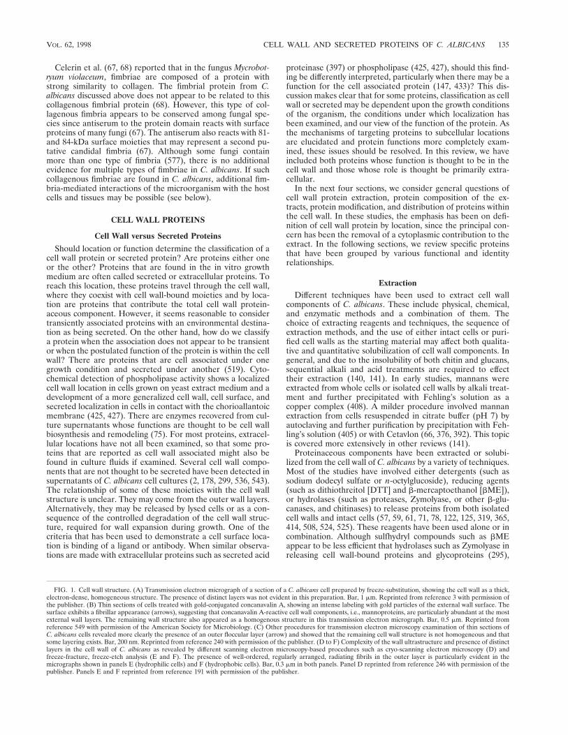
FIG. 1. Cell wall structure. (A) Transmission electron micrograph of a section of a C. albicans cell prepared by freeze-substitution, showing the cell wall as a thick,electron-dense, homogeneous structure. The presence of distinct layers was not evident in this preparation. Bar, 1 mm. Reprinted from reference 3 with permission ofthe publisher. (B) Thin sections of cells treated with gold-conjugated concanavalin A, showing an intense labeling with gold particles of the external wall surface. Thesurface exhibits a fibrillar appearance (arrows), suggesting that concanavalin A-reactive cell wall components, i.e., mannoproteins, are particularly abundant at the mostexternal wall layers. The remaining wall structure also appeared as a homogenous structure in this transmission electron micrograph. Bar, 0.5 mm. Reprinted fromreference 549 with permission of the American Society for Microbiology. (C) Other procedures for transmission electron microscopy examination of thin sections ofC. albicans cells revealed more clearly the presence of an outer floccular layer (arrow) and showed that the remaining cell wall structure is not homogeneous and thatsome layering exists. Bar, 200 nm. Reprinted from reference 240 with permission of the publisher. (D to F) Complexity of the wall ultrastructure and presence of distinctlayers in the cell wall of C. albicans as revealed by different scanning electron microscopy-based procedures such as cryo-scanning electron microscopy (D) andfreeze-fracture, freeze-etch analysis (E and F). The presence of well-ordered, regularly arranged, radiating fibrils in the outer layer is particularly evident in themicrographs shown in panels E (hydrophilic cells) and F (hydrophobic cells). Bar, 0.3 mm in both panels. Panel D reprinted from reference 246 with permission of thepublisher. Panels E and F reprinted from reference 191 with permission of the publisher.
VOL. 62, 1998 CELL WALL AND SECRETED PROTEINS OF C. ALBICANS 135

these chemical agents solubilize a complex array of protein-aceous components from the walls of intact C. albicans cells(57, 58). On the other hand, some b-glucanases used to solu-bilize cell wall moieties actually are enzymatic complexes thatmay contain other unidentified or uncontrolled hydrolytic ac-tivities, which may alter the native characteristics of the re-leased molecules.
Other extraction procedures have been less frequently re-ported and include chemical, enzymatic, and physical methodsalone or in combination. b-Elimination with NaOH has beenused to release putative structural proteins (369). Ethylene-diamine has also been used in structural studies to extract pro-teins (455). Salt (NaCl) was used to extract the surface determi-nant of a MAb (40) and a surface adhesin (225). Homogenizationhas been used to shear fimbriae (583), and a-mannosidase treat-ment followed by sonication has been used to release wall anti-gens (195).
There has been little comparison of the various methodsused to extract the wall proteins. Casanova and Chaffin (57)compared five extracts for yeast cells and germ tubes: (i) bMEextract of intact cells at alkaline pH; (ii) the Zymolyase extractof the treated cells; (iii) bME extract of isolated cell walls atalkaline pH; (iv) the Zymolyase extract of the treated cellwalls; and (v) a sodium dodecyl sulfate (SDS)-bME extract ofisolated cell walls. The extracts were examined by blotting withconcanavalin A, two MAbs (MAb 4C12 to a high-molecular-weight component of germ tubes [59] and MAb 24.17 to amannan epitope of a high-molecular-weight component [72])and antiserum for factor 6. The authors concluded that the twosequential extracts obtained from intact cells were most satis-factory. In addition, although extraction with reducing agents isfrequently thought to release medium to small componentsfrom the cell wall, this study showed that bME also releasedthe high-molecular-weight components. These appeared to belarger than the same component present in Zymolyase ex-tracts. bME and other reducing reagents are believed to solu-bilize mainly components associated with the outermost layersof the cell wall (50, 61, 295). These reagents also increase cellwall porosity and facilitate subsequent action of cell wall de-grading enzymes (103, 589). The hydrolysis of glucan by Zy-molyase or glucanases may release proteins enmeshed or co-valently attached to the glucan. Proteins covalently attached toglucan are postulated to represent species contributing to cellwall structure (59, 122, 123, 125, 482). Covalent attachment ofmannoprotein to glucan, perhaps through phosphodiester link-ages, has been suggested, as noted below (237, 238, 462, 463).
A valid question is whether the proteins found in theseextracts are genuine cell wall components. It has been sug-gested that treatment of intact C. albicans cells with reducingagents (DTT or bME) may release some intracellular macro-molecular components (50). This question has been examinedmost thoroughly for extracts obtained with bME. As discussedlater in this review, receptors or binding proteins for ligandsthat bind to the intact cell are found in such extracts. On theother hand, several proteins previously associated with a cyto-plasmic function have also been found in the wall extracts (6,8, 161, 303, 520). These observations led to additional experi-ments using different approaches to demonstrate that the pro-teins found in the extract were genuine wall components.Transmission electron microscopy demonstrated that each ofthe moieties was indeed present in the cell wall, including thecell wall interior (6, 8, 303). Chaffin and colleagues (6, 303) alsoused a more general method to identify genuine cell wall pro-teins. Intact cells were treated with a derivative of biotin thatdoes not permeate the membrane and therefore does not labelcytoplasmic proteins. The extracted biotinylated proteins in-
cluded those previously thought to be confined to the cyto-plasm. This demonstrated that the proteins were present in thecell wall prior to extraction and that their presence in theextract was not due to cytoplasmic contamination. Support forthe validity of the extraction procedure was also obtained withparental and mutant strains of S. cerevisiae (294). Two mem-bers of the Ssa family of proteins (Ssa1p and Ssa2p) weredetected in the cell wall and cytoplasm of the parental strain,whereas in the mutant strain missing these two proteins theremaining members of the family were detected in the cyto-plasm but not in the cell wall. The failure to find Ssa proteinsin the cell wall of the mutant strain demonstrated that the cellwall extract was not contaminated with the Ssa proteins of thecytoplasm. Hence, current evidence indicates that treatmentwith sulfhydryl compounds is a suitable method to releaseautochthonous cell wall protein and glycoprotein componentswithout substantially altering their biological characteristics (6,60, 61, 293, 295, 296, 300, 302, 377).
Composition
Analysis of the protein and glycoprotein constituents solu-bilized from isolated cell wall preparations and from intactcells of both candidal growth forms by different treatments hasrevealed both a complex array of protein-containing compo-nents and quantitative and qualitative differences in the pro-tein composition of yeast and mycelial cell walls (57–59, 71,125, 295, 319, 320, 324, 363, 414–416, 524) (Fig. 2). Somecomponents have been characterized as high-molecular-weightmannoproteins (HMWM). The identity of these proteins mayvary with the morphology of the organism. Several HMWMare released by treatment with b-glucanases and may be co-valently attached to structural polysaccharides. These HMWMmay play an important role in modulating the organization ofthe different cell wall constituents to obtain the final supramo-lecular structure of the wall specific for each C. albicans mor-phology. In addition, HMWM contain large amounts of car-bohydrate and consequently could be major elicitors ofanticandidal host immunity (59, 62, 123, 159, 160, 297, 301,326, 327, 415, 479, 523). In the medium- to low-molecular-weight range, from 20 to more than 40 polypeptide species(depending on the study considered) have been identified (61,71, 125) (Fig. 2). As discussed above, the evidence suggeststhat these proteins are bona fide cell wall constituents.
There is a growing body of experimental evidence indicatingthat the properties—expression, distribution, and chemicalcharacteristics—of cell wall proteins and glycoproteins ob-served in vitro and in vivo are dependent on multiple factors.These include growth conditions, organism-related factors(such as growth state, morphology of the cells, strain andserotype, phenotypic switching, cell surface hydrophobic orhydrophilic status), and the nature of the biological specimens(intact cells or isolated wall preparations) that are subjected toanalysis (5, 7, 24, 44, 57, 63, 106, 159, 190, 191, 195, 210, 284,297, 301, 326, 416, 422, 477, 513, 529). Iron availability, whichhas been shown to be important for pathogens in establishinginfection (46, 400, 566), affects the cell surface (529). There arequantitative but not qualitative changes in the profile of sur-face proteins associated with growth at different iron concen-trations. Yeast cells of most strains grown in limiting or excessiron do not adhere as well to human buccal epithelial cells asdo organisms grown at intermediate concentrations that sup-port optimum growth. The effects of growth conditions on theexpression of specific proteins are discussed for each protein inlater sections. The cell wall may be thus envisaged as a highlydynamic “organelle.” The fungus is capable of expressing dif-
136 CHAFFIN ET AL. MICROBIOL. MOL. BIOL. REV.

ferentially variable wall constituents that may be useful forswitching between commensal and pathogenic lifestyles and formodulating and/or evading the immune host defense.
ModificationPosttranslational modifications of proteins include glycosyl-
ation, acetylation, prenylation, phosphorylation, ubiquitinationand addition of a GPI moiety. Organisms use these modifica-tions to confer structural options for proteins, to provide reg-ulatory control of their functions and to target proteins tospecific cellular locations. While not all of these modificationshave been described in C. albicans, it is likely that the organismpossesses the ability to modify its proteins by most, if not all, ofthe posttranslational modifications.
Glycosylation. Without any doubt, glycosylation is the mostimportant modification of the proteins in the fungal cell wall.Attachment of sugar moieties to proteins results in the forma-tion of the glycoproteins, which in the case of C. albicans aremainly mannoproteins. The general features of mannoproteinshave been discussed above. However, the presence of nongly-cosylated proteins has also been found in the cell wall of C.albicans (6, 8, 58, 61, 161). Mannosylated proteins can bebroadly divided into two classes. The HMWM, most of whichare larger than 200 kDa, are postulated to have structuralfunctions within the cell wall. Some medium- to low-molecu-lar-weight proteins also react with concanavalin A, indicatingtheir mannan content. Within this broad division, the amountof carbohydrate attached to the same polypeptide may vary(59, 122, 364). Within the high-molecular-weight class, there is
a difference in the size of the side chains associated with mor-phology. Oligosaccharides obtained from mannoproteins fromyeast cells average 600 residues, and those from germ tubesaverage 300 residues (122). Cell wall proteins may also be Oglycosylated with unbranched mannose chains containing oneto a few residues (412). Elorza et al. (123) have suggested thatsome proteins are initially secreted as O-glycosylated proteinsand become cross-linked with glucan and/or other N-glycosy-lated proteins only after incorporation into the cell wall struc-ture.
Phosphorylation. Phosphorus is a minor component of thecell wall of C. albicans (77). It has been assumed that it ispresent in cell wall mannoproteins in phosphodiester linkagesbetween mannose residues (17, 454). Bulk mannan from C.albicans can be fractionated into five fractions that differ in theamount of phosphate (393). Phosphomannoprotein complexesfrom cells of both C. albicans A and B serotypes have beencharacterized (261–263, 494–498). This material contained 0.9to 1.6% phosphate depending on the morphology and strainconsidered, and the authors concluded that b-(1,2) oligoman-nosaccharides were attached by phosphodiester linkage toother branching moieties. b-(1,2) oligomannosidic epitopeswere further observed on a C. albicans 14- to 18-kDa phos-pholipomannan moiety (545), a glycolipid with important im-munologic properties (133–135, 228, 229, 424), whose mannoseresidues may be added differently from mannan (546). Not allthe glycoproteins in the cell wall of C. albicans contain phos-phate, and some proteins may contain phosphate but not car-bohydrate (58).
FIG. 2. Polypeptide composition of cell wall extracts and whole protoplast homogenates from C. albicans as revealed by different electrophoretic techniques (A andB). C. albicans cells from which samples were obtained were incubated in the presence of 14C-labelled protein hydrolysate and subsequently tagged with biotin.Double-labelled cell wall proteins and glycoproteins were extracted from intact blastoconidia (lanes 1 and 3) and germinated blastoconidia (lanes 2 and 4) by sequentialtreatment with bME (lanes 1 and 2) and digestion with Zymolyase 20T (lanes 3 and 4). Samples of protoplast homogenates from blastoconidia and germinatedblastoconidia were run in lanes 5 and 6, respectively. Polypeptides were separated by SDS-PAGE and detected by fluorography (A) or transferred to nitrocellulose anddetected with an avidin-peroxidase conjugate (B; lane S in this panel shows a mixture of prestained molecular weight standards run in parallel). Numbers and lettersare used to identify and compare bands detected in the cell wall extracts by the different experimental procedures used. Although qualitative differences were observed(i.e., some polypeptides exhibited a strong radioactive label but were weakly biotinylated [band 6; star]), surface labelling of cells with biotin appeared to be a suitabletechnique to detect proteins in the wall of C. albicans. Thus, from the complex polypeptide pattern found in the protoplast homogenate samples as revealed byfluorography (A, lanes 5 and 6), only few species were labelled with biotin, indicating that most proteins released by bME and Zymolyase from intact cells are bonafide cell wall components. Brackets indicate a cluster of bands within a molecular weight range where many candidal moieties that represent receptors for host ligandshave been identified (see the text). Reprinted from reference 61 with permission of the American Society for Microbiology. (C) The complexity of the polypeptidepattern of the cell wall extracts was clearly evidenced when analysis was performed by two-dimensional PAGE and silver staining (the polypeptide pattern showncorresponds to the bME extract from blastoconidia). Reprinted from reference 486 with permission of the American Society for Microbiology.
VOL. 62, 1998 CELL WALL AND SECRETED PROTEINS OF C. ALBICANS 137

Finally, there are results suggesting that b-1,6- and b-1,3-glucan moieties present in C. albicans cell wall mannoproteinsmay be connected to a GPI anchor, which is known to bephosphodiester linked to the C-terminal amino acid of themature protein (237, 238, 247, 268). This is in agreement withthe hypothesis that proteins destined to be incorporated intothe cell wall are linked to b-glucan through the glycan part oftheir GPI anchors (104), as has been demonstrated for the S.cerevisiae a-agglutinin (306, 573). The GPI anchor targetsa-agglutinin to the cell wall (573). Pulse-chase experimentsindicate that a plasma membrane-bound form is released toperiplasmic space as an intermediate form that is then incor-porated into the cell wall (305, 306). Recent studies of thelinkage between mannoprotein and glucan suggest that theGPI remnant consists of ethanolamine-phosphate-mannose5,with the terminal mannose attached to the nonreducing end ofb-1,6-glucan (268). A transglycosylation reaction is proposedto effect the linkage.
Ubiquitination. Ubiquitin is a small (approximately 8,500-Da) polypeptide first isolated from bovine thymus (167). Se-quence analysis of various ubiquitin genes has revealed strikingevolutionary conservation among species (138). Ubiquitinplays important roles in protein modification, protein degra-dation, gene transcription, organization of chromatin struc-ture, and stress resistance in higher eukaryotes (138, 139, 197).It is also associated with some cell surface protein and signal-ing functions (69, 368, 404, 504, 553). In yeast, a role forubiquitination in endocytosis and/or turnover of plasma mem-brane protein receptors including a-receptor has been demon-strated (198, 269, 442). The C terminus of ubiquitin is co-valently attached to ε-amino groups of lysine in proteinsubstrates by an enzymatic conjugation system. A large numberof enzymes responsible for the formation and processing ofubiquitin-protein conjugates have been described (138), in-cluding in C. albicans (98). We have cloned a polyubiquitingene (UBI1) of C. albicans that contains three tandem copies,head-to-tail spacerless repeats, of the sequence coding for the76 amino acids of the ubiquitin protein (485). The gene hasalso been cloned from a different strain (15). Northern blotanalysis revealed a single mRNA population of about 1 kbpresent in similar amounts in both yeast and mycelial cells(485). Indirect immunofluorescence demonstrated that ubiq-uitin determinants were located on the cell surface, and West-ern blot analysis of a bME extract demonstrated that severalcell wall proteins contained ubiquitin-like epitopes. The cellwall species that are ubiquitinated are discussed below with theindividual proteins. The role of ubiquitin in the cell wallwhether in protein degradation, stress protection, or perhapseven modulation of activity of receptor-like molecules remainsto be assessed.
Distribution and Expression
As described above, transmission electron microscopy stud-ies of the C. albicans cell wall show the existence of severallayers. The structural appearance of these layers is variable andseems to be related to the strain examined, the growth condi-tions, the morphology (yeast cells or germ tubes) exhibited bythe microorganism, and the sample preparation protocol (50,64, 422) (Fig. 1). After total removal of proteins and manno-proteins by treatment with strong alkali or heating, the cell wallappeared to be significantly thinner with loss of any apprecia-ble layering. These structural changes were paralleled by theabsence of electron-dense components detectable with ordi-nary electron microscopy dyes, concanavalin A binding sites,
and positive staining with reagents specific for mannoproteins(periodate-silver). Thus, the cell wall layering appears attrib-utable to the distribution of mannoproteins at various levelswithin the wall structure (64). Treatment of cells with sulfhy-dryl agents and hydrolytic enzymes, coupled with specific cy-tochemical staining, has consistently shown that mannoproteinconstituents are preferentially located at the outermost layer ofthe wall of C. albicans cells. The surface has a fibrillar orflocculent aspect with thin, delicate filaments or fimbriae (Fig.1; see below). This material is present mostly in virulentstrains, and it is also more abundant in isolates exhibitingincreased adherence to host tissues (64). Proteins whose bio-logical function is at the cell surface or the extracellular envi-ronment may nevertheless be found at the innermost layer ofthe wall and through the wall as they travel from the plasmamembrane and periplasmic space to their destination (478).Thus, some proteins destined for the extracellular environmentmay also be obtained in cell wall extracts. Evidence for thismannoprotein traffic in C. albicans has been reported (423).Immunoelectron microscopy shows that proteins on the cellsurface visualized by indirect immunofluorescence are also de-tected at the cell surface as well as near the plasma membranewith some protein distributed through the wall (6, 303). On theother hand, a protein that is not detected at the cell surface byindirect immunofluorescence but is present in cell wall extractsis located only in the interior of the wall (8). Using a panel ofMAbs to localize proteins, Ponton et al. (416) demonstratedvarious distribution classes of cell wall proteins: (i) expressedonly on the germ tube surface, (ii) expressed on the germ tubesurface and within the yeast cell wall, (iii) expressed on bothyeast cell and germ tube surfaces, and (iv) expressed within thewall of both germ tubes and yeast cells. A fifth category, ex-pressed only on yeast surfaces, is also reported (296). Proteinsthat are associated with b-glucans should be concentrated nearthe plasma membrane with the structural polysaccharide. Inany case, since asymmetry in mannoprotein distribution is ev-ident in C. albicans walls (64), layering is more likely to be theresult of quantitative differences in the proportions of theindividual wall components (b-glucans, chitin, and mannopro-teins) in each layer rather than qualitative differences (389).
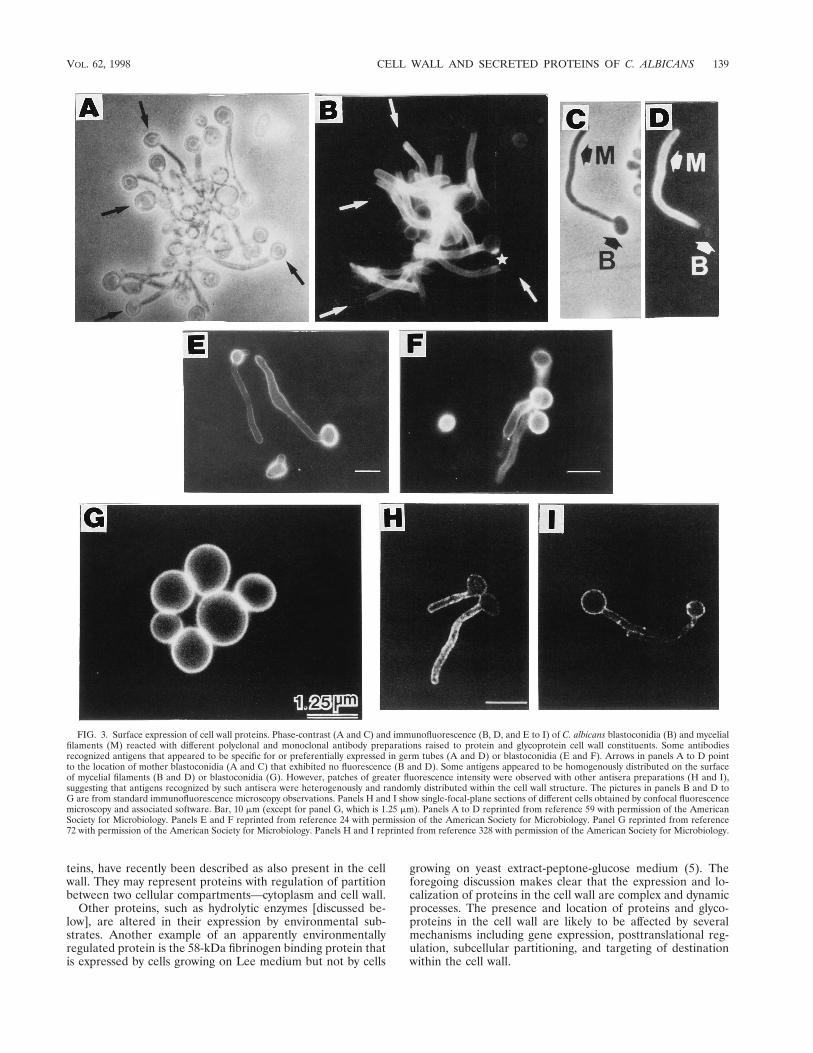
Differences in the distribution of proteins and glycoproteinsat the cell surface are also noted. This asymmetry may berelated to the physiologic role played by each particular moi-ety. High-molecular-weight mannoproteins that may play animportant and active morphogenetic role in modulating theorganization of the cell wall (59, 62, 123, 160, 326, 327, 480) arehomogeneously distributed on the cell surface (59, 62, 159).Some proteins and mannoprotein moieties that are receptorsfor different host ligands exhibit clustering or asymmetric cellsurface distribution (60, 296, 328). The distribution of commonor morphology-associated, homogeneously or heterogeneouslydistributed cell surface antigens of C. albicans as revealed byimmunofluorescence microscopy is shown in Fig. 3.
At least three cell moieties appear to have posttranslationalregulation of their localization in the cell wall. A MAb thatrecognized a hyphal surface protein detected a smaller proteinin the plasma membrane of yeast cells (394). The C3d bindingprotein was detected as a 60-kDa moiety in germ tube cellwalls, while a 50-kDa component was found in yeast cell mem-branes (563). The third example is a 30-kDa protein found inthe cell wall of germ tubes but not yeast cells (5). However, thegene is expressed in yeast cells and presumably translated,although the cellular location of the protein is not known.Another group of proteins to be discussed below, i.e., enolase,hsp70, 3-phosphoglycerate kinase, and glyceraldehyde-3-phos-phate dehydrogenase (GAPDH), well-known cytoplasmic pro-
138 CHAFFIN ET AL. MICROBIOL. MOL. BIOL. REV.
teins, have recently been described as also present in the cellwall. They may represent proteins with regulation of partitionbetween two cellular compartments—cytoplasm and cell wall.
Other proteins, such as hydrolytic enzymes [discussed be-low], are altered in their expression by environmental sub-strates. Another example of an apparently environmentallyregulated protein is the 58-kDa fibrinogen binding protein thatis expressed by cells growing on Lee medium but not by cells
growing on yeast extract-peptone-glucose medium (5). Theforegoing discussion makes clear that the expression and lo-calization of proteins in the cell wall are complex and dynamicprocesses. The presence and location of proteins and glyco-proteins in the cell wall are likely to be affected by severalmechanisms including gene expression, posttranslational reg-ulation, subcellular partitioning, and targeting of destinationwithin the cell wall.
FIG. 3. Surface expression of cell wall proteins. Phase-contrast (A and C) and immunofluorescence (B, D, and E to I) of C. albicans blastoconidia (B) and mycelialfilaments (M) reacted with different polyclonal and monoclonal antibody preparations raised to protein and glycoprotein cell wall constituents. Some antibodiesrecognized antigens that appeared to be specific for or preferentially expressed in germ tubes (A and D) or blastoconidia (E and F). Arrows in panels A to D pointto the location of mother blastoconidia (A and C) that exhibited no fluorescence (B and D). Some antigens appeared to be homogenously distributed on the surfaceof mycelial filaments (B and D) or blastoconidia (G). However, patches of greater fluorescence intensity were observed with other antisera preparations (H and I),suggesting that antigens recognized by such antisera were heterogenously and randomly distributed within the cell wall structure. The pictures in panels B and D toG are from standard immunofluorescence microscopy observations. Panels H and I show single-focal-plane sections of different cells obtained by confocal fluorescencemicroscopy and associated software. Bar, 10 mm (except for panel G, which is 1.25 mm). Panels A to D reprinted from reference 59 with permission of the AmericanSociety for Microbiology. Panels E and F reprinted from reference 24 with permission of the American Society for Microbiology. Panel G reprinted from reference72 with permission of the American Society for Microbiology. Panels H and I reprinted from reference 328 with permission of the American Society for Microbiology.
VOL. 62, 1998 CELL WALL AND SECRETED PROTEINS OF C. ALBICANS 139

Enzymes with Cell Wall FunctionAs discussed above, a number of hydrolytic enzymes have
been recovered from both cell-associated locations (cell walland periplasm) and culture medium whose function is postu-lated to be within the cell wall (Table 1). These enzymes arethought to be involved in cell wall biosynthesis or the remod-eling that accompanies growth and division of cells.
Exo-b-(1,3)-glucanase. Secretory exo-b-glucan hydrolases(b-glucanases or b-glucosidases) are widely occurring enzymesin many yeast and fungal species. Although the exact physio-logical roles of these enzymes are unknown (141, 386), theyparticipate in the metabolism of b-glucan, which is the mainstructural microfibrillar polymer of the cell wall in C. albicans(64). The most widely accepted biological role of glucanases islimited hydrolysis of cell wall glucan during morphogeneticevents (141, 386). b-Glucanases have been described to beassociated with the C. albicans cell wall (387, 428).
Detailed information on the chemical nature of secretoryb-(1,3)-glucanases has been reported mainly for S. cerevisiae,where at least two isoenzymes, arising by differential glycosyl-ation of a primary gene product, are secreted into the medium(430). In C. albicans, exo-b-(1,3)-glucanase activity was foundto be secreted and exported mainly during germ tube forma-
tion. Negligible enzyme was released into the medium whenyeast cells were grown (429). As with b-N-acetylglucosamini-dase, this may be related to the more porous nature of thegerm tube cell wall (519). In contrast to S. cerevisiae, only oneexoglucanase has been detected in C. albicans, and it accountsfor most of the total glucanase activity present in the growthmedium and cell extracts (307, 361, 428). However, there aresome discrepancies between results reported from differentgroups. The enzyme purified from cell extracts of C. albicans1001 was reported to be a heterodimer of subunits with mo-lecular masses of 63 and 44 kDa (362). Subsequently, the majorexoglucanases secreted into the medium by strains 1001 and3153A were found to be identical, single nonglycosylatedpolypeptides, with a molecular mass of about 38 kDa (307).The peptides had significant chemical and immunological sim-ilarity to the major exoglucanase secreted by S. cerevisiae.Cloning and sequencing of the gene EXG (previously XOG1)coding for the exo-b-(1,3)-glucanase of C. albicans (75, 308)revealed high identity to the b-(1,3)-exoglucanase EXG1 genecloned from S. cerevisiae (561). A single transcript was detectedin both yeast and hyphal forms, and the levels of expressionappeared proportional to the growth rate (74). Sequence anal-ysis indicated a signal peptide for secretion and a recognition
TABLE 1. Hydrolytic enzymes and proteins with cell wall and extracellular targets
Enzyme Gene Location Comments Reference(s)
Cell wall substratesExo-b-(1,3)-glucanase EXG (XOG1) Cell wall, extracellular Cell wall morphogenesis 75, 307, 308, 361, 387, 428, 429b-1,3-glucan transferase BGL2 Cell wall Cell wall metabolism 122, 178, 196, 466, 471Chitinase CHT1-3 Periplasm, cell wall,
extracellularHydrolytic enzyme, cell wall
morphogenesis141, 170, 346, 347
b-N-acetylglucosaminidase HEX1 Periplasmic,extracellular
Hydrolytic enzyme, virulencefactor?
55, 221, 365, 426, 519
Transglutaminase Cell wall Covalent cross-links? 456
Extracellular substratesSecreted aspartyl
proteinaseSAP1-9 Extracellular, cell
surfacePutative virulence factor, gene
expression conditiondependent
1, 3, 18, 31, 84, 99–101, 109, 147,157, 205, 206, 214, 215, 232, 233,277, 348–350, 353, 359, 370, 372,378, 379, 381, 397, 433–435, 441,445–448, 450, 451, 509–511, 514,515, 552, 570, 574
PhospholipasePhospholipase A Cell wall, surface,
extracellularHydrolytic enzyme 23, 174, 425, 427
Phospholipase B PLB1 Extracellular Hydrolytic enzyme, putativevirulence factor
19, 217, 473, 533
Phospholipase C Extracellular Hydrolytic enzyme 425, 427Lysophospholipase Cell wall, surface,
extracellularHydrolytic enzyme 19, 23, 174, 355, 425, 427, 533
Lysophospholipase-transacylase
Extracellular Hydrolytic enzyme, putativevirulence factor
19, 217, 533, 534
Esterase Extracellular Hydrolytic enzyme 56, 419, 449, 551Glucoamylase Extracellular Hydrolytic enzyme 82Hemolytic factor Cell wall, extracellular Hydrolytic enzyme 317Acid phosphatase Periplasmic, surface Hydrolytic enzyme 78, 105, 391, 547Lipase LIP1 Extracellular Hydrolytic enzyme 148Hyaluronidase Extracellular Hydrolytic enzyme, virulence
factor?499, 500
Chondroitan sulfatase Extracellular Hydrolytic enzyme, virulencefactor?
499, 500
Metallopeptidase Cell wall,extracellular?
Hydrolytic enzyme 120, 121
Trehalase Cell wall, extracellular Hydrolytic enzyme 362, 454
140 CHAFFIN ET AL. MICROBIOL. MOL. BIOL. REV.

by a KEX2-like protease (75). A mature enzyme of 400 aminoacid residues with no sites for N-linked glycosylation was pre-dicted. These results are consistent with the characteristics(carbohydrate content and molecular mass) of the secretedenzyme previously reported (307). Recombinant exo-b-(1,3)-glucanase of C. albicans purified from S. cerevisiae has beenfound to contain a number of short blocks of sequence homol-ogy to several genes for cellulases of the family A glucanases,including the conserved sequence site NEP, which has previ-ously been shown to be important in the catalytic function ofseveral cellulases (76). Glu-330 has been identified as the cat-alytic nucleophile in the enzyme (308).
b-1,3-Glucan transferase. Many yeasts, including C. albicansand S. cerevisiae, contain a highly conserved protein with a sizeranging from 31.5 to 34 kDa depending on the species (196). Itis a major cell wall mannoprotein in S. cerevisiae (466). In C.albicans, a protein of 34 kDa is secreted by protoplasts andobserved as an aggregate in gel filtration (122). When releasedby Zymolyase, it eluted as a low-molecular-weight species. Theprotein appeared to have a single N-linked oligosaccharide of2.5 kDa and a residual protein moiety of 31.5 kDa. A cell wallprotein with an approximate molecular mass of 34 kDa wasisolated as a by-product during purification of an endo-(1,3)-b-glucanase from the material secreted to the medium by C.albicans (178). The enzyme displayed a unique glucanosyltransferase activity and did not contain any exo- or endo-b-glucanase activity. The authors suggested that this 34-kDa pro-tein is a glucan-branching enzyme responsible for the transfor-mation of the initial linear b-(1,3)-glucan into the branchedb-(1,3)-b-(1,6)-glucan that is found in the cell wall of the fun-gus. The C. albicans BGL2 gene encoding the b-1,3 glucantransferase has been cloned (471) and is similar to the S.cerevisiae BGL2 gene (245). The S. cerevisiae enzyme has beendescribed as an exoglucanase (245) and as an endoglucanase(373). However, more recently, the S. cerevisiae enzyme hasbeen shown to be homologous to the sequence of the C. albi-cans enzyme and corresponds to Bgl2p (165). More sensitiveassays revealed that at low concentrations of glucose oligosac-charides, glucanase activity was observed, while at higher con-centrations, glucosyltransferase activity predominated. The en-zyme transfers b-1,3 glucan oligosaccharides from a donorb-1,3 glucan to an acceptor b-1,3 glucan, forming a linearpolymer joined by a b-1,6 linkage (587). This activity wouldpermit the enzyme to participate in cross-linking or repair ofglucan within the cell wall.
The C. albicans and S. cerevisiae genes have N-terminalsignal sequences but not C-terminal sequences for the additionof a GPI anchor (245, 471). A mutant C. albicans strain wasconstructed by sequential disruption of alleles and was missingthe protein in extracts (468). However, residual non-Bgl2pactivity was detected, suggesting the presence of another trans-ferase in C. albicans. In vivo, the mutant strain was less virulentfor mice and fewer organisms were recovered from the kidneysof infected animals than for the parental strain. The reductionin virulence of C. albicans suggested that there were differ-ences in the cell wall that affected the survival of the mutantstrain in the animal model. In S. cerevisiae, the disruptant strainmissing the protein had no obvious phenotype (245) and noresidual glucosyl transferase activity (165). A strain in whichBgl2p was overproduced had a reduced growth rate (373).These observations suggest that the role of BGL2 in cell wallmetabolism has yet to be fully elucidated.
Chitinases. Chitinases are produced by many organisms in-cluding chitin-containing organisms that produce both chitinsynthases and chitinases. In C. albicans, the most likely role forthese enzymes, like glucanases, is limited hydrolysis of cell wall
chitin during morphogenetic events (141). C. albicans containsthree chitinase genes, CHT1, CHT2, and CHT3 (346). Morethan half the chitinase activity detectable in whole-cell extractsis associated with secreted enzyme (periplasmic and cell wall)(170). Reactivity is detected in supernatants of washed cellsand with intact cells by using a substrate that does not enter thecell (346). Chitinase production increases during exponentialgrowth and is greater in cells grown on yeast extract-peptone-dextrose (YEPD) medium than on a minimal medium. One ofthe roles of chitinase in S. cerevisiae, where chitin is confinedprimarily to the septum, is mother-daughter separation. Dis-ruption of S. cerevisiae CTS1 results in clumps of cells due tofailure of the cells to separate (273). Treatment of C. albicansyeast cells with a chitinase inhibitor also leads to inhibition ofcell separation and clumps of cells (170).
The C. albicans chitinase gene(s) was identified by use ofdegenerate PCR primers based on conserved fungal chitinasesequences (346). The deduced amino acid sequence encodedby the CHT2 open reading frame (ORF) predicted a polypep-tide of 583 amino acids, and that encoded by CHT3 predicteda polypeptide of 567 amino acids. The deduced sequence fromCHT1 consisted of 416 amino acids (347). The C. albicanschitinases were similar (36 to 38%) to that of S. cerevisiae(346). However, the similarity was 55 to 65% in the N-terminalregion containing the putative catalytic domain. There arepotential N-glycosylation sites in the sequences. Near the 39end of the sequences of CHT2 and CHT3 is a sequence en-coding a region that is rich in serine and threonine, which maybe potential sites for O glycosylation. This region is not presentin CHT1, which encodes a smaller putative protein (347). Ex-pression of CHT2 and CHT3 was detected in both yeast cellsand hyphae, although greater expression was associated withyeast growth (346). Expression of CHT1 was not detectedunder the various growth conditions examined. Preliminaryresults using a disruption of CHT2 suggested that the chitinaseparticipated in cell separation as cells tended to form clumpsor clusters.
b-N-Acetylglucosaminidase. Production of b-N-acetylglu-cosaminidase by C. albicans cells is induced by the presence ofGlcNAc in the medium (519). GlcNAc, the b-N-acetylglu-cosaminidase reaction product, also induces the synthesis ofother enzymes required for the metabolism of this amino sugar(171, 492, 519). The enzyme may function as a chitobiase that,in conjunction with chitinase, completes the hydrolysis of chitinto provide both nitrogen and carbon sources from chitin. Theenzyme, for which two molecular forms with different glyco-sylation levels appear to exist (364), is secreted and depositedinto different regions of the cell envelope of both the yeast andmycelial forms (365, 426, 519). The enzyme is also released tothe culture medium of both growth forms but to a greaterextent (at least fourfold greater) during hyphal formation(519). It is suggested that the germ tube wall is more porousthan that of yeast cells and, consequently, that release of ex-tracellular enzymes to the medium is facilitated by the hyphalformation process. b-N-Acetylglucosaminidase acts on a num-ber of natural and synthetic substrates including diacetylchito-biose and triacetylchitotriose. In its native form, the enzymeappears to be a monomer with a molecular mass of 66 kDa(519).
The metabolism of GlcNAc by C. albicans has attractedinterest, since this amino sugar induces the yeast-to-myceliumtransition in this fungal species (493). b-N-Acetylglucosamini-dase from C. albicans is specifically a chitobiase (519). It mayact coordinately with chitinase, which is also present in thisfungus (22), in the hydrolysis of chitin to form GlcNAc. How-ever, the role of these enzymes in cell metabolism is not clear
VOL. 62, 1998 CELL WALL AND SECRETED PROTEINS OF C. ALBICANS 141

(223), but there is no direct role in morphogenesis (383). Invivo, cleaving of GlcNAc from complex carbohydrates by C.albicans b-N-acetylglucosaminidase may provide a suitable car-bon source for the fungus. Alternatively, removal of GlcNAcresidues from glycoproteins of the fungal cell surface maycause conformational changes that modify adhesion of C. al-bicans cells to host tissues (55). Jenkinson and Shepherd (223)reported that a C. albicans mutant defective in the productionof b-N-acetylglucosaminidase was less virulent in an experi-mentally induced candidiasis mouse model than was the wild-type strain, thus suggesting a possible role for this enzyme as acandidal virulence factor. On the other hand, the phenotypicproperties of the mutant suggest that the enzyme is not essen-tial for the growth of C. albicans cells (223). Hence, furtherwork is required to determine the role of this enzyme in patho-genicity. The entire b-N-acetylglucosaminidase gene (HEX1)from C. albicans has been cloned (55). The organism appearedto use alternative transcription termination sites dependingupon growth conditions, since the HEX1 mRNA from cellsgrown on GlcNAc was 200 nucleotides longer than the tran-script from cells grown on glucose. Plasmid-borne HEX1 alsoresponded to GlcNAc induction.
Glutaminyl-peptide-g-glutamylyl-transferase. The activityof the enzyme glutaminyl-peptide-g-glutamylyl-transferase(transglutaminase) has been detected in cell extracts obtainedfrom both growth phases of C. albicans. The activity was asso-ciated mostly with the cell wall fraction, whereas the cytosolcontained almost negligible amounts of enzyme activity (456).This distribution suggested an extracellular (cell wall-bound)location for transglutaminase in intact cells. Although the wallcomponent displaying this enzymatic activity has not beencharacterized, the postulated transglutaminase activity couldbe involved in the formation of covalent bonds between dif-ferent wall proteinaceous moieties (456). In this context, it hasbeen suggested that formation of such covalent linkages couldplay a role in maintaining the spatial organization of the cellwall during the biogenesis and assembly of this structure.
Hydrolytic Enzymes and Proteins withExtracellular Targets
The enzymes in the previous section are postulated to findsubstrates and to have their primary function within the cellwall. This section considers enzymes whose substrates are notassociated with the cell wall but are found in the environment(Table 1). The action of these enzymes may provide access tonutrients for the organism. When hydrolysis of these substratesor action of extracellular proteins affects the function andviability of the host, the enzymes may be considered virulencefactors that contribute to the establishment of infection. Theseproteins are at least transiently associated with the cell wallduring their translocation across the cell wall to the externalenvironment. However, in some cases, the association is lesstransient. The distribution of some of these enzymes is variableand, under some growth conditions, may be primarily cell as-sociated in the periplasm and cell wall. Some of these proteinshave also been localized to the cell surface by the same criteriathat have established a cell surface location for adhesins whosefunction is at the cell surface. One of these extracellular pro-teins, Sap, also found at the cell surface, may contribute toadherence (209, 406). We have also included in this section adiscussion of a secreted protein with immunomodulation prop-erties whose relationship with other secreted moieties has notbeen established. Additional studies of the function, localiza-tion, and molecular mechanisms of targeting proteins to spe-
cific locations will help resolve questions about the cell-asso-ciated and extracellular distribution of these proteins.
Acid proteinase. The extracellular proteolytic activity is oneof the several hydrolytic enzyme activities described for C.albicans (390) and is due to aspartyl proteinase enzymes. Thecandidal secreted aspartyl proteinase was first identified byStaib (514, 515), and over the last 25 years its biological char-acteristics, including its role as a potential virulence factor ofC. albicans, have been studied by a number of laboratories.The proteolytic activity is associated with a 42- to 45-kDa acidcarboxyl proteinase. The enzyme has broad substrate specific-ity and is active in the range of pH 2.0 to 7.0, with the pHoptimum varying from 2.5 to 4.5 depending on the substrate(109, 451). As discussed below, there is a family of aspartylproteinases whose expression appears to be regulated by thestrain, cell type, and environment. As a protease, the enzymemay have a variety of substrates, and these substrates may varydepending upon the host organ, e.g., skin or blood, that iscolonized or infected. This potential spectrum of substratesand expression of isoenzymes may account for the differentroles that have been postulated for the enzyme as a virulencefactor. Purification of Sap1, Sap2, and Sap3 showed isoenzymedifferences in antigenic similarity, thermal stability, and activityat low pH (509). The crystal structure of the enzyme showeddetails of the binding site, suggesting the possibility of struc-tural differences among isoenzymes that might affect substratespecificities (1, 94). The techniques and strains that are beingdeveloped and constructed and understanding isoenzyme dif-ferences should contribute to elucidation of the role of theenzyme in colonization and infection.
Aspartyl proteinases are secreted by pathogenic species ofCandida in vivo during infection (99, 100, 349, 450). The en-zymes are secreted in vitro when the organism is cultured inthe presence of exogenous protein (usually bovine serum al-bumin) as the nitrogen source (18, 109, 206, 434, 441, 447, 448,451). However, exogenous protein in the culture medium wasfound not to be essential for induction of enzyme synthesis(205). Instead, the pH of the medium seems to act directlyupon secretory aspartyl proteinase synthesis and not as a sec-ondary effect of the nitrogen supply from the proteinase-me-diated protein digestion, as initially thought. In any case, in-duction of C. albicans extracellular aspartyl proteinase appearsto involve stimulation of a signal transduction event at theplasma membrane level (281). This stimulation appears to beinduced by large proteins and/or polypeptides (containingeight or more amino acid residues), since internalization ofsmall peptides with less than seven residues by peptide trans-port was not the inducing signal for proteinase production.
Initially designated Cap (Candida aspartyl proteinase), thisenzymatic activity has been given a variety of labels. Thus, thefirst cloned gene in C. albicans was referred to as PEP1, be-cause of its similarity to the pepsinogen gene (372). However,the Candida proteinase is similar to many different aspartylproteinase genes and is not homologous to pepsinogen. Thestudy reporting the cloning of the second gene referred to bothsecreted aspartyl proteinase genes as PRA genes (574), be-cause of their similarity to the Pra gene product of S. cerevisiae.However, Pra proteinase is a vacuolar enzyme that is not se-creted. A related Candida aspartyl proteinase that also is notsecreted is more closely homologous to the Pra proteinase(304), while a Saccharomyces homolog of the Candida secretedaspartyl proteinase is not Pra (571). For these reasons, it wasproposed that the Candida secreted aspartyl proteinase bereferred to as Sap (SAP gene; Sap protein) (311, 571).
The biochemical properties of Sap (i.e., molecular weight,pI, sensitivity to inhibitors, substrate specificity, N-terminal
142 CHAFFIN ET AL. MICROBIOL. MOL. BIOL. REV.

amino acid sequence of the protein) vary depending on thestrain and laboratory (109, 451). The recent cloning and se-quencing of seven distinct SAP genes (from SAP1 to SAP7)(214, 215, 359, 366, 571, 574) may contribute to an explanationof such discrepancies. The seven cloned genes each encode amature protein that is highly conserved and a precursor pep-tide that contains the most evolutionarily divergent region ofthe SAP genes (570). Comparison of the N-terminal proteinsequences suggested the existence of at least one more SAPgene (SAP8) (371). SAP8 was subsequently cloned by Hubeand colleagues (511). Also, a ninth gene (SAP9) has beencloned (461). Hence, a family consisting of at least nine SAPgenes can be drawn upon to produce Sap enzymatic activity inC. albicans. Recently, the levels of the Sap1, Sap2, and Sap3isoenzymes were monitored under a variety of growth condi-tions for several C. albicans strains (570), including strainWO-1, which alternates between two switch phenotypes, white(W) and opaque (O) (510). These studies revealed that thespecific Sap isoenzyme produced is determined by the cell type(strain) whereas the level of Sap production is affected byenvironmental factors, and they showed that both the yeast-to-mycelium transition and phenotypic switching can deter-mine which of the Sap isoenzymes is produced (570). A studyof the expression of seven members of the SAP gene family indifferent strains and phenotypes and under different conditionswas performed by another group (214). SAP1 and SAP3 levelswere regulated during the phenotypic transition between Wand O forms. SAP2 was the dominant transcript in the yeastform, and its expression was autoinduced by peptide productsof its own enzymatic activity and repressed by amino acids.SAP4 and SAP6 expression was observed only at neutral pHduring morphogenetic conversion from yeast to hypha inducedby serum. Expression of SAP7 was not detected under any ofthe experimental conditions used throughout the study. Theauthors concluded that the different members of the SAP genefamily may play distinct roles in colonization and invasion ofthe host (214). SAP8 is the third gene of the family to beexpressed in the opaque phenotype (511).
Regardless of the question of how many C. albicans secretedproteinases exist and what role each may play, evidence sug-gests that aspartyl proteinase either is not glycosylated or isglycosylated at a very low level. One of the reported enzymesdoes not appear to contain any putative N-glycosylation sites inthe deduced amino acid sequence (215). The addition of tuni-camycin (an inhibitor of N glycosylation [112]) had no effect onthe secretion, molecular weight, and activity of aspartyl pro-teinase (370). In addition, the purified enzyme does not reactwith concanavalin A (453) or stain with periodic acid-silverreagent (370). Therefore, if the functional protein does containany oligosaccharide residues, they are believed to be O linkedwith the peptide chain. The Asn-Ala-Thr consensus glycosyla-tion sequence has been found in the prepropeptide region, butthere is no convincing evidence that this site is actually glyco-sylated (215). An immunocytochemistry analysis with affinity-purified antibodies to the enzyme detected reactivity in gran-ules, and this reactivity was inhibited by glycogen, suggestingthat the antibodies cross-reacted with glycogen-like polysac-charides (3). A 45-kDa intracellular form of the secretory as-partyl proteinase that seems to be the precursor of the 43-kDamature enzyme has been reported (205, 206). It has beensuggested that although the mature form is not glycosylated,that glycosylation may occur to some extent in the 45-kDaprecursor (353), which may partly explain the results reportedby Akashi et al. (3).
Proteinase production is believed to enhance the ability ofthe organism to colonize and penetrate host tissues and to
evade the host immune system (31, 96, 109, 209, 232, 433, 435,451, 552). A correlation between virulence and levels of pro-teinase production in both C. albicans clinical isolates (99, 157)and laboratory strains with altered proteinase levels (277, 350,441) supports a potential role in virulence. More virulentstrains of C. albicans can be isolated from patients with AIDSthan from normal subjects, and this characteristic may be as-sociated with an elevated production of aspartyl proteinase(101, 397). In this context, the enzyme has been reported to berare or absent in nonpathogenic strains or species of Candida(348). Mutant strains of C. albicans that do not secrete thisenzyme show significant reductions in lethality for mice (277,350). It has also been suggested that the protease could pro-mote the release of cell wall mannan by cleaving the peptidemoiety of candidal cell wall mannoproteins (381). The releasedmannan may cause stimulation or suppression of cell-mediated(oligosaccharide fragments of mannan appear to be potentinhibitors of cell-mediated immunity) and humoral immunefunctions (381). The enzyme is able to degrade a number ofimportant defensive host proteins such as immunoglobulinsand complement (232, 446, 451). Infection with high-protein-ase-generating C. albicans strains (378) and injection of puri-fied candidal proteinase alone (445) can cause a decrease inthe levels of host defense molecules. The decrease presumablyrenders the host more susceptible to candidiasis. In this con-text, it has also been reported that C. albicans proteinase cantrigger the kininogen-kinin proteolytic cascade in the blood-stream (233). Activation of this and of other interrelated cas-cades that are triggered by this system (complement, fibrino-lysis, and arachidonic cascades) can have a variety of negativeimmunologic consequences for the host. Proteolytic activitymay also play a key role in providing a suitable environmentthat strongly affects the final outcome of Candida infections inburn patients (379), who are more susceptible to fatal candi-diasis than are normal individuals. In vitro, at least Sap2phydrolyzes mucin, and it may contribute in vivo to penetrationof the gastrointestinal mucin barrier and provide access tounderlying cells (84). The protease may also mediate adhesionto endothelial and epithelial cells, since enzyme inhibitors in-hibit binding to endothelial cells and reduce cavitation of yeastcells binding to murine corneocytes (147, 433).
More direct evidence of the implication of Sap proteins invirulence has come from recent studies by Hube, Sanglard andcolleagues (214a, 460a). The authors constructed strains har-boring disruptions in a number of SAP genes, including SAP1,SAP2, and SAP3 (214a) and a triple-knockout of SAP4, SAP5,and SAP6 (460a). In all cases, mutants showed decreased vir-ulence in an animal model of disseminated candidiasis.
Phospholipase. Extracellular (secreted) phospholipase ac-tivities that have been reported for C. albicans include phos-pholipases A, B, and C (19, 23, 174, 217, 425, 427, 533). C.albicans also secretes lysophospholipase and lysophospho-lipase-transacylase (19, 23, 174, 217, 355, 425, 427, 533, 534);however, these activities appear to be associated with the sameenzyme (355, 533, 534). Phospholipase A and lysophospho-lipase activities have been found in the cell wall of yeast cellsand hyphae by cytochemical techniques (425, 427). Enzymeactivity was associated more closely with the walls of olderyeast cells than of younger cells and was more prominent at thetip of growing hyphae than in the lateral walls (425, 427).When yeast cells and germ tubes were grown in the samemedium but at a different pH, the specific activity of extracel-lular phospholipase A was similar for yeast cells and germtubes while that of lysophospholipase was higher for the yeastform (174).
Two forms of lysophospholipase-transacetylase have been
VOL. 62, 1998 CELL WALL AND SECRETED PROTEINS OF C. ALBICANS 143

purified from culture supernatants of yeast cells of one strain(533, 534). The enzymes appeared to be monomers of approx-imately 81 and 41 kDa. Both enzymes had hydrolase andtransacetylase activities, with the hydrolase activity being moreprominent at low substrate concentrations and the transacety-lase activity being more prominent at higher concentrations.The larger species contained proline and higher specific activ-ities for the two reactions than the smaller species. In addition,rabbit polyclonal antiserum against the smaller species did notreact with the larger species in Western blot assays. A singleenzyme was purified from another strain (355). This enzymewas a 84-kDa glycoprotein that had both hydrolase andtransacetylase activities. The enzyme was biochemically dis-tinct from the two enzymes purified in the first studies and didnot react with the antibody prepared to the smaller species inthe previous study.
Since these enzymes are associated with membrane damageof the host cells, adherence, and penetration, they are putativevirulence factors of C. albicans. A correlation between phos-pholipase activity and adhesion to buccal epithelial cells andvirulence in a mouse model has been reported (23). C. albicansstrains that adhered and were virulent were also higher pro-ducers of extracellular phospholipase activity than were otherC. albicans, C. parapsilosis, and S. cerevisiae strains that did notadhere. More recently, the role of phospholipases in the patho-genesis of C. albicans was explored by Ibrahim et al. (217).These authors used three different approaches to demonstratethe contribution and role of these enzymes as virulence factors.First, blood isolates of C. albicans produced higher extracellu-lar phospholipase activities than did commensal isolates. Sec-ond, an isolate with high extracellular phospholipase activitywas invasive in an infant mouse model of candidiasis whereasan isolate with low extracellular phospholipase activity was not.Third, among several selected putative virulence factors exam-ined in an animal model of invasive candidiasis with differentisolates, only the levels of phospholipase activity were found tobe predictive of mortality. Characterization of phospholipasesproduced by three isolates showed the secretion mainly ofphospholipase B and lysophospholipase-transacylase. Theseresults implicated extracellular phospholipases in the patho-genesis of hematogenous infections caused by C. albicans. Apartial PLB1 sequence is available (473).
Esterase. Esterase activity has been found in a variety ofpathogenic Candida species (56, 419, 452). Recently, inductionof an extracellular esterase from C. albicans in culture mediacontaining different Tweens (polyoxyethylenesorbitan com-pounds) as the sole carbon source was reported (551). Levelsof enzyme activity correlated with fungal growth and substrateconcentration. Activity was stimulated by activators of lipaseactivity. The induced esterase was heat labile and had maxi-mum activity at pH 5.5. Thin-layer chromatography of thereaction products suggested that the enzyme is a monoesterhydrolase and thus not a lipase in strict sense. Esterase activitywas present in a variety of Candida clinical isolates, with noapparent correlation between enzymatic activity and relativepathogenicity. Its biological significance remains to be estab-lished.
Glucoamylase. The C. albicans gene encoding secreted glu-coamylase [a-(1,4)-D-glucan glucohydrolase] has been clonedby conferring on S. cerevisiae cells the ability to grow on mediacontaining starch as the sole carbon source. The enzyme isefficiently secreted in the heterologous host (82). Coding andpromoter regions in the sequence were identified. The gene,which contains no introns, codes for a protein of 946 aminoacids with a deduced molecular mass of 105 kDa. However,there are 17 potential sites for N glycosylation, which may
explain the fact that the partially purified enzyme has an ap-parent molecular mass of 200 kDa. Importantly, the first 20amino acid residues are characteristic of a typical signal se-quence found in secreted proteins. Because of the highly effi-cient translation and secretion of the gene product, this genepromoter and its signal sequence have been used for the ex-pression of heterologous proteins in S. cerevisiae (292). Thesequence was also used in vaccine development for the expres-sion of glycoprotein antigens (83).
Hemolytic factor. The ability of pathogenic organisms toacquire iron in the mammalian host has been shown to becritical in establishing infection (46, 400, 566). Iron availabilityis a limiting growth factor for C. albicans in human serum,where iron may be sequestered by transferrin (118, 367). In thiscontext, it has been recently reported that C. albicans exhibitshemolytic activity when grown on glucose-enriched blood agar(317). The activity is present on intact organisms, and hyphaedisplay greater activity than yeast cells. The factor is also se-creted into the culture medium. However, the identity of thecell surface moiety responsible for this activity remains to becharacterized. In any case, this hemolytic activity may be aputative virulence factor for C. albicans, allowing iron acqui-sition from hemoglobin released from lysed host erythrocytesand restoring the ability to grow in human serum.
Acid phosphatase. Among the catalytic mannoproteins de-tected outside the plasma membrane barrier in C. albicans,acid phosphatase was one of the first to be characterized. It isa candidal hydrolase, for which some role in the pathogenesisof candidiasis has been suggested but not confirmed (390, 493).C. albicans acid phosphatase is an inducible enzyme that hasbeen purified to homogeneity; the purified enzyme is a 125- to130-kDa mannoprotein with a pH optimum of 3.6 to 4.5 (78,391). The location (or distribution) within the cell wall struc-ture of acid phosphatase in C. albicans (78, 105, 547) is similarto that of acid phosphatase in S. cerevisiae, where the enzymewas found to be located in the outermost and innermost cellwall layers (137, 290).
Miscellaneous. Lipase activity is secreted by C. albicans cellsgrowing in medium containing Tweens 80, 60, 40, and 20 as thecarbon source (148). A candidal gene, LIP1, conferring lipaseactivity on S. cerevisiae has been isolated. The deduced se-quence contained 351 amino acids with a predicted molecularmass of 38 kDa and five potential N-glycosylation sites. South-ern blot analysis at low stringency suggested the presence of alipase family.
Hyaluronidase and chondroitin sulfatase are produced bymost isolates of C. albicans and secreted to the surroundingenvironment (499). The enzymes are produced by C. tropicalis,C. guilliermondii, C. parapsilosis, and C. krusei but by a smallerproportion of isolates. There was no apparent difference in thefrequency of enzyme production by isolates from individualswith or without oral infection. These enzymes are consideredto be important virulence factors for oral bacterial pathogensand may also contribute to oral candidal infection. A recentreport by Shimizu et al. (500) supported the importance of thecombined action of these two enzymes along with acid protein-ase and phospholipase in the virulence of C. albicans.
Recently, El Moudni et al. showed that C. albicans cellscontain high levels of metallopeptidase (121). The enzymedisplays an electrophoretic mobility equivalent to about 52kDa. By using a metallopeptidase-specific antiserum in indirectimmunofluorescence and immunoelectron microscopy studies,the enzyme was localized in the cell wall and along the plas-malemma (120). Identification of such type of enzymatic ac-tivity could have important implications for the ability of C.
144 CHAFFIN ET AL. MICROBIOL. MOL. BIOL. REV.
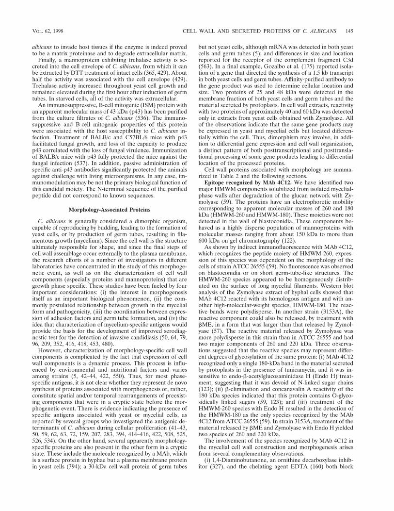
albicans to invade host tissues if the enzyme is indeed provedto be a matrix proteinase and to degrade extracellular matrix.
Finally, a mannoprotein exhibiting trehalase activity is se-creted into the cell envelope of C. albicans, from which it canbe extracted by DTT treatment of intact cells (365, 429). Abouthalf the activity was associated with the cell envelope (429).Trehalase activity increased throughout yeast cell growth andremained elevated during the first hour after induction of germtubes. In starved cells, all of the activity was extracellular.
An immunosuppressive, B-cell mitogenic (ISM) protein withan apparent molecular mass of 43 kDa (p43) has been purifiedfrom the culture filtrates of C. albicans (536). The immuno-suppressive and B-cell mitogenic properties of this proteinwere associated with the host susceptibility to C. albicans in-fection. Treatment of BALB/c and C57BL/6 mice with p43facilitated fungal growth, and loss of the capacity to producep43 correlated with the loss of fungal virulence. Immunizationof BALB/c mice with p43 fully protected the mice against thefungal infection (537). In addition, passive administration ofspecific anti-p43 antibodies significantly protected the animalsagainst challenge with living microorganisms. In any case, im-munomodulation may be not the primary biological function ofthis candidal moiety. The N-terminal sequence of the purifiedpeptide did not correspond to known sequences.
Morphology-Associated Proteins
C. albicans is generally considered a dimorphic organism,capable of reproducing by budding, leading to the formation ofyeast cells, or by production of germ tubes, resulting in fila-mentous growth (mycelium). Since the cell wall is the structureultimately responsible for shape, and since the final steps ofcell wall assemblage occur externally to the plasma membrane,the research efforts of a number of investigators in differentlaboratories have concentrated in the study of this morphoge-netic event, as well as on the characterization of cell wallcomponents (especially proteins and mannoproteins) that aregrowth phase specific. These studies have been fueled by fourimportant considerations: (i) the interest in morphogenesisitself as an important biological phenomenon, (ii) the com-monly postulated relationship between growth in the mycelialform and pathogenicity, (iii) the coordination between expres-sion of adhesion factors and germ tube formation, and (iv) theidea that characterization of mycelium-specific antigens wouldprovide the basis for the development of improved serodiag-nostic test for the detection of invasive candidiasis (50, 64, 79,96, 209, 352, 416, 418, 453, 480).
However, characterization of morphology-specific cell wallcomponents is complicated by the fact that expression of cellwall components is a dynamic process. This process is influ-enced by environmental and nutritional factors and variesamong strains (5, 42–44, 422, 550). Thus, for most phase-specific antigens, it is not clear whether they represent de novosynthesis of proteins associated with morphogenesis or, rather,constitute spatial and/or temporal rearrangements of preexist-ing components that were in a cryptic state before the mor-phogenetic event. There is evidence indicating the presence ofspecific antigens associated with yeast or mycelial cells, asreported by several groups who investigated the antigenic de-terminants of C. albicans during cellular proliferation (41–43,50, 59, 62, 63, 72, 159, 207, 283, 394, 414–416, 422, 508, 525,526, 534). On the other hand, several apparently morphology-specific proteins are also present in the other form in a crypticstate. These include the molecule recognized by a MAb, whichis a surface protein in hyphae but a plasma membrane proteinin yeast cells (394); a 30-kDa cell wall protein of germ tubes
but not yeast cells, although mRNA was detected in both yeastcells and germ tubes (5); and differences in size and locationreported for the receptor of the complement fragment C3d(563). In a final example, Gozalbo et al. (175) reported isola-tion of a gene that directed the synthesis of a 1.5 kb transcriptin both yeast cells and germ tubes. Affinity-purified antibody tothe gene product was used to determine cellular location andsize. Two proteins of 25 and 48 kDa were detected in themembrane fraction of both yeast cells and germ tubes and thematerial secreted by protoplasts. In cell wall extracts, reactivitywith two proteins of approximately 40 and 60 kDa was detectedonly in extracts from yeast cells obtained with Zymolyase. Allof the observations indicate that the same gene products maybe expressed in yeast and mycelial cells but located differen-tially within the cell. Thus, dimorphism may involve, in addi-tion to differential gene expression and cell wall organization,a distinct pattern of both posttranscriptional and posttransla-tional processing of some gene products leading to differentiallocation of the processed proteins.
Cell wall proteins associated with morphology are summa-rized in Table 2 and the following sections.
Epitope recognized by MAb 4C12. We have identified twomajor HMWM components solubilized from isolated mycelial-phase walls after degradation of the glucan network with Zy-molyase (59). The proteins have an electrophoretic mobilitycorresponding to apparent molecular masses of 260 and 180kDa (HMWM-260 and HMWM-180). These moieties were notdetected in the wall of blastoconidia. These components be-haved as a highly disperse population of mannoproteins withmolecular masses ranging from about 150 kDa to more than600 kDa on gel chromatography (122).
As shown by indirect immunofluorescence with MAb 4C12,which recognizes the peptide moiety of HMWM-260, expres-sion of this species was dependent on the morphology of thecells of strain ATCC 26555 (59). No fluorescence was observedon blastoconidia or on short germ-tube-like structures. TheHMWM-260 species appeared to be homogeneously distrib-uted on the surface of long mycelial filaments. Western blotanalysis of the Zymolyase extract of hyphal cells showed thatMAb 4C12 reacted with its homologous antigen and with an-other high-molecular-weight species, HMWM-180. The reac-tive bands were polydisperse. In another strain (3153A), thereactive component could also be released, by treatment withbME, in a form that was larger than that released by Zymol-yase (57). The reactive material released by Zymolyase wasmore polydisperse in this strain than in ATCC 26555 and hadtwo major components of 260 and 220 kDa. Three observa-tions suggested that the reactive species may represent differ-ent degrees of glycosylation of the same protein: (i) MAb 4C12recognized only a single 180-kDa band in the material secretedby protoplasts in the presence of tunicamycin, and it was in-sensitive to endo-b-acetylglucosaminidase H (Endo H) treat-ment, suggesting that it was devoid of N-linked sugar chains(123); (ii) b-elimination and concanavalin A reactivity of the180 kDa species indicated that this protein contains O-glyco-sidically linked sugars (59, 123); and (iii) treatment of theHMWM-260 species with Endo H resulted in the detection ofthe HMWM-180 as the only species recognized by the MAb4C12 from ATCC 26555 (59). In strain 3153A, treatment of thematerial released by bME and Zymolyase with Endo H yieldedtwo species of 260 and 220 kDa.
The involvement of the species recognized by MAb 4C12 inthe mycelial cell wall construction and morphogenesis arisesfrom several complementary observations.
(i) 1,4-Diaminobutanone, an ornithine decarboxylase inhib-itor (327), and the chelating agent EDTA (160) both block
VOL. 62, 1998 CELL WALL AND SECRETED PROTEINS OF C. ALBICANS 145

normal germ tube formation in C. albicans without affectingthe overall cell growth and protein synthesis. Although theremay be other effects, these compounds appear to inhibit thesynthesis and export, respectively, of the wall mannoproteinrecognized by MAb 4C12.
(ii) A C. albicans yeast monomorphic mutant lacks this my-celium-specific epitope (128). Treatment of cells with cysta-mine, an inhibitor of glutamyltransferases postulated to beinvolved in the formation of covalent bonds between cell wallproteins, almost completely blocked the incorporation of thespecies containing the epitope recognized by MAb 4C12 intothe nascent walls of regenerating protoplasts but did not inhibittheir release into the medium (456).
(iii) Fab fragments prepared from MAb 4C12, an immuno-globulin G1 (IgG1) subclass antibody, inhibited germ tubeformation (62). Binding of Fab fragments may alter the nativeconfiguration of the 260-kDa mannoprotein, causing its ab-normal and irregular deposition in the cell wall as revealed byindirect immunofluorescence. Intact MAb 4C12 did not in-hibit germ tube formation, and indirect immunofluorescenceshowed confluent and homogeneous binding of the antibody tothe surface of mycelial cells. In this context, Fab fragmentsfrom a MAb directed against the peptide portion of a cellsurface specific glycoprotein of Dictyostelium discoideum inhib-ited the initial stages of the cell differentiation process in this
slime mold (506). Similarly, the yeast-to-mycelium transition inC. albicans can also be envisaged as a cell differentiation pro-cess. These observations support the contention that HMWMare not mere passive components of the cell wall but may playan important and active morphogenetic role in modulating theorganization of the remainder of the cell wall constituents toobtain the final supramolecular structure specific for each mor-phology (327, 479–482).
Epitope recognized by MAb 3D9. Protein from a germ tubeculture purified by affinity chromatography on fibrinogen wasused as immunogen in the production of MAbs. One of theseantibodies, MAb 3D9 of the IgM class, reacted only with germtube surfaces as judged by indirect immunofluorescence (324).The fluorescence was homogeneous along the germ tube.However, after 48 h of growth, the fluorescence was less in-tense and heterogeneous, with its greatest reactivity at the tip.This observation suggested that expression was greater at thepoint of active growth. Whether the diminution of reactivitybehind the growing tip was a result of shedding of the deter-minant or alterations and rearrangements in the mature hyphalwall is unclear. While the presumed presence of the determi-nant in the culture medium points to the first explanation, thepossibility that excess antigen is secreted to the exocellularenvironment during growth can not be eliminated. Westernblot analysis of EDTA-bME and Zymolyase extracts from
TABLE 2. Morphology-associated and other cell wall proteins
Protein Gene Location Commentsa Reference(s)
Morphology associated4C12 antigen (180
kDa, 260 kDa)Hyphae Structural protein? 59, 62, 122, 128, 160, 327
3D9 antigen (110–170 kDa)
Hyphae Structural protein? 324, 325
DC3:H10 antigen Hyphae Present in complex with otherproteins
87, 300
Hwp1p (34 kDa) HWP1 Hyphae N-terminal signal sequence, tandemrepeats, C terminus ST rich
513
Hyr1p (deduced94 kDa)
HYR1 Hyphae N-terminal signal sequence, Cterminus ST rich, GPI anchorsequence
16
Cell wall protein(30 kDa)
Hyphal cell wall, yeast? Probable posttranslation regulationof location
5
MAb determinants 106Yeast cell (68, 82,
96, 104 kDa)Yeast cells and early germ
tubesProteins have one or two common
epitopesHyphae (104, 117
kDa)Germ tubes and hyphal tip Proteins have one or two common
epitopes
OtherHsp70 (70 kDa) SSA1 (HSP70),
SSA2Yeast cells, hyphae Chaperone?, translocation?;
immunogenic, Ab protective?278, 294, 303
Hsp9047-kDa fragment HSP90 Yeast cells, hyphae? hsp90 degradation product,
immunogenic, Ab protective331, 334, 335, 337–340,
342, 532Enolase (48 kDa) ENO1-2 Yeast cells, hyphae Binds to glucan, internal cell wall,
lacks N-terminal signal sequence,N terminus blocked,immunogenic, antigen and Absmay be diagnostic, Abprotective?
8, 143, 177, 219, 221, 330,356, 420, 520–522, 556,558, 564
Phosphoglyceratekinase (40 kDa)
PGK1 Yeast cells, hyphae Lacks N-terminal signal sequence,enzymatic activity? immunogenic
6
GAPDH (33 kDa) TDH1 Yeast cells, hyphae Enzymatic activity, immunogenic 161ADH ADH1? Cell wall? Fibronectin binding? immunogenic 27, 406, 489Als1p ALS1 Cell wall? N-terminal signal sequence,
variable tandem repeat, GPI-anchor sequence, member family
212, 473
a Abbreviations: S, serine; T, threonine; Ab, antibody.
146 CHAFFIN ET AL. MICROBIOL. MOL. BIOL. REV.

yeast cells and germ tubes confirmed reactivity with germ tubemoieties. The reactive moiety was polydisperse. The determi-nant was larger than 210 kDa in the chemical extract andranged from 110 to 220 kDa in the Zymolyase extract. Thelarger size in extract containing reducing agent parallels thatobserved with the determinant of MAb 4C12, as discussedabove. The sensitivity of the determinant to proteases andinsensitivity to periodate indicated that the epitope was a pep-tide. Analysis by SDS-polyacrylamide gel electrophoresis(PAGE) of the protein purified from Zymolyase extracts ofgerm tubes revealed a single disperse band of 110 to 170 kDathat was reactive with concanavalin A (325). The final productwas enriched 126-fold for the protein and 16-fold for carbohy-drate. MAb 3D9 did not react with Rhodotorula or Saccharo-myces or any of 40 isolates distributed among six other Candidaspecies (324). Reactivity was observed with all 58 isolates of C.albicans examined. The determinant appears to be specific toC. albicans and the hyphal form.
Hwp1p. HWP1 (hyphal wall protein) was cloned by screeninga cDNA library with adsorbed antiserum that reacted only withhyphal surfaces (513). Further analysis showed that the ORFencoded a 234-amino-acid sequence with a calculated molec-ular mass of 22,750 Da containing a typical signal sequence andsignal site for yeast KEX2-encoded endoprotease. More inter-estingly, the sequence revealed a series of tandem repeatscontaining a 10-amino-acid motif rich in proline and glutaminethat covered much of the remaining sequence. No consensusN-glycosylation sites were found. The C terminus was rich inserine and threonine, providing potential O-glycosylation sites.A 2.3-kb mRNA was detected only under growth conditionswhere hyphae were produced. Antiserum raised to the recom-binant protein reacted only with hyphal surfaces. Similar ex-pression of Hwp1p was detected on the surface of 20 other C.albicans strains. The antiserum also reacted with fungi in tissuesections of colonized mice. Again, only hyphal surfaces werereactive. Western blot analysis of Zymolyase extracts of hyphalforms showed a pattern of polydisperse reactivity at approxi-mately 34 kDa. No reactivity was detected with extracts ofyeast cells. A recombinant protein produced in Pichia pastoriswas about twice the calculated size. The authors suggest thatthe differences may be attributable to anomalous migration ofproline-rich proteins, glycosylation, and protease activity ofZymolyase. As a proline-rich protein, Hwp1p may share prop-erties with similar proteins in which proline residues are pro-posed function to maintain polypeptide chains in extendedconformations and to mediate noncovalent interactions be-tween chains. As noted by the authors, salivary proline-richproteins are found in this group. These proteins are a substratefor transglutaminase of buccal epithelial cells to which C. al-bicans can adhere. This protein on candidal surfaces may alsobe a substrate. The repeats also contain cysteine that may beinvolved in covalent linkages.
Hyr1p. A hyphally regulated gene, HYR1, was isolated andcharacterized (16). Northern analysis confirmed that the 3-kbtranscript of the gene was expressed under several conditionsfavoring formation of germ tubes but not under yeast growthconditions. The ORF encoded a predicted 937-amino-acidpolypeptide of 94.1 kDa. The sequence contained an N-termi-nal signal sequence and C-terminal sequence for attachment ofa GPI anchor. The sequence also contained 17 N-glycosylationsites and a domain rich in serine and threonine. Another do-main was rich in serine, glycine, and asparagine and containedseven copies of a 4-amino-acid motif, NEGS, of unknownfunction. The presence of an N-terminal signal sequence, aC-terminal GPI signal sequence, and a serine- and threonine-rich region is shared with several other sequences that are
associated with the S. cerevisiae cell wall (Cwp1p, Cwp2p,Tip1p, Aga1p, Aga1p, and Flo1p) (291, 444, 538, 555) ormembrane sequences (388). Other C. albicans sequences havesimilar structural characteristics: the putative cell wall proteinAls1p (discussed in a subsequent section) (212), the proteinencoded by clone 8M (477), membrane protein Phr1p (467),and possibly the putative Hyr1-like protein associated withHYR99 (473).
A mutant strain containing disruptions of both alleles ofHYR1 showed no obvious phenotype in yeast growth, germtube formation, sensitivity to caffeine or calcofluor white, oraberrant forms microscopically. Although the gene is differen-tially expressed in germ tubes, expression of the gene as afusion protein under the alcohol dehydrogenase promoter inyeast cells did not result in an apparent change in the yeastphenotype or in formation of germ tubes. Although the functionof this protein is not known, the authors suggest that it may playa structural role. The lack of phenotype of the mutant is notinconsistent with this possibility, since the protein may be nones-sential or have a function that overlaps with another protein.
Immunomodulatory 65-kDa Mannoprotein (MP65)Cassone and colleagues (45, 65, 169, 542, 543) have studied
the main mannoprotein components of C. albicans implicatedin immunomodulation of host defense. Among the mannopro-teins present in an acidic extract (MP-F2), a 65-kDa manno-protein (MP65) is a main target of human T-cell response. Theproliferative response stimulated by this component was of anantigenic nature rather than a mitogenic one, and the responsewas targeted mainly to polypeptide epitopes (65, 542). A sim-ilar constituent was present in the material released to theculture medium by C. albicans (45, 543). MP65 was furtherpurified by immunoaffinity chromatography with MAbs gener-ated against putative protein epitopes of the molecule (169).MP65 has a pI of 4.1 and a protein-to-polysaccharide ratio of1.8:1. The moiety appears to contain only O-linked sugar res-idues, as deduced from its sensitivity to dilute alkali but not toseveral endoglycosidases. Nanogram doses of purified MP65induced extensive T-cell proliferation of human peripheralblood mononuclear cells.
Heat Shock ProteinsOne of the most highly evolutionarily conserved features of
living organisms is the ability to respond to a sudden change oftemperature (heat shock response) and to other adverse envi-ronmental conditions. This response involves the increasedproduction of a set of proteins collectively referred to as heatshock proteins (hsps), whose production presumably contrib-utes to the protection and damage repair of cells followingstress (312). Initially identified as proteins whose synthesis wasenhanced by an increase in temperature, several of the majorhsps have been described since then to play important roles inall major growth-related processes, such as cell division, DNAsynthesis, transcription, translation, protein folding and trans-port, and membrane translocation (312). Several hsps are syn-thesized constitutively, reflecting the important cellular func-tions performed by these proteins under nonstress conditions.hsps are also immunodominant antigens and major targets ofhost immune response during different types of infection (240,241, 323, 332, 581). The response to hsps may also be involvedin autoimmunity (241, 332, 582). Several families of hsps existwhich are designated according to their average apparent mo-lecular mass (e.g., hsp90, hsp70, and hsp60). Some of thesefamilies are divided into different subfamilies, and each sub-family may have different members.
VOL. 62, 1998 CELL WALL AND SECRETED PROTEINS OF C. ALBICANS 147

The response of C. albicans to heat shock includes the syn-thesis of a set of proteins with an ample array of molecularmasses (588). Genes coding for proteins belonging to the hsp90and hsp70 families have been found in C. albicans (131, 278,303, 316, 335, 532). Interestingly, the cellular location of someof these gene products is not confined to the cytosolic com-partment, since they are also present in the cell wall and at thecell surface (303, 340). This extracellular localization could bea common feature of several fungal hsps, since it has beendescribed also for Histoplasma capsulatum (168) and S. cerevi-siae (294). As components of fungal cell walls, hsps may playroles similar to those described in the cytoplasm.
hsp90. The structure of hsp90 is highly conserved in organ-isms ranging from bacteria to humans. Members of the hsp90family of proteins play important roles in protein biogenesisand have been referred to as molecular chaperones. hsp90sinteract with a variety of cellular proteins, such as steroidhormone receptors (410) and tyrosine kinases (95), and havebeen associated with morphological changes in pathogenicfungi and protozoa (322, 488, 554). S. cerevisiae contains twogenes encoding hsp90, HSC82 and HSP82. HSC82 is a consti-tutively expressed gene only weakly induced upon stress expo-sure, while HSP82 is expressed at a much lower basal level andis strongly activated after exposure to heat shock (32).
In C. albicans, an immunodominant 47-kDa antigen wasidentified as a heat-stable breakdown product of hsp90 (335).This 47-kDa component was first identified as one of the majortargets of the immune response mounted by patients withsystemic candidiasis (334, 335) and is different from enolase(143, 329, 517). C. albicans hsp90 or its fragments circulate inthe body fluids of patients with disseminated candidiasis. The47-kDa antigen was isolated from patient sera by affinity chro-matography (338), and detection of antigenemia has potentialdiagnostic value (334). Circulating fungal hsp90 may also playa role in pathogenesis (332, 336). Antibodies to the 47-kDaantigen correlated with survival to infection both in humansand in animal models (339, 342). Antibody to the 47-kDaantigen was present in the majority of patients with chronicmucocutaneous candidiasis and AIDS (47, 339), and it hasbeen suggested that it mediates the protective effect of hu-moral immunity in systemic candidiasis (339, 342). Epitopemapping showed that patients recovering from systemic can-didiasis produce antibodies against both fungus-specific andconserved epitopes of hsp90 (342). In particular, a highly con-served epitope, LKVIRK, was recognized by all patients withantibody to the 47-kDa antigen (341), and both patient seraand a murine MAb raised to this epitope when given prophy-lactically reduced mortality in a mouse model of invasive can-didiasis (342). Treatment with a human recombinant antibodyfragment to the same epitope also reduced the mortality ofinfected mice and improved renal function in sublethally chal-lenged mice (331). The authors proposed that the fungal hsp90may bind to host proteins, thereby interfering with host proteinstructure or function, and that antibody may neutralize theability of the fungal hsp90 to bind to host proteins.
Matthews et al. used an affinity-purified monospecific anti-body against the 47-kDa antigen and immunoelectron micros-copy techniques to study the subcellular localization of thismolecule (340). Results showed that it was present in thecytoplasm and the cell wall of both yeast and mycelial cells ofC. albicans. This extracellular location implies that the 47-kDaantigen is naturally exposed and readily available to antibodiesproduced by the host, without the necessity for cellular break-age. This extracellular location may also be responsible for itshigh antigenicity and some of the other properties (mainly
immunoprotection) associated with the immune response tothis molecule.
More recently, a full-length C. albicans HSP90 gene wasisolated by screening cDNA and genomic libraries with a probederived from the S. cerevisiae homolog HSP82 (532). The pre-dicted mass of C. albicans hsp90 was 81 kDa, and the C-terminal portion was identical to the previously described 47-kDa fragment (334). Expression of HSP90 was regulatedduring growth and morphological transition and after heatshock. Southern blotting analysis revealed only one HSP90locus (532). The inability to isolate an homozygous null mutantis consistent with the notion that a single HSP90 locus exists inC. albicans and that hsp90 is essential for viability.
hsp70. hsp70s are highly conserved in organisms rangingfrom bacteria to humans. The DnaK chaperones are the pro-karyotic homologues of hsp70s (437), having about 50% iden-tity to all eukaryotic hsp70s. The eukaryotic proteins are be-tween 50 and 98% identical. Sequence similarity betweenhsp70 proteins extends over the entire protein, but particularlyhighly conserved regions are present in the N-terminal domain(30, 89). The hsp70s are not restricted to heat shock protec-tion; they also play a role in protein folding, translocation ofproteins across membranes, and gene regulation (90, 91, 155,156, 431). In S. cerevisiae, at least 10 HSP70-like genes havebeen described and classified into five families from SSA toSSE (288, 374, 440, 502). Most of the proteins encoded bythese genes are present at considerable levels under all growthconditions, although their synthesis is increased after exposureto stress. The SSA family includes four members, SSA1 toSSA4, with 80 to 97% DNA sequence similarity (218). Cellviability requires moderate to high levels of at least one of theproteins encoded by this family (567). Under normal growthconditions, both SSA1 and SSA2 genes were expressed at mod-erate levels, while heat shock resulted in increased expressionof SSA1 and strong induction of both SSA3 and SSA4 expres-sion (218). Inactivation of either SSA1 or SSA2 did not resultin an obvious phenotype. However, ssa1 ssa2 double mutantswere unable to form colonies at 37°C (88). They were viableand grew more slowly than the parental strain at temperatureslower than 37°C as a result of a high expression of SSA4 (567).Until recently, the gene products of the SSA subfamily of hsp70have been considered to be located exclusively in the cytosol ofS. cerevisiae (91, 312). However, we have described the pres-ence of Ssa1p and Ssa2p in the cell wall of this budding yeast,where they could play a similar role to that in the cytoplasm(294).
In C. albicans, three different genes encoding members ofthe hsp70 protein family have been described (131, 278, 303,316). By screening a C. albicans cDNA library with antibodiesto whole fungal cells, La Valle et al. (278) isolated a cloneencoding a stress protein of C. albicans whose deduced aminoacid sequence was 84% similar to the product of S. cerevisiaeSSA1. As expected, Southern blots of digested DNA probedwith the cloned cDNA under low-stringency conditions indi-cated the presence of other members of the HSP70 gene familyin C. albicans. Confirming this observation, antiserum raised tothe recombinant protein recognized two bands in C. albicanswhole cell extracts with apparent molecular masses of about 70kDa. Northern blot experiments with the cloned cDNA as aprobe showed the presence of 2.4- and 2.2-kb hybridizationbands. Expression of the 2.4-kb constituent was heat inducible.
A second member of the hsp70 family of proteins was iden-tified by our group (303). The nucleotide and deduced aminoacid sequences of a cDNA clone coding for a constitutivelyexpressed 70-kDa cell wall protein (5) showed a high degree ofhomology and identity to the SSA1 and SSA2 sequences of S.
148 CHAFFIN ET AL. MICROBIOL. MOL. BIOL. REV.

cerevisiae. The relationship with the SSA family was furthersupported by the reactivity of the 70-kDa candidal componentwith antibody recognizing the Ssa proteins of S. cerevisiae.Thus, to reflect the existence of multiple members of thisfamily in C. albicans, and in accordance with the nomenclatureof S. cerevisiae, we proposed that the original HSP70 designa-tion of La Valle et al. (278) be replaced with SSA1 and that thegene corresponding to the second member of this family benamed SSA2.
Several lines of evidence demonstrated that a member(s) ofthe hsp70 family of proteins is present in the cell wall of C.albicans (303). Affinity-purified antibody to the Ssa2 fusionprotein produced by the cloned cDNA fragment identified a70-kDa moiety in the bME extracts containing cell wall com-ponents from both morphologies of the organism. Indirectimmunofluorescence demonstrated its presence at the cell sur-face. Immunoelectron microscopy showed that it was presentin both the cytoplasm and the cell wall. Biotinylation of intactcells with a derivative that did not permeate the cell membranedemonstrated that the 70-kDa protein was present outside thecell membrane before extraction. Furthermore, and confirmingthe results obtained with C. albicans, members of the hsp70family of proteins have been found in the cell wall of S. cer-evisiae (294).
Members of the hsp70 family are highly immunogenic pro-teins and are major targets of the host immune response todifferent pathogenic microorganisms, including pathogenicfungi (240, 241, 323). La Valle et al. (278) reported that serafrom three healthy individuals contained antibodies to C. albi-cans Ssa1p and suggested that presence of such antibodiescould contribute to protection against infection. It was alsopointed out that the variability in this generally conservedprotein was predominantly in the C-terminal region, which isalso the immunodominant region (241). Moreover, prelimi-nary experiments suggested that recombinant Ssa1p inducesT-lymphocyte proliferation. In the case of the product of C.albicans SSA2, serum samples from both normal individualsand patients with candidiasis contained antibodies against theC-terminal portion of Ssa2p (303). hsp70 is also recognized bysera of patients with oral and esophageal infection (524). It hasalso been demonstrated that the antibody response in a murinemodel of systemic candidiasis includes reactivity against a 75-kDa hsp (85). Due to the high levels of homology between thedifferent hsp70s, a combination of antibodies to the differentmembers of this family in C. albicans as well as to hsp70s fromother microorganisms may be present in the host serum. Thesereactivity patterns are also likely to be the result of combina-tion of antibodies against conserved epitopes and epitopesunique to individual C. albicans hsps.
In any case, the ubiquitous nature of hsps, along with theirhigh degree of homology, poses interesting challenges to theimmune system of the host. First, the presence of epitopesshared by a number of infectious agents may provide the im-mune system with a universal signal for infection, and antibod-ies to these conserved regions could provide some naturalresistance to infection, somewhat in between innate and ac-quired immunity. Second, epitopes shared by the parasite andthe host may trigger deleterious autoimmune responses (240).This could also be true for immune responses against highlyconserved immunodominant glycolytic enzymes (see below).
Heat shock or stress mannoproteins. Heat shock manno-proteins with approximate molecular masses of 180 to 200, 130to 150, 90 to 110, and 60 to 70 kDa have been identified asbeing involved in the secretory immune response during mu-cosal candidiasis (413, 417). These molecules were expressedafter a temperature shift from 25 to 37°C and were detected by
indirect immunofluorescence. The antigenic determinants rec-ognized by secretory IgAs in saliva and vaginal washings werepolysaccharide in nature (413). Subculturing on agar at 25°Cswitched off expression of the antigens from mucosal isolates,while several transfers were required to reduce expression byisolates from deep infections (417). Since factors other thantemperature can influence the in vitro and in vivo expression,these cell wall components should be considered “stress” pro-teins. The 200-kDa component appeared to be the main com-ponent recognized by vaginal secretory IgA. The 180- to 200-kDa component also enhanced tumor necrosis factor secretionby a murine macrophage cell line (411).
Glycolytic Enzymes
Glycolytic enzymes, which are highly conserved through evo-lution, appear to be important during C. albicans pathogenesis,since they act as major inducers of host immune responses andare major allergens during candidiasis (161, 219, 221, 489, 517,530). Glycolytic enzymes can constitute up to 30% of totalsoluble proteins in S. cerevisiae (201). The presence of glyco-lytic enzymes in the cell wall of C. albicans has been reportedrecently. Enolase was found to be associated with glucan in theinner layers of the cell wall (8). Another report localized thisenzyme on the surface of C. albicans cells, probably due toadventitious binding of enolase released from lysed cells (132).Phosphoglycerate kinase (PGK) was found in the cell wall andat the surface (6). We have also recently reported the presenceof a cell wall-bound form of GAPDH (161). The presence ofalcohol dehydrogenase (ADH) in the cell wall also has beensuggested (406) as the fibronectin receptor. Together, localiza-tion of presumably “intracellular” glycolytic enzymes in the cellwall of C. albicans is rather intriguing, as are the possible rolesthat these molecules could play as part of the cell wall (e.g.,energetic role, antigenicity, surface receptors), as well as themechanisms involved in their secretion. However, the presenceof glycolytic enzymes on microbial surfaces is not unprece-dented. GAPDH has been reported as a major surface proteinon group A streptococci (Streptococcus pyogenes), where it notonly is enzymatically active but also serves as a binding proteinfor fibronectin, lysozyme, myosin, actin, and plasmin (403,572). In Kluyveromyces marxianus, the same enzyme proteinhas been identified as a constitutive protein of the cell wall, andits level increases substantially upon induction of flocculation(136). ADH is also a surface protein of Entamoeba histolytica,where it has been reported to display receptor activities for anumber of extracellular matrix (ECM) components (579).PGK, GAPDH, and triose-phosphate isomerase have beenfound on the surface of Schistosoma mansoni (173, 280, 503)and have been suggested as potential candidates for vaccinedevelopment.
Enolase. Enolase (2-phospho-D-glycerate hydrolyase) is anenzyme of the glycolytic pathway, where it catalyzes the dehy-dration of 2-phosphoglycerate to phosphoenolpyruvate. It alsocatalyzes the reverse reaction during gluconeogenesis. It isamong the most abundant proteins in C. albicans and S. cer-evisiae (201). In S. cerevisiae, enolase is encoded by two geneswhose expression is differentially regulated depending on thecarbon source and growth phase (343) and is coordinatelyregulated with the expression of other glycolytic enzymes(314). Different groups have reported the cloning and charac-terization of the C. albicans enolase gene (143, 330, 521).Although only one gene was initially detected by Southernanalysis (143, 330), further experiments demonstrated thepresence of two enolase gene loci (420). However, it is notknown whether both loci provide functional genes.
VOL. 62, 1998 CELL WALL AND SECRETED PROTEINS OF C. ALBICANS 149
ibqfu_000
Resaltado
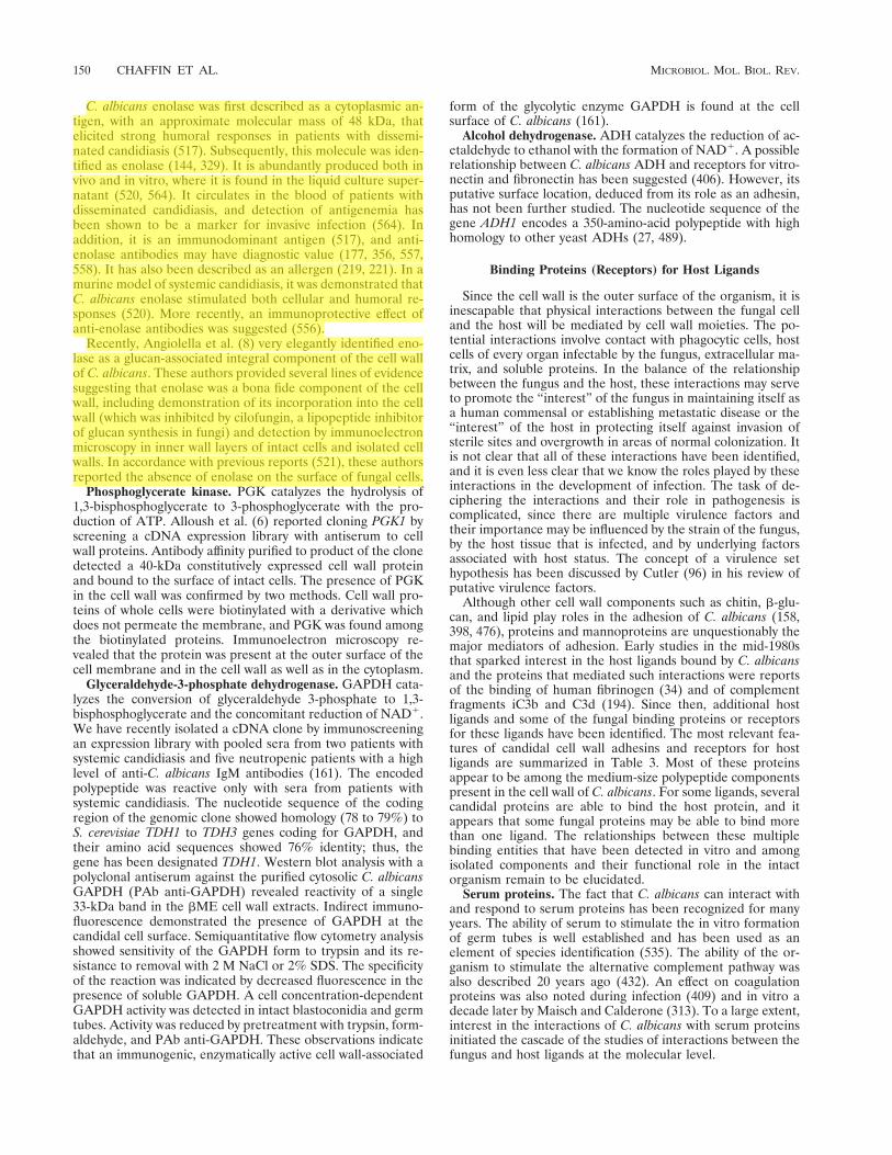
C. albicans enolase was first described as a cytoplasmic an-tigen, with an approximate molecular mass of 48 kDa, thatelicited strong humoral responses in patients with dissemi-nated candidiasis (517). Subsequently, this molecule was iden-tified as enolase (144, 329). It is abundantly produced both invivo and in vitro, where it is found in the liquid culture super-natant (520, 564). It circulates in the blood of patients withdisseminated candidiasis, and detection of antigenemia hasbeen shown to be a marker for invasive infection (564). Inaddition, it is an immunodominant antigen (517), and anti-enolase antibodies may have diagnostic value (177, 356, 557,558). It has also been described as an allergen (219, 221). In amurine model of systemic candidiasis, it was demonstrated thatC. albicans enolase stimulated both cellular and humoral re-sponses (520). More recently, an immunoprotective effect ofanti-enolase antibodies was suggested (556).
Recently, Angiolella et al. (8) very elegantly identified eno-lase as a glucan-associated integral component of the cell wallof C. albicans. These authors provided several lines of evidencesuggesting that enolase was a bona fide component of the cellwall, including demonstration of its incorporation into the cellwall (which was inhibited by cilofungin, a lipopeptide inhibitorof glucan synthesis in fungi) and detection by immunoelectronmicroscopy in inner wall layers of intact cells and isolated cellwalls. In accordance with previous reports (521), these authorsreported the absence of enolase on the surface of fungal cells.
Phosphoglycerate kinase. PGK catalyzes the hydrolysis of1,3-bisphosphoglycerate to 3-phosphoglycerate with the pro-duction of ATP. Alloush et al. (6) reported cloning PGK1 byscreening a cDNA expression library with antiserum to cellwall proteins. Antibody affinity purified to product of the clonedetected a 40-kDa constitutively expressed cell wall proteinand bound to the surface of intact cells. The presence of PGKin the cell wall was confirmed by two methods. Cell wall pro-teins of whole cells were biotinylated with a derivative whichdoes not permeate the membrane, and PGK was found amongthe biotinylated proteins. Immunoelectron microscopy re-vealed that the protein was present at the outer surface of thecell membrane and in the cell wall as well as in the cytoplasm.
Glyceraldehyde-3-phosphate dehydrogenase. GAPDH cata-lyzes the conversion of glyceraldehyde 3-phosphate to 1,3-bisphosphoglycerate and the concomitant reduction of NAD1.We have recently isolated a cDNA clone by immunoscreeningan expression library with pooled sera from two patients withsystemic candidiasis and five neutropenic patients with a highlevel of anti-C. albicans IgM antibodies (161). The encodedpolypeptide was reactive only with sera from patients withsystemic candidiasis. The nucleotide sequence of the codingregion of the genomic clone showed homology (78 to 79%) toS. cerevisiae TDH1 to TDH3 genes coding for GAPDH, andtheir amino acid sequences showed 76% identity; thus, thegene has been designated TDH1. Western blot analysis with apolyclonal antiserum against the purified cytosolic C. albicansGAPDH (PAb anti-GAPDH) revealed reactivity of a single33-kDa band in the bME cell wall extracts. Indirect immuno-fluorescence demonstrated the presence of GAPDH at thecandidal cell surface. Semiquantitative flow cytometry analysisshowed sensitivity of the GAPDH form to trypsin and its re-sistance to removal with 2 M NaCl or 2% SDS. The specificityof the reaction was indicated by decreased fluorescence in thepresence of soluble GAPDH. A cell concentration-dependentGAPDH activity was detected in intact blastoconidia and germtubes. Activity was reduced by pretreatment with trypsin, form-aldehyde, and PAb anti-GAPDH. These observations indicatethat an immunogenic, enzymatically active cell wall-associated
form of the glycolytic enzyme GAPDH is found at the cellsurface of C. albicans (161).
Alcohol dehydrogenase. ADH catalyzes the reduction of ac-etaldehyde to ethanol with the formation of NAD1. A possiblerelationship between C. albicans ADH and receptors for vitro-nectin and fibronectin has been suggested (406). However, itsputative surface location, deduced from its role as an adhesin,has not been further studied. The nucleotide sequence of thegene ADH1 encodes a 350-amino-acid polypeptide with highhomology to other yeast ADHs (27, 489).
Binding Proteins (Receptors) for Host Ligands
Since the cell wall is the outer surface of the organism, it isinescapable that physical interactions between the fungal celland the host will be mediated by cell wall moieties. The po-tential interactions involve contact with phagocytic cells, hostcells of every organ infectable by the fungus, extracellular ma-trix, and soluble proteins. In the balance of the relationshipbetween the fungus and the host, these interactions may serveto promote the “interest” of the fungus in maintaining itself asa human commensal or establishing metastatic disease or the“interest” of the host in protecting itself against invasion ofsterile sites and overgrowth in areas of normal colonization. Itis not clear that all of these interactions have been identified,and it is even less clear that we know the roles played by theseinteractions in the development of infection. The task of de-ciphering the interactions and their role in pathogenesis iscomplicated, since there are multiple virulence factors andtheir importance may be influenced by the strain of the fungus,by the host tissue that is infected, and by underlying factorsassociated with host status. The concept of a virulence sethypothesis has been discussed by Cutler (96) in his review ofputative virulence factors.
Although other cell wall components such as chitin, b-glu-can, and lipid play roles in the adhesion of C. albicans (158,398, 476), proteins and mannoproteins are unquestionably themajor mediators of adhesion. Early studies in the mid-1980sthat sparked interest in the host ligands bound by C. albicansand the proteins that mediated such interactions were reportsof the binding of human fibrinogen (34) and of complementfragments iC3b and C3d (194). Since then, additional hostligands and some of the fungal binding proteins or receptorsfor these ligands have been identified. The most relevant fea-tures of candidal cell wall adhesins and receptors for hostligands are summarized in Table 3. Most of these proteinsappear to be among the medium-size polypeptide componentspresent in the cell wall of C. albicans. For some ligands, severalcandidal proteins are able to bind the host protein, and itappears that some fungal proteins may be able to bind morethan one ligand. The relationships between these multiplebinding entities that have been detected in vitro and amongisolated components and their functional role in the intactorganism remain to be elucidated.
Serum proteins. The fact that C. albicans can interact withand respond to serum proteins has been recognized for manyyears. The ability of serum to stimulate the in vitro formationof germ tubes is well established and has been used as anelement of species identification (535). The ability of the or-ganism to stimulate the alternative complement pathway wasalso described 20 years ago (432). An effect on coagulationproteins was also noted during infection (409) and in vitro adecade later by Maisch and Calderone (313). To a large extent,interest in the interactions of C. albicans with serum proteinsinitiated the cascade of the studies of interactions between thefungus and host ligands at the molecular level.
150 CHAFFIN ET AL. MICROBIOL. MOL. BIOL. REV.
ibqfu_000
Resaltado
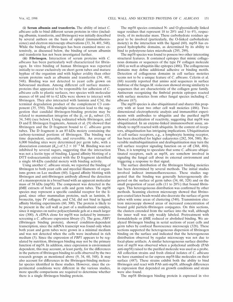
(i) Serum albumin and transferrin. The ability of intact C.albicans cells to bind different serum proteins in vitro (includ-ing albumin, transferrin, and fibrinogen) was initially describedby several authors on the basis of optical (immunofluores-cence) and electron microscopy observations (33, 34, 401, 548).While the binding of fibrinogen has been examined more ex-tensively, as discussed below, the binding of serum albuminand transferrin has not been investigated further.
(ii) Fibrinogen. Interaction of serum proteins with C.albicans has been particularly well characterized for fibrin-ogen. In vitro binding of human fibrinogen was reportedinitially to occur exclusively on short germ tubes and maturehyphae of the organism and with higher avidity than otherserum proteins such as albumin and transferrin (34, 401,548). Binding was not detected to yeast cells grown onSabouraud medium. Among different cell surface manno-proteins that appeared to be responsible for adhesion of C.albicans cells to plastic surfaces, two species with molecularmasses of 68 and 60 to 62 kDa were shown to interact withfibrinogen. They also interacted with laminin and C3d, theterminal degradation product of the complement C3 com-ponent (35, 550). This multiple interaction lead to the sug-gestion that candidal fibrinogen-binding proteins may berelated to mammalian integrins of the b2 or b3 subset (33,34, 548) (see below). Using iodinated whole fibrinogen, andD and E fibrinogen fragments, Annaix et al. (9) showed thatfibrinogen and the D fragment bound to C. albicans germtubes. The D fragment is an 85-kDa moiety containing thecarboxy-terminal portions of fibrinogen. The binding wastime dependent, saturable, and reversible. An average of6,000 binding sites were determined per germ tube, with adissociation constant (Kd) of 5.2 3 1028 M. Binding was notinhibited by several sugars, suggesting that the interactionwas not a lectin type of binding. Ligand affinity blotting of aDTT-iodoacetamide extract with the D fragment identifieda single 68-kDa candidal moiety with binding activity.
Using another C. albicans strain, we reported the binding offibrinogen to both yeast cells and mycelial filaments of organ-isms grown on Lee medium (60). Ligand affinity blotting withfibrinogen and anti-fibrinogen antibody allowed the detectionof a mannoprotein in a broad band with an apparent molecularmass of 58 kDa (mp58). The component was detected in thebME extracts of both yeast cells and germ tubes. The mp58species may represent a specific candidal receptor for the li-gand, since other mammalian proteins, such as laminin, fi-bronectin, type IV collagen, and C3d, did not bind in ligandaffinity blotting experiments (60, 300). The protein is likely tobe present in the cell wall as part of a multisubunit complex,since it migrates on native polyacrylamide gels at a much largersize (300). A cDNA clone for mp58 was isolated by immuno-screening a C. albicans expression library (5). The gene, FBP1(fibrinogen binding protein), showed condition-dependenttranscription, since the mRNA transcript was found only whenboth yeast and germ tubes were grown in a minimal mediumand was not detected when the cells were incubated in richmedium (5). Since the expression of FBP1 appears to be reg-ulated by nutrition, fibrinogen binding may not be the primaryfunction of mp58. In addition, since expression is environmentdependent, this may account, at least partly, for the differencesin the pattern of fibrinogen binding to cells observed by the tworesearch groups as mentioned above (9, 34, 60, 548). It mayalso account for differences in the fibrinogen-binding molecu-lar species identified in these studies. However, since the ex-perimental conditions were different in the various studies,more specific comparisons are required to determine whetherthere is a single fibrinogen binding protein.
The mp58 species contained N- and O-glycosidically linkedsugar residues that represent 18 to 20% and 3 to 4%, respec-tively, of its molecular mass. These carbohydrate residues ap-pear to be involved (particularly, the O-linked carbohydratemoiety) in the interaction with the ligand (60). mp58 has ex-posed hydrophobic domains, as determined by its ability tobind to polystyrene-latex microbeads (295, 299).
The mp58 species was found to possess two other interestingstructural features. It contained epitopes that mimic collage-nous domains or sequences of the type IV collagen molecule(484) as well as ubiquitin-like epitopes (486). The collageneousdomains may define additional adhesive or binding motifs.Detection of collagenous domains in cell surface moietiesseems not to be a unique feature of C. albicans. Celerin et al.(68) recently reported that amino acid sequences in surfacefimbriae of the fungus M. violaceum showed strong similarity tosequences that are characteristic of the collagen gene family.Antiserum recognizing the fimbrial protein epitopes reactedwith surface moieties from other fungal species including C.albicans (67).
The mp58 species is also ubiquitinated and shares this prop-erty with at least two other cell wall moieties (486). Two-dimensional electrophoretic analysis and immunoblot experi-ments with antibodies to ubiquitin and the purified mp58showed colocalization of reactivity, suggesting that mp58 wasubiquitinated. In an enzyme-linked immunosorbent assay, an-tibody to mp58 reacted with ubiquitin. In the context of recep-tors, ubiquitination has intriguing implications. Ubiquitinationof cell surface receptors, e.g., a lymphocyte homing receptor,has been described for higher eukaryotes (282, 504, 580). Re-versible multiubiquitination and deubiquitination may turn thecell surface receptor signaling function on or off (368, 404).Thus, it is tempting to speculate that some C. albicans ubiqui-tinated receptors, such as mp58, are somehow involved insignaling the fungal cell about its external environment andtriggering a response to that signal.
The surface distribution of the fibrinogen binding moietieshas been determined by several methods. The initial studiesinvolved indirect immunofluorescence. These studies sug-gested that the binding was generally heterogeneously dis-persed on the surface of germ tubes (34, 60, 328) and on thesmall proportion of yeast cells (11 to 20%) that bound fibrin-ogen. This heterogeneous distribution was confirmed by othermethods. Scanning electron microscopy showed that fibrino-gen-coated latex beads would also decorate the surface of germtubes with some areas of clustering (548). Transmission elec-tron microscopy showed areas of increased concentration ofbound gold particle-fibrinogen conjugates. On thin sections,the clusters extended from the surface into the wall, althoughthe inner wall was only weakly labeled. Pretreatment withformaldehyde or bME reduced or abolished binding. We an-alyzed fibrinogen binding in serial sections of yeast cells andgerm tubes by confocal fluorescence microscopy (328). Thesesections supported the heterogeneous dispersion of fibrinogenbinding on the surface and indicated that the heterogeneousdistribution observed by regular microscopy was not due tofocal-plane artifacts. A similar heterogeneous surface distribu-tion of mp58 was observed when a polyclonal antibody (PAbanti-mp58) raised to the purified molecule was used as a probe.All collection strains and fresh clinical isolates of C. albicanswe have examined so far express mp58-like molecules on theirsurface (487). These strains exhibit both the ability to bindfibrinogen and react with PAb anti-mp58, although differencesin expression that depended on growth conditions and strainwere also found.
The mp58 fibrinogen binding protein is expressed in vivo
VOL. 62, 1998 CELL WALL AND SECRETED PROTEINS OF C. ALBICANS 151
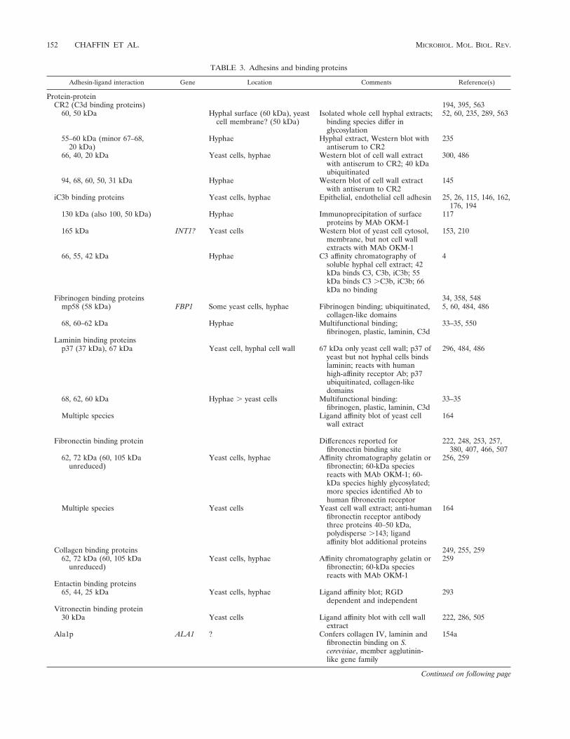
TABLE 3. Adhesins and binding proteins
Adhesin-ligand interaction Gene Location Comments Reference(s)
Protein-proteinCR2 (C3d binding proteins) 194, 395, 563
60, 50 kDa Hyphal surface (60 kDa), yeastcell membrane? (50 kDa)
Isolated whole cell hyphal extracts;binding species differ inglycosylation
52, 60, 235, 289, 563
55–60 kDa (minor 67–68,20 kDa)
Hyphae Hyphal extract, Western blot withantiserum to CR2
235
66, 40, 20 kDa Yeast cells, hyphae Western blot of cell wall extractwith antiserum to CR2; 40 kDaubiquitinated
300, 486
94, 68, 60, 50, 31 kDa Hyphae Western blot of cell wall extractwith antiserum to CR2
145
iC3b binding proteins Yeast cells, hyphae Epithelial, endothelial cell adhesin 25, 26, 115, 146, 162,176, 194
130 kDa (also 100, 50 kDa) Hyphae Immunoprecipitation of surfaceproteins by MAb OKM-1
117
165 kDa INT1? Yeast cells Western blot of yeast cell cytosol,membrane, but not cell wallextracts with MAb OKM-1
153, 210
66, 55, 42 kDa Hyphae C3 affinity chromatography ofsoluble hyphal cell extract; 42kDa binds C3, C3b, iC3b; 55kDa binds C3 .C3b, iC3b; 66kDa no binding
4
Fibrinogen binding proteins 34, 358, 548mp58 (58 kDa) FBP1 Some yeast cells, hyphae Fibrinogen binding; ubiquitinated,
collagen-like domains5, 60, 484, 486
68, 60–62 kDa Hyphae Multifunctional binding;fibrinogen, plastic, laminin, C3d
33–35, 550
Laminin binding proteinsp37 (37 kDa), 67 kDa Yeast cell, hyphal cell wall 67 kDa only yeast cell wall; p37 of
yeast but not hyphal cells bindslaminin; reacts with humanhigh-affinity receptor Ab; p37ubiquitinated, collagen-likedomains
296, 484, 486
68, 62, 60 kDa Hyphae . yeast cells Multifunctional binding:fibrinogen, plastic, laminin, C3d
33–35
Multiple species Ligand affinity blot of yeast cellwall extract
164
Fibronectin binding protein Differences reported forfibronectin binding site
222, 248, 253, 257,380, 407, 466, 507
62, 72 kDa (60, 105 kDaunreduced)
Yeast cells, hyphae Affinity chromatography gelatin orfibronectin; 60-kDa speciesreacts with MAb OKM-1; 60-kDa species highly glycosylated;more species identified Ab tohuman fibronectin receptor
256, 259
Multiple species Yeast cells Yeast cell wall extract; anti-humanfibronectin receptor antibodythree proteins 40–50 kDa,polydisperse .143; ligandaffinity blot additional proteins
164
Collagen binding proteins 249, 255, 25962, 72 kDa (60, 105 kDa
unreduced)Yeast cells, hyphae Affinity chromatography gelatin or
fibronectin; 60-kDa speciesreacts with MAb OKM-1
259
Entactin binding proteins65, 44, 25 kDa Yeast cells, hyphae Ligand affinity blot; RGD
dependent and independent293
Vitronectin binding protein30 kDa Yeast cells Ligand affinity blot with cell wall
extract222, 286, 505
Ala1p ALA1 ? Confers collagen IV, laminin andfibronectin binding on S.cerevisiae, member agglutinin-like gene family
154a
Continued on following page
152 CHAFFIN ET AL. MICROBIOL. MOL. BIOL. REV.

(302). Immunohistochemical techniques with the PAb anti-mp58 were applied to tissue specimens from patients withconfirmed systemic and mucocutaneous candidiasis. Mostyeast cells and hyphal filaments were strongly positive. Al-though the proportion of positive yeast cells was not deter-mined, there is a sense that this proportion may be higher thanthat observed in vitro (60, 328). The in vivo expression of mp58provides support for the contention that fibrinogen bindingmay be important in host interactions. In this context, theability of C. albicans cells to bind fibrinogen may be a viru-lence-related property, since coagulation proteins such as fi-brin appear to be the receptor for C. albicans cells that adhereto blood clots in vivo (34). Disseminated intravascular coagu-lation was reported to occur in parallel, in some instances, withsevere Candida septicemia (409). Moreover, binding to fi-bronectin that is believed to be one of the host ligands forCandida adhesion (51, 443) and binding of platelets to C.albicans cells appears to be mediated by fibrinogen (313, 439,547).
(iii) Complement fragment C3d. The ability of C. albicanscells to rosette antibody-sensitized erythrocytes coated withC3d as well as iC3b complement fragments was first describedby Heidenreich and Dierich (194) and latter reported by otherinvestigators (for a review of this topic, see reference 50).
A C3d-binding protein was purified from extracts of wholehyphal organisms by affinity chromatography. The purified re-ceptor consisted of a doublet of 60 and 66 to 68 kDa. However,the biological activity, i.e., binding of C3d, appeared to beassociated with the 60-kDa component that is expressed at thecell surface of hyphal elements (52, 289, 470, 563). Although it
was initially thought that C. albicans yeast cells did not expressC3d binding proteins, immunoelectron microscopy with aMAb to the purified C3d receptor revealed otherwise (235).An epitope cross-reacting with this MAb was detected on theplasma membrane of blastoconidia. During germination, it be-came associated with the cell wall, mostly at the cell surface ofthe organism. This component was a 50-kDa protein that, inaddition to the hyphal 60-kDa moiety, has been reported toexhibit major C3d binding activity (563). Both the purified 50-and 60-kDa moieties were able to inhibit rosetting of erythro-cytes coated with iC3b (90%) and C3d (50 to 64%) with hy-phae. Other authors have identified additional componentswith different molecular masses that exhibited antigenic cross-reactivity toward antibodies (PAb anti-C3d) to the major cellwall C3d binding protein previously characterized (52, 289). Inone study, a major component of 55 to 60 kDa and minorspecies of 67 to 68 and 20 kDa were detected in extracts fromhyphae (235). In another study, moieties of 66, 40, and 21 kDawere detected in bME extracts from blastoconidia and hyphae(300). In a study of parental and clotrimazole-tolerant clones,reactive species of 94, 68, 60, 50 and 31 kDa were detected inthe DTT extract of the parental strain (145). The differencesobserved in these studies could be due to one or more sources,including extraction procedures, strain and growth conditions,differences in glycosylation, and lack of specificity of the probeused for immunodetection. On the other hand PAb anti-C3dappeared to be a highly specific probe, since it did not cross-react with other well-characterized candidal cell surface recep-tors such as the mp58 fibrinogen binding moiety. This supportsthe contention of a separate identity for the mp58 moiety and
TABLE 3—Continued
Adhesin-ligand interaction Gene Location Comments Reference(s)
Protein-sugarFucose binding protein Yeast cells, hyphae? Buccal, vaginal epithelial cell
adhesin; strain variation37, 53, 92, 93, 345, 544
.15.7 kDa 53, 544GlcNAc binding protein Yeast cells, hyphae? Buccal, vaginal epithelial cell
adhesin; strain variation37, 53, 92, 93, 345, 544
190 kDa Yeast cells Cell wall extracts of galactose-but not glucose-grown cells;not known if is the same proteinin reference 345
129
Protein-glycosphingolipidFimbrial protein66 kDa?
Yeast cells, hyphae? Epithelial cell adhesin;adhesintope shared withP. aeruginosa pili
279, 583–586
Protein-presumed proteinSecreted aspartyl proteinase,
42–45 kDaSAP1-9 Yeast cells, hyphae Binding endothelial cells,
corneocytes147, 433; Table 1
Protein-plasticPlastic binding proteins Binding to many plastics,
environmental influences181, 243, 344, 458
60, 68, 200, .200 kDa Hyphae 60, 68 kDa may be multifunctionalbinding fibrinogen, laminin, C3d
33, 34, 549
30 kDa AAF1 Yeast cells, hyphae Bind polystyrene and epithelialcells
20, 21
Carbohydrate-proteinAcid labile mannan
b-1,2 mannotetraose Yeast cells, hyphae Mannose receptor, splenic, lymphnode macrophage
73, 285
Carbohydrate-unknownAcid-stable mannan
Factor 6 Yeast cells, hyphae BEC adhesion 357Unknown Yeast cells, hyphae Splenic, lymph node macrophage 234
VOL. 62, 1998 CELL WALL AND SECRETED PROTEINS OF C. ALBICANS 153

the C3d receptor (300). C3d binding protein is presumablypresent in the cell wall in association with itself or other pro-teins, since it is found in cell wall extracts in a form with anapparent molecular weight larger than that detected by SDS-PAGE (300, 470).
Both direct and indirect evidence indicate that the 50- and60-kDa species are mannoproteins that differ in the glycosyla-tion of a unique polypeptide (52, 289, 470, 563). Mannopro-teins obtained by concanavalin A affinity chromatography in-hibited rosetting. The two components present in DTT extractsreacted in blotting experiments with concanavalin A and weresensitive to periodate treatment. Differences in the carbohy-drate moiety were shown by the demonstration of differencesin reactivity with sugar-specific reagents (563). The 60-kDa,but not the 50-kDa, mannoprotein reacted with wheat germagglutinin. Endoglycosidase F and N-glycanase altered the 60-kDa species, with the generation of 40- to 45-kDa products(470, 563). Treatment with neuraminidase resulted in loss ofimmunoreactivity of the 60-kDa moiety, presumably due toloss of carbohydrate (563) or possibly contaminating proteaseactivity (as noted above). Treatments that targeted protein butnot those that targeted carbohydrate reduced rosetting. Thus,the carbohydrate moiety is unlikely to be involved in ligandbinding. This is in contrast to mp58 fibrinogen binding species,where O-linked mannose appear to be involved in ligand bind-ing (60).
One of the moieties that was recognized by the PAb anti-C3d receptor (300) also reacted with anti-ubiquitin antibody(486). This 40-kDa moiety, along with the mp58 molecule andthe candidal laminin receptor characterized by our group(296), is a major ubiquitinated protein in the cell wall (see thediscussion of mp58, above).
The relationship, if any, of the candidal and mammalianreceptors for C3d is unclear. Significant differences in the iso-electric point and amino acid composition have been reportedbetween the Candida 60-kDa C3d-binding protein and themammalian C3d receptor (CR2) (470). Both MAbs and PAbsdirected to CR2 failed to react directly with C. albicans cells(115, 117, 162). However, in one report, an anti-human CR2antibody was able to inhibit rosetting of C3d-coated erythro-cytes with C. albicans (115). In any case, due to the functionalsimilarities, the term “CR2” or “CR2-like” has also been usedfor the candidal C3d receptor (149, 563).
The candidal receptor for C3d is expressed in vivo (235).Immunoelectron microscopy was used to examine expressionby fungal cells in kidney tissue and peritoneal lavage fluid frominfected mice. Fungal cells in peritoneal lavage fluid showedintense staining of mother cells of germ tubes, germ tubes, andpseudohyphae. Staining of the parental yeast cells was greatestat the innermost layers of the cell wall and minimal at thesurface, which was consistent with the in vitro distribution. Incontrast, germ tubes and pseudohyphae exhibited stainingthroughout the cell wall. In kidney tissue, the C3d receptor wasexpressed primarily on the cell wall of hyphae and pseudohy-phae. The CR2 protein is also immunogenic in vivo (149). In alymphoblastogeneic assay, lymphocytes from lymph nodes andspleens from infected mice were stimulated to a greater extentthan were lymphocytes from control mice. In an intravenousmurine model of infection, several avirulent clones had re-duced expression of C3d binding ability but not iC3b bindingability (145). These clones were isolated as tolerant to clotrim-azole, and, while all clones grew under conditions favoringhyphal formation, some differences in morphology of the or-ganisms were noted. The clones differed in lipase activity butnot acid proteinase. An analysis of the cell wall proteins of oneclone demonstrated that the components associated with C3d
binding were quantitatively and qualitatively altered. Whilethis correlation with C3d binding but not iC3b binding andvirulence supports a role for C3d binding in infection, it is notsufficient to determine its contribution to or role in pathogen-esis.
The C3d receptor is one of the best-characterized glyco-(manno)proteins present on the surface of C. albicans, yet itsrole in the infectious process is mostly unknown (50, 406). Thefact that the C3d-binding protein is also expressed in vivo (149,235) and the isolation of avirulent C. albicans strains with areduced ability to recognize C3d (145) support a possible rolefor this candidal adhesin as virulence factor. However, themechanism(s) by which it may contribute to pathogenesis re-mains speculative. Clumping between opsonized and nonop-sonized C. albicans cells by using complement receptors maylead to aggregate formation that would protect the fungusfrom phagocytosis. Another possibility is participation in ironacquisition from complement-opsonized erythrocytes, as hasbeen suggested for iC3b binding proteins discussed in the sub-sequent section. In this context, the deposition of complementfragments on any surface, e.g., endothelial cells, would providea potential bridge for adhesion of C. albicans to that surface.
(iv) Complement fragment iC3b. The presence of iC3b-binding components on the surface of hyphal forms of C.albicans was reported by Heidenreich and Dierich (194) andsubsequently confirmed for both yeast and hyphal forms (115,162). Very early in the study of the iC3b binding protein,various laboratories hypothesized that the functional similaritybetween the fungus and host cells in binding iC3b might extendto a structural similarity of the receptors involved. Antibodiesto the human receptors were used as probes of candidal sur-faces. Human complement receptors 3 and 4 (CR3 and CR4)bind iC3b and are two members of the human b2 subset of theintegrin family of receptors (86, 460). These heterodimericproteins differ in their a subunits. CR3 contains the aM sub-unit, and CR4 contains the aX subunit. aM and aX arepolypeptides of 165 and 150 kDa, respectively, and, in mam-mals, they each combine with a common b2 chain of 95 kDa. Atleast eight MAbs that recognize aM exhibit high to moderatereactivity with C. albicans, and one MAb recognizing aX showshigh reactivity with the fungus (25, 115, 117, 146, 162, 210; fora review, see reference 209).
The expression of iC3b binding capacity is altered by severalenvironmental parameters. Expression is temperature depen-dent, since hyphae formed at 30°C strongly bound coatederythrocytes while those produced at 38.5°C did not (117).Initially, it was reported that binding was not Ca21 dependent(117). More recently, this issue has been revisited, and thesestudies indicate that Ca21 is required to stabilize the binding(512). When flow cytometry methods were used to monitor thebinding of anti-aM antibodies as a measure of iC3b bindingcapacity, reactivity was also detected on yeast cells, although ata low level (162). Yeast cells grown in 50 mM glucose boundmore antibody than did cells grown in 5 mM glucose, and theextent of the response was strain dependent. Binding of radio-labeled ligand to yeast cells demonstrated that binding wassaturable and reversible, with an association constant of 2.45 3106 liters/mol. The authors noted that this constant was verysimilar to that for human CR3. Carbon source also affectediC3b binding capacity as yeast cells grown in glucose had a fourto six fold higher expression of the receptor compared to thosegrown in glutamate (210).
Identification of the candidal receptors for iC3b was initiallyattempted by Eigentler et al. (117). Using a monoclonal anti-body to the aM chain (MAb OKM-1), these authors immuno-precipitated a 130-kDa protein along with minor components
154 CHAFFIN ET AL. MICROBIOL. MOL. BIOL. REV.
of 50 and 100 kDa from extracts of C. albicans cells previouslylabeled with 125I. Later, a 165-kDa moiety was detected incytosolic and membrane fractions but not in cell wall extractsof the yeast cells by Western immunoblotting, again with MAbOKM-1 (210). More recently, a number of polypeptides withmolecular masses of 70, 66, 55, and 42 kDa were purified byaffinity chromatography from whole homogenates of hyphalforms of C. albicans (4). The 42-kDa moiety was affinity puri-fied on C3 and reacted with MAb OKM-1. The three othercomponents were isolated by affinity chromatography with an-tiserum prepared to the 42 kDa species. However, the high-molecular-weight components, i.e., the 130- and 165-kDa spe-cies, detected in previous studies were not identified in thiswork. The 66-, 55-, and 42-kDa proteins cross-reacted withMAb OKM-1 in Western immunoblotting experiments. How-ever, differences were observed in the ability of these moietiesto bind C3, C3b, and iC3b. Ligand affinity blots revealed bind-ing of these three complement components to the 42-kDaspecies, whereas the 55-kDa protein reacted with C3 but onlyweakly with C3b and iC3b, and the 66-kDa component failedto bind any C3 ligand. Neuraminidase treatment of the 66-kDacomponent resulted in the appearance of a 42-kDa moiety andof two other proteins between 55 and 66 kDa, presumably dueto loss of carbohydrate or possibly contaminating proteaseactivity (as noted above). The initial 55-kDa species almostcompletely disappeared following enzyme treatment, withsome slight evidence of aggregation products. The 42-kDaentity was essentially unaffected by the treatment. None of thecomponents was affected by treatment with endoglycosidase F.The 55- and 66-kDa moieties were suggested to be glycosylatedforms of the 42-kDa entity. The antiserum against the 42-kDaspecies inhibited the binding of iC3b-coated erythrocytes to thefungus and bound to a human cell line expressing CR3. Theauthors concluded that although candidal CR3-like and humanCR3 receptors have antigenic similarity and bind identical li-gands, they may differ in structure.
The binding of iC3b, the cross-reactivity of intact Candidacells and isolated candidal macromolecular components to-wards a number of MAbs recognizing epitopes on the aX andchains, and the similarity in molecular mass between the aMand aX chains and the 130- and 165-kDa candidal proteinsreactive toward MAb OKM-1 indicate functional antigenic andstructural relationships between the iC3b receptor of C. albi-cans and human CR3 and CR4. Because of these similarities,the C. albicans iC3b receptor is frequently termed the integrinanalog (for reviews, see references 49, 50, 208, 209, and 406).Recently, these similarities have been used to construct probesfor a candidal gene. Gale et al. (153) used as a probe a frag-ment of the transmembrane region of aM to screen a partial C.albicans genomic library. Isolated clones were then probedwith a degenerate oligonucleotide for a conserved cytoplasmicsequence of aX. The isolated sequence, aINT1, revealed anORF encoding 1,164 amino acids (ca. 185 kDa) with no exten-sive database homology. The sequence contains a putativemembrane-spanning region and cytoplasmic tail. Within thededuced amino acid sequence there is an I region, found in fivea-integrin subunits, that has 18% identity to the I domain ofaM, and the region contains three potential partial motifs forbinding divalent cations. PAbs prepared to the peptide corre-sponding to the second cation binding site and to an RGD-containing peptide were prepared. These antisera reacted with82 and 64% of yeast cells, respectively. The gene was expressedin S. cerevisiae under the GAL1-10 promoter, and 19% oftransformed cells reacted with MAb OKM-1, compared to 6%of control cells. However, the binding of iC3b to S. cerevisiaecells expressing the gene was not examined. When the gene
was induced, the S. cerevisiae cells formed structures that re-sembled germ tubes. This is clearly an intriguing gene, andfeatures of the deduced protein proposed to be membraneanchored and exposed at the cell wall surface differ from fea-tures of others identified or proposed as cell wall proteins.
There are a number of questions to be resolved before anunambiguous identity can be assigned to the iC3b receptor anddetermination to what extent it is indeed an integrin analog. Itshould be noted that no evidence has been found for thepresence of a b2 protein in C. albicans. In any case, the rela-tionship of the medium-molecular-weight components reactingwith MAb OKM-1 detected by Alaei et al. (4) and the otherhigh-molecular-weight species, previously detected, that alsocross-react with this antibody (117, 210) is unknown. The pre-dicted unglycosylated size of aInt1p (153) is greater than anyspecies that is detected by MAb OKM-1 in extracts or that isreported to bind ligand (4, 117, 208). There is also some ques-tion associated with the unambiguous subcellular location ofthe iC3b-binding species in intact cells. MAbs to aM and aXare detected by immunofluorescence as binding at the surfaceof C. albicans yeast cells and hyphae (25, 115, 117, 146, 162,176, 210). This observation is consistent with the suspected roleof epitopes recognized by these MAbs as cell surface-boundreceptors for C3b and iC3b. However, immunoblotting andligand affinity-blotting moieties that cross-react with MAbOKM-1 and bind C3 derivatives were detected mostly in cyto-solic extracts or whole homogenates of C. albicans cells (4, 117,210). While the functional and antigenic similarities betweenthe candidal binding protein and the human CR3 and CR4seem to rest on firm ground, the structural relationships be-tween these molecules remain in question and require furtherwork to be established clearly.
As for the C3d binding proteins, several functions have beensuggested for the iC3b receptors of C. albicans in the patho-genesis of candidiasis, although in this case a more clear rela-tionship of these candidal components with virulence andpathogenicity appears to exist. A spontaneous mutant strainselected as being resistant to cerelenin was also reduced in itsability to adhere to fibrin clots and to cause endocarditis orvaginitis in animal models. This strain was examined for thepresence of C3d and iC3b binding proteins (395). Rosetting ofiC3b-coated but not C3d-coated erythrocytes was impaired inthe mutant strain, which showed a 53% reduction. Extractsfrom the wild-type strain but not from the mutant strain wereable to inhibit rosetting. This further supported the loss offunctional iC3b receptors in the mutant strain. The iC3b-bind-ing moieties may participate in the process for acquiring ironfrom opsonized erythrocytes, as suggested by Moors et al.(367). C. albicans activates the alternate pathway of comple-ment, and, in vivo, “bystander” deposition of complement frag-ments on erythrocytes may supply the ligands to mediate bind-ing of the fungus. By rosetting such erythrocytes, C. albicansmay gain access to their iron through the candidal cell surfacehemolysin-mediated lysis (317). Gilmore et al. (162) proposeda protective mechanism in which iC3b that was noncovalentlyattached to the receptor on the surface of C. albicans maycause masking of the recognition site on iC3b for CR3 recep-tors of neutrophils, thus decreasing phagocytosis of iC3b-coated candidal cells. They observed that yeast cells of onestrain coated with MAb Mo1 (an anti-human CR3 antibody)that blocked the receptor showed approximately 65% phago-cytosis compared to 55% for uncoated yeast cells when incu-bated with serum and neutrophils.
The ability of C. albicans to bind to iC3b via a specificreceptor may play an important role in the adherence of thefungus to the host epithelial and endothelial surfaces (25, 26,
VOL. 62, 1998 CELL WALL AND SECRETED PROTEINS OF C. ALBICANS 155

146, 176). Yeast cells grown in glucose express more iC3breceptor than cells grown in glutamate and have increasedbinding to human umbilical vein cells (176). Blocking of thereceptor with iC3b resulted in decreased binding. Among otherCandida spp., C. stellatoidea but not C. tropicalis, C. parapsilo-sis, C. krusei, or C. lusitaniae rosetted iC3b-coated erythrocytes(115, 194). Bendel et al. (26) examined the correlation betweenexpression of the iC3b receptor and adhesion to HeLa cells.The proportion of cells binding MAb OKM-1 was greatest forC. albicans (68%) followed by C. tropicalis (32%), with C.parapsilosis, C. glabrata, C. lusitaniae, C. krusei, and S. cerevisiaeranging from 18 to 1%. A similar hierarchy was obtained foradhesion, since C. albicans and C. tropicalis adhered similarlywith 45 to 50% of yeast cells bound and C. krusei and S.cerevisiae adhered with 10 to 12% of yeast cells bound. Inanother study, it was noted that C. albicans showed greatersurface fluorescence with MAb OKM-1 than did C. tropicalisbut that adhesion of the two species to HeLa cells was similar(25). Incubation of C. albicans with anti-aM MAbs OKM-1 andMo1, iC3b, or several RGD-containing peptides from iC3bsignificantly inhibited the adhesion of C. albicans to HeLa cellswhile C. tropicalis binding was not inhibited by iC3b or itspeptides. On the other hand, purified fibronectin and fibronec-tin-RGD peptides inhibited the adhesion of C. tropicalis butnot C. albicans. After growth in serum-free medium, the pres-ence of iC3b and fibronectin was observed on the surface ofHeLa cells. Treatment of HeLa cells with anti-C3 antibody butnot anti-fibronectin antibody inhibited the adhesion of C. al-bicans, and the reverse was true for C. tropicalis. These obser-vations support different mechanisms for adherence of C. al-bicans and C. tropicalis to epithelial cells. Overall, thesestudies, while implicating iC3b receptors in candidal adhesion,also show that other adhesins are involved, since inhibition ofthe iC3b receptor does not completely abolish the interactionwith host cells.
Extracellular matrix proteins. The initiation of studies onthe ability of C. albicans to bind and adhere to components ofECM is attributable largely to Klotz (248, 249, 251). Klotzshowed that C. albicans yeast cells bound at the junctions ofendothelial cells in confluent layers and that binding was en-hanced when endothelial cells were removed from the sub-strate or contracted, resulting in ECM exposure. ECM pro-teins, collagen type IV, laminin, and fibronectin immobilizedon plastic plates mediated the binding of yeast cells to thecoated wells. ECM components form a network by interactionswith each other. For example, entactin (nidogen) has the abil-ity to interact with other ECM components such as fibronectin,fibrinogen, laminin, and type IV collagen (142, 575, 576). Thus,multiple binding targets are simultaneously present and adja-cent in the ECM. Another potential contributor to the inter-action are the collageneous domains present in candidal pro-teins, e.g., p37 laminin binding protein of yeast cells and mp58fibrinogen binding protein (484). Some ECM proteins havecollagen binding sites, and these may bind to candidal proteinscontaining collagenous domains. Therefore, in vivo binding toECM is likely to involve multiple receptors binding to differentligands. These may be candidal receptors that bind to similarbinding sites on ECM components as do integrins (see below),as well as ECM components that have collagen binding sitesthat bind to candidal proteins via collagenous domains. Ingeneral, the receptors are expressed extensively on hyphae,and this has been postulated as one of the ways by which germtubes contribute to the development of infection. Althoughyeast cells are the most likely form to be hematogenouslydisseminated and extravasate, only a few yeast cells express thevarious binding capacities in vitro. An obvious question, then,
is whether interactions between extravasating yeast cells andECM contribute to establishing and maintaining infection.Three possibilities may be considered. First, not all extravasat-ing yeast cells may need to bind for an infection to be initiated.Second, more cells may express the various receptors in vivo.Third, with the multiplicity of binding interactions that areavailable to the fungus, most yeast cells may express at leastone of these, and that may be sufficient for binding.
In mammalian systems, the majority of molecules involvedin cell adhesion to ECM components are members of thesupergene family of integrins. These binding proteins arehighly conserved receptors that mediate cell-matrix and cell-cell interactions in a wide variety of biological processes in-cluding infectious diseases (216, 457). The parallel betweencandidal complement fragment receptors and host integrinsand candidal ECM receptors and host integrins was not unap-preciated. The presence of candidal moieties exhibiting cross-reactivity with antibodies raised against the a and b chains ofintegrins has been reported (25, 102, 115, 162, 176, 210, 318,464, 465). In this case the b1 subunit found in some mamma-lian integrins mediating binding to ECM was also detected inC. albicans membranes or cell surface (318, 464, 465). It wassuggested that binding of C. albicans to fluid-phase and immo-bilized human basement membrane glycoproteins and endo-thelium, e.g., laminin, fibronectin, entactin, and vitronectin, ismediated, at least partly, by integrin analogs (or perhaps ho-mologs) present at the surface of fungal cells (49, 50, 102, 116,176, 209, 250, 259, 406, 464, 465). The possible existence ofintegrin analogs for complement fragment receptors in C. al-bicans cells has been discussed above.
(i) Laminin. Laminin, a major component of the basementmembrane (539), is a large multidomain glycoprotein thatseems to play a critical role not only in normal cell adhesionbut also during tissue invasion and metastasis by tumor cellsand pathogenic microorganisms. Contemporaneous with thestudy by Klotz (249) on the binding of the fungus to immobi-lized ECM components, Bouchara et al. (35) reported thebinding of laminin to germ tubes grown in medium 199. Asdetermined by indirect immunofluorescence, no binding toyeast cells grown in Lee medium was detected. Immunoelec-tron microscopy showed that this binding was in the outermostfibrillar layer. Radiolabeled laminin was used to show thatbinding was saturable and specific. Further analysis indicatedabout 8,000 binding sites per organism with a Kd of 1.3 3 1029
M. Binding was reduced by fibrinogen but not fibronectin,serum albumin, or several sugars, suggesting that binding wasnot lectin-like.
The presence of receptors in a DTT-iodoacetamide extractof germ tubes was examined by ligand affinity blotting. Reac-tivity was detected with a 68 kDa moiety that was a majorcomponent of the extract and with a doublet of 60 and 62 kDa.The components of the doublet were suggested to be possibledegradation products. These proteins seem to be the samecandidal moieties that were also found to interact with fi-bronectin, fibrinogen, and C3d and that appeared to be re-sponsible for an enhanced adhesion of C. albicans cells toplastic surfaces (33, 34, 253, 548), suggesting the existence of asingle receptor-like molecule at the surface of C. albicans cellsthat may recognize several ligands.
We have reported the characterization of two polypeptidesthat bind laminin (296) and that appear to be different fromthose described by Bouchara et al. (35). A ligand affinity blotwith laminin reacted with a 37-kDa (p37) and 67-kDa moietyonly in extracts from intact yeast cells. The 67-kDa but not the37-kDa component reacted with concanavalin A (61). The sizeof the components was similar to the human high-affinity lami-
156 CHAFFIN ET AL. MICROBIOL. MOL. BIOL. REV.
nin receptor, and antibody to that receptor was used for anal-ysis of the candidal proteins (296). A rabbit polyclonal anti-body (PAb 4160) directed toward the carboxyl terminus of theligand binding domain of the human 67-kDa high-affinity lami-nin receptor and its 37-kDa precursor was used in a Westernblot analysis. PAb 4160 reacted with the 37- and 67-kDa com-ponents in the extract from yeast cells. A second antibody, PAb4056, directed toward the internal domain of the human re-ceptors, reacted with a 37-kDa moiety in extracts from bothyeast cells and germ tubes. The 37-kDa moiety from germtubes did not bind laminin. Under the conditions used, neitherthe 37- nor the 67-kDa species bound fibrinogen, fibronectin,or collagen type IV. Thus, the p37 moiety did not appear to bea multifunctional protein in yeast cells and appeared to benonfunctional in germ tubes.
The localization of the protein on the surface was examinedby indirect immunofluorescence with PAb 4160. A patchybinding pattern was only detected on about 10% of yeast cells.The reactive yeast cells also appeared to be the larger cells inthe population. These observations were confirmed using apolyclonal antibody raised against the purified p37 component(302). Since a heterogeneous surface dispersion was also ob-served for mp58 (328) and since mp58 is present in the cell wallin a multisubunit state (300), we considered the possibility thatboth receptors were present in a multireceptor complex. Usinga dual fluorescence labeling technique, we have found thatalthough both the mp58 and p37 receptors were heteroge-neously distributed on the cell surface, they were not colocal-ized, since the areas of concentration of each receptor weredifferent (302).
The p37 polypeptide was found to have some features incommon with the mp58 species. Thus, p37 also possesses col-lagen-like domains (484) and ubiquitin-like epitopes (486) thatmay define additional structural, regulatory, or binding motifs(see the discussion of mp58, above). The 67-kDa moiety doesnot appear to be ubiquitinated. Thus, although these proteinsappeared to be functionally and antigenically related, only oneof them was ubiquitinated. Since the roles of ubiquitinationinclude targeted protein turnover and receptor regulation, thissuggests that there may be stability or regulatory differencesbetween these proteins.
More recently, Glee et al. (164) have detected by ligandaffinity blotting an even larger number of cell wall proteins withthe ability to bind laminin. The number of reactive moieties inthat study may be more than 10. The determination was madewith extracts that were analyzed without dialysis or other treat-ment. The authors showed that dialysis altered the profile ofproteins recovered from extracts. Also, the profile of fibronec-tin binding proteins differed in dialyzed and undialyzed ex-tracts. In addition to treatment of the extract used for analysis,the rationale for the differences between results reported bydifferent groups with respect to the binding of fibrinogen to C.albicans cells (see above) may also apply here, as may differ-ences in the conditions used for analysis.
The p37 is expressed in vivo. Immunohistochemical analysisof tissue sections from patients with disseminated and super-ficial candidiasis with PAb anti-p37 revealed that the pattern ofmorphological expression was similar to the in vitro pattern.Expression was essentially restricted to yeast cells, with lessthan 50% of the organisms being reactive (302).
Laminin binding proteins are postulated to contribute to thepathogenesis of infection by mediating adhesion to host ligandsand retention of the fungus in host tissue. Support for such arole is provided by several reports from in vitro studies. C.albicans appears to use multiple mechanisms to adhere tokeratinocytes (396). Adhesion was inhibited by several sugars
and peptides, including a synthetic peptide from the laminin Bchain, although laminin itself was not very effective. Models forbinding to ECM have utilized substrates immobilized in wellsof microtiter plates. Yeast cells adhere to immobilized laminin,and binding is inhibited by anti-laminin antibody, RGD-con-taining peptides, and fibronectin (249, 253, 260, 462). Yeastcells also adhere to corneal endothelial cell ECM that containsmultiple ligands (253). The ability of laminin to inhibit thisbinding was not examined, although fibronectin was a signifi-cant inhibitor. As noted previously, yeast cells have enhancedbinding to junctions of endothelial cells in confluent monolay-ers and to ECM when cells are contracted (248, 251). Clearly,the role of adhesion to laminin in pathogenesis has yet to beestablished. This is likely to prove a challenge, since it appearsthat not all binding proteins may have been identified andcharacterized and the number of such proteins will make theconstruction or isolation of mutant strains deficient in lamininbinding difficult.
(ii) Fibronectin. C. albicans and other Candida species ad-here to fibronectin. Fibronectin is a large (440-kDa) dimericglycoprotein that circulates in plasma and is found as part ofECM. Various functional domains of the molecule have beenidentified; they include binding domains denoted as fibrin,collagen, DNA, cell (containing an RGD sequence), and hep-arin. Initial studies of the adhesion of Candida cells to endo-thelium suggest a role for fibronectin as the most prominenthost ligand for candidal adhesion (248, 257, 472, 507), andbinding of C. albicans to immobilized fibronectin was demon-strated by Skerl et al. (507). The binding was inhibited bymannan and was reduced by proteolytic treatment of fungalcell surfaces. The fibronectin receptor appeared to be distinctfrom C3d and iC3b binding activities when fibronectin bindingin strains deficient in the other activities was examined (380).Several studies in different laboratories have examined thecandidal receptor and its ligand binding characteristics. How-ever, there are differences between these studies that mayreflect the presence of multiple adhesins. A contribution bystrains, experimental conditions, or assay methods is also apossible explanation for the differences in results reported.Comparisons between studies are also difficult, since the sameparameters were not determined in each study. While a num-ber of observations are consistent with homology between thecandidal fibronectin receptor(s) and a mammalian integrin,some are not.
The first receptor study by Klotz and Smith (253) showedthat binding of soluble fibronectin to C. albicans yeast cells wassaturable, with approximately 8,000 binding sites per cells anda Kd of 1.1 3 1028 M. Binding occurred rapidly and was notreversible in a 2-h period. In a later study, Negre et al. (380)found two classes of binding sites: a small number (approxi-mately 5,000 per cell) of high-affinity binding sites (Kd, 1.3 31029 M) and a large number (approximately 30,000 per cell) oflow-affinity sites (Kd, 1.2 3 1027 M). The binding in this studyoccurred more slowly and was reversible. This study used abroader concentration range that facilitated the detection oftwo binding classes. Differences were also reported for theeffect of cations, in particular calcium, on binding. Initially,binding was found to be enhanced by two- to more than three-fold by the presence of Ca21 (222, 259). The Ca21 effect wasmore pronounced at pH 7, near the optimal binding pH 6, thanat pH 4 (222). In a subsequent study, fibronectin binding de-creased with increasing concentrations of Ca21 and was en-hanced by the presence of EDTA, since in the presence of thechelator the number of high-affinity binding sites doubled(380). Negre et al. (380) pointed out that the lack of calciumdependence differed from that expected for an integrin-like
VOL. 62, 1998 CELL WALL AND SECRETED PROTEINS OF C. ALBICANS 157
receptor and suggested that previous investigations may havebeen looking at a calcium-dependent effect of binding to plas-tic, the substratum to which fibronectin is bound in assays withinsoluble ligand. However, the Ca21 effect observed by Jakabet al. (222) was with soluble fibronectin.
Some disagreements have been reported both in the identityof fibronectin fragments that interact with C. albicans and inthe inhibitors of binding. Some results support the collagenbinding domain as the site of soluble fibronectin binding, whileothers support the cell binding domain as the major recogni-tion site. The cell binding domain is also suggested to be themajor site of binding to immobilized fibronectin on the basis ofsome observations, while other results support multiple bind-ing sites. Penn and Klotz (407) reported that among proteolyticfibronectin fragments, the 120-kDa soluble cell binding do-main but not the gelatin or heparin binding domain bound tofungal cells. Santoni et al. (465) reported that the fungusbound to the immobilized 120-kDa fragment. On the otherhand, Negre et al. (380) found that the 120-kDa moiety wasless active in inhibiting soluble fibronectin binding than thegelatin/collagen domain but more active than the fibrin andleast active heparin binding domains. Adhesion was less spe-cific with fragments from each of the four binding domainspromoting adhesion of the fungus to acrylic disks coated withany of the fragments. Jakab et al. (222) reported that thecollagen binding fragment of fibronectin was a less effectiveinhibitor (25%) of soluble fibronectin binding than was a frag-ment containing the cell binding domain (85%).
There is also disagreement concerning inhibitors of interac-tions of the fungus with soluble and immobilized fibronectin.Klotz and Smith (253) and Jakab et al. (222) reported thatRGD and related peptides inhibited binding. RGD was themost effective peptide inhibitor (15 to 28%) of soluble fi-bronectin binding (222, 253). The GRGESP and GRGDTPpeptides were less effective inhibitors, and GRGDSP was in-effective (253). However, GRGESP was an effective inhibitor(88%) of binding to immobilized ligand. In contrast, Santoni etal. (465) found that GRGDSP but not GRGESP could inhibitbinding to insoluble fibronectin. The difference with respect toGRGESP and GRGDSP inhibition is of particular interest,since it affects the possibility that the fungal receptor is anintegrin homolog (465). GRGDSP but not GRGESP is aneffective inhibitor with mammalian integrins. In opposition tothese observations, Negre et al. (380) reported that a recom-binant 120-kDa fibronectin fragment with the RGDS sequencedeleted was as good a promoter of C. albicans binding as wasthe native sequence. They also found that RGD peptides didnot block soluble fibronectin binding. The inhibitory effect ofheparin has also differed among studies. Heparin has beenreported to inhibit soluble fibronectin binding (222) but notthe binding of the 120-kDa fragment containing the cell bind-ing domain (407). Heparin and heparan sulfate were found toinhibit binding to immobilized fibronectin, apparently by bind-ing to fibronectin and interfering with recognition or access tothe binding site(s) by C. albicans (254).
Klotz et al. (259) used affinity chromatography to isolate afibronectin binding component(s) from a detergent extract ofyeast cells. Two bound proteins with unreduced molecularmasses of 60 and 105 kDa were detected by SDS-PAGE, andtwo species of 62 and 72 kDa were detected under reducingconditions. The proteins were present in the cell membrane aswell as in the cell wall of yeast cells and were also found ingerm tubes. The proteins present in the yeast cell extract wereeluted from columns of fibronectin or gelatin coupled to aga-rose with EDTA, a-methylmannopyranoside, or peptides con-taining the RGD sequence. Both moieties appeared to be
mannoproteins on the basis of their ability to bind concanava-lin A. The 60-kDa component in extracts was found to exhibitcross-reactivity with antibodies raised to the human integrinreceptor for fibronectin, vitronectin, and complement (MAbsOKM-1 and Mo1 for the aM subunit). The anti-fibronectinreceptor antiserum also identified two other major reactivemoieties of 50 and 90 kDa and several less reactive species inthe same size range. This reactivity further indicated that can-didal cell wall glycoproteins that are probably related to inte-grins may be receptors responsible for yeast cell adherence toECM (259). On the basis of this assumption, a method similarto that used to purify animal integrins was used to isolate thefungal binding protein (256). In SDS-PAGE, this procedureyielded a highly glycosylated protein of approximately 60 kDa,whose N terminus was blocked to Edman degradation. Iteluted from a molecular mass sizing chromatography columnwith an apparent molecular mass of 42 kDa. A similar phe-nomenon was observed when the C3d binding protein wassubjected to gel permeation chromatography, and these au-thors suggested that the apparently anomalous migrationmight be due to some interactions with the column matrixretarding elution (470). These experiences suggest that sizedeterminations for cell wall proteins obtained by gel perme-ation chromatography must be considered carefully if they arenot supported by other procedures.
On the other hand, the presence of a fibronectin receptorantigenically related to the a5b1 mammalian integrin has beenreported for cells of C. albicans and other Candida spp. (465).The binding to C. albicans of antibodies to human integrinsubunits and the human fibronectin receptor was examined byfluorescence flow cytometry. Reactivity was detected with yeastcells with anti-a5 antibodies (MAb SAM1 and marginally withMAb P1D6) and with two anti-fibronectin receptor antiserum.The percentage of positive organisms, approximately 97%, wasgreater when germ tubes were analyzed with one anti-fibronec-tin receptor antiserum. In addition, two MAbs (A1A5 and4B4) for the b1 subunit that were negative with yeast cellsstained 41 to 59% of the germ tubes. Anti-fibronectin antibodycompletely inhibited yeast cell and germ tube adhesion toimmobilized fibronectin, while an RGD-containing peptidepartially inhibited binding. MAb SAM1 (anti-a5) also inhibitedbinding, while MAb 4B4 (anti-b1) partially inhibited binding.These results suggest that the C. albicans receptor is antigeni-cally and functionally related to a5b1 integrin. With the samecommercial anti-human fibronectin receptor antiserum, West-ern blot analysis of undialyzed extract revealed three majorreactive species between approximately 40 and 50 kDa and apolydisperse reactivity above 143 kDa (164). However, thesespecies did not represent all the moieties in the extract thatwere able to bind ligand, since additional species larger than 50kDa and smaller than 35 kDa were detected in a ligand affinityblot.
MAb SAM1 and the two anti-fibronectin receptor antiserawere used to examine for a similar receptor on other species(464). Reactivity was observed with C. tropicalis, C. stellatoideaand C. glabrata, with C. tropicalis exhibiting the greatest activitywith MAb SAM1. C. krusei showed marginal reactivity with theantibodies. C. tropicalis and C. stellatoidea bound to immobi-lized fibronectin to a greater extent than did C. glabrata, whileC. krusei bound only marginally. Thus, a correlation appears toexist between the expression and function of the a5b1 integrin-like fibronectin receptor and the hierarchy of relative virulenceamong the pathogenic Candida spp. Quite recently, DeMuriand Hostetter (102) have examined a b1 fibronectin receptor inC. tropicalis. Binding of fibronectin was saturable and revers-ible, with 854 receptors per cell and a Kd of 2.3 3 1029 M. The
158 CHAFFIN ET AL. MICROBIOL. MOL. BIOL. REV.

receptor density is about 6- to 10-fold lower than estimated forC. albicans (253, 380). An extract of a cell membrane prepa-ration inhibited fibronectin binding (102). A ligand affinity blotof the extract identified a band of approximately 125 kDa.Immunoblotting with an antiserum to a human fibronectinreceptor (a5b1) also identified a similar-sized band, althoughreactivity with smaller bands was detected with both receptorand control antisera. MAb P1D6 (anti-a5) did not react withthe extract, while an antiserum to b1 reacted with the 125-kDamoiety. A fibronectin binding protein of 105 kDa was immu-noprecipitated from a cell extract. These observations sug-gested that C. tropicalis contained the b1 subunit but not acorresponding a subunit, and the authors suggested that onesubunit might be sufficient.
Although the regulation of fibronectin receptor expression isstill poorly understood, expression of the fibronectin recep-tor(s) appears to be regulated in part by environmental con-ditions. The nutrient composition of the growth medium is oneof the most commonly observed parameters affecting bindingcapacity. The binding of fibronectin is influenced by the batchof medium used for fungal growth (380). Differences in bindingbetween organisms grown on Sabouraud dextrose medium insuspension and on agar were noted (222). Organisms grown insuspension at 37°C bound 60% more fibronectin than whengrown at 21°C. When grown under conditions that resulted indifferences in cell surface hydrophobicity (CSH), the morehydrophobic cells showed increased binding to fibronectin.Silva et al. (505) also demonstrated that hydrophobic cellsbound in greater numbers than hydrophilic cells to immobi-lized fibronectin. Yeast cells grown on Sabouraud mediumbound some 2.5 times more fibronectin than did organismsgrown on Lee medium and more than 5 times as much as thosegrown on yeast nitrogen base (578). Metabolically active yeastcells (26 and 37°C) bound more ligand than did cells main-tained at 4°C (253). Recently, hemoglobin has been identifiedas an inducer of fibronectin receptors in C. albicans cells (578).In the presence of hemoglobin, the binding of soluble fibronec-tin was enhanced 20- to 80-fold. The induction was specific forhemoglobin, since hemin, Fe, and protoporphyrin IX could notsubstitute. Thus, the effect was not just an increase in the Feconcentration. A small enhancement of expression was ob-served with globulin, globin, and myoglobin. Cells were re-quired to grow in the presence of hemoglobin to manifest theincreased expression, and the induction was reversible whenhemoglobin was removed. Induced cells also showed increasedbinding to immobilized fibronectin and confluent endothelialcell layers. However, removal of endothelial cells from thewells did not result in an increased binding to the exposedmatrix material. The effect of hemoglobin on adhesive poten-tial is interesting, since, as discussed above, the organism hashemolytic activity (317). Exposure of C. albicans to the vascularcompartment may induce receptors that enhance further in-teractions of the fungus with the host.
Considering all the experimental evidence reported, it seemslikely that there is more than one mechanism mediating thebinding of fibronectin to candidal surfaces. There may be mul-tiple fibronectin receptors. As noted in the discussion of lami-nin binding (see above), the preparation of the extract used toidentify reactive species may profoundly affect the outcome(164). In fresh, undialyzed extracts, multiple fibronectin bind-ing species were identified. In dialyzed extracts, the numberand relative contribution of the species were altered. In addi-tion, only some of the reactive species identified by ligandaffinity blots were detected with antiserum to the human re-ceptor. Antigenic differences between human receptors andcandidal receptors may also contribute to differences, as may
the conditions used for analysis. Santoni et al. (465) found adifference in reactivity with C. albicans and other speciesamong anti-a-subunit MAbs. In a later study that failed toidentify a reactive moiety, the less reactive MAb was used(102). Alternatively, a single receptor could recognize multipledomains with different affinities. There is precedence for thelatter among proteins such as the staphylococcal fibronectinbinding protein (36). A third possibility is that there is a singlereceptor protein that in different forms or states of modifica-tion has affinity for different domains of fibronectin.
The expression of fibronectin binding proteins has not beenexamined specifically in vivo. However, both in vitro and invivo observations support the hypothesis that binding to fi-bronectin is a contributor to host interactions. Since the pref-erential adhesion to ECM as opposed to endothelial cells wasreported (248), adhesion studies have focused on soluble andimmobilized fibronectin as discussed above. These providemodels of exposure to serum fibronectin and ECM. However,several studies have suggested that fibronectin can mediatecellular adhesion (230, 248, 472, 507). Binding to both buccaland vaginal epithelial cells in suspension was reduced by theaddition of fibronectin (230, 507). These epithelial cells weredemonstrated by immunofluorescence techniques to have fi-bronectin on their surfaces. Vaginal epithelial cells could bedemonstrated to be composed of two populations that differedin surface fibronectin (230). More C. albicans yeast cells boundto the high-fibronectin epithelial cells than to the low-fibronec-tin epithelial cells, and adhesion to the high-fibronectin cellswas inhibited by the addition of exogenous fibronectin. Thehigh-fibronectin epithelial cells were more abundant in epithe-lial-cell populations recovered during the first or fourth weekof the menstrual cycle, during pregnancy, and from diabeticwomen (475). In a model of nonbacterial thrombotic endocar-ditis, it was noted that fibronectin was present on treated butnot normal rabbit cardiac valvular endothelium and that therewas a correlation between organisms that bound fibronectinand those that caused infection (472). In vivo evidence of a roleof adhesion to fibronectin or any other ECM ligand in patho-genesis is at best scant. Binding of C. albicans to substrates withRGD recognition motifs contributed to studies to examine theeffect of peptides containing the motif in model systems. Klotzet al. (260) reported that an RGD-containing peptide reducedthe census of yeast cells in several tissues in a rabbit model ofinfection 4 h after intravenous inoculation and proposed thatthe effect could be attributable to blocking the adherence tohost substrates. In a perfused murine liver model, treatment ofyeast cells with RGD-containing peptides increased the trap-ping and killing of yeast cells when they were infused into theliver (469). The authors suggested that binding of the peptideto the yeast surface might serve as an opsonin, promotingkilling by Kupffer cells. Another possibility would be that in-hibition of adherence to other liver cells increased the propor-tion of yeast cells that were available for phagocytosis andkilling. Whether the multiple fibronectin receptors indicatedby several studies result from processing of the product or asingle gene or from the expression of multiple genes, a geneticapproach to examine the contribution interactions with solubleand insoluble fibronectin and role of the various binding moi-eties will be very challenging.
(iii) Entactin. Lopez-Ribot and Chaffin (293) examined theinteraction between C. albicans and entactin (nidogen), a noveladhesion glycoprotein molecule present in the ECM, especiallyin the basement membrane (239). Entactin is a sulfated glyco-protein that contains an RGD sequence and that forms a tightcomplex with laminin and interacts with collagen IV, fibronec-tin, fibrinogen, and itself. The interaction of entactin with
VOL. 62, 1998 CELL WALL AND SECRETED PROTEINS OF C. ALBICANS 159

intact cells and the binding moieties present in cell wall ex-tracts was examined. Cells of both the yeast and the hyphalmorphologies of the fungus were found to bind entactin, asdetected by an indirect immunofluorescence assay. Most germtubes bound entactin, and binding was heterogenously distrib-uted on the hyphal surface with no reactivity on the parentyeast cell. A small percentage of nongerminated yeast cells ina germ tube culture bound entactin, and about 10% of cells ina yeast culture expressed the entactin binding capacity.
Cell wall material in a bME extract from both morphologiesbound to immobilized entactin. The specificity of the bindingwas demonstrated by the ability of either an anti-entactin an-tibody or an antiserum to cell wall components to block thebinding. No morphological differences were associated withthe binding components, since extracts from yeast cells wereable to completely inhibit the binding of germ tube compo-nents to entactin and vice versa. Binding utilized both RGD-dependent and -independent binding sites, since an RGDSpeptide inhibited binding by approximately 50%. Thus, thereare at least two sites on the entactin molecule that are recog-nized by candidal binding proteins. A single RGD sequence ispresent in the central domain of entactin. Preincubation of theextracts with laminin or fibronectin depleted the extracts ofsome entactin binding capacity. This may be due to a promis-cuous receptor that binds multiple ligands, e.g., RGD motif, orto a multisubunit receptor that contains binding proteins forseveral ECM components.
The entactin binding components of the extract were de-tected by ligand affinity blotting with entactin. Moieties withmolecular masses of approximately 25, 44, and 65 kDa presentin bME cell wall extracts from both growth forms reacted withentactin. These bands, which appear to be identical in the twogrowth forms, may represent different stages of modification ofthe same entactin binding protein or different binding proteinsthat may recognize the same or different adhesive domains onentactin. Since entactin has the ability to bind to collagen, asnoted above, one or more of these proteins may contain col-lagenous domains that are interacting with the collagen bind-ing site of entactin. The possible relationship (if any) of thesemolecules to some other receptor-like molecules described forC. albicans within the same molecular weight range remainsundetermined.
(iv) Vitronectin. C. albicans cells can bind vitronectin (se-rum-spreading factor) (222). Vitronectin is a constituent ofvascular walls and dermis. It is present in serum and is involvedin the regulation of blood coagulation (97). It contains a num-ber of domains capable of interacting with microorganisms andmammalian cells, including an RGD sequence that binds eu-karyotic integrin receptors and a glycosaminoglycan-bindingdomain that interacts with complex carbohydrates and glyco-conjugates (182, 528). The characteristics of vitronectin bind-ing to Candida were found to be the same as and to differsignificantly from those of other ECM components such asfibronectin. The binding of vitronectin increased during lateexponential growth of the organism (222). Two studies thathave examined the effect of growth temperature and cell sur-face hydrophobicity (CSH) on the interaction have reachedsomewhat different conclusions. Jakab et al. (222) found thatcells grown at 37°C bound 35% more soluble vitronectin thandid cells grown 21°C and that cells with increased CSH boundsomewhat more vitronectin than did cells with more hydro-philic surfaces. On the other hand, Silva et al. (505) found thathydrophobic cells grown at 23°C did not differ significantly intheir adhesion to immobilized vitronectin from cells grown at37°C. The interaction with soluble vitronectin was optimal atpH 4 and was enhanced substantially by calcium at pH 7 (222).
A reduction in the binding capacity of cells following heatingor treatment with several proteases suggested the presence ofa protein receptor. Among potential binding inhibitors, a 64%reduction was observed with fibronectin and a minimal reduc-tion of 10% with fibrinogen and collagen type I, while no effectwas noted with type IV collagen and gelatin. Unlike otherECM ligands, no clear role for the RGD motifs present in thevitronectin molecule was established, since RGD and RGDSwere ineffective inhibitors while GRGDS was somewhat effec-tive (20% inhibition). Heparin was the most effective inhibitor(50 to 85% inhibition) (222, 286). This suggests that bindingmay be through the glycosaminoglycan binding region of vitro-nectin. Analysis of binding of plasma vitronectin revealed thepresence of both high- and low-affinity receptors (286). For thehigh-affinity receptor, analysis yielded a determination of98,000 binding sites per cell with a Kd of 3.5 3 1027 M. Thisdissociation constant is similar to the low-affinity binding con-stant reported in one study for fibronectin (380), although thenumber of binding sites is larger than reported for any otherligand.
Both carbohydrate and protein receptors for vitronectinhave been reported from the same laboratory. In agreementwith the prediction of a protein receptor (222), ligand affinityblotting with radiolabeled vitronectin identified a 30-kDa spe-cies extracted from cells with SDS and bME (286). In theblotting analysis, heparin completely inhibited vitronectinbinding, although it inhibited the binding to intact cells by only50%. More recently, Olson et al. (398) have reported that fungiinteract with vitronectin through cell wall b-glucan, similarly tothe mechanism described for Pneumocystis carinii (287). Theauthors hypothesized that the glycosaminoglycan binding re-gion of vitronectin bound cell wall b-glucan. Using b-glucanfrom S. cerevisiae, they demonstrated by suspension bindinganalysis that there was a concentration-dependent specificbinding between vitronectin and b-glucan. Assuming that thecandidal vitronectin receptor would be antigenically related tothe human integrin vitronectin receptor, Klotz et al. (259)analyzed an octylglucoside extract of isolated cell walls withantiserum to the human receptor. Moieties of 50, 60, and 90kDa were most reactive, although several other components inthe same molecular weight range were also detected.
Since it has been reported that vitronectin is adsorbed topolymeric biomaterials to a much higher degree than otherECM and serum proteins, attachment of C. albicans to indwell-ing plastic catheters and prostheses (contaminated medicalmaterial is a major cause of nosocomial Candida infections[see below]) could take place through interactions with vitro-nectin. In conclusion, vitronectin binding may represent anovel adherence mechanism of C. albicans cells to host tissuesand biomaterials (222). Binding to vitronectin may also be aprotective mechanism for the host. Interaction with vitronectinappears to increase the binding of C. albicans to a macrophagecell line and phagocytosis (286). Vitronectin-coated b-glucanparticles stimulate tumor necrosis factor alpha release frommacrophages, although at high concentrations the b-glucansuppressed cytokine release (398). Since b-glucan is commonto fungi, this may be a general fungal recognition system.Binding of C. albicans to macrophage may involve multiplemechanisms or may be specific to macrophages from differenttissues. As noted below, moieties of both acid-labile and acid-stable mannan appear to mediate the binding to splenic mac-rophages (234, 285).
(v) Collagens. Klotz (249) showed that C. albicans bound toimmobilized type IV collagen, a component of basement mem-brane, and also to type I collagen. Binding to gelatin (dena-tured type I collagen) was about one-third lower than to the
160 CHAFFIN ET AL. MICROBIOL. MOL. BIOL. REV.

native form in this study, although in a more recent study thebinding to native and denatured collagen was similar (255).Binding to type I collagen was reduced by about half to fivefold by the removal of calcium and magnesium ions (249, 259).Calcium mediated most of the ion enhancement, since removalof calcium effected nearly a threefold reduction (259).
Adhesion of C. albicans to type I and type IV collagen isinhibited almost completely by fibronectin and substantially bya GRGRSP peptide (253). Adhesion to type I but not type IVcollagen is inhibited about 40% by a GRGDTP peptide. Onthe other hand, adhesion to type IV but not type I collagen isinhibited by an RGD peptide. Heparin inhibited the binding totype I and type IV collagen (254). When tested with type Icollagen, heparan sulfate and dextran sulfate were also inhib-itors. These compounds appeared to mediate the inhibition bybinding to the ECM ligand rather than to the fungus. Anunfractionated tryptic digest of gelatin almost completelyabolished the binding of C. albicans to immobilized type IVand type I collagen, gelatin, fibronectin, and laminin (255).Fragments of gelatin produced by CnBr digestion were alsoexamined. Several peptides isolated by high-pressure liquidchromatography were very effective in inhibiting binding toimmobilized gelatin. The only identifiable peptide was a 47-amino-acid fragment with residues 40 to 86 of the a-1 chain oftype I collagen that contains three RGX sequences but notRGE or RGD sequences. Several peptides from the a-1 chainwere synthesized and examined. Only a 10-amino-acid peptide(residues 778 to 787), GQRGVVGLPG-NH2, was active, caus-ing 68% inhibition. The authors suggest that inhibitory frag-ments from gelatin are biocompatible and are potential can-didates for reducing adherence to host proteins in vivo.
Klotz et al. (259) used affinity chromatography to isolatereceptors for gelatin present in an detergent extract of yeastcells. Two proteins of 60 and 105 kDa were obtained. The sametwo proteins were obtained from extracts of germ tubes and byaffinity isolation with a fibronectin matrix. These proteins werediscussed in the section on fibronectin binding proteins, above.
Mannan adhesins and other binding proteins. In addition tothe cell wall receptors that interact with host proteins found inserum and ECM, C. albicans has mannoproteins and proteinsthat bind other host ligands or that contribute to hydrophobicinteractions. Among these additional interactions are recogni-tion between carbohydrates and proteins. The carbohydratepartner may be either the candidal adhesin, as in the mannanmoiety of mannoproteins, or the host ligand recognized bycandidal lectin-like proteins. Furthermore, C. albicans can bindto plastic materials. Adhesion to plastic is not an esotericinteraction, since binding of the fungus to plastic materialsfound in catheters and protheses may contribute to the devel-opment of infection and deterioration of these devices andsince adherent organisms may be more resistant to antifungaldrugs.
(i) Mannan adhesins. In addition to the ability of proteinmoieties to interact with the host, the mannan portion of man-noprotein may be involved in the fungus-host interaction. Twodifferent oligosaccharides of mannan have been implicated inthe adhesion of hydrophilic yeast cells to the marginal zone ofmurine spleen. A mutant strain deficient in the acid-labileportion of mannan was reduced in its ability to bind to mar-ginal zone areas and displayed less specificity in binding, sincecells also adhered to white pulp (73). Cutler and colleaguesdemonstrated that yeast cells bound to macrophages of themarginal zone and that mannan inhibited the binding to spleenand lymph node tissue (236). They identified a b-1,2-linkedmannotetraose as the entity possessing adhesive activity in theacid-labile mannan (285). An additional adhesin that contrib-
utes significantly to the binding of yeast cells to the marginalzone has been found in the acid-stable portion of mannan(234). Factor 6, a mannooligosaccharide that confers serotypeA specificity, also contributes to epithelial adhesion (357). Afactor 6-deficient strain or serotype B strains showed reducedadhesion to exfoliated human buccal epithelial cells comparedto serotype A cells. Mannan from serotype A parental cells butnot from the deficient strain, as well as antiserum to factor 6,inhibited the binding of C. albicans A cells. O-linked mannosylmoieties of cell wall proteins may also play a role. The O-linked oligosaccharides of the 58-kDa fibrinogen binding pro-tein have been implicated in the binding of ligand (60).
(ii) Hydrophobic proteins. CSH in C. albicans appears to berelated to a plethora of host interactions and fungal functions.These interactions and functions include the ability of themicroorganism to adhere to epithelial and endothelial cellsurfaces, ECM proteins, and plastic indwelling devices; toevade the action of phagocytic cells; and to enhance the uptakeof substances from the medium (10, 39, 130, 185–187, 252, 258,298, 309, 500, 549, 562).
Preliminary attempts to characterize cell wall componentsthat may confer a hydrophobic character to the surface of C.albicans suggested that several surface proteins were poten-tially involved in CSH of the organism. Tronchin et al. (549)reported the existence of germ tube-specific proteins with mo-lecular masses of .200,000, 200,000, 68, and 60 kDa involvedin the adherence of C. albicans to plastic as discussed below.Adherence to a plastic substrate suggests that these moietieshave hydrophobic character. Later, Hazen et al. (193) reportedthat treatment of C. albicans cells with lyticase, a b-1,3-glu-canase preparation, released at least four medium- to low-molecular-mass (,65-kDa) surface proteins that were uniqueor more abundant in hydrophobic yeast cells. Another exper-imental approach to detecting this class of proteins was devel-oped based on the preferential binding of hydrophobic pro-teins to latex spheres (295). Material released from the surfaceof intact yeast cells and germ tubes by mild treatment withbME or Zymolyase was adsorbed onto latex-polystyrene mi-crospheres. SDS-PAGE analysis of the adsorbed proteinsshowed that different sets of low- to medium-molecular-weight(from 20 to 67 kDa) proteins and mannoproteins were associ-ated with each of the four fractions. Thus, different proteins,including species released both by the reducing agent and byhydrolysis of glucan linkages, appeared to contribute to hydro-phobic properties of yeast cells and germ tubes. Hydrophobiccomponents appeared to be more abundant in the extractsfrom germ tubes than from yeast cells. This is in agreementwith observations indicating that mycelial filaments of C. albi-cans are invariably hydrophobic regardless of the CSH dis-played by the mother blastoconidia from which the germ tubesemanate (183, 184, 186, 189, 295, 297). Hydrophobic interac-tion chromatography–high-pressure liquid chromatographyanalysis of C. albicans surface proteins indicated that hydro-phobic proteins are usually smaller than 50 kDa while hydro-philic proteins are predominantly larger than 90 kDa (190,191). This difference is most probably due to the distinct gly-cosylation levels between hydrophobic and hydrophilic cellwall proteins (188). On the other hand, freeze-fracture exam-ination of the most external cell surface layers revealed struc-tural differences between hydrophobic and hydrophilic cells.Thus, hydrophilic cells exhibit a dense layer of fibrils, com-posed mostly of high-molecular-weight mannoproteins, that isabsent or scant in hydrophobic cells (190). Hazen and Hazen(191) suggested that the amount of cell wall-bound hydropho-bic proteins remains constant but the amount of hydrophilicmasking proteins varies during growth. Hence, conversion
VOL. 62, 1998 CELL WALL AND SECRETED PROTEINS OF C. ALBICANS 161

from surface hydrophilicity to hydrophobicity by C. albicanscells could be due to changes in the length and concentrationof fibrils present in the external wall layer (190, 191) or tochanges in the glycosylation levels of cell wall mannoproteins(188, 501). Such changes, in turn, may account for the shifts inCSH within a population observed as a consequence of themorphologic transition or of different physiologic and environ-mental conditions.
Overall, the observations in the studies mentioned aboveindicate that CSH of C. albicans cells could be ascribed toprotein and mannoprotein species that appear to be quantita-tively minor wall components. However, detection of a rela-tively large assortment of hydrophobic molecules suggests thatthey may simultaneously play other essential physiologicalroles rather than exclusively confer the hydrophobic characterto the cell surface. In this context, the germ tube proteins withmolecular masses of 68 and 60 kDa (sometimes a doublet of 60and 62 kDa) described by Tronchin et al. (549), which appearto be involved in the adherence of C. albicans cells to plastic,may also act as receptors for laminin, fibrinogen, and C3d (35,549). Evidence suggesting that the mp58 fibrinogen bindingmannoprotein (60) and possibly the p37 laminin receptor (294)contain exposed hydrophobic domains has been reported (295,299, 483). Whether these receptors are hydrophobic orwhether hydrophobic forces facilitate the initial contact andenhanced stability for receptor interactions between fungaland host ligands is not known (185, 187). In the case of themp58 moiety, its postulated hydrophobic character would fa-cilitate self-association of individual molecules within thehighly glycosylated hydrophilic environment of the whole-cell-wall structure, leading to areas of increased density. Self-asso-ciation may thus explain the clustering or asymmetric distribu-tion of the mp58 within the cell wall structure (328). Clusteringof receptors is a phenomenon that, as stated above, couldincrease the security of interaction between the fungal cell andthe host.
Using a polyclonal antiserum against yeast hydrophobic pro-teins, Glee et al. (163) demonstrated that hydrophobic speciesare also exposed on fungal cells present in host tissues. In vivoexpression supports the contention that CSH may play a role incandidal virulence and pathogenesis, since hydrophobic cellsare more virulent in an animal model (10). In agreement withprevious reports, this antiserum recognized hydrophobic pro-teins predominantly in the low-molecular-mass range (163).Several C. albicans secreted enzymes and receptor-like mole-cules have been characterized in this size range. However,distinct antibodies to the candidal secreted aspartyl proteinaseand exo-b-(1,3)-glucanase were negative for moieties recog-nized by the polyclonal antiserum anti-hydrophobic proteins(163). These two enzyme proteins appear not to be among thehydrophobic moieties or to share determinants with them.Hydrophobic cells showed increased binding to several ECMcomponents, i.e., fibronectin, laminin, fibrinogen, and type IVcollagen, compared to hydrophilic cells (505). Among the pro-teins recognized by antiserum to hydrophobic proteins aresome proteins that also react with anti-human fibronectin re-ceptor antiserum (164). Enhanced binding to the ECM byhydrophobic proteins may be a contributor to the enhancedvirulence of hydrophobic cells. On the other hand, the rate ofaccumulation and the amount of bound complement factor C3were similar for hydrophilic and hydrophobic yeast cells (270).Thus, there may be a difference in glycosylation patterns be-tween proteins that bind complement and those that bindECM.
(iii) Fimbriae. The presence of long, thin filamentous pro-tein cell surface appendages termed fimbriae in C. albicans
strains was initially reported by Gardiner et al. (154). Fimbriaemay mediate adhesive interactions between the fungus and thehost. As discussed above, further characterization of purifiedfimbrial preparations revealed that the major structural sub-unit of the fimbriae is a glycoprotein with a molecular mass ofapproximately 66 kDa (583). The glycoprotein consists of 80 to85% carbohydrate (primarily D-mannose) and 10 to 15% pro-tein composed of 50% hydrophobic amino acid residues. It isunclear whether there are additional minor components thatcontribute to the fimbrial structure and function. The fimbriaebind directly to buccal epithelial cells (BECs), and purifiedfimbrial preparations inhibit C. albicans binding to BECs(583). The ability of C. albicans yeast cells to bind specificallyto the glycosphingolipid lactosylceramide has been reported(226). In this regard, C. albicans fimbriae also bind to asialo-GM1 (gangliotetraosylceramide) in a saturable and concentra-tion-dependent manner (584). Therefore, the fimbrial proteinappears to be the moiety mediating binding to glycosphingo-lipids displayed on the surface of human BECs and couldrepresent an adhesion motif for fungal cells (226). Bacterialpili may also bind to the glycosphingolipid asialo-GM1, andantibodies to Pseudomonas aeruginosa pili cross-reacted withfimbriae (585). Recent studies showed that the epitope thatconfers receptor-binding properties (adhesintope) to C. albi-cans fimbriae appears to be identical to that present in P.aeruginosa pili (279, 586). Antibodies prepared to the bacterialadhesintope inhibited binding (43 to 50%) of the fungus toBECs. Two synthetic peptides from the P. aeruginosa adhesin-tope were competitive inhibitors of binding to asialo-GM1 (58to 69%) and BECs (48 to 59%). The smaller peptide, DEQ-GIPK, that was the central region of the larger peptide wassomewhat less effective than the larger peptide. These con-served adhesive motifs present in adhesins from evolutionarilydistant microorganisms may account for the recognition ofhost ligands bearing bGalNAc(1-4)bGal groups. It has beendemonstrated that a number of pulmonary pathogens are ableto utilize the bGalNAc(1-4)bGal sequence as the ligand orreceptor to attach to host tissues (271). Hence, this carbohy-drate motif may also play a role in the genesis of Candidainfections.
As discussed previously, there is a class of fimbriae com-posed of a protein with strong similarity to collagen that iswidely distributed among fungi (67). Antiserum to proteinepitopes of this collagen-type fungal fimbria reacts with surfacemoieties of C. albicans (67), although it is unclear whether thisrepresents a putative second class of candidal fimbria. Celerinet al. (68) reported that native fungal fimbriae can function likea mammalian ECM component, acting as a substratum thatallows the adherence, spreading, and proliferation of cells of ahuman melanoma cell line. They predicted that collagen-likefungal fimbriae have a role in fungus-host interactions. Alongwith other candidal cell surface moieties, including the mp58and p37 receptors (see above), that contain collagenous do-mains, such a putative collagenous fimbrial protein may defineadditional binding motifs for the fungal cells.
(iv) Plastic binding proteins. The ability of C. albicans cellsto adhere to plastic medical devices was established some timeago. After adherence the organism may propagate and estab-lish biofilms. The release of microorganisms from biofilmsformed on different types of medical implants, prostheses, orcatheters may contribute to or initiate acute disseminated nos-ocomial infections, whose frequency has increased dramati-cally in the last years due precisely to this reason (119, 166). Inthis context, a large number of nosocomial infections caused byCandida species derive from the use of different types of med-ical catheters and prostheses (390). Organisms may also colo-
162 CHAFFIN ET AL. MICROBIOL. MOL. BIOL. REV.

nize denture materials and adhere to saliva-coated plastic, asnoted below. Organisms adhering to plastic may also be lesssusceptible to antifungal drugs (180, 231), and colonizationmay contribute to deterioration of the devices (48, 172, 321,559).
It was suggested in early studies on plastic adherence thatCSH plays a major role in this process (258, 354, 358). How-ever, the nature of candidal cell surface moieties potentiallyinvolved in the attachment of C. albicans to plastic remainedundetermined in these studies. McCourtie and Douglas (344)demonstrated that yeast cells grown in galactose were moststrongly adherent to acrylic. Galactose-grown cells have anadditional surface layer that may mediate the adherence toplastic. In later studies from their laboratory (discussed in thefollowing section), similar conditions were shown to promotebinding to epithelial cells, and material accumulating in themedium inhibited epithelial adhesion (92, 93, 345, 544). Therelationship between material promoting epithelial adhesionand adherence to plastic is not known. Subsequently, Tronchinet al. (549) reported that germ tubes of C. albicans had afibrillar surface layer that was responsible for enhanced adher-ence of the microorganism to polystyrene plastic. Analysis bySDS-PAGE of the material retained on plastic surfaces afterremoval of attached fungal cells allowed further characteriza-tion of the adhesins present in the fibrillar layer that couldmediate binding to plastic. Two components of 68 and 60 kDaand two high-molecular-mass components (a 200-kDa moietyand another species with a molecular mass higher than 200kDa) were solubilized from the plastic surface. The 68-kDacomponent appeared to be one of the major constituents of thegerm tube surface layers. Further evidence for the role of cellwall surface moieties in binding to plastic has recently beenreported (459). The authors described differential effects ofseveral MAbs on germination and adhesion. MAb 3D9, dis-cussed previously as identifying a germ tube protein, had noeffect on either germination or adhesion. Incubation of yeastcells in the presence of MAb B9E decreased both germinationand binding to polystyrene, whereas MAb 21E6 decreasedgermination but enhanced adhesion. Among Candida spp., C.albicans is less adherent than C. krusei and more adherent thanC. glabrata (179).
Adhesion to plastic is likely to involve additional proteins.First, adhesion of C. albicans to plastic is regulated by envi-ronmental conditions and the phenotypic state of the organism(243, 344, 458). Second, as discussed in previous sections, theexpression of cell wall proteins appears to be a very dynamicprocess. One may expect that the pattern of surface compo-nents with the ability to interact with plastic will vary depend-ing on the strain, the morphologic transition process, or thenutritional and environmental shifts (e.g., temperature, com-position of the culture medium, or phenotypic switching), fac-tors that are known to affect the protein and mannoproteincomposition of the cell wall of C. albicans (6, 59, 63, 72, 159,195, 297, 301, 326, 422, 525). Hence, adhesion of C. albicans toplastic could be mediated by cell wall moieties that play otherphysiological roles (as suggested for protein and mannoproteinspecies conferring CSH) (295) rather than by specific surfacecomponents.
Because of their molecular mass and their topological loca-tion at the most external layers of the cell wall structure, thetwo high-molecular-weight species with affinity for plastic, de-tected by Tronchin et al. (549), could be equivalent to theHMWM that form the dense layer of fibrils present on thesurface of hydrophilic cells (190). However, one may expectthat these fibrillar proteins will display a hydrophilic character,while hydrophobic interactions appear to be essential for the
attachment of C. albicans cells to plastic materials (258, 354,358). These large plastic-adhering proteins were minor com-ponents and were not found in the culture medium (549).However, we have detected the presence of highly glycosylatedHMWM in cell wall extracts from C. albicans (295) and in thematerial secreted to the culture medium by cells of bothgrowth phases that were specifically retained on the surface onpolystyrene-latex microspheres (299). These observations sug-gest that these species have exposed hydrophobic domains,most probably present in the peptide moiety. It is the peptideportion of the mannoprotein molecules that predominantlyexhibited the ability to bind to plastic through hydrophobicbonds (540).
C. albicans is able to form biofilms (181). Biofilms consist ofa monolayer or multilayer of cells embedded within a matrix ofextracellular polymeric material (351). Three observations mayaccount for the ability of this fungus to form such biofilms. (i)C. albicans cells can release into the medium protein andmannoprotein species chemically and antigenically related tothe components bound to the cell wall that have exposed hy-drophobic domains (2, 299, 543). (ii) Some wall componentsare not tightly bound to the wall structure since they are easilyretained on the substratum where the cells attach (549). (iii) Asnoted in the following section, there may be complementarymolecules in the released and surface-bound material thatmediate adhesion. Hawser and Douglas (181) showed that theextent of biofilm formation depended on the type of cathetermaterial. Biofilm formation was greatest on latex, which isfrequently used in urinary catheters, followed by silicone elas-tomer and polyvinyl chloride, which is frequently found incentral venous catheters. It was substantially lower on polyure-thane and 100% silicone. In addition, strains differed in theextent of biofilm formation. Scanning electron microscopyshowed that biofilm consists of a dense network of the variouscandidal morphological forms as well as extracellular material.The fungus can bind to silicone found in denture lining andvoice prostheses (48, 565). Adhesion to denture soft liningmaterials was strain variable, greater to an acrylic base than tothe liner, and reduced after coating with saliva (565). Changesin the material may offer potentiation of adhesion, since ad-herence to experimental silicone materials was lower than to acommercial lining material. Binding to silicone voice prosthe-ses differed between two strains, with the strain having themore negative zeta potential binding more slowly to the neg-atively charged silicone rubber (48). Coating the silicone withsaliva reduced binding, and bound organisms detached whenan air bubble was passed through the flow chamber. Microor-ganisms may exist in mixed biofilms containing both bacteriaand yeasts, as observed on silicone voice prostheses, where C.albicans strains were reported to have hydrophilic surfaceswhile the bacterial hydrophobicities were variable (382).
(v) Epithelial binding lectin-like protein. The adhesion of C.albicans to BECs or vaginal epithelial cells facilitates coloni-zation and can be regarded as the first step in the pathogenesisof Candida infections. Several categories of candidal adhesinsappear to be involved in this process: (i) integrin analogs, (ii)fimbrial adhesins, (iii) lectin-like adhesins, and (iv) factor 6(49, 50, 96, 110, 111, 209, 406). Of these, lectin-like interactionsbetween the protein moiety of a candidal lectin-like manno-protein adhesin and a carbohydrate receptor on the surface ofthe host cell seem to be a major mechanism of adhesion toepithelial cells. The presence of candidal lectin-like adhesinswith specificity for L-fucose or GlcNAc has been reported (37,53, 92, 93, 544). Fucose was the major inhibitor of binding ofone strain of C. albicans to human BECs, while glucosamineand GlcNAc were major inhibitors of the binding of another
VOL. 62, 1998 CELL WALL AND SECRETED PROTEINS OF C. ALBICANS 163

strain (93). Synthesis of the lectin-like proteins was increasedwhen yeasts were grown in galactose (345). While two differentlectin-like proteins were produced, each strain appeared toproduce only a single such protein with affinity for either L-fucose or GlcNAc (53, 93). None of these adhesins have beenfully characterized, and there is no information on the molec-ular mass of the native moiety bearing the adhesion motif.However, a fucoside-binding protein fragment of an adhesinwas purified from culture supernatants of C. albicans yeast cellsby affinity adsorption chromatography on the trisaccharide de-terminant of the H blood group antigen that terminates in anL-fucose residue (53, 544). Before chromatography, the proteinwas treated with papain, N-glycanase, and dilute alkali. Thepurified adhesin fragment was devoid of carbohydrate, al-though no further biochemical characteristics of this moleculewere reported. Analysis of the purified biotinylated adhesin bySDS-PAGE and Western blotting with 125I-streptavidin andsubsequent autoradiography revealed a band corresponding toa protein with a molecular mass of 15.7 kDa (53). However, theintact mannoprotein moiety containing the fucoside-bindingfragment is likely to be considerably larger.
These lectin-like proteins can mediate epithelial adhesion.Binding to human BECs and vaginal epithelial cells was exam-ined with strains with different sugar specificities (93). Theextracellular material recognizing L-fucose led to a more than50% inhibition of binding of the homologous strain(s) produc-ing the L-fucose lectin-binding material. Strains producing pro-teins recognizing GlcNAc were inhibited less than 30%. Thematerial recognizing GlcNAc had no effect on the strains pro-ducing the L-fucose binding material. The purified fragment ofthe L-fucose binding protein inhibited the binding of yeast cellsto BECs by up to 80% (53, 544). The failure to obtain completeinhibition suggests that other adhesins contributed to binding.A chromatography overlay assay was used to examine the bind-ing of yeast cells, culture supernatant material, and purifiedfragment to glycosphingolipids extracted from human BECsand sheep erythrocytes (53). All bound to the same com-ponents that contained fucose. However, yeast cells but notthe adhesin preparation bound to a component containingGlcNAc, possibly in the form of an A blood group antigen,suggesting that all adhesins may not be recovered in the culturesupernatant. Binding to other extract components was not de-tected. This study also indicated that blood group antigens canact as epithelial cell receptors for C. albicans.
More recently, Enache et al. (129) have examined the bind-ing of C. albicans to an esophageal cell line (HET1-A) that ismediated in part by a lectin-like interaction. Yeast cells grownin galactose bound more extensively than did cells grown inglucose. With this strain of C. albicans, GlcNAc and glu-cosamine inhibited binding by about 40%. A 190-kDa specieswas detected in the bME extracts of galactose-grown but notglucose-grown yeast cells by Aurodyne-stained blots of SDS-PAGE-separated proteins. This species was postulated to beassociated with the increased adherence of galactose-growncells. Whether this species is a cell wall-bound form of thematerial found by McCourtie and Douglas (345) is not known.
Such lectin-like interactions may contribute to other host orself interactions. Although binding to intact erythrocytes me-diated by the lectin-like moiety was not examined by Cameronand Douglas (53), the recognition of sugar ligands on erythro-cyte surfaces may contribute to a fungal system to obtain iron,as discussed above. The presence on the cell surface of ligandsthat are recognized by this lectin-like protein offers the possi-bility that fungal cell association is mediated, at least in part, bythese complementary sugar-lectin moieties. In any case, fur-ther work is required to fully characterize these lectin-like
candidal adhesins and to establish their relationship, if any, toother cell surface-bound components (e.g., the fimbrial adhe-sin) of the fungus, which are known to recognize other carbo-hydrate-containing molecules displayed on the surface of hu-man epithelial cells such as glycosphingolipids (see above).
(vi) Agglutinin-like proteins. Several genes have been iden-tified that have homology to fungal agglutinin genes, in partic-ular S. cerevisiae AGa1 encoding a-agglutinin (291). The pro-teins encoded by these genes are candidates for surfacebinding interactions. In C. albicans, the ALS (agglutinin-likesequence) family consists of at least four genes that may en-code cell wall proteins (212, 473). In S. cerevisiae, a-agglutininmediates cell-cell interaction during the mating of haploidyeast. It is possible that the ALS genes are remnants of such acycle in C. albicans or encode proteins that have been sub-verted to another function. Like Aga1p, the Als proteins hadhydrophobic N and C termini, suggestive of processing throughthe secretory pathway and addition of a GPI anchor. Recently,the GPI anchor of Aga1p was shown to be lost during covalentattachment of the protein to cell wall glucan (306). One un-usual property of the Als proteins was a central domain of atandemly repeated 36-amino-acid motif. These tandem repeatswere not found in Aga1p. The number of copies of this re-peated motif was highly variable, depending on which C. albi-cans strain was examined. In addition, ALS alleles in the samestrain may exhibit different numbers of copies of the repeatmotif. The repeat sequences were rich in serine, threonine, andproline and contained a consensus N-glycosylation site, sug-gesting that the repeat region would be highly N and O glyco-sylated. If indeed Als1p is in the cell wall, along with Hwp1p(513) (discussed above), it will be the second cell wall proteinto contain a repeat motif. The repeat motifs of the Als proteinsand Hwp1p are not the same. While ALS genes were originallyisolated due to their expression in RPMI 1640-grown hyphaebut not in YEPD-grown yeast forms, the genes do not appearto be regulated by the yeast-hyphal transition. Instead, regu-lation is dependent on components of the culture media.
Recently, Gaur and Klotz described the isolation of a gene,ALA1 (for agglutinin-like adhesin) encoding a C. albicans ad-hesin for extracellular matrix components and BECs that ap-pears to be another member of the family (154a). A C. albicansgenomic library was transformed into S. cerevisiae, and adher-ent clones were selected by using ECM protein-coated mag-netic beads. Transformed S. cerevisiae bound to collagen IV,fibronectin, or laminin-coated beads. Plasmids conferring ad-herent properties were rescued, and sequencing revealed anORF encoding a protein of 1,419 amino acid residues. Thesequence predicted surface localization. Ala1p shows homol-ogy to C. albicans Als1p and S. cerevisiae Aga1, especially atthe N terminus, where it shows characteristics reminiscent of Igdomains that may be implicated in binding. Its relationship toother ECM binding moieties described above remains to beestablished.
(vii) Adherence to Streptococcus spp. and other bacteria. Asa commensal of the oral cavity, C. albicans must adhere tosurfaces and cells of the oral cavity to prevent clearance by theflushing action of saliva. In addition to adhesins that mediatethe binding to BECs discussed in other sections, the organismcan bind salivary proteins and oral and nonoral bacteria. Theseinteractions appear to be effected through multiple mecha-nisms. Macroscopic and microscopic aggregation or agglutina-tion of isolates with C. albicans has been observed with strainsof Streptococcus sanguis, S. salivarius, S. mutans, S. mitis, Fu-sobacterium nucleatum, Actinomyces viscosus, Lactobacillusamylovorus, and Bacteroides gingivalis (13, 213). In general,agglutination with bacteria other than streptococcal species
164 CHAFFIN ET AL. MICROBIOL. MOL. BIOL. REV.
was sensitive to heat treatment of the bacteria and to inhibitionby a sugar. For these species, the interaction suggested a lectin-like interaction with candidal surface carbohydrate. The strep-tococcal coagglutination appeared to involve a different mech-anism, which has been examined more extensively insubsequent studies by Jenkinson and colleagues.
C. albicans and C. tropicalis but not C. kefyr and C. kruseiwere able to bind or coaggregate with S. gordonii (202). Thisinteraction appeared to be a general property of the fungus,since numerous strains exhibited the ability to bind to thebacterium. In addition to S. gordonii, C. albicans yeast cellsbound to S. oralis and S. sanguis but not to S. mutans and S.salivarius when the bacteria were immobilized in microtiterplate wells or cosedimented (203, 224). These results differ tosome extent from the previous study in that binding to S.mutans and S. salivarius was not observed. Binding was greaterfor yeast cells grown at 37°C than at 28°C (224). The greatestcoaggregation was observed when the cells were starved forglucose. The increase appeared to be due to synthesis of a newprotein, since the response was inhibited by the presence oftrichodermin or amphotericin B. The change was not related tohydrophobicity, since no change in this property was noted.The binding was inhibited by the addition of EDTA and heator proteolytic treatment of yeast cells. Addition of calcium ionsor treatment of yeast cells with bME did not alter coaggrega-tion. Thus, candidate adhesins are among the proteins that canbe released by b-glucanase treatment, although many proteinsare present in both chemical and enzymatic extracts. Cell wallpolysaccharide from S. gordonii inhibited the reaction (203).Antibody to the polysaccharide also reduced binding. Thispolysaccharide contained rhamnose, glucose, N-acetylgalac-tosamine, glucose, and GlcNAc. The binding was not inhibitedby sugars found in the polysaccharide or fucose or mannose(203, 224), suggesting that the candidal recognition motif is amore complex oligosaccharide structure. The failure of sugarsto inhibit the reaction suggests that the fungal adhesin is notthe epithelial binding lectin-like adhesin discussed elsewherein this review; on the other hand, the bacterial polysaccharidecontains some of the same sugars recognized by this lectin-likeadhesin.
However, the interactions are not restricted to recognitionof bacterial carbohydrates (204). Treatment of the bacteriawith alkali only partially reduced the interaction with the fun-gus and suggested the participation of alkali-insensitive moi-eties. Candidates for this interaction were found among sur-face proteins that were already known to contribute tobacterial interactions. Inactivation in S. gordonii of cshA thatspecified a high-molecular-mass protein resulted in about a50% reduction in the binding of C. albicans cells. Antibody tothe amino-terminal nonrepetitive domain of the protein butnot the C-terminal portion containing repetitive amino acidblocks partially inhibited the binding of C. albicans. When thenonrepetitive and repetitive domains were immobilized inplate wells, they both supported candidal adhesion. Inactiva-tion of the bacterial sspA or sspA and sspB genes, encodingantigen I/II adhesins, resulted in a greater decrease in binding(about 80%). When a recombinant Enterococcus faecalis strainexpressed the sspB gene, binding of C. albicans was increasedthreefold compared to that for the nontransformed strain. Themutant strains of S. gordonii retained expression of the poly-saccharide, supporting the contribution of the proteins to bind-ing. Similar proteins are also expressed by S. oralis and S.sanguis but not by S. mutans and E. faecalis, providing a basisfor differences in coadherence among bacterial species. Thesevarious studies support multiple interactions between strepto-cocci and C. albicans.
(viii) Adherence to salivary proteins. C. albicans can bindseveral salivary proteins, although very little is known aboutthe cell wall components mediating the binding. C. albicansadheres to human salivary proteins when these proteins are useto coat dental acrylic or lining material or hydroxyapatitebeads, a model for pellicle formation on tooth surfaces (54,113, 385, 560). Several bacterial species, e.g., S. sanguis and A.viscosus, can bind to pellicle. Several C. albicans strains ad-hered to the beads, although differences in the extent of bind-ing were noted (54). Glucose starvation of yeast cells grown at28°C enhanced adhesion. Treatment of the bound salivary pro-teins but not the fungus with neuraminidase increased binding.Deglycosylation of salivary proteins bound to glass slides re-duced the binding of the fungus (384). These observationssuggest that carbohydrates may be involved in binding. Neur-aminidase secreted by oral bacteria could increase the avail-ability or identity of ligands participating in binding by expos-ing sugar residues that are recognized by candidal adhesins,e.g., the fucose-binding protein discussed above.
More than one salivary component may be involved. Frac-tionation of saliva showed that proline-rich proteins (PRPs)and statherin promoted binding to beads (54). In addition, anunknown component of another fraction of parotid saliva sup-ported binding. Parotid saliva that is rich in PRPs significantlyincreases the binding to denture acrylic compared to that forsubmandibular/sublingual saliva (560). A number of PRPshave been described and implicated in the binding of somebacteria to pellicle (108). Binding of radiolabeled yeast cells tomembranes containing electrophoretically separated salivaproteins identified four major reactive species (17, 20, 24, and27 kDa) (399). Purification and partial-sequence analysis indi-cated that these were basic PRPs. PRPs may also becomebound to oral epithelial cells and may contribute to the bindingto those cells.
Mucins in human submandibular and sublingual saliva bindto both yeast cells and germ tubes, as detected by their pres-ence in an extract of candidal and bound saliva proteins (113).Little binding was detected with components of parotid saliva.Similarly, the submandibular and sublingual saliva promotedyeast binding when bound to polymethylmethacrylate beads.On the other hand, in the same study, purified mucin failed tobind yeast cells to coated beads or to replace mucin-depletedsaliva, suggesting that purification may alter the recognitionstructure. Mucins do not bind to the hydroxyapatite beads, andmucin fractions were not active in the study by Cannon et al.(54). In a solid-phase overlay assay with electrophoreticallyseparated salivary components from either human or rat salivatransferred to nitrocellulose, a single reactive component wasdetected (199). The reactive component of human saliva wasidentified as a low-molecular-weight mucin, MG2 in humansaliva and rat submandibular gland (RSMG) mucin in rat sa-liva. The purified RSMG component had characteristics ofboth mucin and proteoglycan (200). The binding moiety con-tained glycosaminoglycans and human blood group A oligo-saccharide, which are generally associated with proteoglycansand mucins, respectively. Enzyme sensitivity assays suggestedthe presence of both chondroitin sulfate and heparan sulfate.The binding capacity was associated primarily with the heparansulfate side chains. This finding contrasts with a previous re-port that while heparan sulfate inhibited fungal binding toECM, it did so by binding to ECM and not directly to thefungus (254). In contrast to the observation that mucin was theonly reactive species in an overlay assay of separated salivarycomponents (199), as noted above, multiple PRPs were iden-tified as reactive species in a recent study (399). These differ-ences have yet to be resolved.
VOL. 62, 1998 CELL WALL AND SECRETED PROTEINS OF C. ALBICANS 165

Binding of salivary components to silicone surfaces of den-ture soft lining material or voice prostheses may reduce bind-ing compared to the uncoated material, as discussed previously(48, 565).
(ix) Miscellaneous. A DNA sequence of C. albicans, AAFI,that confers enhanced adhesion to both polystyrene and epi-thelial cells as well as autoaggregation properties to S. cerevi-siae transformants has been isolated (20, 114). A 30-kDa pro-tein that may be responsible for these phenomena in S.cerevisiae transformants was identified on the surface of bothyeast and hyphal C. albicans cells (21). The authors suggestedthat this moiety may be related to the cysteine-rich 30-kDahydrophobins that have been isolated from the cell surface ofSchizophyllum and Aspergillus species (568, 569). The ability ofthe 30-kDa protein to promote autoaggregation in S. cerevisiaetransformants indicates that the cell surface molecule may alsobe involved in the interaction among the fungal cells, acting asan aggregation factor. In fact, the formation of macroscopicaggregates is commonly observed in C. albicans mycelial cul-tures. Further work is obviously required to determine the roleof this protein in candidal biology and host interactions.
A 49-kDa component that was able to inhibit the binding ofC. albicans to human BECs by about 40% was obtained fromtwo strains of C. albicans yeast cells grown on galactose (225).The component that was present in culture supernatants orextracted from cells by salt and heat was about 96% protein-aceous with 4% carbohydrate and minor amounts of phos-phate and sulfate. Although the similarity in size to enolaseand the 47-kDa hsp90 fragment found in the cell wall wasnoted, the study did not examine the effect of sugars or otherligands or antibodies on the binding to epithelial cells. Therelationship of this species to the lectin-like binding protein,iC3b receptor, or other epithelial cell adhesin is unknown.
WHERE ARE WE GOING? THE MYSTERIESAND CHALLENGES
A term that has been used in studies about cell wall proteinsand in this review is “dynamic.” This term has been used toremind us that the cell wall is not a static, invariant structurebut, rather, a structure that can change during the life of asingle cell in response to the growth state and a variety ofenvironmental stimuli. This plasticity of an organelle that isrequired to maintain the integrity of the cell in its normalmilieu is perhaps surprising. However, the budding cycle, cellgrowth, and hyphal formation require that this protective en-velope be modulated in even its structural role. As demon-strated by the studies considered in this review, the regulationof expression of components in the cell wall occurs at severallevels. Even in the glucan and chitin component, the fractionthat one may be tempted to consider most invariant, there istemporal regulation of synthesis during the cell cycle involvingmultiple enzymes (for recent reviews, see references 80 and246).
In the previous sections of this review, we have focusedprimarily on individual components of the cell wall. Theseobservations suggest the direction of studies in the near future.Thus, we can anticipate additional studies to identify cell wallproteins and their functions in vitro, particularly if they arepostulated as being contributors to pathogenesis, and we canexpect the extension of studies of identified proteins to dem-onstrate the role of specific components in host interactions.These latter studies are likely to take advantage of the increas-ing experience with molecular genetics with this organism andemerging views of host immune response. In these concludingremarks, we would like to summarize many of the observations
from the standpoint of the questions they have raised about themechanisms required to produce and regulate the structureand function of the proteins and mannoproteins in the cellwall. Some of the recent observations are quite surprising andraise questions that we would not have considered a few yearsago.
The mannoprotein and protein fraction contains some 40 ormore species that, in itself, introduces a measure of complexityto the cell wall. Redundancy appears to be common amongbinding proteins or receptors, with several moieties bindingeach ligand or plastic. Identification of proteins in cell wall orcell extracts is not synonymous with surface localization, whichis a requirement to be the mediator of the binding observed inintact cells. Thus, with the exception of the plastic bindingproteins that were isolated by a method that implies exposureof the fungal surface to the substratum, it is not clear that eachof these components is present at the cell surface. For most ofthese proteins that bind the same ligand, we do not knowwhether they represent products of different genes, are relatedby processing, or both. Another complicating factor is thepossibility that processing alters the ligand specificity or bind-ing site used by the binding protein.
Another related issue is the suggestion made several yearsago that candidal binding proteins are multifunctional. Sup-porting this contention is the similarity of apparent molecularsize for some components, the competition among ligands forisolated components, and the analogy to mammalian integrinsthat may bind more than one ligand. On the other hand, someligands bind to more than one integrin. In contrast to theseobservations, there are some proteins that seem to bind asingle ligand, e.g., the mp58 fibrinogen binding protein and thep37 laminin receptor moieties. Two studies that directly ad-dressed this point determined that the C3d binding and mp58fibrinogen binding proteins were different and that laminin andfibrinogen binding were not colocalized in yeast cells (300,302). The fungus also appears to use multiple interactions tobind to host cells and substrates. Two mannan adhesins havebeen identified in mediating the binding to splenic macro-phage. C. albicans can bind to multiple ligands present in ECMand do so via more than one binding site, i.e., RGD-peptidedependent and independent. Multiple binding interactionshave also been suggested to mediate the binding to epithelialcells, e.g., mannan factor 6, lectin-like protein, and iC3b bind-ing protein. Unraveling the individual contribution of the var-ious binding proteins, those that are singular and those that arepromiscuous in their ligand specificity, to the complex sub-strates presented by the host tissue compared to the use ofpurified ligands in vitro will be challenging.
Multiple mechanisms affect the expression of various cellwall mannoproteins and proteins. First, expression may regu-lated at the level of gene transcription as judged from North-ern blot analysis. Among the genes whose expression is regu-lated by nutrient conditions are the genes for mp58 (FBP1) andALS1 (5, 212). At least two genes, HWP1 and HYR1, appear tobe expressed only in hyphae (16, 513). There are also proteinswhose level of expression responds to in vitro growth condi-tions. Thus, more iC3b binding protein is detected at elevatedglucose levels and more lectin-like protein is detected whencells are grown on galactose rather than glucose (162, 210,345). There are also proteins and protein modifications thatare expressed on some cells but not others in the population(60, 72, 296). Some determinants are also expressed by a vari-able number of cells in the population, apparently dependingon the stage of culture growth (41, 42, 72).
Expression of the SAP gene family clearly is subject to avariety of controls of environment, strain, and morphology.
166 CHAFFIN ET AL. MICROBIOL. MOL. BIOL. REV.

Determination of the conditions affecting locus-specific expres-sion is under way, and elucidation of the regulatory circuits willsurely follow (214, 359). Miyasaki et al. (359) have suggestedthat there may be different trans-acting elements present indifferent strains or whose expression is controlled by morphol-ogy that contribute to differences in locus expression. For otherproteins, variable expression within a population exposed tothe same environmental conditions poses some interestingquestions. The phenomenon is similar to penetrance, in whichnot all organisms with an appropriate genotype express thephenotype. Nonexpressing yeast cells can give rise to express-ing germ tubes, and subsequent yeast cultures have a popula-tion of variable expression. For the mp58 and p37 fibrinogenand laminin binding proteins, the expressing yeast cells wereamong the largest cells in the population. Since older cells aregenerally larger than young ones (70), an intriguing possibilityis that yeast cell expression is associated either with age or withsome alteration in control of cell size.
Multiple posttranslational mechanisms determine cell wallprotein expression. Proteins whose cellular location is mor-phology dependent are examples of one type of regulation (5,394, 563). Since at least two of these proteins differ in size inthe different locations, a posttranslational processing-depen-dent mechanism of targeting may be hypothesized. Thus, whenprocessed in one way, the protein bears a signal that targetsone destination, and when processed in another way, it bears adifferent destination signal. Such a change could also be ef-fected by a regulation of mRNA splicing that removed signalsfor processing and targeting under one condition but not theother. In the case of the one protein where gene expression hasbeen examined (5), no obvious difference was noted in the sizeof the transcript in a Northern blot.
Another type of regulation is represented by proteins thatare differentially distributed within the cell wall. First, there areproteins that are secreted through the cell wall to the exocel-lular environment, and then there are proteins that are re-tained within the cell wall. However, retained proteins candiffer in their location. Some proteins are homogeneously dis-tributed at the surface, and others are heterogeneously distrib-uted (59, 62, 293, 296, 328). At least one protein, enolase, islocated internally and is not expressed at the surface (8). Someproteins are thought to be covalently linked to glucan (237,238, 247, 268, 462, 463). However, proteins that are postulatedto be covalently linked, i.e., releasable by b-glucanases, mayalso be released by treatment with reducing agents (57).Among the proteins that have signal sequences are proteinssuch as Sap, whose presumed major destination is the exocel-lular environment, as well as proteins that remain predomi-nantly wall associated. Therefore a signal sequence is not suf-ficient to determine the extracellular location. The retention orrelease mechanisms have yet to be explored fully.
These examples suggest the likely existence of multiplemethods for targeting cell wall proteins to specific locationsand retention of proteins in the cell wall. Gene sequencesclearly indicate that some proteins are secreted by the classicalendoplasmic reticulum-Golgi pathway. Proteins such as Hwp1,Hyr1p, Sap proteins, b-N-acetylglucosaminidase, and putativeAls1p contain a signal peptide sequence associated with rout-ing through this pathway (55, 215, 292, 359, 571). On the otherhand, other cell wall proteins do not appear to contain such asignal peptide. These proteins include hsp70, enolase, PGK,and GAPDH, which that have been demonstrated to be bonafide cell wall components. A few exceptions of secreted pro-teins without such a signal sequence have been noted in plants,animals, and microbes (272). These proteins in prokaryoticorganisms and the model yeast S. cerevisiae appear to be se-
creted by substrate-specific transport systems (ATP bindingcassette [ABC] transporters), while the mechanisms for suchproteins in mammalian cells are unknown. The secretion ofa-factor in S. cerevisiae involves the ABC transporter Ste6p.Other cell wall proteins appear to be secreted by as yet un-known pathways (81). If this is a valid analogy, the existence ofseveral such systems for this group of C. albicans proteinswould be predicted. The presence of an hsp70 among thisgroup raises another possibility. Among functions associatedwith cytoplasmic hsp70 is translocation across intracellularmembranes. hsp70 may assist in its own translocation acrossthe plasma membrane or may contribute to the translocationof other proteins. If members of this group of proteins provideimportant or essential functions for C. albicans, substrate-spe-cific transporters may constitute a new target for antifungalagents.
One area in which we have minimal information, even fromthe model system of S. cerevisiae, is how proteins are targetedto specific cell wall locations. As noted above, there are severalaspects to localization of a protein within the cell or cell wall.In S. cerevisiae, a-agglutinin is modified by addition of a GPIanchor that is lost during covalent attachment to cell wallglucan (306) and a truncated protein missing the GPI anchor issecreted to the medium (573). To date, no C. albicans cell wallproteins have been demonstrated to have a GPI anchor, al-though Hyr1p and the putative cell wall protein Als1p containa likely sequence. Two proteins, determinants of which arerecognized by MAb 4C12 or 3D9, are both released by b-glu-canases, but both may also be found in bME extracts. If theenzymatically released moieties are covalently attached to glu-can, there are several possibilities to explain this partitioning.First, the moieties could be secreted in both a GPI-tailed form(enzymatically released) and an unmodified form (chemicallyreleased). A second possibility would be that some of thebound form is subsequently released endogenously within thewall. A third possibility is that excess moiety is secreted andonly a portion is attached.
Another subcellular partitioning problem is found with thehsp70, PGK, GAPDH, and enolase proteins that are foundboth in the cytoplasm and the cell wall. There must be somemechanism to apportion the protein between the two sites. Forenolase, two genes have been identified, and the function ofthe two loci is unknown (420). One possibility is that the locispecify intra- and extracellular enolase. The cell wall enolaseprotein is blocked at its N terminus, and this type of modifi-cation could be used as a partitioning mechanism. Unlike eno-lase, only one locus is proposed for PGK (309). Subcellularpartitioning may influence the regulatory circuit(s) affectingthe regulation of the gene. The promoter region of C. albicansPGK1 did not contain some of the regulatory sequences foundin S. cerevisiae (6), and the noncoding regions of other glyco-lytic enzymes are less highly conserved than the coding regions(530). These differences support the potential for differentregulation between the two species that could be related inpart to the subcellular partitioning in C. albicans.
A homogeneous and heterogeneous distribution of moietieson the cell surface has been demonstrated. However, mecha-nisms to effect such differences in distribution have not beenaddressed. Since there is thought to be minimal mobility withinthe cell wall, such an asymmetric distribution is likely to occurduring cell wall synthesis. Indeed, surfaces of formaldehyde-fixed cells show the same distribution pattern of fibrinogenbinding as do viable cells (60). Thus, this asymmetry is notinduced by ligand binding. Furthermore, the mp58 and p37binding proteins, which are both asymmetrically distributed,are not codistributed in cells that express both activities (302).
VOL. 62, 1998 CELL WALL AND SECRETED PROTEINS OF C. ALBICANS 167

Such a subcellular localization could be introduced by an asym-metric delivery of the proteins to the growing cell wall or byself-aggregation following delivery. However, the last possibil-ity would suggest that different proteins are separately deliv-ered or have different aggregation mechanisms.
Although none of the glycolytic enzyme proteins present inthe cell wall have been demonstrated to also bind host ligands,there is a precedent among other microbial systems for thisactivity. With several proteins of the glycolytic pathwaypresent, at least two of which appear to be enzymatically active,the possibility that the function of these proteins is indeedenzymatic will have to be considered. This is particularly in-teresting, since the reactions are able to generate ATP andNADH, which could be used for extracellular reactions, suchas covalent linkages between cell wall components. However,this would also require that the substrates for the reaction beavailable.
Lastly, there is tantalizing evidence that cell wall adhesins/binding proteins are environmental sensors capable of initiat-ing an alteration in fungal gene expression. Bailey et al. (14)showed that the profile of abundant proteins was altered byadherence to HeLa cells and that after adhesion, at least oneprotein appeared to become phosphorylated. Alterations ofgene expression are mediated by some integrins (216). If thefunctional analogy of some surface proteins with integrins ex-tends beyond the identity of the ligand, binding of ligands suchas ECM may also initiate a cascade of signals that alter geneexpression. If this suggestion is substantiated, a whole newavenue of investigation will open to determine the genes and
functions altered by adhesion and the adhesive interactionsthat are able to initiate such events. In this context, it is pro-vocative that the major ubiquitinated proteins of the cell wallalso are binding proteins. There are suggestions that ubiquiti-nation may be involved in the regulation of receptor signalingfor some mammalian receptors (368, 404).
FINAL COMMENT AND OUTLOOK
Our investigation into the proteins and mannoproteins ofthe cell wall of C. albicans is little more than a decade old.From these studies, along with those of the glucan and chitincomponent, has emerged a picture of a complex organelle. Thenumber of proteins and mannoproteins found in wall in itselfprovides some measure of complexity. To this is added theevidence that proteins and mannoproteins collectively and in-dividually do not have the same patterns of distribution withinthe wall, at the cell surface, or secreted to the extracellularenvironment. Furthermore, proteins are not retained in thewall by the same mechanisms, i.e., covalent or noncovalentattachment, and this again applies to some individual proteinsthat appear to have both mechanisms. A schematic modelshowing some of these aspects of distribution of cell wall com-ponents and the distribution of various protein and glycopro-tein species in the wall structure is presented in Fig. 4. Theidentification and function of some these extracellular proteinsas hydrolyases, adhesins, and putative structural proteins werenot unexpected. However, the identity of some proteins tointracellular enzymes, the antigenic, functional, or structural
FIG. 4. Schematic diagram of the cell wall (CW) structure of C. albicans, showing the presence of different layers enriched in particular components. Themicrofibrillar polymers of b-glucans (b-g, ) and chitin (c, ) appear to be more heavily concentrated in the inner cell wall domains; b-glucan–chitin complexes thatappear to be formed by glycosidic linkages between both polymers will be located adjacent to the plasma membrane (PM) and the periplasmic space (ps). Proteins andglyco(manno)proteins (gp) appear to be dominant in the outermost cell wall layer, although they are also distributed through the entire wall structure. Once secretedthrough the plasma membrane, some protein and glycoproteins species will remain at the periplasmic space, possibly playing enzymatic roles (‚); some others willestablish functional (i.e., b-glucanases [�]) or structural covalent associations with b-glucans and possibly also with chitin (v—v) adjacent to the plasma membrane;and, finally, other moieties will constitute the most external layer, where the different molecular entities may be homogeneously (h) or heterogeneously (fimbriae,cluster of receptor-like molecules, etc. [E]) distributed or specifically released (i.e., extracellular enzymes) to the extracellular medium (EM) (F, ■). Proteins andglycoprotein species in the outermost wall layer (h, E) may establish different types of covalent (disulfide linkages) and noncovalent (hydrophobic and hydrogen ionicbonds) interactions. During their passage through the wall from the plasma membrane and periplasmic space to the outermost cell wall layers (h, E) and possibly theextracellular environment (F, ■), proteins and glycoproteins are most likely to be in equilibrium with other proteinaceous constituents, thus contributing, at least froma functional point of view, to the cell wall layering. In any case, protein and glycoprotein species other than those specifically secreted to the exocellular medium mayalso be released to such locations by dying (lysed) cells or as a consequence of unbalanced processes of synthesis and degradation of the cell wall structure, requiredfor wall expansion during cell growth. To simplify the scheme, some aspects such as possible interactions of cell wall components with the plasma membrane andproteins retained in the cell wall, apparently by either covalent or noncovalent linkages, are not depicted.
]
168 CHAFFIN ET AL. MICROBIOL. MOL. BIOL. REV.

relationships that some proteins appear to have with mamma-lian integrins, and the redundancy of proteins with apparentlysimilar functions were not anticipated. The cell wall proteinsare subject to regulation at the transcriptional and posttrans-lational levels by mechanisms that are responsive to environ-mental conditions and growth state. In turn, some cell wallproteins may be sensors of environmental contacts that initiatea signal for alterations in fungal gene expression. The localiza-tion of proteins within the cell wall is apparently subject toregulatory mechanisms that we are only now recognizing andhave not yet begun to explore. This organelle is integral tofungal biology and pathogenesis of candidal infection, andunraveling its mysteries will provide challenges for the nextdecade.
ACKNOWLEDGMENTS
Work in the laboratory of W.L.C. was supported by Public HealthService grant AI23416 from the National Institutes of Health. Work inthe laboratory of J.P.M. was supported by grants from DIGICyT(PM92-0246) and CICyT (SAF95-0595), Ministerio de Educacion yCiencia, and FISSS (93/0801), Ministerio de Sanidad y Consumo,Spain. The support of grant CRG 931457 (NATO Collaborative Re-search Grants Programme) to W.L.C. and J.P.M. is also acknowl-edged. J.L.L.R. acknowledges support from Public Health Servicegrant 1 R29 AI42401-01 and a Pfizer Medical Education Grant.
We thank Frans Klis for providing manuscripts prior to publication.
REFERENCES
1. Abad-Zapatero, C., R. Goldman, S. W. Muchmore, C. Hutchins, K. Steart,J. Navaza, C. D. Payne, and T. L. Ray. 1996. Structure of a secreted asparticprotease from C. albicans complexed with a potent inhibitor: implicationsfor the design of antifungal agents. Protein Sci. 5:640–652.
2. Aguiar, J. M., F. Baquero, and J. M. Jones. 1993. Candida albicans exocel-lular antigens released into a synthetic culture medium: characterizationand serological response in rabbits. J. Gen. Microbiol. 139:3005–3010.
3. Akashi, T., M. Homma, T. Kanbe, and K. Tanaka. 1993. Ultrastructure ofproteinase-secreting cells of Candida albicans studied by alkaline bismuthstaining and immunocytochemistry. J. Gen. Microbiol. 139:2185–2195.
4. Alaei, S., C. Larcher, C. Ebenbichler, W. M. Prodinger, J. Janatova, andM. P. Dierich. 1993. Isolation and biochemical characterization of the iC3breceptor of Candida albicans. Infect. Immun. 61:1395–1399.
5. Alloush, H. M., J. L. Lopez-Ribot, and W. L. Chaffin. 1996. Dynamicexpression of cell wall proteins of Candida albicans revealed by probes fromcDNA clones. J. Med. Vet. Mycol. 34:91–97.
6. Alloush, H. M., J. L. Lopez-Ribot, B. J. Masten, and W. L. Chaffin. 1997.3-Phosphoglycerate kinase: a glycolytic enzyme present in the cell wall ofCandida albicans. Microbiology 143:321–330.
7. Anderson, J., R. Mihalik, and D. R. Soll. 1990. Ultrastructure and antige-nicity of the unique cell wall pimple of the Candida opaque phenotype. J.Bacteriol. 172:224–235.
8. Angiolella, L., M. Facchin, A. Stringaro, B. Maras, N. Simonetti, and A.Cassone. 1996. Identification of a glucan-associated enolase as a main cellwall protein of Candida albicans and an indirect target of lipopeptideantimycotics. J. Infect. Dis. 173:684–690.
9. Annaix, Y., J. R. Bouchara, G. Tronchin, J. M. Senet, and R. Robert. 1990.Structures involved in binding human fibrinogen to Candida albicans germtubes. FEMS Microbiol. Immunol. 64:147–154.
10. Antley, P. P., and K. C. Hazen. 1988. Role of yeast cell growth temperatureon Candida albicans virulence in mice. Infect. Immun. 56:2884–2890.
11. Ashman, R. B., and J. M. Papadimitriou. 1995. Production and function ofcytokines in natural and acquired immunity to Candida albicans infection.Microbiol. Rev. 59:646–672.
12. Ataoglu, H., J. Zueco, and R. Sentandreu. 1993. Characterization ofepitopes recognized by Candida factor 1 and 9 antisera by use of Saccha-romyces cerevisiae mnn mutants. Infect. Immun. 61:3313–3317.
13. Bagg, J., and R. W. Silverwood. 1986. Coagglutination reactions betweenCandida albicans and oral bacteria. J. Med. Microbiol. 22:165–169.
14. Bailey, A., E. Wadsworth, and R. A. Calderone. 1995. Adherence of Can-dida albicans to human buccal epithelial cells: host-induced protein synthe-sis and signalling events. Infect. Immun. 63:569–572.
15. Bailey, D. A., N. A. R. Gow, and A. J. P. Brown. 1995. GenBank accessionno. Z544197.
16. Bailey, D. A., P. J. F. Feldman, M. Bovey, N. A. R. Gow, and A. J. P. Brown.1996. The Candida albicans HYR1 gene, which is activated in response tohyphal development, belongs to a gene family encoding yeast cell wallproteins. J. Bacteriol. 178:5353–5360.
17. Ballou, C. E. 1976. Structure and biosynthesis of the mannan component ofthe yeast cell envelope. Adv. Microb. Physiol. 14:93–158.
18. Banerjee, A., K. Ganesan, and A. Datta. 1991. Induction of secretory acidproteinase in Candida albicans. J. Gen. Microbiol. 137:2455–2461.
19. Banno, Y., T. Yamada, and Y. Nozawa. 1985. Secreted phospholipases ofthe dimorphic fungus Candida albicans: separation of three enzymes andsome biological properties. Sabouraudia 23:47–54.
20. Barki, M., Y. Koltin, M. Yanko, A. Tamarkin, and M. Rosenberg. 1993.Isolation of a Candida albicans DNA sequence conferring adhesion andaggregation on Saccharomyces cerevisiae. J. Bacteriol. 175:5683–5689.
21. Barki, M., Y. Koltin, M. van Wetter, and M. Rosenberg. 1994. A Candidaalbicans surface antigen mediating adhesion and autoaggregation in Sac-charomyces cerevisiae. Infect. Immun. 62:4107–4111.
22. Barrett-Bee, K., and M. Hamilton. 1984. The detection and analysis ofchitinase activity from the yeast form of Candida albicans. J. Gen. Micro-biol. 130:1857–1861.
23. Barret-Bee, K., Y. Hayes, R. G. Wilson, and J. F. Riley. 1985. A comparisonof phospholipase activity, cellular adherence and pathogenicity of yeasts.J. Gen. Microbiol. 131:1217–1221.
24. Barturen, B., J. Bikandi, R. San Millan, M. D. Moragues, P. Regulez, G.Quindos, and J. Ponton. 1995. Variability in expression of antigens respon-sible for serotype specificity in Candida albicans. Microbiology 141:1535–1543.
25. Bendel, C. M., and M. K. Hostetter. 1993. Distinct mechanisms of epithelialadhesion for Candida albicans and Candida tropicalis. J. Clin. Invest. 92:1840–1849.
26. Bendel, C. M., J. St. Sauver, S. Carlson, and M. K. Hostetter. 1995.Epithelial adhesion in yeast species: correlation with surface expression ofthe integrin analog. J. Infect. Dis. 171:1660–1663.
27. Bertram, G., R. K. Swoboda, G. W. Gooday, N. A. R. Gow, and A. J. P.Brown. 1996. Structure and regulation of the Candida albicans ADH1 geneencoding an immunogenic alcohol dehydrogenase. Yeast 12:115–127.
28. Bobichon, H., D. Gache, and P. Bouchet. 1994. Ultrarapid cryofixation ofCandida albicans: evidence for a fibrillar reticulated external layer andmannan channels within the cell wall. Cryo-Lett. 15:161–172.
29. Bodey, G. P. 1993. Candidiasis: pathogenesis, diagnosis, and treatment, 2nded. Raven Press, New York, N.Y.
30. Boorstein, W., T. Ziegelhoffer, and E. A. Craig. 1994. Molecular evolutionof the HSP70 multigene family. J. Mol. Evol. 38:1–17.
31. Borg, M., and R. Ruchel. 1988. Expression of extracellular acid protease byproteolytic Candida spp. during experimental infection of oral mucosa.Infect. Immun. 56:626–631.
32. Borkovich, K., F. Farrelly, D. Finkelstein, J. Taulien, and S. Lindquist.1989. Hsp82 is an essential protein that is required in higher concentrationsfor growth of cells at higher temperature. Mol. Cell. Biol. 9:3919–3930.
33. Bouali, A., R. Robert, G. Tronchin, and J.-M. Senet. 1986. Binding ofhuman fibrinogen to Candida albicans in vitro: a preliminary study. J. Med.Vet. Mycol. 24:345–348.
34. Bouali, A., R. Robert, G. Tronchin, and J. M. Senet. 1987. Characterizationof binding of human fibrinogen to the surface of germ-tubes and myceliumof Candida albicans. J. Gen. Microbiol. 133:545–551.
35. Bouchara, J. P., G. Tronchin, V. Annaix, R. Robert, and J. M. Senet. 1990.Laminin receptors on Candida albicans germ tubes. Infect. Immun. 58:48–54.
36. Bozzini, M., L. Visai, P. Pignatti, T. E. Petersen, and P. Spezial. 1992.Multiple binding sites in fibronectin and the staphylococcal fibronectinreceptor. Eur. J. Biochem. 207:327–333.
37. Brassart, D., A. Woltz, M. Golliard, and J. R. Neeser. 1991. In vitro inhi-bition of adhesion of Candida albicans clinical isolates to human buccalepithelial cells by Fuca132Galb-bearing complex carbohydrates. Infect.Immun. 59:1605–1613.
38. Braun, B. R., and A. D. Johnson. 1997. Control of filament formation inCandida albicans by the transcriptional repressor TUP1. Science 277:105–109.
39. Braun, P. C. 1994. Surface hydrophobicity enhances cortiscosterone incor-poration in Candida albicans. Infect. Immun. 62:4087–4090.
40. Brawner, D. L., and J. E. Cutler. 1984. Variability in expression of a cellsurface determinant on Candida albicans as evidenced by an agglutinatingmonoclonal antibody. Infect. Immun. 43:966–972.
41. Brawner, D. L., and J. E. Cutler. 1986. Ultrastructural and biochemicalstudies of two dynamically expressed cell surface determinants on Candidaalbicans. Infect. Immun. 51:327–336.
42. Brawner, D. L., and J. E. Cutler. 1986. Variability in expression of cellsurface antigens of Candida albicans during morphogenesis. Infect. Immun.51:337–343.
43. Brawner, D. L., and J. E. Cutler. 1987. Cell surface and intracellularexpression of two Candida albicans antigens during in vitro and in vivogrowth. Microb. Pathog. 2:249–257.
44. Brawner, D. L., J. E. Cutler, and W. L. Beaty. 1990. Caveats in the inves-tigation of form-specific molecules of Candida albicans. Infect. Immun.58:378–383.
45. Bromuro, C., A. Torosantucci, M. J. Gomez, F. Urbani, and A. Cassone.
VOL. 62, 1998 CELL WALL AND SECRETED PROTEINS OF C. ALBICANS 169

1994. Differential release of an immunodominant 65 kDa mannoproteinantigen from yeast and mycelial forms of Candida albicans. J. Med. Vet.Mycol. 32:447–459.
46. Bullen, J. J. 1981. The significance of iron in infection. Rev. Infect. Dis.3:1127–1138.
47. Burford-Mason, A. P., R. C. Matthews, and J. R. B. Williams. 1987. Tran-sient abrogation of immunosuppression in a patient with chronic mucocu-taneous candidiasis following vaccination with Candida albicans. J. Infect.14:147–157.
48. Busscher, H. J., G. I. Geertsema-Doornbusch, and H. C. van der Mei. 1997.Adhesion to silicone rubber of yeast and bacteria isolated from voiceprostheses: influence of salivary conditioning films. J. Biomed. Mater. Res.34:201–209.
49. Calderone, R. A. 1993. Molecular interactions at the interface of Candidaalbicans and host cells. Arch. Med. Res. 24:275–279.
50. Calderone, R. A., and P. C. Braun. 1991. Adherence and receptor relation-ships of Candida albicans. Microbiol. Rev. 55:1–20.
51. Calderone, R. A., and W. M. Scheld. 1987. Role of fibronectin in thepathogenesis of candidal infections. Rev. Infect. Dis. 9(Suppl. 4):400–403.
52. Calderone, R. A., L. Linehan, E. Wadsworth, and A. L. Sandberg. 1988.Identification of C3d receptors on Candida albicans. Infect. Immun. 56:252–258.
53. Cameron, B. J., and L. J. Douglas. 1996. Blood group glycolipids as epi-thelial cell receptors for Candida albicans. Infect. Immun. 64:891–896.
54. Cannon, R. D., A. K. Nand, and H. F. Jenkinson. 1995. Adherence ofCandida albicans to human salivary components adsorbed to hyroxyapatite.Microbiology 141:213–219.
55. Cannon, R. D., K. Niimi, H. F. Jenkinson, and M. G. Shepherd. 1994.Molecular cloning and expression of the Candida albicans b-N-acetylglu-cosaminidase (HEX1) gene. J. Bacteriol. 176:2640–2647.
56. Casal, M., and M. J. Linares. 1983. Contribution to the study of theenzymatic profiles of yeast organisms with medical interest. Mycopatholo-gia 81:155–159.
57. Casanova, M., and W. L. Chaffin. 1991. Cell wall glycoproteins of Candidaalbicans as released by different methods. J. Gen. Microbiol. 137:1045–1051.
58. Casanova, M., and W. L. Chaffin. 1991. Phosphate-containing proteins andglycoproteins of the cell wall of Candida albicans. Infect. Immun. 59:808–813.
59. Casanova, M., M. L. Gil, L. Cardenoso, J. P. Martınez, and R. Sentandreu.1989. Identification of wall-specific antigens synthesized during germ tubeformation by Candida albicans. Infect. Immun. 57:262–271.
60. Casanova, M., J. L. Lopez-Ribot, C. Monteagudo, A. Llombart-Bosch, R.Sentandreu, and J. P. Martınez. 1992. Identification of a 58-kilodalton cellsurface fibrinogen-binding mannoprotein from Candida albicans. Infect.Immun. 60:4221–4229.
61. Casanova, M., J. L. Lopez-Ribot, J. P. Martınez, and R. Sentandreu. 1992.Characterization of cell wall proteins from yeast and mycelial cells ofCandida albicans by labelling with biotin: comparison with other tech-niques. Infect. Immun. 60:4898–4906.
62. Casanova, M., J. P. Martınez, and W. L. Chaffin. 1990. Fab fragments froma monoclonal antibody against a germ tube mannoprotein block the yeast-to-mycelium transition in Candida albicans. Infect. Immun. 58:3810–3812.
63. Casanova, M., J. P. Martınez, and W. L. Chaffin. 1991. Identification ofgerm tube cell wall antigens of Candida albicans. J. Med. Vet. Mycol.29:269–272.
64. Cassone, A. 1989. Cell wall of Candida albicans: its functions and its impacton the host. Curr. Top. Med. Mycol. 3:248–314.
65. Cassone, A., A. Torosantucci, C. Palma, M. J. Gomez, C. M. Ausiello, andJ. Y. Djeu. 1992. Cell wall constituents of Candida albicans as biologicalresponse modifiers, p. 159–166. In A. L. Goldstein and E. Garaci (ed.),Combination therapies. Plenum Press, New York, N.Y.
66. Cawley, T. N., M. G. Harrington, and R. Letters. 1972. A study of thephosphate linkages in phosphomannan in cell walls of Saccharomyces cer-evisiae. Biochem. J. 129:711–720.
67. Celerin, M., A. W. Day, A. J. Castle, and D. E. Laudenbach. 1995. Aglycosylation pattern that is unique to fimbriae from the taxon Microbot-ryales. Can. J. Microbiol. 41:452–460.
68. Celerin, M., J. M. Ray, N. J. Schisler, A. W. Day, W. G. Stetler-Stevenson,and D. E. Laudenbach. 1996. Fungal fimbriae are composed of collagen.EMBO J. 15:4445–4453.
69. Cenciarelli, C., D. Hou, K. C. Hsu, B. L. Rellahan, D. L. Wiest, H. T. Smith,V. A. Fried, and A. M. Weissman. 1992. Activation-induced ubiquitinationof the T cell antigen receptor. Science 257:795–797.
70. Chaffin, W. L. 1984. The relationship between yeast cell size and celldivision in Candida albicans. Can. J. Microbiol. 30:192–203.
71. Chaffin, W. L., and D. M. Stocco. 1983. Cell wall proteins of Candidaalbicans. Can. J. Microbiol. 29:1438–1444.
72. Chaffin, W. L., J. Skudlarek, and K. J. Morrow. 1988. Variable expressionof a surface determinant during proliferation of Candida albicans. Infect.Immun. 56:302–309.
73. Chaffin, W. L., B. Collins, J. N. Marx, G. T. Cole, and K. J. Morrow, Jr.
1993. Characterization of mutant strains of Candida albicans deficient inexpression of a surface determinant. Infect. Immun. 61:3449–3458.
74. Chambers, R. S., and P. A. Sullivan. 1993. Expression of the exoglucanasegene in yeast and hyphal forms of Candida albicans. FEMS Microbiol. Lett.111:63–67.
75. Chambers, R. S., M. J. Broughton, R. D. Cannon, A. Carne, G. W. Emer-son, and P. A. Sullivan. 1993. An exo-b-(1,3)-glucanase of Candida albi-cans: purification of the enzyme and molecular cloning of the gene. J. Gen.Microbiol. 139:325–334.
76. Chambers, R. S., A. R. Walden, G. S. Brooke, J. F. Cutfield, and P. A.Sullivan. 1993. Identification of a putative active site residue in the exo-b-(1,3)-glucanase of Candida albicans. FEBS Lett. 327:366–369.
77. Chattaway, F. W., M. R. Holmes, and A. J. E. Barlow. 1968. Cell wallcomposition of the mycelial and blastospore forms of Candida albicans.J. Gen. Microbiol. 51:367–376.
78. Chattaway, F. W., S. Shenolikar, and A. J. E. Barlow. 1974. Release of acidphosphatase and polysaccharide- and protein-containing components fromsurface of dimorphic forms of Candida albicans by treatment with dithio-threitol. J. Gen. Microbiol. 83:423–426.
79. Chaturvedi, V. P., R. Vanegas, and W. L. Chaffin. 1994. Coordination ofgerm tube formation and surface antigen expression in Candida albicans.FEMS Microbiol. Lett. 134:99–106.
80. Cid, V. J., A. Duran, F. del Rey, M. P. Snyder, C. Nombela, and M. Sanchez.1995. Molecular basis of cell integrity and morphogenesis in Saccharomycescerevisiae. Microbiol. Rev. 59:345–386.
81. Cleves, A. E., D. N. W. Cooper, S. H. Barondes, and R. B. Kelly. 1996. A newpathway for protein export in Saccharomyces cerevisiae. J. Cell Biol. 133:1017–1026.
82. Cohen, J., F. Groyne, and M. Merchez. 1986. Nucleotide sequence andtranscriptional analysis in Candida albicans and Saccharomyces cerevisiae ofthe C. albicans gene coding for a secreted glucoamylase. Abstracts of the13th International Conference on Yeast Genetics and Molecular Biology.Yeast 2:S67.
83. Cohen, J., M. Francotte, C. Thiriart, F. van Wijnendaele, C. Bruck, and M.de Wilde. 1988. Expression of the HIV-1 ENV (gp160) in the yeast Sac-charomyces cerevisiae via expression/secretion vectors and partial charac-terization of gene product, p. 64. In Cold Spring Harbor Symposium onModern Approaches to New Vaccines Including Prevention of AIDS. ColdSpring Harbor Laboratory, Cold Spring Harbor, N.Y.
84. Colina, A., F. Aumont, N. Deslaureirs, P. Belhemeru, and L de Repentigny.1996. Evidence for degradation of gastrointestinal mucin by Candida albi-cans secretory aspartyl proteinase. Infect. Immun. 64:4514–4519.
85. Constantino, P. J., K. M. Franklyn, N. F. Gare, and J. R. Warmington.1994. Production of antibodies to antigens of Candida albicans in CBA/Hmice. Infect. Immun. 62:1400–1405.
86. Corbi, A. L., T. K. Kishimoto, L. J. Miller, and T. A. Springer. 1988. Thehuman leukocyte adhesion glycoprotein Mac-1 (complement receptor type3, CD11b) a subunit. Cloning, primary structure, and relation to the inte-grins, von Willebrand factor and factor B. J. Biol. Chem. 263:12403–12411.
87. Cortlandt, D. A., J. L. Lopez-Ribot, K. J. Morrow, D. C. Straus, and W. L.Chaffin. 1995. Dynamic expression of a cell surface determinant of Candidaalbicans. Mycopathologia. 132:1–8.
88. Craig, E. A., and K. Jacobsen. 1984. Mutations of the heat-inducible 70kilodalton genes of yeast confer temperature-sensitive growth. Cell 38:841–849.
89. Craig, E. A., and K. Jacobsen. 1985. Mutations in cognate gene of Saccha-romyces cerevisiae HSP70 result in reduced growth at low temperatures.Mol. Cell. Biol. 5:3517–3524.
90. Craig, E., P. J. Kang, and W. Boorstein. 1990. A review of the role of 70kDa heat shock proteins in protein translocation across membranes. An-tonie Leeuwenhoek 58:137–146.
91. Craig, E. A., B. D. Gambill, and R. J. Nelson. 1993. Heat shock proteins:molecular chaperones of protein biogenesis. Microbiol. Rev. 57:402–414.
92. Critchley, I. A., and L. J. Douglas. 1987. Isolation and partial characteriza-tion of an adhesin from Candida albicans. J. Gen. Microbiol. 133:629–636.
93. Critchley, I. A., and L. J. Douglas. 1987. Role of glycosides as epithelial cellreceptors for Candida albicans. J. Gen. Microbiol. 133:637–643.
94. Cutfield, S. M., E. J. Dodson, B. F. Anderson, P. C. Moody, C. J. Marshall,P. A. Sullivan, and J. F. Cutfield. 1995. The crystal structure of a majorsecreted aspartic proteinase from Candida albicans in complexes with twoinhibitors. Structure 3:1261–1271.
95. Cutforth, T., and G. M. Rubin. 1994. Mutations in hsp83 and cdc37 impairsignaling by the sevenless receptor tyrosine kinase in Drosophila. Cell 77:1027–1036.
96. Cutler, J. E. 1991. Putative virulence factors of Candida albicans. Annu.Rev. Microbiol. 45:187–218.
97. Dahlback, K., H. Lofberg, and B. Dahlback. 1987. Immunohistochemicaldemonstration of vitronectin in association with elastin and amyloid depos-its in human kidney. Histochemistry 87:511–515.
98. Damagnez, V., M. Rolfe, and G. Cottarel. 1995. Schizosaccharomyces pombeand Candida albicans cDNA homologues of the Saccharomyces cerevisiaeUBC4 gene. Gene 155:137–138.
170 CHAFFIN ET AL. MICROBIOL. MOL. BIOL. REV.
99. De Bernardis, F., L. Agatensi, I. K. Ross, G. W. Emerson, R. Lorenzini,P. A. Sullivan, and A. Cassone. 1990. Evidence for a role for secretedaspartate proteinase of Candida albicans in vulvovaginal candidiasis. J. In-fect. Dis. 161:1276–1283.
100. De Bernardis, F., A. Cassone, J. Sturtevant, and R. A. Calderone. 1995.Expression of Candida albicans SAP1 and SAP2 in experimental vaginitis.Infect. Immun. 63:1887–1892.
101. De Bernardis, F., P. Chiani, M. Ciccozzi, G. Pellegrini, T. Ceddia, G.DıOffizzi, I. Quinti, P. A. Sullivan, and A. Cassone. 1996. Elevated asparticproteinase secretion and experimental pathogenicity of Candida albicansisolates from oral cavities of subjects infected with human immunodefi-ciency virus. Infect. Immun. 64:466–471.
102. DeMuri, G. P., and M. K. Hostetter. 1996. Evidence for a b1 integrinfibronectin receptor in Candida tropicalis. J. Infect. Dis. 174:127–132.
103. de Nobel, J. G., C. Dijkers, E. Hooijberg, and F. M. Klis. 1989. Increasedcell wall porosity in Saccharomyces cerevisiae after treatment with dithio-threitol or EDTA. J. Gen. Microbiol. 135:2077–2084.
104. de Nobel, H., and P. N. Lipke. 1994. Is there a role for GPIs in yeast cell wallassembly? Trends Cell Biol. 4:41–45.
105. De Nollin, S., F. Thone, and M. Borgers. 1975. Enzyme cytochemistry ofCandida albicans. J. Histochem. Cytochem. 23:758–765.
106. Deslauriers, N. J. Michaud, B. Carre, and C. Leveillee. 1996. Dynamicexpression of cell-surface antigens probed with Candida albicans-specificmonoclonal antibodies. Microbiology 142:1239–1248.
107. Domer, J. E. 1989. Candida cell wall mannan: a polysaccharide with diverseimmunologic properties. Crit. Rev. Microbiol. 17:33–51.
108. Douglas, C. W. 1994. Bacterial-protein interactions in the oral cavity. Adv.Dent. Res. 8:254–262.
109. Douglas, L. J. 1988. Candida proteinases and candidosis. Crit. Rev. Bio-technol. 8:121–129.
110. Douglas, L. J. 1992. Mannoprotein adhesins of Candida albicans, p. 34–50.In J. E. Bennet, R. J. Hay, and P. K. Peterson (ed.), New strategies in fungaldisease. Churchill Livingstone, Inc., New York, N.Y.
111. Douglas, L. J. 1995. Adhesin-receptor interactions in the attachment ofCandida albicans to host epithelial cells. Can. J. Bot. 73(Suppl. 1):S1147–S1153.
112. Duksin, D., and W. C. Mahoney. 1982. Relationship of the structure andbiological activity of the natural homologues of tunicamycin. J. Biol. Chem.257:3105–3109.
113. Edgerton, M., F. A. Scannapieco, M. S. Reddy, and M. J. Levine. 1993.Human submandibular-sublingual saliva promotes adhesion of Candidaalbicans to polymethylmethacrylate. Infect. Immun. 61:2644–2452.
114. Edwards, J. E. 1994. GenBank accession no. U18983.115. Edwards, J. E., T. A. Gaither, J. J. O’Shea, D. Rotrosen, T. J. Lawley, S. A.
Wright, and M. M. Frank. 1986. Expression of specific binding sites onCandida with functional and antigenic characteristics of human comple-ment receptors. J. Immunol. 137:3577–3583.
116. Edwards, J. E., C. L. Mayer, S. G. Filler, E. Wadsworth, and R. A. Cal-derone. 1992. Cell extracts of Candida albicans block adherence of theorganisms to endothelial cells. Infect. Immun. 60:3087–3091.
117. Eigentler, A., T. F. Schulz, C. Larcher, E. M. Breitwieser, B. L. Myones,A. L. Petzer, and M. P. Dierich. 1989. C3bi-binding protein on Candidaalbicans: temperature-dependent expression and relationship to humancomplement receptor type 3. Infect. Immun. 60:3087–3091.
118. Ellin, R. J., and S. M. Wolff. 1973. Effect of pH and iron concentration ongrowth of Candida albicans in human serum. J. Infect. Dis. 127:705–708.
119. Elliot, T. S. J. 1988. Intravascular-device infections. J. Med. Microbiol.27:161–167.
120. El Moudni, B., M. H. Rodier, P. Babin, and J. L. Jacquemin. 1997. Met-allopeptidase in Candida albicans: immunolocalization in yeast. J. Mycol.Med. 7:5–9.
121. El Moudni, B., M. H. Rodier, C. Barrault, M. Ghazali, and J. L. Jacquemin.1995. Purification and characterisation of a metallopeptidase of Candidaalbicans. J. Med. Microbiol. 43:282–288.
122. Elorza, M. V., A. Marcilla, and R. Sentandreu. 1988. Wall mannoproteinsof the yeast and mycelial cells of Candida albicans: nature of the glycosidicbonds and polydispersity of their mannan moieties. J. Gen. Microbiol.134:2393–2404.
123. Elorza, M. V., S. Mormeneo, F. Garcıa de la Cruz, C. Gimeno, and R.Sentandreu. 1989. Evidence for the formation of covalent bonds betweenmacromolecules in the domain of the wall of Candida albicans mycelialcells. Biochem. Biophys. Res. Commun. 162:1118–1125.
124. Elorza, M. V., A. Murgui, H. Rico, and R. Sentandreu. 1987. Formation ofa new cell wall by protoplasts of Candida albicans: effect of papulacandin B,tunicamycin and nikkomycin. J. Gen. Microbiol. 133:2315–2325.
125. Elorza, M. V., A. Murgui, and R. Sentandreu. 1985. Dimorphism in Can-dida albicans: contribution of mannoproteins to the architecture of yeastand mycelial cells. J. Gen. Microbiol. 131:2209–2216.
126. Elorza, M. V., H. Rico, and R. Sentandreu. 1983. Calcofluor white alters theassembly of chitin fibrils in Saccharomyces cerevisiae and Candida albicanscells. J. Gen. Microbiol. 129:1577–1582.
127. Elorza, M. V., H. Rico, D. Gozalbo, and R. Sentandreu. 1983. Cell wall
composition and protoplast regeneration in Candida albicans. AntonieLeeuwenhoek J. Microbiol. Serol. 49:457–469.
128. Elorza, M. V., R. Sentandreu, and J. Ruiz-Herrera. 1994. Isolation andcharacterization of yeast monomorphic mutants of Candida albicans. J.Bacteriol. 176:2318–2325.
129. Enache, E., T. Eskandari, L. Borja, E. Wadsworth, B. Hoxter, and R.Calderone. 1996. Candida albicans adherence to a human oesophageal cellline. Microbiology 142:2741–2746.
130. Ener, B., and L. J. Douglas. 1992. Correlation between cell-surface hydro-phobicity of Candida albicans and adhesion to buccal epithelial cells. FEMSMicrobiol. Lett. 78:37–42.
131. Eroles, P., M. Sentandreu, M. V. Elorza, and R. Sentandreu. 1995. Cloningof a DNA fragment encoding part of a 70 kDa heat shock protein ofCandida albicans. FEMS Microbiol. Lett. 128:95–100.
132. Eroles, P., M. Sentandreu, M. V. Elorza, and R. Sentandreu. 1997. Thehighly immunogenic enolase and Hsp70p are adventitious Candida albicanscell wall proteins. Microbiology 143:313–320.
133. Faille, C., D. W. R. Mackenzie, J. C. Michalski, and D. Poulain. 1992. Theevaluation of an enzyme immunoassay using neoglycolipids constructedfrom Candida albicans oligomannosides to define the specificity of anti-mannan antibodies. Eur. J. Clin. Microbiol. Infect. Dis. 11:438–446.
134. Faille, C., J. C. Michalski, G. Strecker, D. W. R. Mackenzie, D. Camus, andD. Poulain. 1990. Immunoreactivity of neoglycolipids constructed fromoligomannosidic residues of Candida albicans cell wall. Infect. Immun.58:3537–3544.
135. Faille, C., J. M. Wieruszeski, G. Lepage, J. C. Michalski, D. Poulain, andG. Strecker. 1991. 1H-NMR spectroscopy of manno-oligosaccharides of theb-1,2-linked series released from the phosphopeptidomannan of Candidaalbicans VW32 (serotype A). Biochem. Biophys. Res. Commun. 181:1251–1258.
136. Fernandes, P. A., J. N. Keen, J. B. C. Findlay, and P. Moradas-Ferreira.1992. A protein homologous to glyceraldehyde-3-phosphate dehydrogenaseis induced in the cell wall of a flocculent Kluyveromyces marxianus. Biochim.Biophys. Acta 1159:67–73.
137. Field, C., and R. Schekman. 1980. Localized secretion of acid phosphatasereflects the pattern of cell surface growth in Saccharomyces cerevisiae. J.Cell Biol. 86:123–128.
138. Finley, D., and V. Chau. 1991. Ubiquitination. Annu. Rev. Cell Biol. 7:25–69.
139. Finley, D., E. Ozkaynak, and A. Varshavsky. 1987. The yeast polyubiquitingene is essential for resistance to high temperatures, starvation and otherstresses. Cell 48:1035–1046.
140. Fleet, G. H. 1985. Composition and structure of yeast cell walls. Curr. Top.Med. Mycol. 1:24–56.
141. Fleet, G. H. 1991. Cell walls, p. 199–277. In A. H. Rose, and J. S. Harrison(eds.), The yeast, 2nd ed., vol. 4. Academic Press, Ltd., London, UnitedKingdom.
142. Fox, J. F., U. Mayer, R. Nischt, M. Aumailley, R. Reinhardt, H. Wiede-mann, K. Maun, R. Timpl, T. Krieg, J. Engel, and M. L. Chu. 1991.Recombinant nidogen consists of three globular domains and mediatesbinding of laminin to collagen type IV. EMBO J. 10:3137–3146.
143. Franklyn, K. M., and J. R. Warmington. 1993. Cloning and nucleotidesequence analysis of the Candida albicans enolase gene. FEMS Microbiol.Lett. 111:101–108.
144. Franklyn, K. M., J. R. Warmington, A. K. Ott, and R. B. Ashman. 1990. Animmunodominant antigen of Candida albicans shows homology to the en-zyme enolase. Immunol. Cell Biol. 68:173–178.
145. Franzke, S., R. A. Calderone, and K. Schaller. 1993. Isolation of avirulentclones of Candida albicans with reduced ability to recognize the CR2 ligandC3d. Infect. Immun. 61:2662–2669.
146. Frey, C. L., J. M. Barone, and D. J. Drutz. 1990. The role of the Candidaalbicans iC3b receptor in fungal adherence to endothelial cells, abstr. F-101,p. 425. In Abstracts of the 90th General Meeting of the American Societyfor Microbiology 1990. American Society for Microbiology, Washington,D.C.
147. Frey, C. L., J. M. Barone, G. Dreyer, Y. Koltin, S. R. Petteway, Jr., and D. J.Drutz. 1990. Synthetic protease inhibitors inhibit Candida albicans extra-cellular acid protease activity and adherence to endothelial cells, abstr.F-102, p. 425. In Abstracts of the 90th General Meeting of the AmericanSociety for Microbiology 1990. American Society for Microbiology, Wash-ington, D.C.
148. Fu, Y., A. S. Ibrahim, W. Fonzi, X. Zhoud, C. F. Ramos, and M. A.Ghannoum. 1997. Cloning and characterization of a gene (LIP1) whichencodes a lipase from the pathogenic yeast Candida albicans. Microbiology143:331–340.
149. Fukayama, M., E. Wadsworth, and R. A. Calderone. 1992. Expression of theC3d-binding protein (CR2) from Candida albicans during experimentalcandidiasis as measured by lymphoblastogenesis. Infect. Immun. 60:8–12.
150. Fukazawa, Y. 1989. Antigenic structure of Candida albicans, p. 37–62. In E.Kurstak, G. Marquis, P. Auger, L. de Repentigny, and S. Montplaisir (ed.),Immunology of fungal diseases. Marcel Dekker, Inc., New York.
151. Fukazawa, Y., and K. Kagaya. 1997. Molecular basis of adhesion of Can-
VOL. 62, 1998 CELL WALL AND SECRETED PROTEINS OF C. ALBICANS 171
dida albicans. J. Med. Vet. Mycol. 35:87–99.152. Fukazawa, Y., A. Nishikawa, M. Suzuki, and T. Shinoda. 1980. Immuno-
chemical basis of the specificity of the yeast: immunochemical determinantsof several antigenic factors of yeast, p. 126–136. In H. J. Preusser (ed.),Medical mycology. Gustav Fischer Verlag, Stuttgart, Germany.
153. Gale, G., D. Finkel, M. Meinke, M. McClellan, J. Olson, K. Kendrick, andM. Hostetter. 1996. Cloning and expression of a gene encoding an integrin-like protein in Candida albicans. Proc. Natl. Acad. Sci. USA 93:357–361.
154. Gardiner, R. B., M. Canton, and A. W. Day. 1982. Serological studies on thefimbriae of yeast and yeast-like species. Bot. Gaz. 143:534–541.
154a.Gaur, N. K., and S. A. Klotz. 1997. Expression, cloning, and characteriza-tion of a Candida albicans gene, ALA1, that confers adherence propertiesupon Saccharomyces cerevisiae for extracellular matrix proteins. Infect. Im-mun. 65:5289–5294.
155. Georgopoulus, C., and W. Welch. 1993. Roles of the major heat shockproteins as molecular chaperones. Annu. Rev. Cell. Biol. 9:601–634.
156. Gething, M. J., and J. Sambrook. 1992. Protein folding in the cell. Nature355:33–45.
157. Ghannoum, M., and K. Abu-Elteen. 1986. Correlative relationship betweenproteinase production, adherence, and pathogenicity of various strains ofCandida albicans. J. Med. Vet. Mycol. 24:407–413.
158. Ghannoum, M. A., G. R. Burns, K. A. Elteen, and S. S. Radwan. 1986.Experimental evidence for the role of lipids in adherence of Candida spp.to human buccal epithelial cells. Infect. Immun. 54:189–193.
159. Gil, M. L., M. Casanova, J. P. Martınez, and R. Sentandreu. 1991. Anti-genic cell wall mannoproteins in Candida albicans isolates and in otherCandida species. J. Gen. Microbiol. 137:1053–1061.
160. Gil, M. L., M. Casanova, and J. P. Martınez. 1994. Changes in the cell wallglycoprotein composition of Candida albicans associated to the inhibition ofgerm tube formation by EDTA. Arch. Microbiol. 161:489–494.
161. Gil-Navarro, I., M. L. Gil, M. Casanova, J. P. Martınez, and D. Gozalbo.1997. The glycolytic enzyme glyceraldehyde-3-phosphate-dehydrogenase ofCandida albicans is a surface antigen. J. Bacteriol. 179:4992–4999.
162. Gilmore, B. J., E. M. Retsinas, J. S. Lorenz, and M. K. Hostetter. 1988. AniC3b receptor on Candida albicans. J. Infect. Dis. 157:38–46.
163. Glee, P. M., P. Sundstrom, and K. C. Hazen. 1995. Expression of surfacehydrophobic proteins by Candida albicans in vivo. Infect. Immun. 63:1373–1379.
164. Glee, P. M., J. Masouka, W. T. Ozier, and K. C. Hazen. 1996. Presence ofmultiple laminin- and fibronectin-binding protein in cell wall extract ofCandida albicans: influence of dialysis. J. Med. Vet. Mycol. 34:57–61.
165. Goldman, R. C., P. A. Sullivan, D. Zakula, and J. O. Capobianco. 1995.Kinetics of b-1,3 glucan interaction at the donor and acceptor sites of thefungal glucosyltransferase encoded by the BGL2 gene. Eur. J. Biochem.227:372–378.
166. Goldmann, D. A., and G. B. Pier. 1993. Pathogenesis of infections relatedto intravascular catheterization. Clin. Microbiol. Rev. 6:176–192.
167. Goldstein, G., M. Scheid, U. Hammerling, D. H. Schlesinger, H. D. Niall,and W. A. Boyse. 1975. Isolation of a polypeptide that has lymphocyte-differentiating properties and is probably represented universally in livingcells. Proc. Natl. Acad. Sci. USA 72:11–15.
168. Gomez, F. J., A. M. Gomez, and G. S. Deepe, Jr. 1992. An 80-kilodaltonantigen from Histoplasma capsulatum that has homology to heat shockprotein 70 induces cell mediated immune responses and protection in mice.Infect. Immun. 60:2565–2571.
169. Gomez, M. J., A. Torosantucci, S. Arancia, B. Maras, L. Parisi, and A.Cassone. 1996. Purification and biochemical characterization of a 65-kilo-dalton mannoprotein (MP65), a main target of anti-Candida cell-mediatedimmune responses in humans. Infect. Immun. 64:2577–2584.
170. Gooday, G. W., W.-Y. Zhu, and R. W. O’Donnell. 1992. What are the rolesof chitinases in the growing fungus? FEMS Microbiol. Lett. 100:387–392.
171. Gopal, P., P. A. Sullivan, and M. G. Shepherd. 1982. Enzymes of N-acetylglucosamine metabolism during germ-tube formation in Candida al-bicans. J. Gen. Microbiol. 128:2319–2326.
172. Gottlieb, K., and S. Mobarhan. 1994. Review: microbiology of the gastros-tomy tube. J. Am. Coll. Nutr. 13:311–313.
173. Goudot-Crozel, V., D. Caillol, M. Djabali, and A. J. Dessein. 1989. Themajor parasite surface antigen associated with human resistance to schis-tosomiasis is a 37 kDa glyceraldehyde-3P-dehydrogenase. J. Exp. Med.170:2065–2080.
174. Goyal, S., and G. K. Khuller. 1992. Phospholipid composition and subcel-lular distribution in yeast and mycelial forms of Candida albicans. J. Med.Vet. Mycol. 30:355–362.
175. Gozalbo, D., M. V. Elorza, R. Sanjuan, A. Marcilla, E. Valentın, and R.Sentandreu. 1993. Critical steps in fungal cell wall synthesis: strategies fortheir inhibition. Pharmacol. Ther. 60:337–345.
176. Gustafson, K. S., G. M. Vercellotti, C. M. Bendel, and M. K. Hostetter.1991. Molecular mimicry in Candida albicans. Role of an integrin analoguein adhesion of the yeast to human endothelium. J. Clin. Invest. 87:1896–1902.
177. Gutierrez, J., C. Maroto, E. Piedrola, E. Martın, and J. A. Perez. 1993.
Circulating Candida antigens and antibodies: useful markers of candidemia.J. Clin. Microbiol. 31:2550–2552.
178. Hartland, R. P., G. W. Emeson, and P. A. Sullivan. 1991. A secretedb-glucan-branching enzyme from Candida albicans. Proc. R. Soc. LondonSer. B 246:155–160.
179. Hawser, S. 1996. Adhesion of different Candida spp. to plastic: XTTformazan determinations. J. Med. Vet. Mycol. 34:407–410.
180. Hawser, S. 1996. Comparisons of the susceptibilities of planktonic andadherent Candida albicans to antifungal agents: a modified XTT tetrazo-lium assay using synchronized C. albicans cells. J. Med. Vet. Mycol. 34:149–152.
181. Hawser, S. P., and L. J. Douglas. 1994. Biofilm formation by Candidaspecies on the surface of catheter materials in vitro. Infect. Immun. 62:915–921.
182. Hayman, E. G., M. D. Pierschbacher, S. Suzuki, and E. Ruoslahti. 1985.Vitronectin: a major cell attachment-promoting protein in fetal bovineserum. Exp. Cell Res. 160:245–258.
183. Hazen, B. W., and K. C. Hazen. 1988. Dynamic expression of cell surfacehydrophobicity during initial yeast cell growth and before germ tube for-mation of Candida albicans. Infect. Immun. 56:2521–2525.
184. Hazen, B. W., R. E. Liebert, and K. C. Hazen. 1988. Relationship of cellsurface hydrophobicity to morphology of monomorphic and dimorphicfungi. Mycologia 80:348–355.
185. Hazen, K. C. 1989. Participation of yeast cell surface hydrophobicity inadherence of Candida albicans to human epithelial cells. Infect. Immun.57:1894–1900.
186. Hazen, K. C. 1990. Cell surface hydrophobicity of medically importantfungi, specially Candida species, p. 249–295. In R. J. Doyle and M. Rosen-berg (ed.), Microbial cell surface hydrophobicity. American Society forMicrobiology, Washington, D.C.
187. Hazen, K. C., D. L. Brawner, M. H. Riesselman, J. E. Cutler, and M. A.Jutila. 1991. Differential adherence of hydrophobic and hydrophilic Can-dida albicans yeast cells to mouse tissues. Infect. Immun. 59:907–912.
188. Hazen, K. C., and P. M. Glee. 1994. Hydrophobic cell wall glycosylation bythe pathogenic fungus Candida albicans. Can. J. Microbiol. 40:266–272.
189. Hazen, K. C., and B. W. Hazen. 1987. A polystyrene microsphere assay fordetecting surface hydrophobicity variations within Candida albicans popu-lations. J. Microbiol. Methods 6:289–299.
190. Hazen, K. C., and B. W. Hazen. 1992. Hydrophobic surface protein maskingby the opportunistic fungal pathogen Candida albicans. Infect. Immun.60:1499–1508.
191. Hazen, K. C., and B. W. Hazen. 1993. Surface hydrophobic and hydrophilicprotein alterations in Candida albicans. FEMS Microbiol. Lett. 107:83–88.
192. Hazen, K. C., B. W. Hazen, and M. M. Allietta. 1995. Cell wall anchoring tocytoplasmic membrane of Candida albicans. FEMS Microbiol. Lett. 125:143–148.
193. Hazen, K. C., J. G. Lay, B. W. Hazen, R. C. G. Fu, and S. Murthy. 1990.Partial biochemical characterization of cell surface hydrophobicity and hy-drophilicity of Candida albicans. Infect. Immun. 58:3469–3476.
194. Heidenreich, F., and M. P. Dierich. 1985. Candida albicans and Candidastellatoidea, in contrast to other Candida species, bind iC3b and C3d but notC3b. Infect. Immun. 50:598–600.
195. Hernando, F. L., J. J. Estevez, M. Cebrian, D. Poulain, and J. Ponton. 1993.Identification of Candida albicans cell wall antigens lost during subculturein synthetic media. J. Med. Vet. Mycol. 31:227–237.
196. Herrero, E. P. Sanz, and R. Sentandreu. 1987. Cell wall proteins liberatedby Zymolyase from several ascomycetous and imperfect yeasts. J. Gen.Microbiol. 133:2895–2903.
197. Hershko, A., and A. Ciechanover. 1992. The ubiquitin system for proteindegradation. Annu. Rev. Microbiol. 61:761–807.
198. Hicke, L., and H. Riezman. 1996. Ubiquitination of a yeast plasma mem-brane receptor signals its ligand-stimulated endocytosis. Cell 84:277–287.
199. Hoffman, M. P., and C. G. Haidaris. 1993. Analysis of Candida albicansadhesion to salivary mucin. Infect. Immun. 61:1940–1949.
200. Hoffman, M. P. and C. G. Haidaris. 1994. Identification and characteriza-tion of a Candida albicans-binding proteoglycan secreted from rat subman-dibular salivary glands. Infect. Immun. 62:828–836.
201. Holland, M. J., and J. P. Holland. 1978. Isolation and identification of yeastmessenger ribonucleic acids coding for enolase, glyceraldehyde-3-phos-phate dehydrogenase, and phosphoglycerate kinase. Biochemistry 17:4900–4907.
202. Holmes, A. R., R. D. Cannon, and H. R. Jenkinson. 1995. Interactions ofCandida albicans with bacterial and salivary molecules in oral biofilms.J. Ind. Microbiol. 15:208–213.
203. Holmes, A. R., P. K. Gopal, and H. F. Jenkinson. 1995. Adherence ofCandida albicans to a cell surface polysaccharide receptor on Streptococcusgordonii. Infect. Immun. 63:1827–1834.
204. Holmes, A. R., R. McNab, and H. F. Jenkinson. 1996. Candida albicansbinding to the oral bacterium Streptococcus gordonii involves multiple ad-hesin-receptor interactions. Infect. Immun. 64:4680–4685.
205. Homma, M., H. Chibana, and K. Tanaka. 1993. Induction of extracellularproteinase in Candida albicans. J. Gen. Microbiol. 139:1187–1193.
172 CHAFFIN ET AL. MICROBIOL. MOL. BIOL. REV.
206. Homma, M., T. Kanbe, H. Chibana, and K. Tanaka. 1992. Detection ofintracellular forms of secretory aspartic proteinase in Candida albicans.J. Gen. Microbiol. 138:627–633.
207. Hopwood, V. D., D. Poulain, B. Fortier, G. Evans, and A. Vernes. 1986. Amonoclonal antibody to a cell wall component of Candida albicans. Infect.Immun. 54:222–227.
208. Hostetter, M. K. 1991. Complement receptors in Candida albicans. Clin.Immunol. Newsl. 11:1–3.
209. Hostetter, M. K. 1994. Adhesins and ligands involved in the interaction ofCandida spp. with epithelial and endothelial cells. Clin. Microbiol. Rev.7:29–42.
210. Hostetter, M. K., J. S. Lorenz, L. Preus, and K. E. Kendrick. 1990. TheiC3b receptor on Candida albicans: subcellular localization and modulationof receptor expression by glucose. J. Infect. Dis. 161:761–768.
211. Howlett, J. A., and C. Squier. 1980. Candida albicans ultrastructure: colo-nization and invasion of oral epithelium. Infect. Immun. 29:252–260.
212. Hoyer, L. L., S. Scherer, A. R. Shatzman, and G. P. Livi. 1995. Candidaalbicans ALS1: domains related to a Saccharomyces cerevisiae sexual agglu-tinin separated by a repeating motif. Mol. Microbiol. 15:39–54.
213. Hsu, L. Y., G. E. Minah, D. E. Peterson, J. R. Wingard, W. G. Merz, V.Altomonte, and C. A. Tylenda. 1990. Coaggregation of oral Candida isolateswith bacterial from bone marrow transplant recipients. J. Clin. Microbiol.28:2621–2626.
214. Hube, B., M. Monod, D. A. Schofield, A. J. P. Brown, and N. A. R. Gow.1994. Expression of seven members of the gene family encoding secretoryaspartyl proteinases in Candida albicans. Mol. Microbiol. 14:87–99.
214a.Hube, B., D. Sanglard, F. C. Odds, D. Hess, M. Monod, W. Schafer, A. J. P.Brown, and N. A. R. Gow. 1997. Disruption of each of the secreted aspartylproteinase genes SAP1, SAP2, and SAP3 of Candida albicans attenuatesvirulence. Infect. Immun. 65:3529–3538.
215. Hube, B., C. J. Turver, F. C. Odds, H. Eiffert, G. J. Boulnois, H. Kochel, andR. Ruchel. 1991. Sequence of the Candida albicans gene encoding thesecretory aspartate proteinase. J. Med. Vet. Mycol. 29:129–132.
216. Hynes, R. O. 1992. Integrins: versatility, modulation and signalling in celladhesion. Cell 69:11–25.
217. Ibrahim, A. S., F. Mirbod, S. G. Filler, Y. Banno, G. T. Cole, Y. Kitajima,J. E. Edwards, Jr., Y. Nozawa, and M. A. Ghannoum. 1995. Evidenceimplicating phospholipase as a virulence factor of Candida albicans. Infect.Immun. 63:1993–1998.
218. Ingolia, T. D., M. R. Slater, and E. A. Craig. 1982. Saccharomyces cerevisiaecontains a complex multigene family related to the major heat-shock-in-ducible gene of Drosophila. Mol. Cell. Biol. 2:1388–1398.
219. Ishiguro, A., M. Homma, S. Torii, and K. Tanaka. 1992. Identification ofCandida albicans antigens reactive with immunoglobulin E antibody ofhuman sera. Infect. Immun. 60:1550–1557.
220. Ishii, N. M. Yamamoto, F. Yoshihara, M. Arisawa, and Y. Aoki. 1997.Biochemical and genetic characterization of Rbf1p, a putative transcriptionfactor of Candida albicans. Microbiology 143:429–435.
221. Ito, K., A. Ishiguro, T. Kanbe, K. Tanaka, and S. Torii. 1995. Detection ofIgE antibody against Candida albicans enolase and its crossreactivity toSaccharomyces cerevisiae enolase. Clin. Exp. Allergy 25:522–528.
222. Jakab, E., M. Paulsson, F. Ascencio, and A. Ljungh. 1993. Expression ofvitronectin and fibronectin binding by Candida albicans yeast cells. APMIS101:187–193.
223. Jenkinson, H. F., and M. G. Shepherd. 1987. A mutant of Candida albicansdeficient in b-N-acetylglucosaminidase (Chitobiase). J. Gen. Microbiol.133:2097–2106.
224. Jenkinson, H. F., H. C. Lala, and M. G. Shepherd. 1990. Coaggregation ofStreptococcus sanguis and other streptococci with Candida albicans. Infect.Immun. 58:1429–1436.
225. Jenq, W., C. L. Chen, C. C. Chang, and R. F. E. Crang. 1994. Character-ization of a major cell wall antigen and potential adhesin in three strains ofCandida albicans. Arch. Microbiol. 162:33–40.
226. Jimenez-Lucho, V., V. Ginsburg, and H. C. Krivan. 1990. Cryptococcusneoformans, Candida albicans, and other fungi bind specifically to the gly-cosphingolipid lactosylceramide (Galb1-4Glcb1-1Cer), a possible adhesionreceptor for yeast. Infect. Immun. 58:2085–2090.
227. Jones, L., C. Hobden, and P. O’Shea. 1995. Use of a real time fluorescentprobe to study the electrostatic properties of the cell surface of Candidaalbicans. Mycol. Res. 99:969–976.
228. Jouault, T., A. Bernigaud, G. Lepage, P. A. Trinel, and D. Poulain. 1994.The Candida albicans phospholipomannan induces in vitro production oftumor necrosis factor-a from human and murine macrophages. Immunol-ogy 83:268–273.
229. Jouault, T., G. Lepage, A. Bernigaud, P. A. Trinel, C. Fradin, M. Wierusz-eski, G. Strecker, and D. Poulain. 1995. b-1,2-linked oligomannosides fromCandida albicans act as signals for tumor necrosis factor alpha production.Infect. Immun. 63:2378–2381.
230. Kalo, A., E. Segal, E. Sahar, and D. Dayan. 1988. Interaction of Candidaalbicans with genital mucosal surfaces: involvement of fibronectin in adher-ence. J. Infect. Dis. 157:1253–1256.
231. Kalya, A. V., and D. G. Ahearn. 1995. Increased resistance to antifungal
antibiotics of Candida spp. adhered to silicone. J. Ind. Microbiol. 14:451–455.
232. Kaminishi, H., H. Miyaguchi, T. Tamaki, N. Suenaga, M. Hisamatsu, I.Mihashi, H. Matsumoto, H. Maeda, and Y. Hagihara. 1995. Degradation ofhumoral host defense by Candida albicans proteinase. Infect. Immun. 63:984–988.
233. Kaminishi, H., M. Tanaka, T. Cho, H. Maeda, and Y. Hagihara. 1990.Activation of the plasma kallikrein-kinin system by Candida albicans pro-teinase. Infect. Immun. 58:2139–2143.
234. Kanbe, T., and J. E. Cutler. 1994. Evidence for adhesin activity in theacid-stable moiety of the phosphomannoprotein cell wall complex of Can-dida albicans. Infect. Immun. 62:1662–1668.
235. Kanbe, T., R. K. Li, E. Wadsworth, R. A. Calderone, and J. E. Cutler. 1991.Evidence for the expression of the C3d receptor of Candida albicans in vitroand in vivo by immunofluorescence and immunoelectron microscopy. In-fect. Immun. 59:1832–1838.
236. Kanbe, T., Y. Han, B. Redgrave, M. H. Riesselman, and J. E. Cutler. 1993.Evidence that mannans of Candida albicans are responsible for adherenceof yeast forms to spleen and lymph nodes tissue. Infect. Immun. 61:2578–2584.
237. Kapteyn, J. C., R. C. Montijn, G. J. Dijkgraaf, and F. M. Klis. 1994.Identification of b-1,6-glucosylated cell wall proteins in yeast and hyphalforms of Candida albicans. Eur. J. Cell Biol. 65:402–407.
238. Kapteyn, J. C., R. C. Montijn, G. J. Dijkgraaf, H. van den Ende, and F. M.Klis. 1995. Covalent association of b-1,3-glucan with b-1,6-glucosylatedmannoproteins in cell walls of Candida albicans. J. Bacteriol. 177:3788–3792.
239. Katz, A., A. J. Fish, M. M. Kleppel, S. G. Hagen, A. F. Michael, and R. J.Butkwoski. 1991. Renal entactin (nidogen): isolation, characterization andtissue distribution. Kidney Int. 40:643–652.
240. Kauffmann, S. H. E. 1990. Heat shock proteins and the immune response.Immunol. Today 11:129–136.
241. Kauffmann, S. H. E., and B. Schoel. 1994. Heat shock proteins as antigenin immunity against infection and self, p. 495–531. In R. I. Morimoto, A.Tissieres, and G. Georgopoulos (ed.), The biology of heat shock proteinsand molecular chaperones. Cold Spring Harbor Laboratory Press, ColdSpring Harbor, N.Y.
242. Kennedy, M. J., and R. E. Sandin. 1988. Influence of growth conditions onCandida albicans adhesion, hydrophobicity and cell wall ultrastructure.J. Med. Vet. Mycol. 26:79–92.
243. Kennedy, M. J., A. L. Rogers, and R. J. Yancey, Jr. 1989. Environmentalalterations and phenotypic regulation of Candida albicans adhesion to plas-tic. Infect. Immun. 57:3876–3881.
244. Kerridge, D. 1993. Fungal dimorphism: a sideways look, p. 3–10. In H.Vanden Bossche, F. C. Odds, and D. Kerridge (ed.), Dimorphic fungi inbiology and medicine. Plenum Press, New York, N.Y.
245. Klebl, F., and W. Tanner. 1989. Molecular cloning of a cell wall exo-b-1,3-glucanase from Saccharomyces cerevisiae. J. Bacteriol. 171:6259–6264.
246. Klis, F. M. 1994. Review: cell wall assembly in yeast. Yeast 10:851–869.247. Klis, F. M., L. H. P. Caro, J. H. Vossen, J. C. Kapteyn, A. RF. J. Ram, R. C.
Montijn, M. A. A. Van Berkel, and H. Van Den Ende. 1997. Identificationand characterization of a major building block in the cell wall of Saccha-romyces cerevisiae. Biochem. Soc. Trans. 25:856–860.
248. Klotz, S. A. 1987. The adherence of Candida yeast to human and bovinevascular endothelium and subendothelial extracellular matrix. FEMS Mi-crobiol. Lett. 48:201–205.
249. Klotz, S. A. 1990. Adherence of Candida albicans to components of thesubendothelial extracellular matrix. FEMS Microbiol. Lett. 68:249–254.
250. Klotz, S. A. 1994. Plasma and extracellular matrix proteins mediate in thefate of Candida albicans in the human host. Med. Hypotheses 42:328–334.
251. Klotz, S. A., and R. D. Maca. 1988. Endothelial cell contraction increasesCandida adherence to exposed extracellular matrix. Infect. Immun. 56:2495–2498.
252. Klotz, S. A., and R. L. Penn. 1987. Multiple mechanisms may contribute tothe adherence of Candida yeasts to living cells. Curr. Microbiol. 16:119–122.
253. Klotz, S. A., and R. L. Smith. 1991. A fibronectin receptor on Candidaalbicans mediates adherence of the fungus to extracellular matrix. J. Infect.Dis. 163:604–610.
254. Klotz, S. A., and R. L. Smith. 1992. Glycosaminoglycans inhibit Candidaalbicans adherence to extracellular matrix proteins. FEMS Microbiol. Lett.99:205–208.
255. Klotz, S. A., and R. L. Smith. 1995. Gelatin fragments block adherence ofCandida albicans to extracellular matrix proteins. Microbiology 141:2681–2684.
256. Klotz, S. A., R. C. Chen, R. L. Smith, and J. B. Rouse. 1994. The fibronectinadhesin of Candida albicans. Infect. Immun. 62:4679–4681.
257. Klotz, S. A., D. J. Drutz, J. L. Harrison, and M. Huppert. 1983. Adherenceand penetration of vascular endothelium by Candida yeasts. Infect. Immun.42:374–384.
258. Klotz, S. A., D. J. Drutz, and J. E. Zajic. 1985. Factors governing adherenceof Candida species to plastic surfaces. Infect. Immun. 50:97–101.
259. Klotz, S. A., M. J. Rutten, R. L. Smith, S. R. Babcock, and M. D. Cunning-
VOL. 62, 1998 CELL WALL AND SECRETED PROTEINS OF C. ALBICANS 173

ham. 1993. Adherence of Candida albicans to immobilized extracellularmatrix proteins is mediated by calcium-dependent surface glycoproteins.Microb. Pathog. 14:133–147.
260. Klotz, S. A., R. L. Smith, and B. W. Stewart. 1992. Effect of an arginine-glycine-aspartic acid-containing peptide on hematogenous candidal infec-tions. Antimicrob. Agents Chemother. 36:132–136.
261. Kobayashi, H., P. Giummelly, S. Takahashi, M. Ishida, J. Sato, M. Takaku,Y. Nishidate, N. Shibata, Y. Okawa, and S. Suzuki. 1991. Candida albicansserotype A strains grow in yeast extract added Sabouraud liquid medium atpH 2.0, elaborating mannans without b-1,2 linkage and phosphate group.Biochem. Biophys. Res. Commun. 175:1003–1009.
262. Kobayashi, H., N. Shibata, H. Mitobe, Y. Ohkubo, and S. Suzuki. 1989.Structural study of phosphomannan of yeast-form cells of Candida albicansJ-1012 strain with special reference to application of mild acetolysis. Arch.Biochem. Biophys. 272:364–375.
263. Kobayashi, H., N. Shibata, M. Nakada, S. Chaki, K. Mizugami, Y. Ohkubo,and S. Suzuki. 1990. Structural analysis of cell wall phosphomannan ofCandida albicans NIH B-792 (serotype B) strain, with special reference to1H and 13C NMR analyses of acid-labile oligomannosyl residues. Arch.Biochem. Biophys. 278:195–204.
264. Kobayashi, H., M. Takaku, Y. Nishidate, S. Takahashi, M. Takikawa, N.Shibata, Y. Okawa, and S. Suzuki. 1992. Structure of the D-mannan of thepathogenic yeasts, Candida stellatoidea ATCC 20408 (type II) strain, incomparison with that of C. stellatoidea ATCC 36232 (type I) strain. Carbo-hydr. Res. 231:105–116.
265. Kobayashi, H., N. Shibata, and S. Suzuki. 1992. Evidences for oligoman-nosyl residues containing both b-1,2 and a-1,2 linkages as a serotype A-specific epitope(s) in mannans of Candida albicans. Infect. Immun. 60:2106–2109.
266. Kobayashi, H., S. Tanaka, J. Susuki, Y. Kiuchi, N. Shibata, S. Suzuki, andY. Okawa. 1997. Amended structure of side chains in a cell wall mannanfrom Candida albicans serotype A strain grown in yeast extract-Sabouraudliquid medium under acidic conditions: detection of the branched sidechains corresponding to antigenic factor 4. FEMS Microbiol. Lett. 152:235–242.
267. Kogan, G., V. Pavliak, and L. Masler. 1988. Structural studies of mannan ofthe cell wall of pathogenic yeast Candida albicans serotypes A and B andCandida parapsilosis. Carbohydr. Res. 172:243–253.
268. Kollar, R., B. B. Reinhold, E. Petrakova, H. J. C. Yeh II, G. Ashwell, J.Drgonova, J. C. Kapteyn, F. M. Klis, and E. Cabib. Architecture of theyeast cell wall. J. Biol. Chem., in press.
269. Kolling, R., and C. P. Hollenberg. 1994. The ABC-transporter Ste6 accu-mulates in the plasma membrane in a ubiquitinated form in endocytosismutants. EMBO J. 13:3261–3271.
270. Kozel, T. R., L. C. Weinhold, and D. M. Lupan. 1996. Distinct character-istics of initiation of the classical and alternative complement pathways byCandida albicans. Infect. Immun. 64:3360–3368.
271. Krivan, H. C., D. D. Roberts, and V. Ginsburg. 1988. Many pulmonarypathogenic bacteria bind specifically to the carbohydrate sequencebGalNAc(1-4)bGal found in some glycolipids. Proc. Natl. Acad. Sci. USA85:6157–6161.
272. Kuchler, K. 1993. Unusual routes of protein secretion: the easy way out.Trends Cell. Biol. 3:421–426.
273. Kuranda, M. J., and P. W. Robbins. 1991. Chitinase is required for cellseparation during growth of Saccharomyces cerevisiae. J. Biol. Chem. 266:19758–19767.
274. Kurtz, M. B., R. Kelly, and D. R. Kirsch. 1990. Molecular genetics ofCandida albicans, p. 21–74. In D. R. Kirsch, R. Kelly, and M. B. Kurtz (ed.),The genetics of Candida. CRC Press, Inc., Boca Raton, Fla.
275. Kurtz, M. B., D. R. Kirsch, and R. Kelly. 1988. The molecular genetics ofCandida albicans. Microbiol. Sci. 5:58–63.
276. Kusamichi, M., T. Monodane, M. Tokunaga, and H. Koike. 1990. Influenceof surrounding media on preservation of cell wall structure of Candidaalbicans revealed by low temperature scanning electron microscopy. J.Electron Microsc. 39:477–486.
277. Kwon-Chung, K. J., D. Lehman, C. Good, and P. T. Magee. 1985. Geneticevidence for role of extracellular proteinase in virulence of Candida albi-cans. Sabouraudia 23:81–83.
278. La Valle, R., C. Bromuro, L. Ranucci, H. M. Muller, A. Crisanti, and A.Cassone. 1995. Molecular cloning and expression of a 70-kilodalton heatshock protein of Candida albicans. Infect. Immun. 63:4039–4045.
279. Lee, K. K., L. Yu, D. L. Macdonald, W. Paranchych, R. S. Hodges, and R. T.Irvin. 1996. Anti-adhesin antibodies that recognize a receptor-binding mo-tif (adhesintope) inhibit pilus/fimbrial-mediated adherence of Pseudomonasaeruginosa and Candida albicans to asialo-GM1 receptors and human buc-cal epithelial cell surface receptors. Can. J. Microbiol. 42:479–486.
280. Lee, K. W., K. A. Shalaby, A. Thakur, A. Medhat, A. M. Karim, and P. T.LoVerde. 1995. Cloning of the gene for phosphoglycerate kinase fromSchistosoma mansoni and characterization of its gene product. Mol. Bio-chem. Parasitol. 71:221–231.
281. Lerner, C. G., and R. C. Goldman. 1993. Stimuli that induce production of
Candida albicans extracellular aspartyl proteinase. J. Gen. Microbiol. 139:1643–1651.
282. Leung, D. W., S. A. Spencer, G. Cachianes, R. G. Hammonds, C. Collins,W. J. Henzel, R. Barnard, M. J. Waters, and W. I. Wood. 1987. Growthhormone receptor and serum binding protein: purification, cloning andexpression. Nature 330:537–543.
283. Leusch, H. G. 1989. Detection and characterization of two antigens specificfor cell walls of Candida albicans mycelial growth phase. Curr. Microbiol.19:193–198.
284. Li, R. K., and J. E. Cutler. 1991. A cell surface/plasma membrane antigenof Candida albicans. J. Gen. Microbiol. 137:455–464.
285. Li, R.-K., and J. E. Cutler. 1993. Chemical definition of an epitope/adhesinmolecule on Candida albicans. J. Biol. Chem. 268:18293–18299.
286. Limper, A. H., and J. E. Standing. 1994. Vitronectin interacts with Candidaalbicans and augments organism attachment to the NR8383 macrophagecell line. Immunol Lett 42:139–144.
287. Limper, A. H., J. E. Standing, O. A. Hoffman, M. Castro, and L. W. Neese.1993. Vitronectin binds to Pneumocystis carinii and mediates organismattachment to cultured epithelial cells. Infect. Immun. 61:4302–4309.
288. Lindquist, S., and E. A. Craig. 1988. The heat-shock proteins. Annu. Rev.Genet. 22:631–677.
289. Linehan, L., E. Wadsworth, and R. A. Calderone. 1988. Candida albicansC3d receptor, isolated by using a monoclonal antibody. Infect. Immun.56:1981–1986.
290. Linnemans, W. A. M., P. Boer, and P. F. Elbers. 1977. Localization of acidphosphatase in Saccharomyces cerevisiae: a clue to cell wall formation. J.Bacteriol. 131:638–644.
291. Lipke, P. N., D. Wojciechowicz, and J. Kurjan. 1989. AGa1 is the structuralgene for the Saccharomyces cerevisiae a-agglutinin, a cell surface glycopro-tein involved in cell-cell interactions during mating. Mol. Cell. Biol. 9:3155–3165.
292. Livi, G. P., J. S. Lillquist, L. M. Miles, A. Ferrara, G. M. Sathe, P. L. Simon,C. A. Meyers, J. A. Gorman, and P. R. Young. 1991. Secretion of N-glycosylated interleukin-1b in Saccharomyces cerevisiae using a leader pep-tide from Candida albicans. Effect of N-linked glycosylation on biologicalactivity. J. Biol. Chem. 266:15348–15355.
293. Lopez-Ribot, J. L., and W. L. Chaffin. 1994. Binding of the extracellularmatrix component entactin to Candida albicans. Infect. Immun. 62:4564–4571.
294. Lopez-Ribot, J. L., and W. L. Chaffin. 1996. Members of the hsp70 familyof proteins in the cell wall of Saccharomyces cerevisiae. J. Bacteriol. 178:4724–4726.
295. Lopez-Ribot, J. L., M. Casanova, J. P. Martınez, and R. Sentandreu. 1991.Characterization of cell wall proteins of yeast and hydrophobic mycelialcells of Candida albicans. Infect. Immun. 59:2324–2332.
296. Lopez-Ribot, J. L., M. Casanova, C. Monteagudo, P. Sepulveda, and J. P.Martınez. 1994. Evidence for the presence of a high-affinity laminin recep-tor-like molecule on the surface of Candida albicans yeast cells. Infect.Immun. 62:742–746.
297. Lopez-Ribot, J. L., D. Navarro, P. Sepulveda, J. M. Nogueira, M. Casanova,and J. P. Martınez. 1994. A comparative study on cell wall antigens and cellsurface hydrophobicity in clinical isolates of Candida albicans. Mycopatho-logia 127:1–13.
298. Lopez-Ribot, J. L., M. N. Vespa, and W. L. Chaffin. 1994. Adherence ofCandida albicans germ tubes to murine tissues in an ex vivo assay. Can. J.Microbiol. 40:77–81.
299. Lopez-Ribot, J. L., D. Gozalbo, P. Sepulveda, M. Casanova, and J. P.Martınez. 1995. Preliminary characterization of the material released to theculture medium by Candida albicans yeast and mycelial cells. AntonieLeeuwenhoek 68:195–201.
300. Lopez-Ribot, J. L., J. P. Martınez, and W. L. Chaffin. 1995. Comparativestudy of the C3d receptor and 58-kilodalton fibrinogen-binding mannopro-teins of Candida albicans. Infect. Immun. 63:2126–2132.
301. Lopez-Ribot, J. L., M. Casanova, M. L. Gil, and J. P. Martınez. 1996.Common and form-specific cell wall antigens of Candida albicans as re-leased by chemical and enzymatic treatments. Mycopathologia 134:13–20.
302. Lopez-Ribot, J. L., C. Monteagudo, P. Sepulveda, M. Casanova, J. P.Martınez, and W. L. Chaffin. 1996. Expression of the fibrinogen bindingmannoprotein and the laminin receptor of Candida albicans in vitro and ininfected tissues. FEMS Microbiol. Lett. 142:117–122.
303. Lopez-Ribot, J. L., H. M. Alloush, B. Masten, and W. L. Chaffin. 1996.Evidence for the presence in the cell wall of Candida albicans of a proteinrelated to the hsp70 family. Infect. Immun. 64:3333–3340.
304. Lott, T. J., L. S. Page, P. Boiron, J. Benson, and E. Reiss. 1989. Nucleotidesequence of the Candida albicans aspartyl proteinase gene. Nucleic AcidsRes. 17:1779.
305. Lu, C. F., J. Kurjan, and P. N. Lipke. 1994. A pathway for cell wallanchorage of Saccharomyces cerevisiae a-agglutinin. Mol. Cell Biol. 14:4825–4833.
306. Lu, C. F., R. C. Montijn, J. L. Brown, F. M. Klis, J. Kurjan, H. Bussey, andP. N. Lipke. 1995. Glycosyl phosphatidylinositol-dependent cross-linking of
174 CHAFFIN ET AL. MICROBIOL. MOL. BIOL. REV.
a-agglutinin and b-1,6-glucan in the Saccharomyces cerevisiae cell wall. J.Cell Biol. 128:333–340.
307. Luna-Arias, J. P., E. Andaluz, J. C. Ridruejo, I. Olivero, and G. Larriba.1991. The major exoglucanase from Candida albicans: a non-glycosylatedsecretory monomer related to its counterpart from Saccharomyces cerevi-siae. Yeast 7:833–841.
308. Mackenzie, L. F., G. S. Brooke, J. F. Cutfield, P. A. Sullivan, and S. G.Withers. 1997. Identification of glu-330 as the catalytic nucleophile ofCandida albicans exo-b-(1,3)-glucanase. J. Biol. Chem. 272:3161–3167.
309. Macura, A. B. 1987. Hydrophobicity of Candida albicans related to theiradherence to mucosal epithelial cells. Zentralbl. Bakteriol. Mikrobiol. Hyg.Ser. A 266:491–496.
310. Magee, P. T. 1997. Which came first, the hypha or the yeast? Science277:52–53.
311. Magee, B. B., B. Hube, R. J. Wright, P. A. Sullivan, and P. T. Magee. 1993.The genes encoding the secreted aspartyl proteinases of Candida albicansconstitute a family with at least three members. Infect. Immun. 61:3240–3243.
312. Mager, W. H., and P. Moradas-Ferreira. 1993. Stress response of yeast.Biochem. J. 290:1–13.
313. Maisch, P. A., and R. A. Calderone. 1980. Adherence of Candida albicansto a fibrin-platelet matrix formed in vitro. Infect. Immun. 27:650–656.
314. Maitra, P. K., and Z. Lobo. 1971. A kinetic study of glycolytic enzymesynthesis in yeast. J. Biol. Chem. 246:475–488.
315. Manavathu, E. K. 1993. Chromosomal localization of phosphoglyceratekinase (PGK) gene in Candida albicans using a heterologous probe. IndianJ. Exp. Biol. 31:206–209.
316. Maneu, V. E., A. M. Cervera, J. P. Martınez, and D. Gozalbo. 1997. Mo-lecular cloning of a Candida albicans gene (SSB1) coding for a proteinrelated to the hsp70 family. Yeast 13:677–681.
317. Manns, J. M., D. M. Mosser, and H. R. Buckley. 1994. Production of ahemolytic factor by Candida albicans. Infect. Immun. 62:5154–5156.
318. Marcantonio, E. E., and R. O. Hynes. 1988. Antibodies to the conservedcytoplasmic domain of the integrin b1 subunit react with proteins in ver-tebrates, invertebrates, and fungi. J. Cell Biol. 106:1765–1772.
319. Marcilla, A., M. V. Elorza, S. Mormeneo, H. Rico, and R. Sentandreu. 1991.Candida albicans mycelial cell wall structure: supramolecular complexesreleased by Zymolyase, chitinase and b-mercaptoethanol. Arch. Microbiol.155:312–319.
320. Marcilla, A., S. Mormeneo, M. V. Elorza, J. J. Manclus, and R. Sentandreu.1993. Wall formation by Candida albicans yeast cells: synthesis, secretionand incorporation of two types of mannoproteins. J. Gen. Microbiol. 139:2985–2993.
321. Marcuard, S. P., J. L. Finley, and K. C. MacDonald. 1993. Large-borefeeding tube occulsion by yeast colonies. J. Parenter. Enteral Nutr. 17:187–190.
322. Maresca, B., and G. S. Kobayashi. 1989. Dimorphism in Histoplasma cap-sulatum: a model for the study of cell differentiation in pathogenic fungi.Microbiol. Rev. 53:186–209.
323. Maresca, B., and G. S. Kobayashi. 1994. Hsp70 in parasites as an inducibleprotective protein and as an antigen. Experientia 50:1067–1074.
324. Marot-Leblond, A., R. Robert, J. Aubry, P. Ezcurra, and J.-M. Senet. 1993.Identification and immunochemical characterization of a germ tube specificantigen of Candida albicans. FEMS Immunol. Med. Microbiol. 7:175–186.
325. Marot-Leblond, A., R. Robert, J.-M. Senet, and T. Palmer. 1995. Purifica-tion of a Candida albicans germ tube specific antigen. FEMS Immunol.Med. Microbiol. 12:127–136.
326. Martınez, J. P., M. L. Gil, M. Casanova, J. L. Lopez-Ribot, J. Garcıa deLomas, and R. Sentandreu. 1990. Wall mannoproteins in cells from colo-nial phenotypic variants of Candida albicans. J. Gen. Microbiol. 136:2421–2432.
327. Martınez, J. P., J. L. Lopez-Ribot, M. L. Gil, R. Sentandreu, and J. R.Ruiz-Herrera. 1990. Inhibition of the dimorphic transition of Candida al-bicans by the ornithine decarboxylase inhibitor 1,4-diaminobutanone: alter-ations in the glycoprotein composition of the cell wall. J. Gen. Microbiol.136:1937–1943.
328. Martınez, J. P., J. L. Lopez-Ribot, and W. L. Chaffin. 1994. Heterogeneoussurface distribution of the fibrinogen-binding protein on Candida albicans.Infect. Immun. 62:709–712.
329. Mason, A. B., M. E. Brandt, and H. R. Buckley. 1989. Enolase activityassociated with a cytoplasmic antigen of Candida albicans. Yeast 5(Suppl.):S231–S239.
330. Mason, A. B., H. R. Buckley, and J. A. Gorman. 1993. Molecular cloningand characterization of the Candida albicans enolase gene. J. Bacteriol.175:2632–2639.
331. Matthews, R., S. Hodgetts, and J. Burnie. 1995. Preliminary assessment ofa human recombinant antibody fragment to hsp90 in murine invasive can-didiasis. J. Infect. Dis. 171:1668–1671.
332. Matthews, R. C. 1992. Candida albicans hsp90: link between protection andautoimmunity. J. Med. Microbiol. 36:367–370.
333. Matthews, R. C. 1994. Pathogenicity determinants of Candida albicans:potential targets for immunotherapy? Microbiology 140:1505–1511.
334. Matthews, R. C., and J. P. Burnie. 1988. Diagnosis of systemic candidiasisby an enzyme-linked dot immunobinding assay for a circulating immuno-dominant 47-kilodalton antigen. J. Clin. Microbiol. 26:459–63.
335. Matthews, R. C., and J. P. Burnie. 1989. Cloning of a DNA sequenceencoding a major fragment of the 47 kilodalton stress protein homologue ofCandida albicans. FEMS Microbiol. Lett. 60:25–30.
336. Matthews, R. C., and J. P. Burnie. 1992. The role of hsp90 in fungalinfection. Immunol. Today 13:345–348.
337. Matthews, R. C., J. P. Burnie, and S. Tabaqchali. 1984. Immunoblot anal-ysis of the serological response in systemic candidosis. Lancet ii:1415–1418.
338. Matthews, R. C., J. P. Burnie, and S. Tabaqchali. 1987. Isolation of immu-nodominant antigens from the sera of patients with systemic candidosis andcharacterization of the serological response to Candida albicans. J. Clin.Microbiol. 25:230–237.
339. Matthews, R. C., J. P. Burnie, D. Smith, I. Clark, J. Midgley, M. Conolly,and B. Gazzard. 1988. Candida and AIDS: evidence for protective anti-body. Lancet ii:263–266.
340. Matthews, R. C., C. Wells, and J. P. Burnie. 1988. Characterisation andcellular localisation of the immunodominant 47-kDa antigen of Candidaalbicans. J. Med. Microbiol. 27:227–232.
341. Matthews, R. C., J. P. Burnie, and W. Lee. 1991. The application of epitopemapping to the development of a new serological test for systemic candido-sis. J. Immunol. Methods 143:73–79.
342. Matthews, R. C., J. P. Burnie, D. Howat, T. Rowland, and F. Walton. 1991.Autoantibody to hsp90 can mediate protection against systemic candidosis.Immunology 74:20–24.
343. McAlister, L., and M. J. Holland. 1982. Targeted deletion of a yeast enolasestructural gene: identification and isolation of yeast enolase isozymes.J. Biol. Chem. 257:7181–7188.
344. McCourtie, J., and L. J. Douglas. 1981. Relationship between cell surfacecomposition of Candida albicans and adherence to acrylic after growth ondifferent carbon sources. Infect. Immun. 32:1234–1241.
345. McCourtie, J., and L. J. Douglas. 1985. Extracellular polymer of Candidaalbicans: isolation, analysis and role in adhesion. J. Gen. Microbiol. 131:495–503.
346. McCreath, K. J., C. A. Specht, and P. W. Robbins. 1995. Molecular cloningand characterization of chitinase genes from Candida albicans. Proc. Natl.Acad. Sci. USA 92:2544–2548.
347. McCreath, K. J., C. A. Specht, Y. Liu, and P. W. Robbins. 1996. Molecularcloning of a third chitinase gene (CHT1) from Candida albicans. Yeast12:501–504.
348. McDonald, F. 1984. Secretion of inducible proteinase by pathogenic Can-dida species. Sabouraudia 22:79–82.
349. McDonald, F., and F. C. Odds. 1980. Inducible proteinase of Candidaalbicans in diagnostic serology and in the pathogenesis of systemic candido-sis. J. Med. Microbiol. 13:423–435.
350. McDonald, F., and F. C. Odds. 1983. Virulence for mice of a proteinase-secreting strain of Candida albicans and a proteinase-deficient mutant.J. Gen. Microbiol. 129:431–438.
351. Melo, L. F., T. R. Bott, M. Fletcher, and B. Capdeville. 1992. Biofilms—science and technology. Kluwer Academic Publishers, Dordrecht, TheNetherlands.
352. Merson-Davies, L. A., V. Hopwood, R. Robert, A. Marot-Leblond, J. M.Senet, and F. C. Odds. 1991. Reaction of Candida albicans cells of differentmorphology index with monoclonal antibodies specific for the hyphal form.J. Med. Mycol. 35:321–324.
353. Milewski, S., F. Mignini, I. Covelli, and E. Borowski. 1994. Specific inhibi-tion of acid proteinase secretion in Candida albicans by Lys-Nva-FMDP. J.Med. Vet. Mycol. 32:1–11.
354. Minagi, S., Y. Miyake, K. Inagagi, H. Tsuru, and H. Suginaka. 1985.Hydrophobic interaction in Candida albicans and Candida tropicalis adher-ence to various denture base resin materials. Infect. Immun. 47:11–14.
355. Mirbod, F., Y. Banno, M. A. Ghannoum, A. S. Ibrahim, S. Nakashima, Y.Katajima, G. T. Cole, and Y. Nozawa. 1995. Purification and characteriza-tion of lysophospholipase-transacylase (h-LPTA) from a highly virulentstrain of Candida albicans. Biochim. Biophys. Acta 1257:181–188.
356. Mitsutake, K., S. Kohno, T. Miyazaki, H. Miyazaki, S. Maesaki, and H.Koga. 1994. Detection of Candida enolase antibody in patients with candi-diasis. J. Clin. Lab. Anal. 8:207–210.
357. Miyakawa, Y., T. Kuribayashi, K. Kagaya, M. Suzuki, T. Nakase, and Y.Fukazawa. 1992. Role of specific determinant in mannan of Candida albi-cans serotype A in adherence to human buccal epithelial cells. Infect.Immun. 60:2493–2499.
358. Miyake, Y., Y. Fujita, S. Minagi, and H. Suginaka. 1986. Surface hydro-phobicity and adherence of Candida to acrylic surfaces. Microbios 46:7–14.
359. Miyasaki, S. H., T. C. White, and N. Agabian. 1994. A fourth secretedaspartyl proteinase gene (SAP4) and a CARE2 repetitive element are lo-cated upstream of the SAP1 gene in Candida albicans. J. Bacteriol. 176:1702–1710.
360. Molano, J., B. Bowers, and E. Cabib. 1980. Distribution of chitin in theyeast cell wall. An ultrastructural and chemical study. J. Cell Biol. 85:199–212.
VOL. 62, 1998 CELL WALL AND SECRETED PROTEINS OF C. ALBICANS 175
361. Molina, M., R. Cenamor, and C. Nombela. 1987. Exo-1,3-b-glucanase ac-tivity in Candida albicans: effect of the yeast to mycelium transition. J. Gen.Microbiol. 133:609–617.
362. Molina, M., R. Cenamor, M. Sanchez, and C. Nombela. 1989. Purificationand some properties of Candida albicans exo-1,3-b-glucanase. J. Gen. Mi-crobiol. 135:309–314.
363. Molloy, C., M. G. Shepherd, and P. A. Sullivan. 1989. Identification ofenvelope proteins of Candida albicans by vectorial iodination. Microbios57:73–83.
364. Molloy, C., R. D. Cannon, P. A. Sullivan, and M. G. Shepherd. 1994.Purification and characterization of two forms of N-acetylglucosaminidasefrom Candida albicans showing widely different outer chain glycosylation.Microbiology 140:1543–1553.
365. Molloy, C., M. G. Shepherd, and P. A. Sullivan. 1995. Differential extrac-tion of N-acetylglucosaminidase and trehalase from the cell envelope ofCandida albicans. Exp. Mycol. 19:178–185.
366. Monod, M., G. Togni, B. Hube, and D. Sanglard. 1994. Multiplicity of genesencoding secreted aspartic proteinases in Candida species. Mol. Microbiol.13:357–368.
367. Moors, M. A., T. L. Stull, K. J. Blank, H. R. Buckley, and D. M. Mosser.1992. A role for complement receptor-like molecules in iron acquisition byCandida albicans. J. Exp. Med. 175:1643–1651.
368. Mori, S., C. H. Heldin, and L. Claesson-Welsh. 1992. Ligand-inducedpolyubiquitination of the platelet-derived growth factor b-receptor. J. Biol.Chem. 267:6429–6434.
369. Mormeneo, S., A. Marcilla, M. Iranzo, and R. Sentandreu. 1994. Structuralmannoproteins released by b-elimination from Candida albicans cell walls.FEMS Microbiol. Lett. 123:131–136.
370. Morrison, C. J., S. F. Hurst, S. L. Bragg, R. J. Kuykendall, H. Diaz, D. W.McLaughlin, and E. Reiss. 1993. Purification and characterization of theextracellular aspartyl proteinase of Candida albicans: removal of extraneousproteins and cell wall mannoprotein and evidence for lack of glycosylation.J. Gen. Microbiol. 139:1177–1186.
371. Morrison, C. J., S. F. Hurst, S. L. Bragg, R. J. Kuykendall, H. Diaz, J. Pohl,and E. Reiss. 1993. Heterogeneity of the purified extracellular aspartylproteinase from Candida albicans: characterization with monoclonal anti-bodies and N-terminal amino acid sequence analysis. Infect. Immun. 61:2030–2036.
372. Morrow, B., T. Srikantha, and D. R. Soll. 1992. Transcription of the genefor a pepsinogen, PEP1, is regulated by white-opaque switching in Candidaalbicans. Mol. Cell. Biol. 12:2997–3005.
373. Mrsa, V., F. Klebl, and W. Tanner. 1993. Purification and characterizationof the Saccharomyces cerevisiae BGL2 gene product, a cell wall endo-b-1,3-glucanase. J. Bacteriol. 175:2102–2106.
374. Mukai, H., T. Kuno, H. Tanaka, D. Hirata, T. Miyakawa, and C. Tanaka.1993. Isolation and characterization of SSE1 and SSE2, new members ofthe HSP70 multigene family. Gene 132:57–66.
375. Murgui, A., M. V. Elorza, and R. Sentandreu. 1986. Tunicamycin andpapulacandin B inhibit incorporation of specific mannoproteins into thewall of Candida albicans regenerating protoplasts. Biochim. Biophys. Acta884:550–558.
376. Nakajima, T., and C. E. Ballou. 1974. Characterization of the carbohydratefragments obtained from Saccharomyces cerevisiae mannan by alkaline deg-radation. J. Biol. Chem. 249:7679–7684.
377. Navarro, D., E. Monzonis, J. L. Lopez-Ribot, P. Sepulveda, M. Casanova,J. M. Nogueira, and J. P. Martınez. 1993. Diagnosis of systemic candidiasisby enzyme immunoassay detection of specific antibodies to mycelial phasecell wall and cytoplasmic candidal antigens. Eur. J. Clin. Microbiol. Infect.Dis. 12:839–846.
378. Neely, A. N., C. M. Childress, and I. A. Holder. 1991. Effect of challengewith Candida albicans of different virulence on plasma proteins in burnedmice. Infect. Immun. 59:1576–1578.
379. Neely, A. N., M. M. Orloff, and I. A. Holder. 1992. Candida albicans growthstudies: a hypothesis for the pathogenesis of Candida infections in burns.J. Burn Care Rehabil. 13:323–329.
380. Negre, E., T. Vogel, A. Levanon, R. Guy, T. J. Walsh, and D. D. Roberts.1994. The collagen binding domain of fibronectin contains a high affinitybinding site for Candida albicans. J. Biol. Chem. 269:22039–22045.
381. Nelson, R. D., N. Shibata, R. P. Podzorski, and M. J. Herron. 1991. Candidamannan: chemistry, suppression of cell-mediated immunity, and possiblemechanisms of action. Clin. Microbiol. Rev. 4:1–19.
382. Neu, T. R., G. J. Verkerke, I. F. Herrmann, H. K. Schutte, H. V. Van derMei, and H. J. Busscher. 1994. Microflora on explanted silicone rubbervoice prostheses: taxonomy, hydrophobicity and electrophoretic mobility.J. Appl. Bacteriol. 76:521–528.
383. Niimi, M., K. Niimi, and R. D. Cannon. 1997. Temperature related expres-sion of the vacuolar aspartic proteinase (APR1) gene and b-N-acetylglu-cosaminidase (HEX1) gene during Candida albicans morphogenesis. FEMSMicrobiol. Lett. 148:247–254.
384. Nikawa, H., S. Sadamori, T. Hama, and K. Okuda. 1992. Factors involvedin the adherence of Candida albicans and Candida tropicalis to protein-
adsorbed surfaces. An in vitro study using immobilized protein. Mycopatha-logia 118:139–145.
385. Nikawa, H., S. Hayashi, Y. Nikawa, T. Hamada, and L. P. Samaranayake.1993. Interactions between denture lining material, protein pellicles andCandida albicans. Arch. Oral Biol. 38:631–634.
386. Nombela, C., M. Molina, R. Cenamor, and M. Sanchez. 1988. Yeast b-glu-canases: a complex system of secreted enzymes. Microbiol. Sci. 5:328–332.
387. Notario, V. 1982. b-Glucanases from Candida albicans: purification, char-acterization, and the nature of their attachment to cell wall components.J. Gen. Microbiol. 128:747–759.
388. Nouffer, C., P. Jeno, A. Cozelman, and H. Riezman. 1991. Determinants forglycophospholipid anchoring of the Saccharomyces cerevisiae GAS1 proteinto the plasma membrane. Mol. Cell. Biol. 11:27–37.
389. Odds, F. C. 1985. Morphogenesis in Candida albicans. Crit. Rev. Microbiol.12:45–93.
390. Odds, F. C. 1988. Candida and candidosis. 2nd ed. Bailliere Tindall, Lon-don, United Kingdom.
391. Odds, F. C., and J. C. Hierholzer. 1973. Purification and properties of aglycoprotein acid phosphatase from Candida albicans. J. Bacteriol. 114:257–266.
392. Okubo, Y., N. Shibata, T. Ichikawa, S. Chaki, and S. Suzuki. 1981. Immu-nochemical study on bakers’ yeast mannan prepared by fractional precipi-tation with cetyltrimethylammonium bromide. Arch. Biochem. Biophys.212:204–215.
393. Okubo, Y., Y. Homma, and S. Suzuki. 1979. Relationship between phos-phate content and serological activities of the mannans of Candida albicansstrains NIH A-207, NIH B-792, and J-1012. J. Bacteriol. 137:677–680.
394. Ollert, M. W., and R. A. Calderone. 1990. A monoclonal antibody thatdefines a surface antigen on Candida albicans hyphae cross-reacts with yeastcell protoplasts. Infect. Immun. 58:625–631.
395. Ollert, M. W., E. Wadsworth, and R. A. Calderone. 1990. Reduced expres-sion of the functionally active complement receptor for iC3b but not C3d onan avirulent mutant of Candida albicans. Infect. Immun. 58:909–913.
396. Ollert, M. W., R. Sonchen, H. C. Korting, U. Ollert, S. Brautigam, and W.Brautigam. 1993. Mechanisms of adherence of Candida albicans to culturedhuman epidermal keratinocytes. Infect. Immun. 61:4560–4568.
397. Ollert, M. W., C. Wende, M. Gorlich, C. G. McMullan-Vogel, M. Borg-vonZepelin, C. W. Vogel, and H. C. Korting. 1995. Increased expression ofCandida albicans secretory proteinase, a putative virulence factor, in iso-lates from human immunodeficiency virus-positive patients. J. Clin. Micro-biol. 33:2583–2589.
398. Olson, E. J., J. E. Standing, N. Griego-Harper, O. A. Hoffman, and A. H.Limper. 1996. Fungal b-glucan interacts with vitronectin and stimulatestumor necrosis factor alpha release from macrophages. Infect. Immun.64:3548–3554.
399. O’Sullivan, J. M., R. D. Cannon, P. A. Sullivan, and H. F. Jenkinson. 1997.Identification of salivary basic proline-rich proteins as receptors for Can-dida albicans adhesion. Microbiology 143:341–348.
400. Otto, B. R., A. M. J. J. Verweij-van Vught, and D. M. MacLaren. 1992.Transferrins and heme compounds as iron sources for pathogenic bacteria.Crit. Rev. Microbiol. 18:217–273.
401. Page, S., and F. C. Odds. 1988. Binding of plasma membrane proteins toCandida species in vitro. J. Gen. Microbiol. 134:2693–2702.
402. Palma, C., D. Serbousek, A. Torosantucci, A. Cassone, and J. Y. Djeu. 1992.Identification of a mannoprotein fraction from Candida albicans that en-hances human polymorphonuclear leukocyte (PMNL) functions and stim-ulates lactoferrin in PMNL inhibition of candidal growth. J. Infect. Dis.166:1103–1112.
403. Pancholi, V., and V. A. Fischetti. 1992. A major surface protein on group Astreptococci is a glyceraldehyde-3-phosphate dehydrogenase with multiplebinding activity. J. Exp. Med. 176:415–426.
404. Paolini, R., and J. P. Kinet. 1993. Cell surface control of the multiubiquiti-nation and deubiquitination of high-affinity immunoglobulin E receptors.EMBO J. 12:779–786.
405. Peat, S., W. J. Whelan, and T. E. Edwards. 1961. Polysaccharides of baker’syeast. IV. Mannan 1:29–34.
406. Pendrak, M. L., and S. A. Klotz. 1995. Adherence of Candida albicans tohost cells. FEMS Microbiol. Lett. 129:103–114.
407. Penn, C., and S. A. Klotz. 1994. Binding of plasma fibronectin to Candidaalbicans occurs through the cell binding domain. Microb. Pathog. 17:387–393.
408. Phaff, H. J. 1963. Cell wall of yeasts. Annu. Rev. Microbiol. 17:15–30.409. Philippidis, P., J. L. Naiman, M. S. Sibinga, and M. A. Valdesdapnea. 1971.
Disseminated intravascular coagulation in Candida albicans septicemia.J. Pediatr. 78:683–686.
410. Picard, D., B. Khurshed, M. J. Garabedian, M. G. Fortin, S. Linquist, andK. R. Yamamoto. 1990. Reduced levels of hsp90 compromise steroid re-ceptor action in vivo. Nature (London) 348:166–168.
411. Pitzurra, L., L. Polonelli, C. Cantelli, M. Gerloni, J. Ponton, J. Bikandi,and E. Blasi. 1996. Candida albicans stress mannoprotein, SMP200, en-hances tumour necrosis factor secretion in the murine macrophage cell lineANA-I. J. Med. Vet. Mycol. 34:219–222.
176 CHAFFIN ET AL. MICROBIOL. MOL. BIOL. REV.
412. Podzorski, R. P., G. R. Gray, and R. D. Nelson. 1990. Different effects ofCandida albicans mannan and mannan-derived oligosaccharides on anti-gen-stimulated lymphoproliferation in vitro. J. Immunol. 144:707–716.
413. Polonelli, L., M. Gerloni, S. Conti, P. Fisicaro, C. Cantelli, P. Portincasa,F. Almondo, P. L. Barea, F. L. Hernando, and J. Ponton. 1994. Heat-shockmannoproteins as targets of secretory IgA in Candida albicans. J. Infect.Dis. 169:1401–1405.
414. Ponton, J., and J. M. Jones. 1986. Analysis of cell wall extracts of Candidaalbicans by sodium dodecyl sulfate-polyacrylamide gel electrophoresis andWestern blot techniques. Infect. Immun. 53:565–572.
415. Ponton, J., and J. M. Jones. 1986. Identification of two germ-tube-specificcell wall antigens of Candida albicans. Infect. Immun. 54:864–868.
416. Ponton, J., A. Marot-Leblond, P. A. Ezkurra, B. Barturen, R. Robert, andJ. M. Senet. 1993. Characterization of Candida albicans cell wall antigenswith monoclonal antibodies. Infect. Immun. 61:4842–4847.
417. Ponton, J., F. L. Hernando, M. D. Moragues, P. L. Barea, M. Gerloni, S.Conti, P. Fisicaro, C. Cantelli, and L. Polonelli. 1996. Candida albicansstress mannoproteins expression in superficial and systemic candidiasis.Mycopathologia 133:89–94.
418. Ponton, J., G. Quindos, M. C. Arilla, and D. W. Mackenzie. 1994. Simplifiedadsorption method for detection of antibodies to Candida albicans germtubes. J. Clin. Microbiol. 32:217–219.
419. Pospisil, L., and A. Kabatova. 1976. Lipolytic activity in some Candidastrains. Zentralbl. Bakteriol. Parasitenkd. Infektionskr. Hyg. 131:692–696.
420. Postlethwait, P., and P. Sundstrom. 1995. Genetic organization and mRNAexpression of enolase genes of Candida albicans. J. Bacteriol. 177:1772–1779.
421. Poulain, D., G. Tronchin, J. F. Dubremetz, and J. Biguet. 1978. Ultrastruc-ture of the cell wall of Candida albicans blastospores: study of its constitu-tive layers by the use of a cytochemical technique revealing polysaccharides.Ann. Microbiol. (Inst. Pasteur) 129:140–153.
422. Poulain, D., V. Hopwood, and A. Vernes. 1985. Antigenic variability ofCandida albicans. Crit. Rev. Microbiol. 12:223–270.
423. Poulain, D., J. C. Cailliez, and J. F. Dubremetz. 1989. Secretion of glyco-proteins through the cell wall of Candida albicans. Eur. J. Cell Biol. 50:94–99.
424. Poulain, D., C. Faille, C. Delaunoy, P. Jaquinot, P. A. Trinel, and D.Camus. 1993. Probable presence of b(1-2)-linked oligomannosides that actas human immunoglobulin G3 epitopes and are distributed over Candidaalbicans 14- to 18-kDa antigen. Infect. Immun. 61:1164–1166.
425. Pugh, D., and R. A. Cawson. 1975. The cytochemical localization of phos-pholipase A and lysophospholipase in Candida albicans. Sabouraudia 13:110–115.
426. Pugh, D., and R. A. Cawson. 1977. The cytochemical localization of acidhydrolases in four common fungi. Cell. Mol. Biol. (Oxford) 22:125–132.
427. Pugh, D., and R. A. Cawson. 1977. The cytochemical characterization ofphospholipase in Candida albicans infecting the chick chorioallantoic mem-brane. Sabouraudia 15:29–35.
428. Ram, S. P., K. H. Hynes, L. K. Romana, M. G. Shepherd, and P. A.Sullivan. 1988. The b-glucanases and b-glucosidase of Candida albicans.Life Sci. Adv. Biochem. 7:379–383.
429. Ram, S. P., L. K. Romana, M. G. Shepherd, and P. A. Sullivan. 1984.Exo-(1-3)-b-glucanase, autolysin and trehalase activities during yeastgrowth and germ-tube formation in Candida albicans. J. Gen. Microbiol.130:1227–1236.
430. Ramırez, M., M. D. Munoz, R. D. Basco, G. Gimenez-Gallego, L. M.Hernandez, and G. Larriba. 1990. Two glycosylation patterns for a singleprotein (exoglucanase) in Saccharomyces cerevisiae. FEMS Microbiol. Lett.71:43–48.
431. Rassow, J., W. Voos, and N. Pfanner. 1995. Partner proteins determinemultiple functions of Hsp70. Trends Cell Biol. 5:207–212.
432. Ray, T. L., and D. Wuepper. 1976. Activation of the alternative (properdin)pathway of complement by Candida albicans and related species. J. Invest.Dermatol. 67:700–703.
433. Ray, T. L., and C. D. Payne. 1988. Scanning electron microscopy of epider-mal adherence and cavitation in murine candidiasis: a role for Candida acidproteinase. Infect. Immun. 56:1942–1949.
434. Ray, T. L., and C. D. Payne. 1990. Comparative production and rapidpurification of Candida acid proteinase from protein-supplemented cul-tures. Infect. Immun. 58:508–514.
435. Ray, T. L., C. D. Payne, and B. J. Morrow. 1991. Candida albicans acidproteinase: characterization and role in candidiasis. Adv. Exp. Med. Biol.306:173–183.
436. Reiss, E. 1993. Immunochemistry of fungal antigens (part B): opportunisticpathogens, p. 439–468. In J. W. Murphy, H. Friedman, and M. Bendinelli(ed.), Fungal infections and immune responses. Plenum Press, New York,N.Y.
437. Rensing, S. A., and U. G. Maier. 1994. Phylogenetic analysis of the stress-70protein family. J. Mol. Evol. 39:80–86.
438. Rico, H., F. Miragall, and R. Sentandreu. 1985. Abnormal formation ofCandida albicans walls produced by calcofluor white: an ultrastructural andstereologic study. Exp. Mycol. 9:241–253.
439. Robert, R., C. Mahaza, M. Miegeville, J. Ponton, A. Marot-Leblond, andJ. M. Senet. 1996. Binding of resting platelets to Candida albicans germtubes. Infect. Immun. 64:3752–3757.
440. Rose, M. D., L. M. Misra, and J. P. Vogel. 1989. KAR2, a karyogamy gene,is the yeast homologue of the mammalian BiP/GRP78 gene. Cell 57:1211–1221.
441. Ross, I. K., F. De Bernardis, G. W. Emerson, A. Cassone, and P. A.Sullivan. 1990. The secreted aspartate proteinase of Candida albicans:physiology of secretion and virulence of a proteinase deficient mutant.J. Gen. Microbiol. 136:687–694.
442. Roth, A. F., and N. G. Davis. 1996. Ubiquitination of the yeast a-factorreceptor. J. Cell Biol. 134:661–674.
443. Rotrosen, D., R. A. Calderone, and J. E. Edwards. 1986. Adherence ofCandida species to host tissues and plastic surfaces. Rev. Infect. Dis. 8:73–85.
444. Roy, A., C. F. Lu, D. L. Marykwas, P. N. Lipke, and J. Kurjan. 1991. TheAGA1 product is involved in cell surface attachment of the Saccharomycescerevisiae cell adhesion glycoprotein a-agglutinin. Mol. Cell. Biol. 11:4196–4206.
445. Ruchel, R. 1984. A variety of Candida proteinases and their possible targetsof proteolytic attack in the host. Zentralbl. Bakteriol. Mikrobiol. Hyg. Ser.A 257:266–274.
446. Ruchel, R. 1986. Cleavage of immunoglobulins by pathogenic yeasts of thegenus Candida. Microbiol. Sci. 3:316–319.
447. Ruchel, R., R. Tegeler, and M. Trost. 1982. A comparison of secretoryproteinases from different strains of Candida albicans. Sabouraudia 20:233–244.
448. Ruchel, R., K. Uhlemann, and B. Boning. 1983. Secretion of acid protein-ases by different species of the genus Candida. Zentralbl. Bakteriol. Mik-robiol. Hyg. Ser. A 255:537–548.
449. Ruchel, R., B. Boning, and M. Borg. 1986. Characterization of a secretoryproteinase of Candida parapsilosis and evidence for the absence of enzymeduring infection in vitro. Infect. Immun. 53:411–419.
450. Ruchel, R., F. Zimmermann, B. Boning-Stutzer, and U. Helmchen. 1991.Candidiasis visualized by proteinase-directed immunofluorescence. Vir-chows Arch. A 419:199–202.
451. Ruchel, R., F. de Bernardis, T. L. Ray, P. A. Sullivan, and G. T. Cole. 1992.Candida acid proteinases. J. Med. Vet. Mycol. 30(Suppl. 1):123–132.
452. Rudek, W. 1978. Esterase activity in Candida species. J. Clin. Microbiol.8:756–759.
453. Ruiz-Herrera, J. 1992. Fungal cell wall: structure, synthesis and assembly.CRC Press, Inc., Boca Raton, Fla.
454. Ruiz-Herrera, J., and R. Sentandreu. 1989. Fungal cell wall synthesis andassembly. Curr. Top. Med. Mycol. 3:168–217.
455. Ruiz-Herrera, J., S. Mormeneo, P. Vanaclocha, J. Font de Mora, M. Iranzo,I. Puertes, and R. Sentandreu. 1994. Structural organization of the com-ponents of the cell wall from Candida albicans. Microbiology 140:1513–1523.
456. Ruiz-Herrera, J., M. Iranzo, M. V. Elorza, R. Sentandreu, and S. Morme-neo. 1995. Involvement of transglutaminase in the formation of covalentcross-links in the cell wall of Candida albicans. Arch. Microbiol. 164:186–193.
457. Ruoslahti, E. 1991. Integrins. J. Clin. Invest. 87:1–5.458. Samaranayake, L. P., and T. W. MacFarlane. 1980. An in-vitro study of the
adherence of Candida albicans to acrylic surfaces. Arch. Oral Biol. 25:603–609.
459. San Millan, R., P. A. Ezkurra, G. Quindos, R. Robert, J. M. Senet, and J.Ponton. 1996. Effect of monoclonal antibodies directed against Candidaalbicans cell wall antigens on the adhesion of the fungus to polystyrene.Microbiology 142:2271–2277.
460. Sanchez-Madrid, F., J. A. Nagy, E. Robbins, P. Simon, and T. A. Springer.1983. A human leukocyte differentiation antigen family with distinct a-sub-units and a common b-subunit. J. Exp. Med. 158:1785–1803.
460a.Sanglard, D., B. Hube, M. Monod, F. C. Odds, and N. A. R. Gow. 1997. Atriple deletion of the secreted aspartyl proteinase genes SAP4, SAP5, andSAP6 of Candida albicans causes attenuated virulence. Infect. Immun.65:3539–3546.
461. Sanglard, D., and M. Monod. 1997. Personal communication.462. Sanjuan, R., J. Zueco, J. Perez, C. Penarroja, and R. Sentandreu. 1996. A
comparative study of the incorporation of a 1,6-b-glucan and an O-glyco-sylated protein epitope into the cell wall of Candida albicans. Microbiology142:2255–2262.
463. Sanjuan, R., J. Zueco, R. Stock, J. Font de Mora, and R. Sentandreu. 1995.Identification of glucan-mannoprotein complexes in the cell wall of Can-dida albicans using a monoclonal antibody that reacts with a (1,6)-b-glucanepitope. Microbiology 141:1545–1551.
464. Santoni, G., P. Birarelli, L. Jin-Hong, A. Gamero, J. Y. Djeu, and M.Piccoli. 1995. An a5b1-like integrin receptor mediates the binding of lesspathogenic Candida species to fibronectin. J. Med. Microbiol. 43:360–367.
465. Santoni, G., A. Gismondi, J. H. Liu, A. Punturieri, A. Santoni, L. Frati, M.Piccoli, and J. Y. Djeu. 1994. Candida albicans expresses a fibronectin
VOL. 62, 1998 CELL WALL AND SECRETED PROTEINS OF C. ALBICANS 177
receptor antigenically related to a5b1 integrin. Microbiology 140:2971–2979.
466. Sanz, P., E. Herrero, and R. Sentandreu. 1987. Secretory pattern of a majorintegral mannoprotein of the yeast cell wall. Biochim. Biophys. Acta 924:193–203.
467. Saporito-Irwin, S. M., C. E. Birse, P. S. Sypherd, and W. A. Fonzi. 1995.PHR1, a pH-regulated gene of Candida albicans, is required for morpho-genesis. Mol. Cell. Biol. 15:601–613.
468. Sarthy, A. V., T. McGonigal, M. Coen, D. J. Frost, J. Muelbroek, and R. C.Goldman. 1997. Phenotype in Candida albicans of a disruption of the BGL2gene encoding a 1,3-b-glucosyltransferase. Microbiology 143:367–376.
469. Sawyer, R. T., R. E. Garner, and J. A. Hudson. 1992. Arg-Gly-Asp (RGD)peptides alter hepatic killing of Candida albicans in the isolated perfusedmouse liver model. Infect. Immun. 60:213–218.
470. Saxena, A., and R. A. Calderone. 1990. Purification and characterization ofthe extracellular C3d-binding protein of Candida albicans. Infect. Immun.58:309–314.
471. Scadden, A. D., and P. A. Sullivan. 1994. GenBank accession no. U12975.472. Scheld, W. M., R. W. Strunk, G. Balian, and R. A. Calderone. 1985. Mi-
crobial adhesion to fibronectin in vitro correlates with production of endo-carditis in rabbits. Proc. Soc. Exp. Biol. Med. 180:474–482.
473. Scherer, S. Candida albicans information. http://alces.med.umn.edu/Candida.html.
474. Scherer, S., and P. T. Magee. 1990. Genetics of Candida albicans. Micro-biol. Rev. 54:226–241.
475. Segal, E., A. Soroka, and A. Schecter. 1984. Correlative relationship be-tween adherence of Candida albicans to human vaginal epithelial cells invitro and candidal vaginitis. Sabouraudia 22:191–200.
476. Segal, E., I. Kremer, and D. Dayan. 1992. Inhibition of adherence ofCandida albicans to acrylic by a chitin derivative. Eur. J. Epidemiol. 8:350–355.
477. Sentandreu, M., M. V. Elorza, R. Sentandreu, and W. A. Fonzi. 1996. Anovel pH-regulated cell surface protein of C. albicans, abstr. A13. In Ab-stracts of the ASM Conference on Candida and Candidiasis: biology, patho-genesis, and management. American Society for Microbiology, Washing-ton, D.C.
478. Sentandreu, R., E. Herrero, J. P. Martınez, and G. Larriba. 1984. Biogen-esis of the yeast cell wall. Subcell. Biochem. 10:193–235. Plenum Press, NewYork, N.Y.
479. Sentandreu, R., E. Herrero, J. P. Martınez, and M. V. Elorza. 1991. Yeastcell wall glycoproteins. NATO ASI Ser. H53:229–239.
480. Sentandreu, R., J. P. Martınez, M. V. Elorza, and S. Mormeneo. 1991.Relationships between dimorphism, cell wall structure, and surface activi-ties in Candida albicans, p. 72–88. In R. Prasad (ed.), Candida albicans:cellular and molecular biology. Springer-Verlag KG, Berlin, Germany.
481. Sentandreu, R., M. V. Elorza, S. Mormeneo, R. Sanjuan, and M. Iranzo.1993. Possible roles of mannoproteins in the construction of Candida albi-cans cell wall, p. 169–175. In H. Vanden Bosche, F. C. Odds, and D.Kerridge (ed.), Dimorphic fungi in biology and medicine, Plenum Press,New York, N.Y.
482. Sentandreu, R., S. Mormeneo, and J. Ruiz-Herrera. 1994. Biogenesis of thefungal cell wall, p. 111–124. In J. G. H. Wessels and F. Meinhardt (ed.), TheMycota I: growth, differentiation and sexuality. Springer-Verlag KG, Berlin,Germany.
483. Sepulveda, P. 1995. Caracterizacion de adhesinas y marcadores antigenicosde la superficie celular de Candida albicans. Ph.D. thesis dissertation. Uni-versity of Valencia, Valencia, Spain.
484. Sepulveda, P., A. Murgui, J. L. Lopez-Ribot, M. Casanova, J. Timoneda,and J. P. Martınez. 1995. Evidence for the presence of collagenous domainsin Candida albicans cell surface proteins. Infect. Immun. 63:2173–2179.
485. Sepulveda, P., A. M. Cervera, J. L. Lopez-Ribot, W. L. Chaffin, J. P.Martınez, and D. Gozalbo. 1996. Cloning and characterization of a cDNAcoding for Candida albicans polyubiquitin. J. Med. Vet. Mycol. 34:315–322.
486. Sepulveda, P., J. L. Lopez-Ribot, D. Gozalbo, A. M. Cervera, J. P. Martınez,and W. L. Chaffin. 1996. Ubiquitin-like epitopes associated with Candidaalbicans cell surface receptors. Infect. Immun. 64:4406–4408.
487. Sepulveda, P., J. L. Lopez-Ribot, M. Casanova, D. Navarro, E. Canton, andJ. P. Martınez. Candida albicans fibrinogen binding mannoprotein: expres-sion in clinical strains and immunogenicity in patients with systemic candi-diasis. Microbiologia, in press.
488. Shapira, M., J. G. McEwen, and C. L. Jaffe. 1988. Temperature effects onmolecular processes which lead to stage differentiation in Leishmania.EMBO J. 7:2895–2901.
489. Shen, H. D., K. B. Choo, H. H. Lee, J. C. Hsieh, W. L. Lin, W. R. Lee, andS. H. Han. 1991. The 40-kilodalton allergen of Candida albicans is analcohol dehydrogenase: molecular cloning and immunological analysis us-ing monoclonal antibodies. Clin. Exp. Allergy 21:675–681.
490. Shepherd, M. G. 1987. Cell envelope of Candida albicans. Crit. Rev. Mi-crobiol. 15:7–25.
491. Shepherd, M. G., and P. K. Gopal. 1993. Nature and control of cell wallbiosynthesis. p. 153–167. In H. Vanden Bossche, F. C. Odds, and D. Ker-
ridge (ed.), Dimorphic fungi in biology and medicine. Plenum Press, NewYork, N.Y.
492. Shepherd, M. G., H. M. Ghazali, and P. A. Sullivan. 1980. N-acetyl-D-glucosamine kinase and germ-tube formation in Candida albicans. Exp.Mycol. 4:147–159.
493. Shepherd, M. G., R. T. M. Poulter, and P. A. Sullivan. 1985. Candidaalbicans: biology, genetics, and pathogenicity. Annu. Rev. Microbiol. 39:579–614.
494. Shibata, N., H. Kobayashi, M. Tojo, and S. Suzuki. 1986. Characterizationof phosphomannan-protein complexes isolated from viable cells of yeastand mycelial forms of Candida albicans NIH B-792 strain by action ofZymolyase 100T. Arch. Biochem. Biophys. 251:697–701.
495. Shibata, N., S. Fukasawa, H. Kobayashi, M. Tojo, T. Yonezu, A. Ambo, Y.Ohkubo, and S. Suzuki. 1989. Structural analysis of phospho-D-mannan-protein complexes isolated from yeast and mold form cells of Candidaalbicans NIH A-270 serotype A strain. Carbohydr. Res. 187:239–253.
496. Shibata, N., H. Kobayashi, S. Takahashi, Y. Okawa, K. Hisamichi, S.Suzuki, and S. Suzuki. 1991. Structural study on a phosphorylated man-notetraose obtained from the phosphomannan of Candida albicans NIHB-792 strain by acetolysis. Arch. Biochem. Biophys. 290:535–542.
497. Shibata, N., M. Arai, E. Haga, T. Kikuchi, M. Najima, T. Satoh, H. Koba-yashi, and S. Suzuki. 1992. Structural identification of an epitope of anti-genic factor 5 in mannans of Candida albicans NIH B-792 (serotype B) andJ-1012 (serotype A) as b-1,2-linked oligomannosyl residues. Infect. Immun.60:4100–4110.
498. Shibata, N., K. Hisamichi, T. Kikuchi, H. Kobayashi, Y. Okawa, and S.Suzuki. 1992. Sequential nuclear magnetic resonance assignment of b-1,2-linked mannooligosaccharides isolated from the phosphomannan of thepathogenic yeast Candida albicans NIH B-792 strain. Biochemistry 31:5680–5686.
499. Shimizu, M. T., A. O. Jorge, C. S. Unterkircher, V. Fantinato, and C. R.Paulo. 1995. Hyaluronidase and chondroitin sulphatase production by dif-ferent species of Candida. J. Med. Vet. Mycol. 33:27–31.
500. Shimizu, M. T., N. Q. Almeida, V. Fantinato, and C. S. Unterkircher. 1996.Studies on hyaluronidase, chondroitin sulphatase, proteinase and phospho-lipase secreted by Candida species. Mycoses 39:161–167.
501. Shimokawa, O. and H. Nakayama. 1986. A Candida albicans rough-typemutant with increased cell surface hydrophobicity and a structural defect inthe cell wall mannan. J. Med. Vet. Mycol. 24:165–168.
502. Shirayama, M., K. Kawakami, Y. Matsui, K. Tanaka, and A. Tohe. 1993.MSI3, a multicopy suppressor of mutants hyperactivated in the RAS-cAMPpathway, encodes a novel HSP70 protein of Saccharomyces cerevisiae. Mol.Gen. Genet. 240:323–332.
503. Shoemaker, C., A. Gross, A. Gebremichael, and D. Harn. 1992. cDNAcloning and functional expression of the Schistosoma mansoni protectiveantigen triose-phosphate isomerase. Proc. Natl. Acad. Sci. USA 89:1842–1846.
504. Siegelman, M., M. W. Bond, M. W. Gallatin, T. S. John, H. T. Smith, V. A.Fried, and I. L. Weissman. 1986. Cell surface molecule associated withlymphocyte homing is a ubiquitinated branched-chain glycoprotein. Science231:823–829.
505. Silva, T. M., P. M. Glee, and K. C. Hazen. 1995. Influence of cell surfacehydrophobicity on attachment of Candida albicans to extracellular matrixproteins. J. Med. Vet. Mycol. 33:117–122.
506. Siu, C. H., T. Y. Lam, and A. H. C. Choi. 1985. Inhibition of cell-cell bindingat the aggregation stage of Dictyostelium discoideum development by mono-clonal antibodies directed against an 80,000-dalton surface glycoprotein.J. Biol. Chem. 260:16030–16036.
507. Skerl, K. G., R. A. Calderone, E. Segal, T. Sreevalsan, and W. M. Scheld.1984. In vitro binding of Candida albicans yeast cells to human fibronectin.Can. J. Microbiol. 30:221–227.
508. Smail, E. H., and J. M. Jones. 1984. Demonstration and solubilization ofantigens expressed primarily on the surfaces of Candida albicans germtubes. Infect. Immun. 45:74–81.
509. Smolenski, G., P. A. Sullivan, and S. M. Cutfield. 1997. Analysis of secretedaspartic proteinases for Candida albicans: purification and characterizationof individual Sap1, Sap2, and Sap3 isoenzymes. Microbiology 143:349–356.
510. Soll, D. R. 1992. High-frequency switching in Candida albicans. Clin. Mi-crobiol. Rev. 5:183–203.
511. Soll, D. R. 1996. The emerging molecular biology of switching in Candidaalbicans. ASM News 62:415–420.
512. Spotl, L., J. Most, and M. P. Dierich. 1994. Ca ions stabilize the binding ofcomplement factor iC3b to the pseudohyphal form of Candida albicans.Infect. Immun. 62:1125–1127.
513. Staab, J. F., C. A. Ferrer, and P. Sundstrom. 1996. Developmental expres-sion of a tandemly repeated, proline- and glutamine-rich amino acid motifon hyphal surfaces of Candida albicans. J. Biol. Chem. 271:6298–6305.
514. Staib, F. 1965. Serum-proteins as nitrogen source for yeast-like fungi. Sa-bouraudia 4:187–193.
515. Staib, F. 1969. Proteolysis and pathogenicity of Candida albicans strains.Mycopathol. Mycol. Appl. 37:345–348.
516. Stoldt, V. R., A. Sonneborn, C. E. Leuker, and J. F. Ernst. 1997. Efg1p, an
178 CHAFFIN ET AL. MICROBIOL. MOL. BIOL. REV.
essential regulator of morphogenesis of the human pathogen Candida al-bicans, is a member of a conserved class of bHLH proteins regulatingmorphogenetic processes in fungi. EMBO J. 16:1982–1991.
517. Strockbine, N. A., M. T. Largen, S. M. Zweibel, and H. R. Buckley. 1984.Identification and molecular weight characterization of antigens from Can-dida albicans that are recognized by human sera. Infect. Immun. 43:715–721.
518. Sullivan, P. A., Y. Y. Chiew, C. Molloy, M. D. Templeton, and M. G.Shepherd. 1983. An analysis of the metabolism and cell wall composition ofCandida albicans during germ-tube formation. Can. J. Microbiol. 29:1514–1525.
519. Sullivan, P. A., N. J. McHugh, L. K. Romana, and M. G. Shepherd. 1984.The secretion of N-acetylglucosaminidase during germ-tube formation inCandida albicans. J. Gen. Microbiol. 130:2213–2218.
520. Sundstrom, P., J. Jensen, and E. Balish. 1994. Humoral and cellular im-mune responses to enolase after alimentary tract colonization or intrave-nous immunization with Candida albicans. J. Infect. Dis. 170:390–395.
521. Sundstrom, P. M., and G. R. Aliaga. 1992. Molecular cloning of cDNA andanalysis of protein secondary structure of Candida albicans enolase, anabundant, immunodominant glycolytic enzyme. J. Bacteriol. 174:6789–6799.
522. Sundstrom, P. M., and G. R. Aliaga. 1994. A subset of proteins found inculture supernatants of Candida albicans includes the abundant, immuno-dominant, glycolytic enzyme enolase. J. Infect. Dis. 169:452–456.
523. Sundstrom, P. M., and G. E. Kenny. 1984. Characterization of antigensspecific to the surface of germ tubes of Candida albicans by immunofluo-rescence. Infect. Immun. 43:850–855.
524. Sundstrom, P. M., and G. E. Kenny. 1985. Enzymatic release of germtube-specific antigens from cell walls of Candida albicans. Infect. Immun.49:609–614.
525. Sundstrom, P. M., E. J. Nichols, and G. E. Kenny. 1987. Antigenic differ-ences between mannoproteins of germ tubes and blastospores of Candidaalbicans. Infect. Immun. 55:616–620.
526. Sundstrom, P. M., M. R. Tam, E. J. Nichols, and G. E. Kenny. 1988.Antigenic differences in the surface mannoproteins of Candida albicans asrevealed by monoclonal antibodies. Infect. Immun. 56:601–606.
527. Surarit, R., P. K. Gopal, and M. G. Shepherd. 1988. Evidence for a glyco-sidic linkage between chitin and glucan in the cell wall of Candida albicans.J. Gen. Microbiol. 134:1723–1730.
528. Suzuki, S., M. D. Pierschbacher, E. G. Hayman, K. Nguyen, Y. Ohgren, andE. Ruoslahti. 1984. Domain structure of vitronectin. J. Biol. Chem. 259:15307–15314.
529. Sweet, S. P., and L. J. Douglas. 1991. Effect of iron deprivation on surfacecomposition and virulence determinants of Candida albicans. J. Gen. Mi-crobiol. 137:859–865.
530. Swoboda, R. K., G. Bertram, H. Hollander, D. Greenspan, J. S. Greenspan,N. A. R. Gow, G. W. Gooday, and A. J. P. Brown. 1993. Glycolytic enzymesof Candida albicans are nonubiquitous immunogens during candidiasis.Infect. Immun. 61:4263–4271.
531. Swoboda, R. K., S. Miyasaki, D. Greenspan, and J. S. Greenspan. 1993.Heat-inducible ATP-binding proteins of Candida albicans are recognizedby sera of infected patients. J. Gen. Microbiol. 139:2995–3003.
532. Swoboda, R. K., G. Bertram, S. Budge, G. W. Gooday, N. A. R. Gow, andA. J. P. Brown. 1995. Structure and regulation of the HSP90 gene from thepathogenic fungus Candida albicans. Infect. Immun. 63:4506–4514.
533. Takahashi, M., Y. Banno, and Y. Nozawa. 1991. Secreted Candida albicansphospholipases: purification and characterization of two forms of lysophos-pholipase-transacylase. J. Med. Vet. Mycol. 29:193–204.
534. Takahashi, M., Y. Banno, Y. Shikano, S. Mori, and Y. Nozawa. 1991.Purification and characterization of lysophospholipase-transacylase ofpathogenic fungus Candida albicans. Biochim. Biophys. Acta 1082:161–169.
535. Taschdijian, C., J. Burchall, and P. Kozinn. 1960. Rapid identification ofCandida albicans by filamentation on serum and serum substitutes. Am. J.Dis. Child. 99:212.
536. Tavares, D., A. Salvador, P. Ferreira, and M. Arala-Chaves. 1993. Immu-nological activities of a Candida albicans protein which plays an importantrole in the survival of the microorganism in the host. Infect. Immun. 61:1881–1888.
537. Tavares, D., P. Ferreira, M. Vilanova, A. Videira, and M. Arala-Chaves.1995. Immunoprotection against systemic candidiasis in mice. Int. Immu-nol. 7:785–796.
538. Teunissen, A. W. R. H., E. Holub, J. Van Der Hucht, J. S. Van den Berg, andH. Y. Steensma. 1993. Sequence of the open reading frame of the FLO1gene from Saccharomyces cerevisiae. Yeast 9:423–437.
539. Timpl, R., H. Rohde, P. Gehron-Robey, S. I. Rennard, J. M. Foidart, and G.Martin. 1979. Laminin—a glycoprotein from basement membranes. J. Biol.Chem. 254:9933–9937.
540. Tojo, M., N. Shibata, T. Mikami, M. Suzuki, and S. Suzuki. 1987. Partic-ipation of peptide moieties in adhesive behavior of antigenic mannans ofCandida albicans to the plastic microtiter plate in enzyme-linked immu-nosorbent assay. Clin. Chem. 33:1925–1928.
541. Torosantucci, A., M. Boccanera, I. Casalinuovo, G. Pellegrini, and A. Cas-
sone. 1990. Differences in the antigenic expression of immunomodulatorymannoprotein constituents on yeasts and mycelial forms of Candida albi-cans. J. Gen. Microbiol. 136:1421–1428.
542. Torosantucci, A., C. Bromuro, M. J. Gomez, C. M. Ausiello, F. Urbani, andA. Cassone. 1993. Identification of a 65-kDa mannoprotein as a main targetof human cell-mediated immune response to Candida albicans. J. Infect.Dis. 168:427–435.
543. Torosantucci, A., M. J. Gomez, C. Bromuro, I. Casalinuovo, and A. Cas-sone. 1991. Biochemical and antigenic characterization of mannoproteinconstituents released from yeast and mycelial forms of Candida albicans.J. Med. Vet. Mycol. 29:361–372.
544. Tosh, F. D., and L. J. Douglas. 1992. Characterization of a fucoside-bindingadhesin of Candida albicans. Infect. Immun. 60:4734–4739.
545. Trinel, P. A., M. Borg-von-Zepelin, G. Lepage, T. Jouault, D. W. R. Mack-enzie, and D. Poulain. 1993. Isolation and preliminary characterization ofthe 14- to 18-kDa Candida albicans antigen as phospholipomannan con-taining b-1,2 linked oligomannosides. Infect. Immun. 61:4398–4405.
546. Trinel, P. A., C. Cantelli, A. Bernigaud, T. Jouault, and D. Poulain. 1996.Evidence for different mannosylation processes involved in the associationof b-1,2-linked oligomannosidic epitopes in Candida albicans mannan andphospholipomannan. Microbiology 142:2263–2270.
547. Tronchin, G., D. Poulain, and J. Biguet. 1980. Localisation ultrastructuralede l’activite phosphatasique acide chez Candida albicans. Biol. Cell. (Ivrysur Seine) 38:147–152.
548. Tronchin, G., R. Robert, A. Bouali, and J. M. Senet. 1987. Immunocyto-chemical localization of in vitro binding of human fibrinogen to Candidaalbicans germ tube and mycelium. Ann. Inst. Pasteur Microbiol. 138:177–187.
549. Tronchin, G., J. P. Bouchara, R. Robert, and J. M. Senet. 1988. Adherenceof Candida albicans germ tubes to plastic: ultrastructural and molecularstudies of fibrillar adhesins. Infect. Immun. 56:1987–1993.
550. Tronchin, G., J. P. Bouchara, and R. Robert. 1989. Dynamic changes of thecell wall surface of Candida albicans associated with germination and ad-herence. Eur. J. Cell Biol. 50:285–290.
551. Tsuboi, R., H. Komatsuzaki, and H. Ogawa. 1996. Induction of an extra-cellular esterase from Candida albicans and some of its properties. Infect.Immun. 64:2936–2940.
552. Tsushima, H., H. Mine, Y. Kawakami, F. Hyodoh, and A. Ueki. 1994.Candida albicans aspartic proteinase cleaves and inactivates human epider-mal cysteine proteinase inhibitor, cystatin a. Microbiology 140:167–171.
553. van de Rijn, M., I. L. Weissman, and M. Siegelman. 1990. Biosynthesispathway of gp90MEL-14, the mouse lymph node-specific homing receptor.J. Immunol. 145:1477–1482.
554. Van der Ploeg, L. H. T., S. H. Giannini, and C. R. Cantor. 1985. Heat-shockgenes: regulatory role for differentiation in parasitic protozoa. Science228:1443–1445.
555. van der Vaart, J. M., L. H. P. Caro, J. W. Chapman, R. M. Klis, and C. T.Verrips. 1995. Identification of three mannoproteins in the cell wall ofSaccharomyces cerevisiae. J. Bacteriol. 177:2953–2955.
556. van Deventer, A. J. M., W. H. F. Goessens, H. J. A. van Vliet, and H. A.Verbrugh. 1996. Anti-enolase antibodies protective against systemic candi-diasis in mice, abstr. B50. In Abstracts of the ASM Conference on Candidaand Candidiasis: biology, pathogenesis, and management. American Soci-ety for Microbiology, Washington, D.C.
557. van Deventer, A. J. M., H. J. A. van Vliet, W. C. J. Hop, and W. H. F.Goessens. 1994. Diagnostic value of anti-Candida enolase antibodies.J. Clin. Microbiol. 32:17–23.
558. van Deventer, A. J. M., H. J. A. van Vliet, L. Voogd, W. C. J. Hop, andW. H. F. Goessens. 1993. Increased specificity of antibody detection insurgical patients with invasive candidiasis with cytoplasmic antigens de-pleted of mannan residues. J. Clin. Microbiol. 31:994–997.
559. van Weissenbruch, R., S. Bouckaert, J. P. Remon, H. J. Nelis, R. Aerts, andF. W. Albers. 1997. Chemoprophylaxis of fungal deterioration of the Provoxsilicone tracheoesophageal prosthesis in postlaryngectomy patients. Ann.Otol. Rhinol. Laryngol. 106:329–337.
560. Vasilas, A., L. Molina, M. Hoffman, and C. G. Haidaris. 1992. The influ-ence of morphological variation on Candida albicans adhesion to dentureacrylic in vitro. Arch. Oral Biol. 37:613–622.
561. Vasquez de Aldana, C. R., J. Correa, P. San Segundo, A. Bueno, A. R.Nebreda, E. Mendez, and F. Del Rey. 1991. Nucleotide sequence of theexo-1,3-b-glucanase-encoding gene, EXG1, of the yeast Saccharomyces cer-evisiae. Gene 97:173–182.
562. Vespa, M. N., J. L. Lopez-Ribot, and W. L. Chaffin. 1995. Adherence ofgerm tubes of Candida albicans to tissues from immunocompromised mice.FEMS Immunol. Med. Microbiol. 11:57–64.
563. Wadsworth, E., S. C. Prasad, and R. A. Calderone. 1993. Analysis ofmannoprotein from blastoconidia and hyphae of Candida albicans with acommon epitope recognized by anti-complement receptor type 2 antibod-ies. Infect. Immun. 61:4675–4681.
564. Walsh, T. J., J. W. Hathorn, J. D. Sobel, W. G. Merz, V. Sanchez, S. M.Maret, H. R. Buckley, M. A. Pfaller, R. Schaufele, C. Sliva, E. Navarro, P.Lecciones, P. Chandrasekar, J. Lee, and P. A. Pizzo. 1991. Detection of
VOL. 62, 1998 CELL WALL AND SECRETED PROTEINS OF C. ALBICANS 179
circulating Candida enolase by immunoassay in patients with cancer andinvasive candidiasis. N. Engl. J. Med. 324:1026–1031.
565. Waters, M. G., D. W. Williams, R. G. Jagger, and M. A. Lewis. Adherenceof Candida albicans to experimental denture soft lining materials. J. Pros-thet. Dent. 77:306–312.
566. Weinberg, E. D. 1978. Iron and infection. Microbiol. Rev. 42:45–66.567. Werner-Washburne, M., D. E. Stone, and E. A. Craig. 1987. Complex
interactions among members of an essential subfamily of hsp70 genes inSaccharomyces cerevisiae. Mol. Cell. Biol. 7:2568–2577.
568. Wessels, J. G. H., O. M. H. de Vries, S. A. Asgeirdottir, and F. H. J.Schuren. 1991. Hydrophobin genes involved in formation of aerial hyphaeand fruit bodies in Schizophyllum. Plant Cell 3:793–799.
569. Wessels, J. G. H., O. M. H. de Vries, S. A. Asgeirdottir, and J. Springer.1991. The thn mutation of Schizophyllum commune, which suppresses for-mation of aerial hyphae, affects expression of the Sc3 hydrophobin gene.J. Gen. Microbiol. 137:2439–2445.
570. White, T. C., and N. Agabian. 1995. Candida albicans secreted aspartylproteinases: isoenzyme pattern is determined by cell type, and levels aredetermined by environmental factors. J. Bacteriol. 177:5215–5221.
571. White, T. C., S. H. Miyasaki, and N. Agabian. 1993. Three distinct secretedaspartyl proteinases in Candida albicans. J. Bacteriol. 175:6126–6133.
572. Winram, S. B., and R. Lottenberg. 1996. The plasmin-binding protein Plr ofgroup A streptococci is identified as glyceraldehyde-3-phosphate dehydro-genase. Microbiology 142:2311–2320.
573. Wojciechowicz, D., C-F. Lu, J. Kurjan, and P. N. Lipke. 1993. Cell surfaceanchorage and ligand-binding domains of the Saccharomyces cerevisiae celladhesion protein a-agglutinin, a member of the immunoglobulin superfam-ily. Mol. Cell Biol. 13:2554–2563.
574. Wright, R. J., A. Carne, A. D. Hieber, I. L. Lamont, G. W. Emerson, andP. A. Sullivan. 1992. A second gene for a secreted aspartate proteinase inCandida albicans. J. Bacteriol. 174:7848–7853.
575. Wu, C., and A. E. Chung. 1991. Potential role of entactin in hemostasis.J. Biol. Chem. 266:18802–18807.
576. Wu, C., J. Reing, and A. E. Chung. 1991. Entactin forms a complex withfibronectin and co-localizes in the extracellular matrix of the embryonalcarcinoma-derived 4CQ cell line. Biochem. Biophys. Res. Commun. 178:1219–1225.
577. Xu, J., and A. W. Day. 1992. Multiple forms of fimbriae on the sporidia ofcorn smut, Ustilago maydis. Int. J. Plant Sci. 153:531–540.
578. Yan, S., E. Negre, J. A. Cashel, N. Guo, C. A. Lyman, T. J. Walsh, and D. D.
Roberts. 1996. Specific induction of fibronectin binding activity by hemo-globin in Candida albicans grown in defined media. Infect. Immun. 64:2930–2935.
579. Yang, W., E. Li, T. Kairong, and S. L. Stanley, Jr. 1994. Entamoeba histo-lytica has an alcohol dehydrogenase homologous to the multifunctionaladhE gene product of Escherichia coli. Mol. Biochem. Parasitol. 64:253–260.
580. Yarden, Y., J. A. Escobedo, W. J. Kuang, F. T. Yang, T. O. Daniel, P. M.Tremble, E. Y. Chen, M. E. Ando, R. N. Harkins, U. Francke, V. A. Fried,A. Ullrich, and L. T. Williams. 1986. Structure of the receptor for platelet-derived growth factor helps define a family of closely related growth factorreceptors. Nature 232:226–232.
581. Young, D., R. Lathigra, R. Hendrix, D. Sweetser, and R. A. Young. 1988.Stress proteins are immune targets in leprosy and tuberculosis. Proc. Natl.Acad. Sci. USA 85:4267–4270.
582. Young, D. B. 1992. Heat-shock proteins-immunity and autoimmunity. Curr.Opin. Immunol. 4:396–400.
583. Yu, L., K. K. Lee, K. Ens, P. C. Doig, M. R. Carpenter, W. Staddon, R. S.Hodges, W. Paranchych, and R. T. Irvin. 1994. Partial characterization of aCandida albicans fimbrial adhesin. Infect. Immun. 62:2834–2842.
584. Yu, L., K. K. Lee, H. B. Sheth, P. Lane-Bell, G. Srivastava, O. Hindsgaul,W. Paranchych, R. S. Hodges, and R. T. Irvin. 1994. Fimbria-mediatedadherence of Candida albicans to glycosphingolipid receptors on humanbuccal epithelial cells. Infect. Immun. 62:2843–2848.
585. Yu, L., K. K. Lee, R. S. Hodges, W. Panachych, and R. T. Irvin. 1994.Adherence of Pseudomonas aeruginosa and Candida albicans to glycosphin-golipis (asialo-GM1) receptors is achieved by a conserved receptor-bindingdomain present on their adhesins. Infect. Immun. 62:5213–5219.
586. Yu, L., K. K. Lee, W. Paranchych, R. S. Hodges, and R. T. Irvin. 1996. Useof synthetic peptides to confirm that the Pseudomonas aeruginosa PAK pilusadhesin and the Candida albicans fimbrial adhesin possess a homologousreceptor-binding domain. Mol. Microbiol. 19:1107–1116.
587. Yu, L., R. Goldman, P. Sullivan, G. F. Walker, and S. W. Fesik. 1993.Heteronuclear NMR studies of 13C-labeled yeast cell wall b-glucan oligo-saccharides. J. Biomol. NMR 3:429–441.
588. Zeuthen, M. L., and D. H. Howard. 1989. Thermotolerance and the heat-shock response in Candida albicans. J. Gen. Microbiol. 135:2509–2518.
589. Zlotnik, H., M. P. Fernandez, B. Bowers, and E. Cabib. 1984. Saccharomy-ces cerevisiae mannoproteins form an external cell wall layer that deter-mines wall porosity. J. Bacteriol. 159:1018–1026.
180 CHAFFIN ET AL. MICROBIOL. MOL. BIOL. REV.