
(Cell structure ) mf
132
Bacterial Cell Structure
Transcript of (Cell structure ) mf
Bacterial Cell Structure

Microorganisms have a tremendous impact on all life and chemical make up of our planet
Chemical elements cycling Photosynthesis 5×1030 microbial cells 90% of biomass 90% of the cells in our body
Prokaryotes Eukaryotes

Prokaryotes:
Eubacteria
Archaebacteria
Bacterial Cell Structure
• Nucleoid• Cytoplasmic Structures• Cell Envelope• Capsule & Glycocalyx• Flagella• Pili (Fimbriae)• Endospore
The nucleoid
No true nucleus
Nucleoid
absence of mitotic apparatus
Binary fission
single circular DNA molecule
Haploid

Exceptions:
Planktomycetes (aquatic bacteria) have two nuclear membranes
A few bacteria have 2, 3 or 4 dissimilar chromosomes
Vibrio cholerae and Brucella melitensis have 2 dissimilar chromosomes
Some prokaryotes have a linear chromosome Borrelia burgdorferi and several Streptomyces spp.
Cytoplasmic Structures
reserve materials store in the form of insoluble granules
Inclusion bodies
Bounded by a thin nonunit membrane consisting of lipids
One of the most common inclusion bodies consists of poly β-hydroxybutyric acid (PBH)
when the source of nitrogen, sulfate, or phosphorous is limited, or when the pH is low
And there is excess carbon
starch and glycogen ( polymers of glucose )
sulfur granules
polyphosphate granules (reserves of inorganic phosphate) volutin granules or metachromatic
granules (Corynebacteria)
protein bounded vesicles:
carboxysomes (CO2 fixation enzyme)
magnetosome (magnetite [Fe3O4] ) magnetotaxis
gas vesicles (buoyancy)
Ribosome 70S
No Endoplasmic Reticulum
The cell envelope
The Cell membrane The cell wall Capsule or S-layer
The Cell Membrane
A. Structure
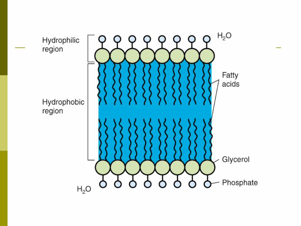
“unit membrane”
composed of phospholipids and proteins
sterols are absent (except: mycoplasmas)

mesosomes (invaginations of CM)
2types : septal mesosome (cross wall formation) lateral mesosome (electron transport enzyme)
bacterial chromosome is attached to a septal mesosome

B- Function
1) selective permeability2) electron transport and oxidative phosphorylation3) excretion of hydrolytic exoenzymes4) biosynthetic functions5) chemotactic systems and other sensory transduction
systems
1- selective permeability
hydrophobic barrier
transport systems (nutrient, waste products)
require energy in some forms
transport systems:
a) Passive transport b) Active transport c) Group translocation d) Special transport processes

a) Passive transport
the only form that does not require energy simple diffusion neither speed nor selectivity dissolved oxygen, carbon dioxide, water
Facilitated diffusion selective channel proteins glycerol
b) Active transport
I) Ion coupled transportII) ABC transport
I) Ion coupled transport
Move a molecule across the CM at the expense of proton motive force (pmf)
transport protein

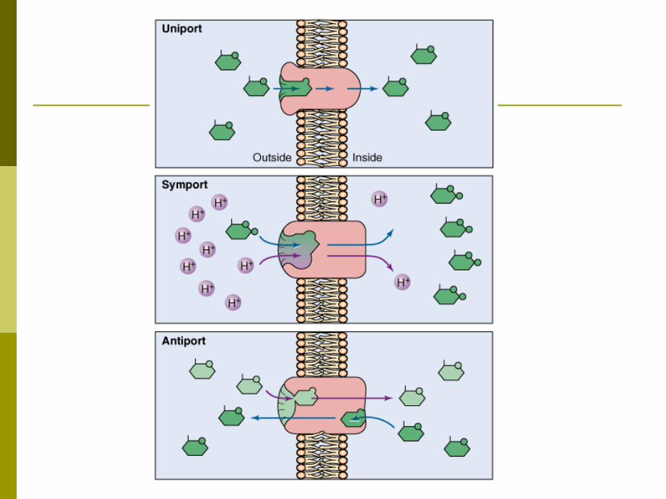
3 basic types: uniport (independent of any coupled ion)
symport (simultaneous transport of a solute [negative charge or neutral] and a positively charged ion, [H+] in the same direction )
antiport (simultaneous transport of two like-charged compounds in the opposite directions: H+, Na+ )
II) ABC transport ATP-binding cassette transport
binding proteins (BP)
Then BPs transfer the bound substrate to a membrane-bound protein complex
ATP hydrolyzed and the energy is used to open the membrane pore
Allow the unidirectional movement of the substrate into the cell
c) Group translocation (vectorial metabolism)
Change in structure certain sugars (eg, glucose and mannose) phosphorylated during the transport
"Phosphotransferase system“
Coupled transport with metabolism
d. Special transport processes
Iron (Fe) is an essential nutrient no free Fe in internal compartments of
animals Transferrin and lactoferrin
Siderophores
Some pathogenic bacteria Specific receptors Bind host transferrin and lactoferrin Fe is removed

2) electron transport and oxidative phosphorylation cytochromes and other enzymes functional analog of the mitochondrial
membrane (theory: mitochondria have evolved from
symbiotic bacteria)

3) excretion of hydrolytic exoenzymes
some nutrients are polymers hydrolytic enzymes degrade these polymers In Gram positive bacteria: external medium In Gram negative bacteria: periplasmic space
4) biosynthetic functions
enzymes for the cell wall biosynthesis enzymes for phospholipid synthesis some proteins of the DNA replicating
complex (septal mesosome)
carrier lipids
5) chemotactic systems and other sensory transduction systems
attractants and repellents bind to specific receptors in CM
Regulate the behavior in the environment
The Cell Wall
Outer of the CM
internal osmotic pressure 5-20 atm burst the cell Peptidoglycan (PG) (mucopeptide, murein)
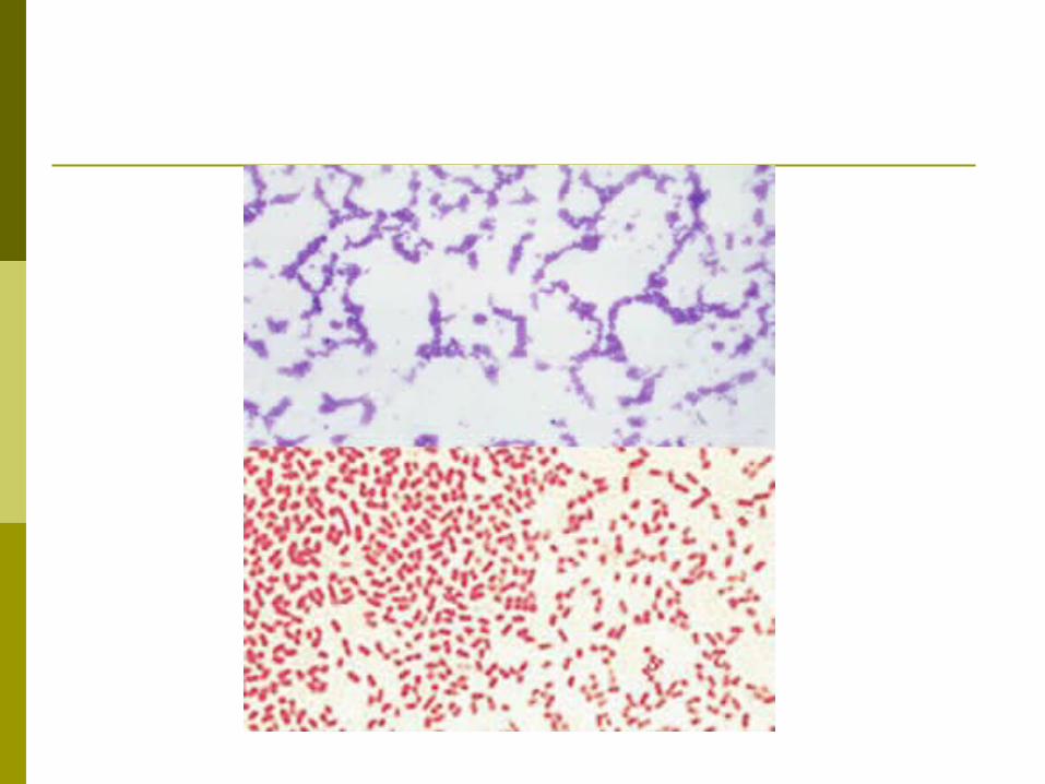
Hans Christian Gram
Based on the composition of the cell wall bacteria are classified as:
Gram positive Gram negative
Cell wall functions
osmotic protection
nonselectively permeable
plays an essential role in cell division
serving as a primer for its own biosynthesis
antigenic properties
Bacterial shapes
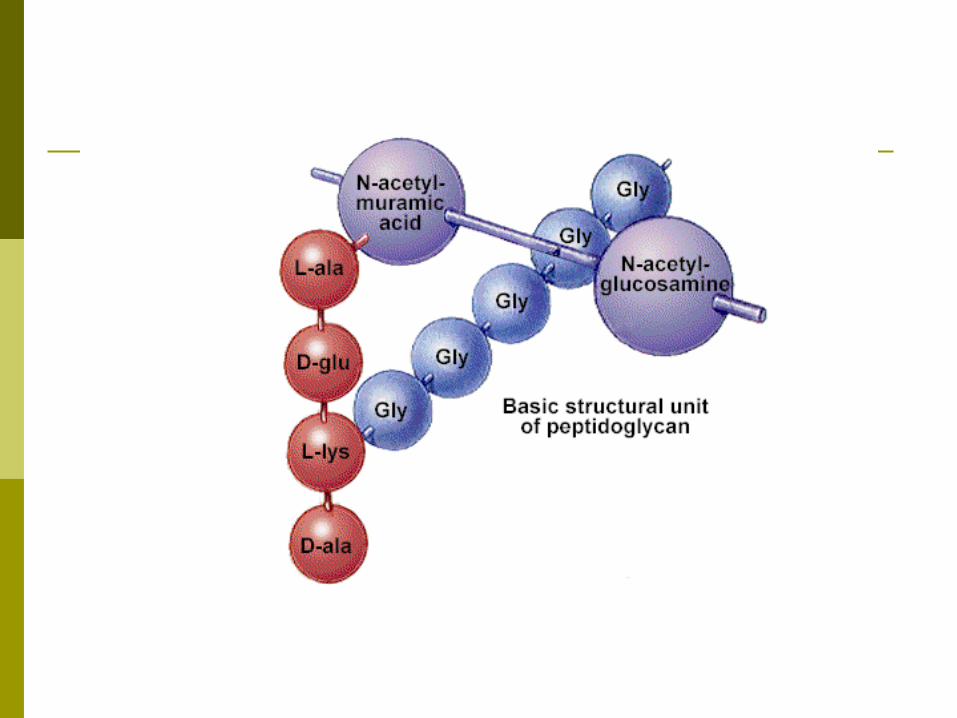
A- The Peptidoglycan Layer complex polymer consisting of three parts: 1) a backbone (alternating N-acetylglucosamine(NAG) and N-acetylmuramic acid(NAM)) 2) tetrapeptide side chains attached to NAM
3) cross-bridges
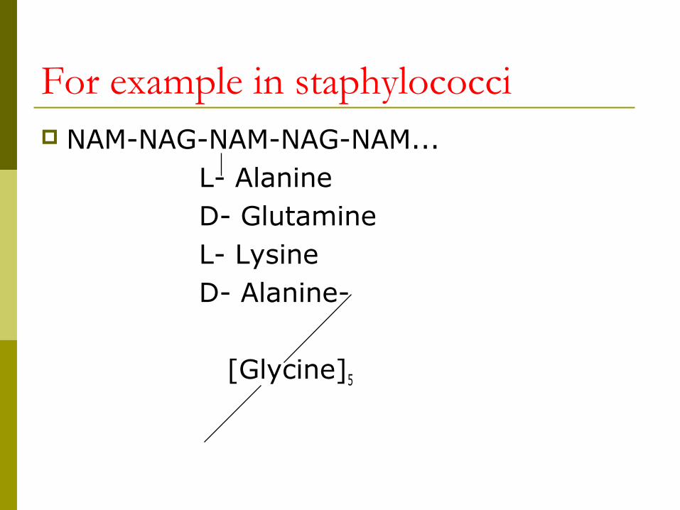
For example in staphylococci NAM-NAG-NAM-NAG-NAM... L- Alanine D- Glutamine L- Lysine D- Alanine-
[Glycine]5
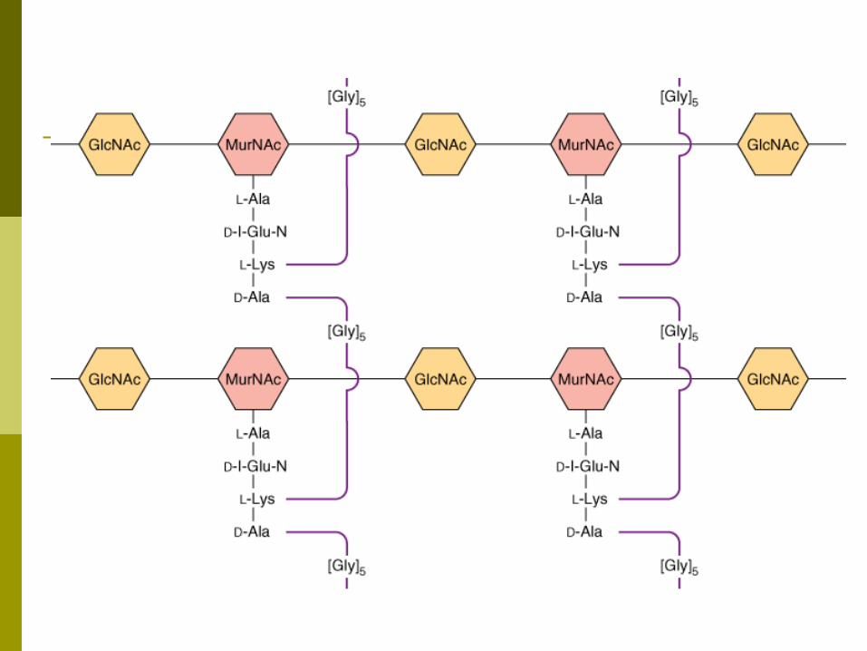

the backbone is the same in all bacteria
the tetrapeptide side chains and cross-bridges vary from species to species

the tetrapeptide side chains of all species:
a) most have L-alanine at position 1
b) D-glutamate at position 2
c) D-alanine at position 4
d) position 3 is the most variable one in G+ L-lysin , in G- mostly Diaminopimelic acid
Diaminopimelic acid( DAP) is the immediate precursor of Lysine
DAP is a unique element of prokaryotic cell walls
cross bridges varies among different species
for example in Staphylococcus, pentaglycin
in many gram negative a direct peptide linkage between the DAP of one side chain and
the alanine of a second side chain
In gram-positive bacteria, there are as many as 40 sheets of PG
In gram-negative bacteria there are only one or two sheets
B- Special components of Gram-Positive Cell Walls
1- Teichoic acids
2- polysaccharide molecules

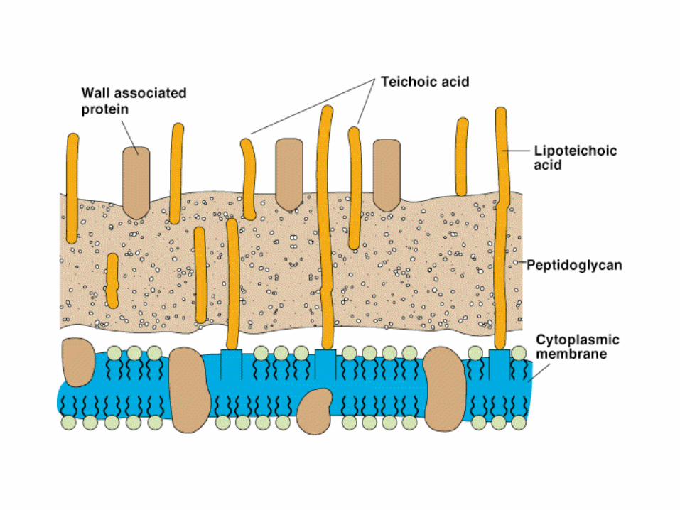
Teichoic acids
Polymers containing ribitol or glycerol residues
there are 2 types of teichoic acids a) wall teichoic acid b) membrane teichoic acid (lipoteichoic acid) linked to membrane glycolipid
the teichoic acid constitute major surface antigens of G+
Streptococcus pneumoniae forssman antigen
Streptococcus pyogenes , attachment of S. pyogenes to animal cells
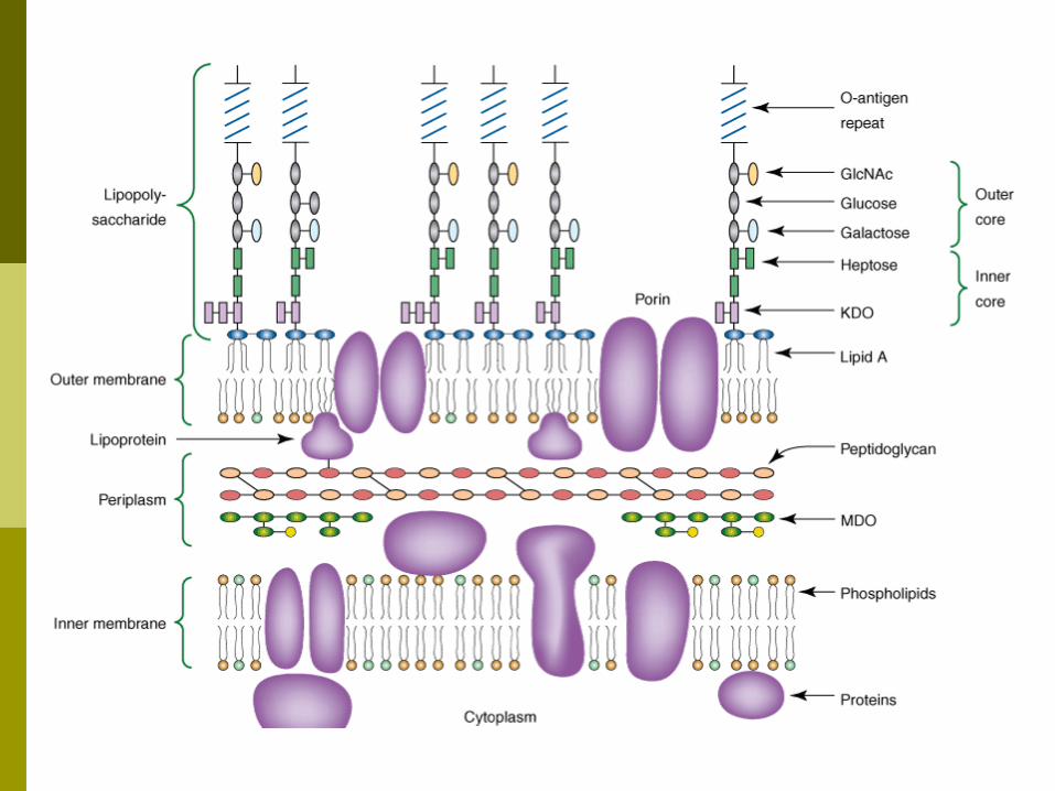
C- Special components of Gram-Negative Cell Walls
1- Outer membrane (OM)3- Lipoproteins4. The periplasmic space
1- Outer membrane (OM)
Chemically distinct bilayered structure the inner leaflet resembles in
composition that of cell membrane while the phospholipids of the outer
leaflet are replaced by Lipopolysaccharide (LPS) molecules
OM exclude hydrophilic molecules, because of its lipid nature
OM exclude hydrophobic molecules (unusual in biologic membranes)
protect the cells from the bile salts (enterics)
special protein channels (porins) Permit passive diffusion of hydrophilic compounds
G- bacteria are relatively high antibiotic resistance
Because large Antibiotic molecules penetrate the OM relatively slowly
major proteins of the OM (omp) receptors for several bacteriophages diffusion of nutrients
OmpA protein anchoring of the OM to PG sex pillus receptor (conjugation)
Minor proteins transport of specific molecules (vit B12 and iron siderophore complexes)
phospholipases and proteases
zone of adhesion (Bayer junction) between CM and OM
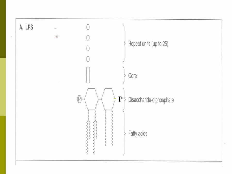
Lipopolysaccharide (LPS) lipid A polysaccharide
Lipid A consists of:
phosphorylated glucosamine disaccharide
fatty acids
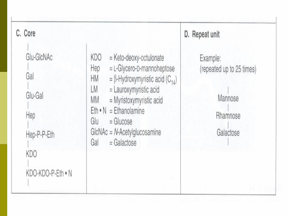
Polysaccharide is made up of:
a core
a terminal series of repeat units
P
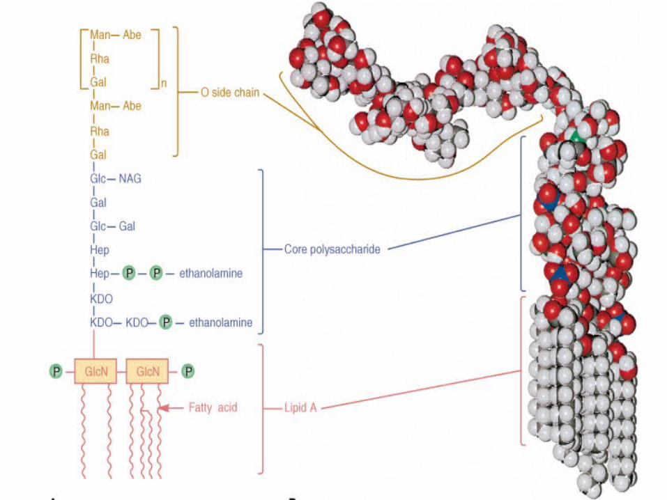
The polysaccharide core, is similar in all gram-negative species
But, Each species contains a unique repeat unit

LPS is extremely toxic to animals,
Endotoxin
because it is firmly bound to the cell surface and is released only when the cells are lysed.
When LPS is split into lipid A and polysaccharide,
all of the toxicity is associated with the Lipid A
The polysaccharide is a major surface antigen
0 antigen

Antigenic specificity is conferred by the terminal repeat units
Over 1000 antigenic types in Salmonella
2- Lipoproteins cross link the outer membrane and
peptidoglycan layers
its function is to stabilize the OM and anchor it to the PG layer
3. The periplasmic space
The space between the inner and outer membranes, called the periplasmic space,
Contains: the peptidoglycan layer and a gel-like solution of proteins.

The periplasmic proteins include:
Binding proteins for specific substrates (eg, amino acids, sugars, vitamins, and ions)
hydrolytic enzymes that break down non-transportable substrates into transportable ones
detoxifying enzymes that inactivate certain antibiotics (eg, β-lactamase).

the Gram positive cell envelope
cell membrane thick Peptidoglycan (PG) Teichoic Acid capsule or S layer

the Gram negative cell envelope
C.M (inner membrane) single sheet of PG outer membrane periplasmic space capsule or S layer


ENZYMES THAT ATTACK CELL WALLS
Lysozyme hydrolyze the linkage between NAG-NAM
found in animal secretions (tears, saliva, nasal secretions) as well as in egg white
autolysins
Bacteria themselves possess a number of, hydrolytic enzymes that attack peptidoglycan,
including glycosidases, amidases, and peptidases.
cell growth
division
autolysis after death
protozoa and the phagocytic cells
Have enzymes that degrade bacterial cell walls
L- form
Gram-positive bacteria:
In osmotically protected media
Lysozyme
L- form
protoplast
Gram-Negative bacteria:
In osmotically protected media,
The outer membrane prevents access of lysozyme
ethylenediaminetetraacetic acid (EDTA)
cells treated with EDTA+lysozyme
L- form
spheroplast
Some L forms can revert to the normal form
The factor that determines their capacity to revert is the presence of residual peptidoglycan
Some bacteria produce L- form in hosts:
Spontaneously Antibiotics
The spontaneous or antibiotic-induced formation of L forms in the host may produce chronic infections

they present special problems in chemotherapy.
relapses of the infection.
Capsule & Glycocalyx
extracellular polymers
Made of polysaccharide
known exception: Bacillus anthracis and Bacillus licheniformis
capsules are made of poly-D-glutamic acid
Glycocalyx more inclusive term
capsule Condensed Well defined layer Closely surrounding the cell Exclude particles, such as India ink
slime layer Loosly associated with the cell Does not exclude particles

The capsule contributes to the invasiveness of pathogenic bacteria
protection from phagocytosis
Until anticapsular antibody is formed
The glycocalyx plays a role in the adherence of bacteria to surfaces
Resistance to desiccation (bind water)
S-layer Many bacteria (G+, G- ) Have a surface layer composed of Protein
or glycoprotein molecules
As the outermost component of the cell envelope
Generally composed of a single molecular species
Function: Protection against: Wall-degrading enzymes
Invasion by: -predatory bacteria Bdellovibrio bacteriovorus -Bacteriophages
Host Cell adhesion
Flagella Thread-like appendages composed entirely
of protein
Motility

protein subunits called flagellin.
H antigens

Basal body
Hook
Filament
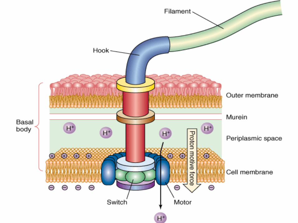

Flagellum has a Counterclockwise rotation
Rotation is driven by proton motive force
chemotaxis
chemical attractants (such as a sugar or an amino acid) or repellants bind to specific receptors located in the cell membrane
The mechanism by which a change in cell behavior is brought about in response to a change in the environment is called sensory transduction
sensory transduction chemotaxis aerotaxis (movement toward the optimal
oxygen concentration), phototaxis (movement of photosynthetic
bacteria toward the light), electron acceptor taxis (movement of
respiratory bacteria toward alternative electron acceptors, such as nitrate and fumarate).

4 types of arrangement are known:
Monotrichous (single polar flagellum)
Amphitrichous ( flagella at both poles)
Lophotrichous (multiple polar flagella)
Peritrichous (flagella distributed over the entire cell)
Pili (Fimbriae) In many gram-negative, in some Gram positive bacteria surface appendages called pili (L “hairs”) or fimbriae (L “Fringes”)
They are shorter and finer than flagella
and do not rotate (twitching motility)
composed of structural protein subunits termed pilins
Two classes :
ordinary pili for adherence “colonization antigens”
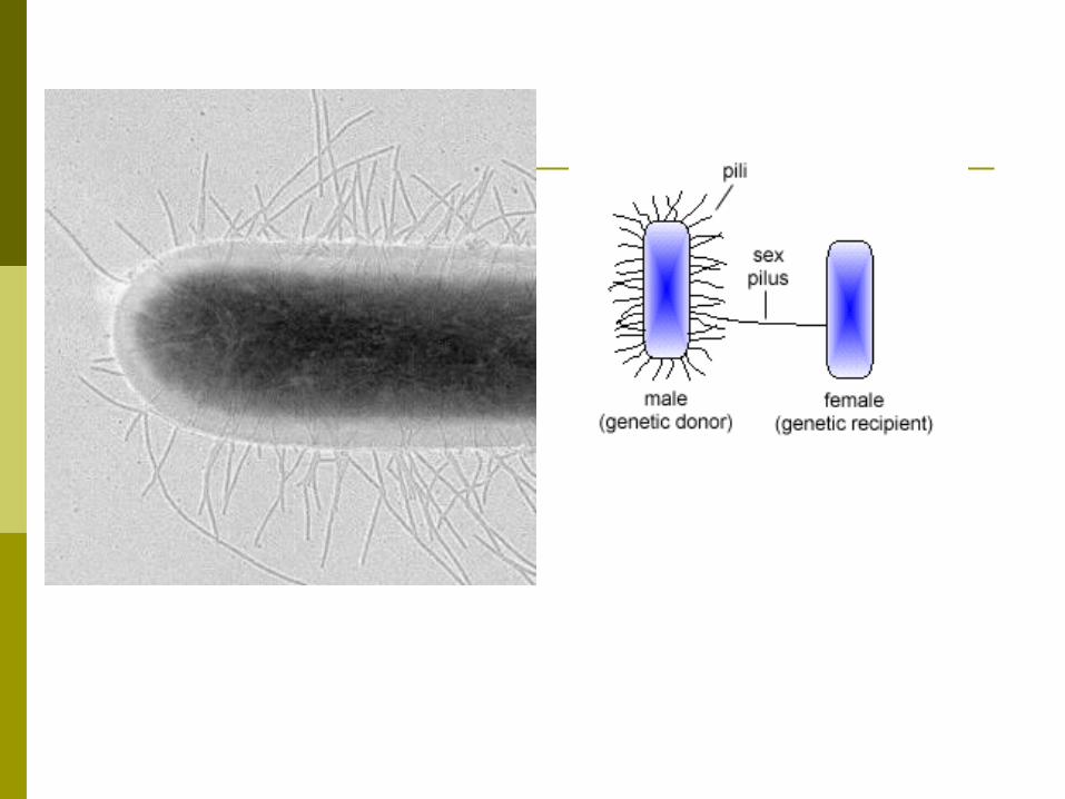
sex pili used in bacterial conjugation

Like capsule , pili inhibit the phagocytic ability of leukocytes
Endospore endospores form in several bacterial
genera
The two most common are gram-positive rods:
Bacillus
Clostridium
The other bacteria known to form endospores are:
the gram-positive coccus sporosarcina
possibly the rickettsial agent of Q fever, Coxiella burnetii
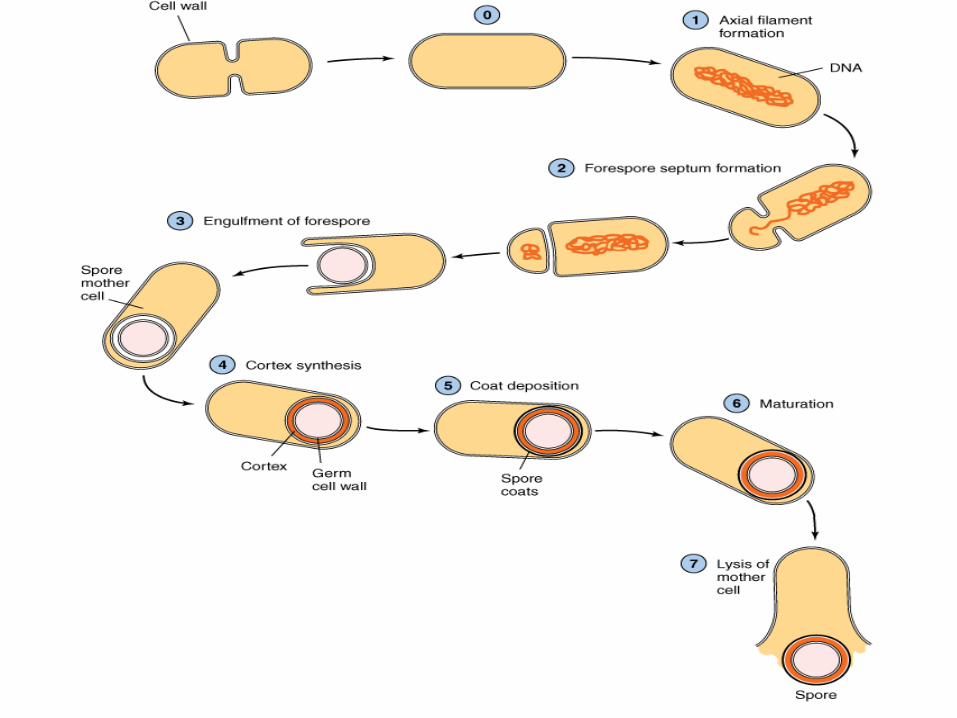
Vegetative cells
Mother cell: spore forming cell
The spore is a: resting cell, highly resistant to desiccation, heat, and
chemical agents
A. SPORULATION
begins when nutritional conditions become unfavorable,
depletion of the nitrogen or carbon source (or both) being the most significant factor
Sporulation involves:
the production of many new structures, enzymes, and metabolites
the disappearance of many vegetative cell components.
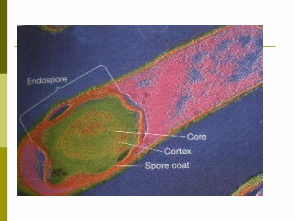
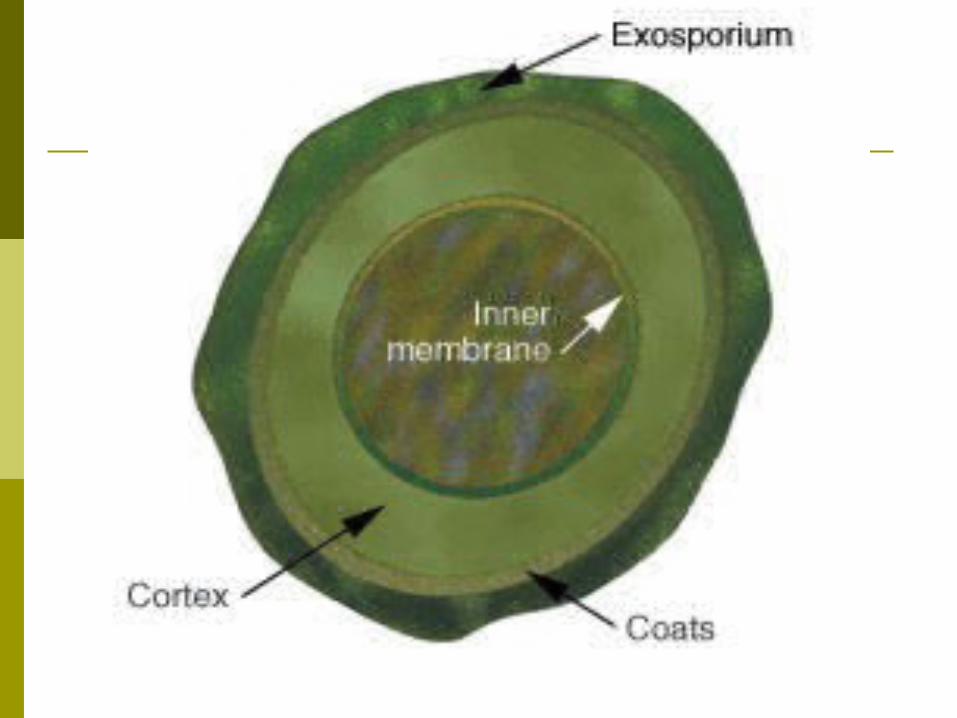
B- Properties of Endospores
1. Core The core is the spore protoplast.
It contains: -a complete chromosome, -the protein synthesizing apparatus, -an energy generating system based on glycolysis.
some unique enzymes are formed (eg, dipicolinic acid synthetase)
The energy for germination is stored as 3PGA rather than as ATP
The heat resistance of spores is due to:
their dehydrated state
the presence of calcium dipicolinate
result in the stabilization of the spore enzymes
2. Spore wall
It contains normal peptidoglycan
3. Cortex The cortex is the thickest layer of the spore
envelope. It contains an unusual type of peptidoglycan
Cortex peptidoglycan is extremely sensitive to lysozyme,
and its autolysis plays a role in spore germination.
4. Coat The coat is composed of a keratin-like
protein
The impermeability of this layer confers on spores their relative resistance to antibacterial chemical agents.
5. Exosporium
The exosporium is a lipoprotein membrane containing some carbohydrate.
C. GERMINATION
The germination process occurs in three stages:
activation, initiation, outgrowth.

1. Activation
Some agents can overcome spore dormancy:
heat, abrasion, acidity, compounds containing free sulfhydryl groups
(SH)
2. Initiation
Once activated, a spore will initiate germination if the environmental conditions are favorable.
Spores have receptors that recognize different effectors as signaling a rich medium
Such as: L-alanine adenosine
Binding of the effector activates an autolysin that rapidly degrades the cortex peptidoglycan.
Water is taken up,
calcium dipicolinate is released,
3. Outgrowth
Degradation of the cortex and outer layers results in the emergence of a new vegetative cell
A period of active biosynthesis follows;
this period, which terminates in cell division, is called outgrowth.