Cell Structure and Function. Consists of Plasma membrane Barrier that surrounds the cytoplasm of the...
36
Cell Structure and Function Chapter 2
-
Upload
lorraine-martin -
Category
Documents
-
view
226 -
download
5
Transcript of Cell Structure and Function. Consists of Plasma membrane Barrier that surrounds the cytoplasm of the...
Cell Structure and Function
Chapter 2

Consists of Plasma membrane
Barrier that surrounds the cytoplasm of the cell Organelles
Internal structures that carry out specialized functions
Bounded by a membrane (exception ribosome, centriole)
CytosolLiquid portion of the cell
Cell Structure (Eukaryotes)

Plant and Animal Cell

Almost all eukaryotes have a nucleus Contains
Nucleoplasm Viscous fluid similar to cytosol
Nucleolus Assembly of ribosomal subunits from proteins and RNA
Nuclear envelope Contains almost all of the DNA in a eukaryotic cell (exceptions,
mitochondria and chloroplast) Protects DNA from metabolic reactions
Nucleus
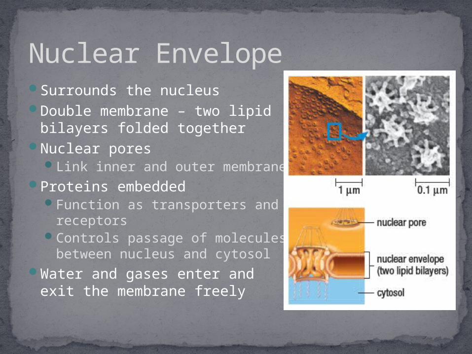
Surrounds the nucleus Double membrane – two lipid
bilayers folded togetherNuclear pores
Link inner and outer membraneProteins embedded
Function as transporters and receptors
Controls passage of molecules between nucleus and cytosol
Water and gases enter and exit the membrane freely
Nuclear Envelope

Group of interacting organelles between the nucleus and plasma membrane
FunctionsBuild lipids, enzymes, and other proteins Transportation Destroy toxins Recycle waste
The Endomembrane System

Extension of the nuclear envelopeContinuous compartment that folds
repeatedly into flattened sacs and tubesTwo Types
Rough ERContains ribosomes
Synthesis of polypeptide chains Once synthesized, proteins fold and take
shape inside ERSmooth ER
Contains no ribosomes Enzymes formed from Rough ER found
here Produce most of cells membrane lipids Break down carbohydrates, fatty acids, and
drugs and poisons
Endoplasmic Reticulum

Membrane-enclosed, saclike organelles Form from budding from other organelles or
plasma membraneFucntions
Transport proteins Digest fatty acids and amino acids (use of
enzymes)Peroxisome – inactivates hydrogen peroxide from
fatty acid breakdownVacuoles
Dispose of waste, debris and toxic materialsLysosomes – fuse with vacuole to dispose of worn out cell
components (use of enzymes)Keep turgor pressure inside plant cell
Vesicles

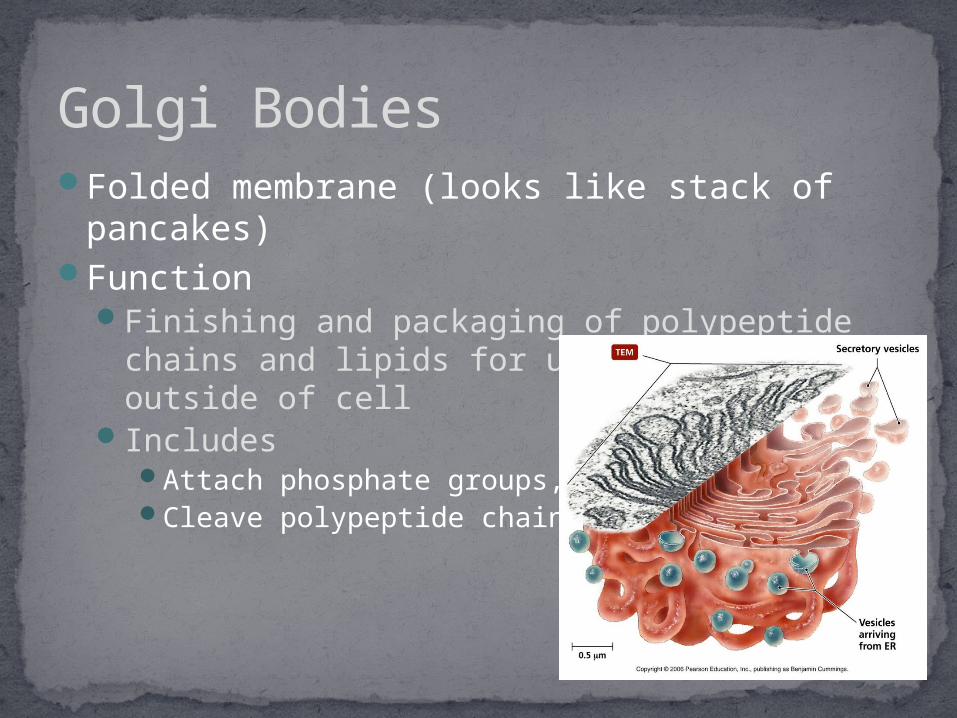
Folded membrane (looks like stack of pancakes)
Function Finishing and packaging of polypeptide chains
and lipids for use inside or outside of cellIncludes
Attach phosphate groups, sugarsCleave polypeptide chains
Golgi Bodies

Double bound membrane organelle
Two compartmentsMitochondrial
matrixIntermembrane
spaceContain their own
DNADivide independently
of cellFunction
Synthesize ATP
Mitochondria

TypesChloroplasts
Double membrane bound organelle
Stroma – semifluid interior, contains enzymes and DNA
Specialize in photosynthesis Contain chlorophyll
ChromoplastsStore orange and red
carotenoidsResponsible for beautiful
colours in autumnAmyloplasts
Store starch grains
Plastids (Plant Cells)

Interconnected system of many protein filaments
FunctionsReinforceOrganizeMove cell structures
Include MicrotubulesMicrofilaments
Cytoskeleton
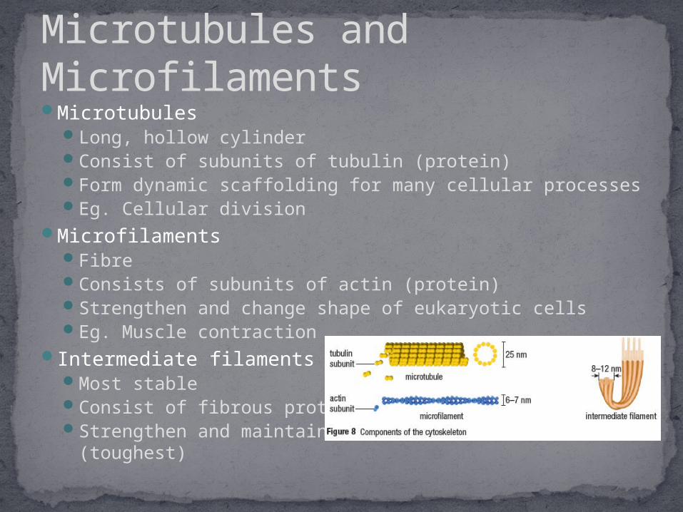
MicrotubulesLong, hollow cylinderConsist of subunits of tubulin (protein)Form dynamic scaffolding for many cellular processes Eg. Cellular division
MicrofilamentsFibreConsists of subunits of actin (protein)Strengthen and change shape of eukaryotic cells Eg. Muscle contraction
Intermediate filamentsMost stable Consist of fibrous proteins Strengthen and maintain cell and tissue structure
(toughest)
Microtubules and Microfilaments

Organized array of microtubulesFlagella
Whiplike structure Propels cells (sperm)
CiliaHair like structure Sweeping action moves particles (lining of airway)
Pseudopod (false feet)Lobe formations Amoeba
Cilia, Flagella, Pseudopods

Located around plasma membranePorous structure Protects supports and gives shape to the cell Waxy cuticle
Covers plant cells and protects exposed surfaces of soft parts of the plant
Limits water loss on hot/dry days
Cell Wall (Plant Cells)

Non-living mixture of fibrous proteins and polysaccharides that surround cells
Functions Supports and anchors cells Separates tissues Cell signalling
Plants Cellulose
Fungi Chitin
Animals Carbohydrates and proteins Bone (Collagen)
Cells interact via cell junctions
Extracellular Matrix (ECM)
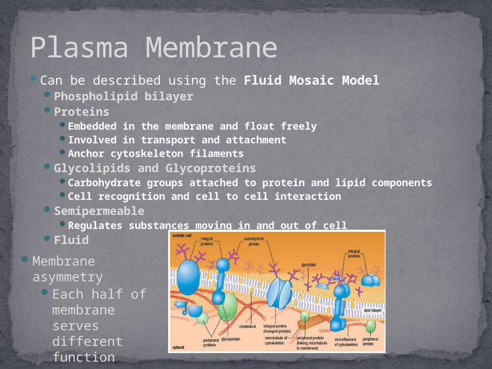
Can be described using the Fluid Mosaic Model Phospholipid bilayer Proteins
Embedded in the membrane and float freely Involved in transport and attachment Anchor cytoskeleton filaments
Glycolipids and Glycoproteins Carbohydrate groups attached to protein and lipid components Cell recognition and cell to cell interaction
Semipermeable Regulates substances moving in and out of cell
Fluid
Plasma Membrane
Membrane asymmetry
Each half of membrane serves different function

Hydrophilic HeadPhosphate
groupPolar group
Hydrophobic TailFatty acids
Align themselves in aqueous solution
Phospholipids

Depends on how densely individual lipid molecules can pack together.
Influenced by:Composition of lipid molecules
Saturated vs Unsaturated hydrocarbons Temperature
Low temperatures decrease fluidity – viscous semisolid gelTemperature related to degree of unsaturated lipids in
membrane Increase in unsaturated membrane lower gelling temperature
SterolsCholesterol – membrane stabilizer
Fluidity

TransportHydrophilic protein channelShape shifting
Enzymatic Activity Respiration and Photosynthesis
Triggering Signals Bind to specific chemicals (hormones) Binding changes the inner surface of
membrane
Attachment and Recognition Exposed to both internal and external
surfaces of membrane Serve as attachment points for
cytoskeleton elements Immune response
Membrane Proteins (Function)

Integral Membrane Proteins (transmembrane)Exposed to aqueous environment on both sides of
the membrane
Peripheral Membrane Proteins Located on surface of a membrane No interaction with hydrophobic core Held to surface by hydrogen / ionic bondsEg. Cytoskeleton
Membrane Protein (Integral/Peripheral)
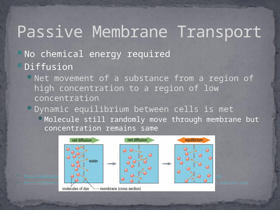
No chemical energy requiredDiffusion
Net movement of a substance from a region of high concentration to a region of low concentration
Dynamic equilibrium between cells is metMolecule still randomly move through membrane but
concentration remains same
http://highered.mcgraw-hill.com/sites/9834092339/student_view0/chapter5/how_diffusion_works.html
http://highered.mcgraw-hill.com/sites/9834092339/student_view0/chapter5/diffusion_through_cell_membranes.html
Passive Membrane Transport
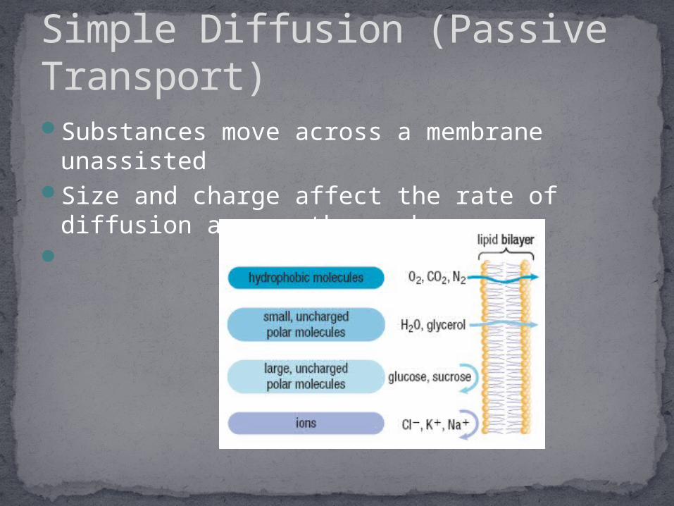
Substances move across a membrane unassisted
Size and charge affect the rate of diffusion across the membrane
Simple Diffusion (Passive Transport)

Diffusion of larger molecules with the help of a transport protein (integral membrane protein)
Stops when equilibrium is reachedTwo types of Transport Proteins
Channel proteinsCarrier proteins
Facilitated Diffusion

Channel ProteinsForm hydrophilic pathways in the membrane Water and certain ions can passVoltage-gated channels
Open or closed by changes in voltage across the membrane or by binding molecules
Eg. Muscle contractions
Facilitated Diffusion

Carrier Proteins Form pathways
through the membrane
Bind to a specific solute (glucose, amino acid)
Carrier protein changes shape allowing solute to move from one side of the membrane to the other
Facilitated Diffusion
http://highered.mcgraw-hill.com/sites/9834092339/student_view0/chapter5/how_facilitated_diffusion_works.html
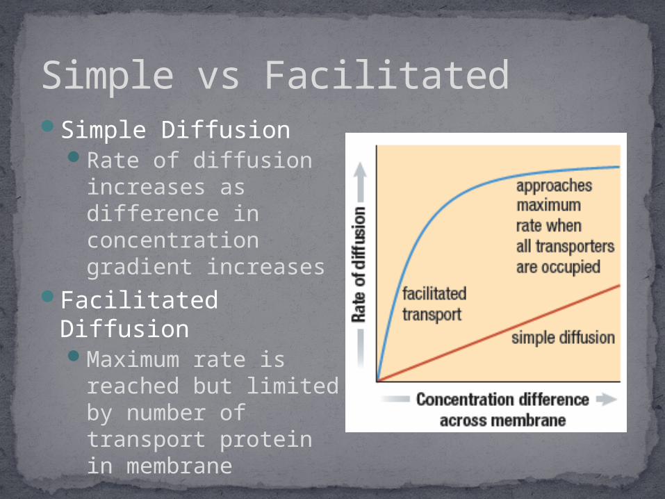
Simple Diffusion Rate of diffusion
increases as difference in concentration gradient increases
Facilitated Diffusion Maximum rate is
reached but limited by number of transport protein in membrane
Simple vs Facilitated
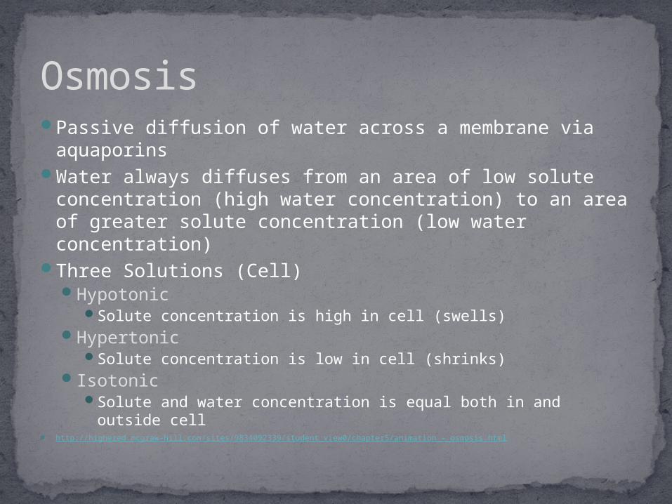
Passive diffusion of water across a membrane via aquaporins
Water always diffuses from an area of low solute concentration (high water concentration) to an area of greater solute concentration (low water concentration)
Three Solutions (Cell) Hypotonic
Solute concentration is high in cell (swells)Hypertonic
Solute concentration is low in cell (shrinks) Isotonic
Solute and water concentration is equal both in and outside cell
http://highered.mcgraw-hill.com/sites/9834092339/student_view0/chapter5/animation_-_osmosis.html
Osmosis

Osmosis

Substance carried across a membrane from an area of low concentration to an area of high concentration
Use of pumpATP used as energy source Two Types
Primary Active TransportSecondary Active Transport
Active Membrane Transport

Pumps that moves positively charged ions across membranes (H⁺. Ca²⁺, Na⁺, K⁺)ATP is hydrolyzedPhosphate group attaches to pump allowing ion to
bind to protein transporterProtein transporter undergoes a folding change
exposing ion to opposite side of membrane Ion is released to side of higher concentrationPhosphate group is released
This creates an electrochemical gradientEffect of voltage and difference in ion concentrationStored potential energy
http://highered.mcgraw-hill.com/sites/9834092339/student_view0/chapter5/primary_active_transport.html
Primary Active Transport

Primary Active Transport

http://highered.mcgraw-hill.com/sites/9834092339/student_view0/chapter5/cotransport__symport_and_antiport_.html
Uses concentration gradient of an ion as its energy sourceThis gradient was already established by primary pump. Transport solute across membraneTwo ways
SymportSolute moves through channel in the same direction as ion
AntiportSolute moves in opposite direction as ion
Secondary Active Tranport

Vesicles are used to transportATP is requiredExocytosis
Transport of proteins and waste material from cytosol to exterior of cell
EndocytosisTransport proteins and large molecules into
cytosol of the cell.
http://highered.mcgraw-hill.com/olcweb/cgi/pluginpop.cgi?it=swf::535::535::/sites/dl/free/0072437316/120068/bio02.swf::Endocytosis%20and%20Exocytosis
Exocytosis and Endocytosis


![J250/01 Paper 1 (Foundation Tier) Sample Question Paper · B Cell membrane, chloroplast, nucleus C Cell wall, cytoplasm, mitochondria D Cell wall, cytoplasm, nucleus Your answer [1]](https://static.fdocuments.us/doc/165x107/5e9fa71c2bc0006f2a48962b/j25001-paper-1-foundation-tier-sample-question-b-cell-membrane-chloroplast.jpg)