Cell Stem Cell Perspective Stem Cell Therapy and the... · Cell Stem Cell Perspective Cardiac Stem...
10
Cell Stem Cell Perspective Cardiac Stem Cell Therapy and the Promise of Heart Regeneration Jessica C. Garbern 1 and Richard T. Lee 2, * 1 Department of Medicine, Boston Children’s Hospital, Boston, MA 02115, USA 2 Harvard Stem Cell Institute, the Brigham Regenerative Medicine Center and the Cardiovascular Division, Department of Medicine, Brigham and Women’s Hospital and Harvard Medical School, Boston, MA 02115, USA *Correspondence: [email protected] http://dx.doi.org/10.1016/j.stem.2013.05.008 Stem cell therapy for cardiac disease is an exciting but highly controversial research area. Strategies such as cell transplantation and reprogramming have demonstrated both intriguing and sobering results. Yet as clinical trials proceed, our incomplete understanding of stem cell behavior is made evident by numerous unresolved matters, such as the mechanisms of cardiomyocyte turnover or the optimal therapeutic strategies to achieve clinical efficacy. In this Perspective, we consider how cardiac stem cell biology has led us into clinical trials, and we suggest that achieving true cardiac regeneration in patients may ultimately require resolution of critical controversies in experimental cardiac regeneration. Introduction The race is on: throughout the world, basic and clinical investiga- tors want to be the first to identify new approaches to regenerate cardiac tissue and to prove the effects of these therapies in patients with heart disease. Despite substantial progress in treating many types of heart disease, the worldwide heart failure burden will remain enormous through this century. The potential of stem cells and the scope of the heart failure problem have fueled a stampede to be the first to achieve human heart regen- eration. Cell transplantation approaches are attractive given their relative ease of use and good safety profile to date, but repro- ducible results endorsing a specific strategy for routine patient care are lacking. Meanwhile, cellular reprogramming strategies are appealing because they potentially allow precise control over cellular behavior, but much work remains before the safety of reprogramming allows clinical testing. Current clinical trials focus largely on injection of cells with cardiomyogenic potential into the heart; however, given the limitations of this approach, we wonder: is this the path to take right now? As we consider the current state of the heart regeneration field, it is worth pausing to reflect on the 1960s, when heart transplan- tation emerged. Initial excitement over heart transplantation led to over 100 heart transplantations worldwide in 1967 and 1968. However, disappointing results soon followed, with only a quarter of the patients surviving more than a few months (Kant- rowitz, 1998). Renowned cardiologist Helen Taussig expressed concern in 1969 that it was not yet time for human trials, warning, ‘‘.our hope should be that physicians and surgeons will pro- ceed with extreme caution until such time as a cardiac transplant will not announce the imminence of death but offer the patient the probability of a return to a useful life for a number of years’’ (Taussig, 1969). During the 1970s, few human heart transplants occurred as the number of surgeons willing to perform heart transplants dwindled due to high mortality in the first year after transplants (Kantrowitz, 1998). Only after rigorous research in organ rejection and immunosuppression in the 1980s did heart transplantation become the accepted medical practice that it is today (Kantrowitz, 1998). Unfortunately, limitations in organ supply and other issues allow transplantation in only a minority of patients with heart failure, and transplantation will not be a solution for the growing problem of heart disease. Half a century after the first human heart transplant, we are now confronted with the new challenge of regenerating damaged hearts in the growing number of patients with heart failure. Will we be following a similar path to that of cardiac trans- plantation? Despite the enormous potential, it is not clear whether we know enough fundamentals to move forward clini- cally or how fast we should go. Some investigators contend that we know all we need to know to move forward, while others are less confident. In this Perspective, we consider both estab- lished principles and ongoing controversies that guide cardiac regeneration research. Established Principles We believe that three fundamental principles of cardiac regener- ative biology have now been established. First, multipotent cardiac progenitor cells (CPCs) exist in the embryonic mamma- lian heart (Moretti et al., 2006; Wu et al., 2006); second, there is creation of a limited number of new heart cells after birth in mam- mals (Beltrami et al., 2003; Bergmann et al., 2009; Malliaras et al., 2013; Mollova et al., 2013; Senyo et al., 2013); and third, some vertebrates, such as newts (Oberpriller and Oberpriller, 1974), zebrafish (Jopling et al., 2010; Poss et al., 2002), and neonatal mice (Porrello et al., 2011), can regenerate myocardium following experimental injury. In an often-controversial field, the establish- ment of these three principles from different lines of evidence by different laboratories represents seminal progress. Multipotent CPCs Exist in the Mammalian Embryo During embryonic development, CPCs arise from a subpopula- tion of mesodermal precursors that can be modeled from in vitro differentiated embryonic stem cells (ESCs) (Kouskoff et al., 2005). The expression of FLK1 marks a panmesodermal cell population that can give rise to cells in both the primary and secondary heart fields (Kattman et al., 2006) as well as skeletal muscles in the head, neck, and trunk (Motoike et al., 2003). For the primary heart field, a population of bipotential Cell Stem Cell 12, June 6, 2013 ª2013 Elsevier Inc. 689
Transcript of Cell Stem Cell Perspective Stem Cell Therapy and the... · Cell Stem Cell Perspective Cardiac Stem...
Cell Stem Cell
Perspective
Cardiac Stem Cell Therapyand the Promise of Heart Regeneration
Jessica C. Garbern1 and Richard T. Lee2,*1Department of Medicine, Boston Children’s Hospital, Boston, MA 02115, USA2Harvard Stem Cell Institute, the Brigham Regenerative Medicine Center and the Cardiovascular Division, Department of Medicine,Brigham and Women’s Hospital and Harvard Medical School, Boston, MA 02115, USA*Correspondence: [email protected]://dx.doi.org/10.1016/j.stem.2013.05.008
Stem cell therapy for cardiac disease is an exciting but highly controversial research area. Strategies such ascell transplantation and reprogramming have demonstrated both intriguing and sobering results. Yet asclinical trials proceed, our incomplete understanding of stem cell behavior is made evident by numerousunresolvedmatters, such as themechanisms of cardiomyocyte turnover or the optimal therapeutic strategiesto achieve clinical efficacy. In this Perspective, we consider how cardiac stem cell biology has led us intoclinical trials, and we suggest that achieving true cardiac regeneration in patients may ultimately requireresolution of critical controversies in experimental cardiac regeneration.
IntroductionThe race is on: throughout the world, basic and clinical investiga-
tors want to be the first to identify new approaches to regenerate
cardiac tissue and to prove the effects of these therapies in
patients with heart disease. Despite substantial progress in
treating many types of heart disease, the worldwide heart failure
burden will remain enormous through this century. The potential
of stem cells and the scope of the heart failure problem have
fueled a stampede to be the first to achieve human heart regen-
eration. Cell transplantation approaches are attractive given their
relative ease of use and good safety profile to date, but repro-
ducible results endorsing a specific strategy for routine patient
care are lacking. Meanwhile, cellular reprogramming strategies
are appealing because they potentially allow precise control
over cellular behavior, but much work remains before the safety
of reprogramming allows clinical testing. Current clinical trials
focus largely on injection of cells with cardiomyogenic potential
into the heart; however, given the limitations of this approach,
we wonder: is this the path to take right now?
Aswe consider the current state of the heart regeneration field,
it is worth pausing to reflect on the 1960s, when heart transplan-
tation emerged. Initial excitement over heart transplantation led
to over 100 heart transplantations worldwide in 1967 and 1968.
However, disappointing results soon followed, with only a
quarter of the patients surviving more than a few months (Kant-
rowitz, 1998). Renowned cardiologist Helen Taussig expressed
concern in 1969 that it was not yet time for human trials, warning,
‘‘.our hope should be that physicians and surgeons will pro-
ceedwith extreme caution until such time as a cardiac transplant
will not announce the imminence of death but offer the patient
the probability of a return to a useful life for a number of years’’
(Taussig, 1969). During the 1970s, few human heart transplants
occurred as the number of surgeons willing to perform heart
transplants dwindled due to high mortality in the first year after
transplants (Kantrowitz, 1998). Only after rigorous research in
organ rejection and immunosuppression in the 1980s did heart
transplantation become the accepted medical practice that it
is today (Kantrowitz, 1998). Unfortunately, limitations in organ
supply and other issues allow transplantation in only a minority
of patients with heart failure, and transplantation will not be a
solution for the growing problem of heart disease.
Half a century after the first human heart transplant, we
are now confronted with the new challenge of regenerating
damaged hearts in the growing number of patients with heart
failure. Will we be following a similar path to that of cardiac trans-
plantation? Despite the enormous potential, it is not clear
whether we know enough fundamentals to move forward clini-
cally or how fast we should go. Some investigators contend
that we know all we need to know to move forward, while others
are less confident. In this Perspective, we consider both estab-
lished principles and ongoing controversies that guide cardiac
regeneration research.
Established PrinciplesWe believe that three fundamental principles of cardiac regener-
ative biology have now been established. First, multipotent
cardiac progenitor cells (CPCs) exist in the embryonic mamma-
lian heart (Moretti et al., 2006; Wu et al., 2006); second, there is
creation of a limited number of new heart cells after birth in mam-
mals (Beltrami et al., 2003; Bergmann et al., 2009;Malliaras et al.,
2013; Mollova et al., 2013; Senyo et al., 2013); and third, some
vertebrates, such as newts (Oberpriller and Oberpriller, 1974),
zebrafish (Jopling et al., 2010; Poss et al., 2002), and neonatal
mice (Porrello et al., 2011), can regeneratemyocardium following
experimental injury. In an often-controversial field, the establish-
ment of these three principles from different lines of evidence by
different laboratories represents seminal progress.
Multipotent CPCs Exist in the Mammalian Embryo
During embryonic development, CPCs arise from a subpopula-
tion of mesodermal precursors that can be modeled from
in vitro differentiated embryonic stem cells (ESCs) (Kouskoff
et al., 2005). The expression of FLK1 marks a panmesodermal
cell population that can give rise to cells in both the primary
and secondary heart fields (Kattman et al., 2006) as well as
skeletal muscles in the head, neck, and trunk (Motoike et al.,
2003). For the primary heart field, a population of bipotential
Cell Stem Cell 12, June 6, 2013 ª2013 Elsevier Inc. 689

Table 1. Estimated Rates of Cardiomyocyte Renewal in Adult Mammals
Annual Rate of Cardiomyocyte Renewal Species Method Reference
0.5%–1.9% human 14C, accelerator mass spectrometry Bergmann et al., 2009
10%–40% human Ki67, phospho-H3, Aurora B, and IdU Kajstura et al., 2010
7%–23% human 14C, accelerator mass spectrometry Kajstura et al., 2012
0.04%–4.5% human Phopho-H3 Mollova et al., 2013
1.3%–4% mouse BrdU Malliaras et al., 2013
0.74% mouse 15N, imaging mass spectrometry Senyo et al., 2013
1.09% mouse [3H]thymidine Soonpaa and Field, 1997; Soonpaa et al., 2013
Cell Stem Cell
Perspective
KIT+ (also referred to as c-kit+)/NKX2.5+ progenitor cells gives
rise to myocardial and smooth muscle cells (Wu et al., 2006).
For the secondary heart field, ISL1+ progenitor cells have been
described to undergo multilineage differentiation into myocar-
dial, smooth muscle, and endothelial cells (Moretti et al., 2006).
Taken together, these studies provide unequivocal evidence
for the existence of multipotent progenitor cells in the developing
embryo heart. Understanding the mechanisms of embryonic
development—in particular, identifying the signals that initiate
and terminate heart development—will be crucial to establishing
therapeutic regenerative approaches that utilize similar molecu-
lar pathways.
Postnatal Cardiomyocyte Renewal Occurs in Mammals,
Including Humans
The classic 20th century teaching was that mammalian cardio-
myocytes cease replication soon after birth, with subsequent
growth of the heart attributed to cardiomyocyte hypertrophy
rather than hyperplasia. In the 1990s, the Anversa laboratory
provided crucial evidence that mammalian cardiomyocytes not
only enter the cell cycle in adulthood, but can also subsequently
undergo karyokinesis and cytokinesis (Kajstura et al., 1998;
Quaini et al., 1994). Recent studies definitively demonstrate
that cardiomyocyte turnover occurs throughout life in mammals,
including humans, although estimates of the rate of cardiomyo-
cyte turnover vary dramatically.
Perhaps themost stunning evidence for cardiomyocyte regen-
eration in humans was revealed by retrospective isotope dating
studies. Taking advantage of the dramatic spike and decline of
worldwide atmospheric carbon-14 (14C) levels during the
1950s to 1960s due to above ground nuclear bomb testing,
Frisen and colleagues developed an ingenious approach to
determine the birth date of cardiomyocytes in humans by
measuring nuclear 14C content (Bergmann et al., 2009). Their
data showed that new cardiomyocytes form in human myocar-
dium at a rate of approximately 1.5% per year at age 25 years,
decreasing substantially in the latter half of life (Bergmann
et al., 2009).
Using the 14Cmethod developed by the Frisen group, Anversa
and colleagues arrived at much higher values for cardiomyocyte
turnover in humans (7%–23% per year); in addition, they re-
ported the surprising finding that cardiogenesis increases with
age (Kajstura et al., 2012). Mathematical modeling assumptions
in the 14C method could explain some of the differences in the14C studies.
Multiple additional lines of evidence support a low rate of
mammalian cardiogenesis and that the rate declines further
with age (Table 1). Earlier studies using [3H]thymidine in adult
690 Cell Stem Cell 12, June 6, 2013 ª2013 Elsevier Inc.
mice estimated an annual renewal rate of approximately 1%
per year (Soonpaa and Field, 1997), almost identical to the rates
of cardiogenesis estimated by more recent mouse studies
(Malliaras et al., 2013; Senyo et al., 2013). A similar rate of cardio-
genesis in young human adults was recently confirmed (1.9% at
20 years) using an imaged-based assay in tissue samples pro-
cured from donor hearts prior to transplantation (Mollova et al.,
2013). Thus, while all studies reveal cardiomyocyte renewal in
postnatal mammals, the majority of studies indicate that this
rate is very low, on the order of 1% per year, and that the rate
declines with age.
Myocardial Regeneration Occurs after Injury in Certain
Vertebrates
Critical insight into how we might regenerate human hearts has
arisen from vertebrates that can indisputably regenerate
myocardium following injury. Urodele amphibians such as newts
can survive after amputation of the apical myocardium and
demonstrate cardiomyocyte regeneration by 30 days postampu-
tation (Oberpriller and Oberpriller, 1974). Similarly, in zebrafish,
amputation of the apex of the heart leads to complete regenera-
tion (Poss et al., 2002). This dramatic regeneration in urodele
amphibians and zebrafish is thought to be due to limited dediffer-
entiation of mature cardiomyocytes and reentry into the cell
cycle (Laube et al., 2006). This is supported by evidence of
sarcomere disassembly (Jopling et al., 2010) as well as expres-
sion of Gata4, a transcription factor that is normally expressed
during embryonic development to regulate myocardial formation
(Kikuchi et al., 2010).
Studies investigating mammalian cardiomyocyte mitosis after
injury can be found as early as the 1970s (Rumyantsev, 1974),
although more definitive evidence for the potential of embryonic
and neonatal mammalian myocardium to regenerate has
recently emerged. Using an elegant mouse model to effectively
damage 50% of the developing cardiac tissue by inactivating
the gene encoding holocytochrome c synthase, Cox and
colleagues demonstrated that lost myocardium is replaced by
healthy tissue during fetal development, resulting in only 10%
of the cardiac volume occupied by diseased tissue at birth
(Drenckhahn et al., 2008). Furthermore, Sadek and colleagues
showed that the 1-day-old neonatal mouse heart is capable of
regeneration after resection of approximately 15% of the
ventricle at the apex (Porrello et al., 2011). This neonatal mouse
heart regeneration appears to occur as a result of dedifferentia-
tion followed by proliferation of preexisting cardiomyocytes.
However, the ability to regenerate myocardium is rapidly lost
by 7 days after birth; instead, the heart develops fibrotic scars
similar to the response observed following myocardial injury in

Figure 1. Mammalian Cardiogenesis duringAgingMultiple lines of evidence exist for the refreshmentof cardiomyocytes during aging in mammals, withtwo predominant mechanisms proposed toexplain the source of new cardiomyocytes duringaging: (1) progenitor cells that give rise to newcardiomyocytes exist in the heart throughout life or(2) mature cardiomyocytes undergo partial dedif-ferentiation, reenter the cell cycle, and proliferateinto new cardiomyocytes. Results from themajority of investigators suggest that this turnoverrate occurs at a low level (approximately 1% peryear in young adults) and declines even furtherwith age.
Cell Stem Cell
Perspective
adult mice and humans (Porrello et al., 2011). These experiments
raise the critical question of what prevents mouse heart regener-
ation after the first days of life, and point to this first week of life as
a crucial period for understanding inherent regenerative mecha-
nisms in mammals.
Unresolved QuestionsThough not all encompassing, here we discuss five substantial
controversies that will require resolution as we push forward to
achieve true cardiac regeneration in a clinical setting. First, we
must understand the source of regenerated cardiomyocytes
during aging and injury. Second, we must establish the ideal
cell source for cell transplantation. Third, we must describe the
mechanism by which cell transplantation clinical trials have
Cell Stem Ce
demonstrated some efficacy. Fourth, we
must identify the best therapeutic
approach for clinical cardiac regenera-
tion. Finally, we must determine the ideal
method to promote stable differentiation
of nonmyocytes into cardiac myocytes.
What Is the Source of Regenerated
Cardiomyocytes?
Two theories emerged over the past
decade to explain the origin of new
cardiomyocytes in adult mammals: (1) a
progenitor or stem cell gives rise to new
cardiomyocytes, or (2) mature cardio-
myocytes reenter the mitotic cell cycle
to give rise to new cardiomyocytes (Fig-
ure 1). There are data to support both of
these hypotheses: putative adult progen-
itor cells in the myocardium have been
identified by multiple markers, including
c-kit (Beltrami et al., 2003; Fransioli
et al., 2008), SCA1 (Oh et al., 2003), and
the so-called ‘‘side population’’ cells
(Pfister et al., 2005) (more extensively re-
viewed by Bollini et al., 2011). However,
other data suggest that the dominant
mechanism of cardiomyocyte generation
is not from progenitor cells, but instead
from preexisting cardiomyocytes (Senyo
et al., 2013). Although these hypotheses
are not mutually exclusive, it is likely that
one mechanism will ultimately prove dominant in the uninjured
mammalian heart.
It is possible that theories of cardiomyocyte refreshment will
parallel those of other fields influenced by the explosion of
stem cell science, where early reports of adult stem cells as
the source of renewal were not supported by later lineage
mapping experiments. For example, pancreatic beta cells were
thought to arise from progenitor cells, but rigorous lineage
mapping studies revealed that beta cells themselves are the
dominant source of new beta cells (Dor et al., 2004). Lineage
mapping experiments using several markers for putative cardiac
progenitors are now underway in many laboratories, and it is
likely that these experiments in aggregate will reveal or exclude
an important role for adult CPCs in mammals.
ll 12, June 6, 2013 ª2013 Elsevier Inc. 691

Figure 2. Proposed Mechanisms for Generation of New Cardiomyocytes after InjuryFour potential mechanisms of the heart’s response to injury may lead to a regenerative response (clockwise from top): (1) paracrine factors are released bynoncardiomyocyte cells to promote the proliferation of existing cardiomyocytes; (2) progenitor cells activate, proliferate, and undergo differentiation into newcardiomyocytes; (3) mature cardiomyocytes undergo dedifferentiation, reenter the cell cycle, and proliferate into new cardiomyocytes; or (4) injury results inactivation of the epicardium, leading to growth of new blood vessels and/or proliferation of new cardiomyocytes.
Cell Stem Cell
Perspective
The mechanism for cardiomyocyte homeostasis in normal
mammalian myocardium is potentially different from regenera-
tion after injury, which could trigger a cascade of signals that
activate dormant progenitor cells or induce proliferation of exist-
ing cardiomyocytes (Figure 2). There is growing evidence for
dedifferentiation of existing cardiomyocytes as the primary
pathway for cell renewal both in injury models and during aging
(Porrello et al., 2011; Senyo et al., 2013), while the magnitude of
response is perhaps related to signals activated after injury.
In addition, activation of the surrounding epicardium, the thin
layer of connective tissue and nonmyocytes on the outer surface
of the heart, may contribute to myocardial repair after injury
(Huang et al., 2012). Epicardial cells that demonstrate an epithe-
lial-to-mesenchymal transition may lead to myocardial revas-
cularization and perhaps to cardiomyocyte formation as well
(Lepilina et al., 2006; Zhou et al., 2008a). Pretreatment of mice
with thymosin beta-4 appears to enhance the formation of new
692 Cell Stem Cell 12, June 6, 2013 ª2013 Elsevier Inc.
cardiomyocytes derived from epicardial progenitor cells (Smart
et al., 2011). However, a subsequent study in which mice were
treated with thymosin beta-4 after myocardial infarction showed
that injury led to epicardial activation, which resulted in angio-
genesis, but not cardiogenesis (Zhou et al., 2012). Whether the
epicardium in the mammalian heart is able to give rise to cardio-
myocytes is a topic that remains actively discussed.
What Is the Ideal Cell Type for Cell Transplantation
Approaches?
The majority of cardiac regenerative approaches in clinical trials
to date have involved transplantation or infusion of cells with po-
tential progenitor features into infarcted myocardium. Types of
stem cells considered for exogenous delivery include embry-
onic, inducible pluripotent, and adult progenitor (including car-
diac, bone marrow, and skeletal myoblast) stem cells. While
there are encouraging signals of benefit in some very rigorously
designed and well-performed studies, there is no consensus on

Cell Stem Cell
Perspective
the ideal cell type to use for cell transplantation (or whether it
might be advantageous to use a combination of cell types, for
example, to facilitate both vasculogenesis and cardiomyogene-
sis). Ultimately, selection of a cell type that allows for autologous
transplantation, rapid expansion in vitro, and specific differenti-
ation into cardiomyocytes is desired.
ESCs. Since the first isolation of human ESCs in 1998 (Thom-
son et al., 1998), the possibility of an unlimited supply of cardio-
myocytes has driven progress in deriving cardiomyocytes in vitro
from human ESCs. When human ESCs are exposed to activin A
and bone morphogenic protein 4, one can generate a highly pu-
rified population of human ESC-derived cardiomyocytes that,
when subsequently transplanted in a prosurvival cocktail,
demonstrate enhanced survival properties in vivo (Laflamme
et al., 2007). Furthermore, by sorting cells based on differences
in glucose and lactate metabolism, cardiomyocyte populations
of up to 99% purity have been isolated from human ESC precur-
sors (Tohyama et al., 2013). Human ESC-derived cardiomyo-
cytes can also electromechanically couple with host cells to
allow synchronous contraction between the grafted cells and
the host tissue (Shiba et al., 2012). While human ESC trans-
plantation into human myocardium has not yet been studied,
teratoma formation was observed when incompletely purified
human ESC-derived cardiomyocytes were transplanted into im-
munosuppressed Rhesus monkeys (Blin et al., 2010). Ultimately,
ethical concerns may prevent the use of human ESCs for clinical
cardiac regeneration; however, human ESCs remain an impor-
tant laboratory tool for understanding differentiation and pluripo-
tency in the cardiogenesis process.
Induced Pluripotent Stem Cells (iPSCs). The discovery that
embryonic and mature mouse fibroblasts (Takahashi and Yama-
naka, 2006) can be induced to become pluripotent stem cells by
retroviral transduction of four transcription factors, OCT3/4,
SOX2, c-MYC, and KLF4, revolutionized regenerative biology.
Creation of iPSCs from human fibroblasts (Takahashi et al.,
2007; Yu et al., 2007) heightened clinical appeal and led to rapid
implementation of iPSCs as a source of cardiomyocytes (Davis
et al., 2012; Nelson et al., 2009). Like ESCs, iPSCs are multipo-
tent and clonogenic. However, iPSCs circumvent many of the
ethical issues surrounding ESCs, and the ability to create autol-
ogous iPSCs from a skin biopsy, hair follicle cells, or blood
(Aasen and Izpisua Belmonte, 2010) allows potential disease
modeling as well as the generation of large numbers of auto-
logous cardiomyocytes. However, developing procedures to
efficiently and cost-effectively produce sufficient quantities of
autologous cells for transplantation within a therapeutic time
frame remains a challenge. Different types of cardiomyocytes,
including atrial-, ventricular-, and nodal-like cells, can form by
differentiation of iPSCs with distributions similar to that seen
with ESC-derived cardiomyocytes (Zhang et al., 2009). Alterna-
tive methods to create iPSCs that avoid the use of viral vectors
have been developed to address tumorigenicity concerns (Okita
et al., 2008). An important issue concerning cardiogenesis with
iPSCs is achieving the long-term stability and integration into
the myocardium, as many cell types derived from iPSCs are
incompletely differentiated compared to the mature cell.
Skeletal Myoblasts. Skeletal myoblasts were among the first
cells tested for cardiac cell therapy applications. However, the
MAGIC clinical trial had disappointing efficacy results and an
increased incidence of arrhythmias in patients who received in-
tramyocardial injection of autologous skeletal myoblasts ob-
tained via thigh muscle biopsy (Leobon et al., 2003). Because
of these discouraging results, combined with the recent avail-
ability of more attractive cell sources, skeletal myoblast studies
have declined in recent years.
Bone-Marrow-Derived Stem Cells. Bone-marrow-derived
cells are able to differentiate in vitro into a wide variety of cells,
including cardiomyocytes and vascular endothelial cells (Ohnishi
et al., 2007). They can also be harvested for autologous trans-
plantation and have shown relatively safe profiles in animal and
early clinical trials (Amado et al., 2005; Hare et al., 2012). A
meta-analysis of 33 randomized controlled trials studying trans-
plantation of adult bone-marrow-derived cells to improve car-
diac function after myocardial infarction revealed substantial
heterogeneity between trials, but a statistically significant
improvement in left ventricular ejection fraction (LVEF) in
response to progenitor cell therapy that was not associated
with significant improvements in morbidity or mortality (Clifford
et al., 2012).
In a well-done randomized and blinded clinical trial, autolo-
gous bone marrow cells led to improved outcomes and ventric-
ular function in patients after myocardial infarction at 2 years
posttransplantation (Assmus et al., 2010) (REPAIR-AMI trial).
However, two recent clinical trials evaluating the safety and effi-
cacy of bone-marrow-derived cell therapies have been some-
what discouraging (Marban and Malliaras, 2012). The TIME trial
did not show any improvement in ventricular function after intra-
coronary delivery of autologous bone marrow cells (Traverse
et al., 2012). Similarly, the POSEIDON trial, while demonstrating
a reassuring safety profile, did not show an improvement in
global ventricular function (as determined by LVEF) after trans-
endocardial delivery of bone-marrow-derived cells in patients
with ischemic cardiomyopathy (Hare et al., 2012). Whether
bone marrow cells can reduce mortality after myocardial infarc-
tion is now being studied in a large multinational trial in Europe
(BAMI trial).
CPCs.Many reports have described CPCs asmultipotent, clo-
nogenic cells that can differentiate into cardiomyocytes and
vascular cells (Beltrami et al., 2003; Messina et al., 2004). In
some publications, the presence of the c-kit marker is used as
a definition of CPCs (Bearzi et al., 2007; Bolli et al., 2011). These
putative progenitors can be isolated from cardiac tissue ob-
tained during heart surgery or endocardial biopsy and then
expanded in culture for use in autologous transplantation (Smith
et al., 2007). The use of a single marker to isolate CPCs from
adult mammalianmyocardium is problematic and highly suscep-
tible to contamination from nonprogenitor cells.
Two prominent clinical trials have reported early results after
transplantation of autologous cells with human progenitor char-
acteristics. The SCIPIO phase 1 trial demonstrated a 12.3%
improvement in LVEF in patients 1 year after intracoronary injec-
tion with autologous c-kit+, lineage– CPCs following myocardial
infarction (Bolli et al., 2011). In the CADUCEUS phase 1 trial, pa-
tients 2–4 weeks postmyocardial infarction were randomized to
receive an intracoronary injection of cardiosphere-derived autol-
ogous stem cells or standard of care (Makkar et al., 2012). While
there was no significant difference between the two groups in
measures of global function, such as LVEF, there was a
Cell Stem Cell 12, June 6, 2013 ª2013 Elsevier Inc. 693
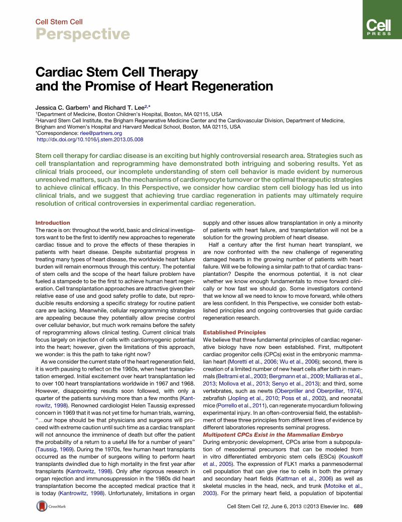
Figure 3. Approaches to CardiacRegeneration after InjuryMultiple strategies are under investigation to pro-mote cardiac regeneration in diseased hearts(clockwise from top): (1) cell therapy with culturedcells injected into the myocardium or coronaryarteries is in clinical trials, with hopes that thesecells may become functional cardiomyocytes; (2)tissue engineering approaches that combine cellswith biomaterials to create functional tissue in vitrofor transplantation into the heart; (3) reprogram-ming noncardiomyocytes into cardiomyocytesin situ may be accomplished with viruses, smallmolecules, or microRNAs; and (4) small moleculessuch as growth factors or microRNAs that aredelivered to promote wound healing via car-diomyocyte proliferation or angiogenesis.
Cell Stem Cell
Perspective
reduction of the scar mass and an increase of viable tissue and
regional contractility when evaluated by cardiac magnetic reso-
nance imaging (MRI) at 6 months (Makkar et al., 2012). No
adverse events related to cell transplantation were reported in
either study at 1 year (SCIPIO) or 6 months (CADUCEUS). To
date, no single cell type has proven itself to meet sufficient
criteria for widespread use in clinical applications, a fact that
may ultimately hinder progress in cell transplantation ap-
proaches.
What Is the Mechanism of Action by which Cell
Transplantation Demonstrates Clinical Efficacy?
The mechanism by which exogenous administration of autolo-
gous progenitor cells contributes to improving cardiac function
remains unclear. It is possible that these autologous cells are
leading to regeneration, but it is also plausible that paracrine
effects or changes in the myocardial response to injury are
responsible. The available technology for imaging cell fate and
myocardium does not allow determination of true regeneration;
therefore we must rely on surrogate measures of efficacy.
Prominent claims that bone marrow cells can become cardio-
myocytes after transplantation into myocardium (Orlic et al.,
2001) have not been replicated by other laboratories (Loffredo
et al., 2011; Murry et al., 2004; Wagers et al., 2002). This conflict
is responsible for some of the ongoing confusion in the field
(Limbourg and Drexler, 2005). The use of bone marrow cells for
prevention and treatment of heart failure has had varied clinical
success to date but remains under intense clinical investigation
as described above.
Extensive data indicate that most cells transplanted into the
heart do not survive long-term, and thus the concept of paracrine
694 Cell Stem Cell 12, June 6, 2013 ª2013 Elsevier Inc.
effects from injected cells has become
popular despite only indirect evidence
for this theory (Govaert et al., 2009; Lof-
fredo et al., 2011). In addition to the mod-
ulation of the extracellular milieu in vitro
(Baffour et al., 2006), the effect of trans-
planted bone-marrow-derived cells on
improving cardiac function may be due
primarily to a paracrine effect (Gnecchi
et al., 2005; Iso et al., 2007; Loffredo
et al., 2011; Williams and Hare, 2011).
Even in the case of human cardio-
sphere-derived cells, which are derived
from human myocardium, the benefits of cell therapy may be
paracrine (Li et al., 2012). The factors secreted or released
from injected cells that benefit cardiac function remain to be
identified. If there is a specific combination of multiple factors
from a defined population of cells, then unraveling the paracrine
cocktail may be very challenging. Furthermore, as improved
methods to enhance cell survival and engraftment are devel-
oped, distinguishing between independent cell effects and para-
crine effects will become even more difficult.
A major challenge in cell therapy approaches is how to
improve engraftment. An excellent review by Terrovitis and col-
leagues describes methods to both evaluate and optimize
engraftment (Terrovitis et al., 2010). Methods to quantify engraft-
ment remain controversial, and correlation of engraftment to im-
provements in morbidity andmortality remain unclear. Surrogate
measures of success such as global heart function with LVEF
may not provide adequate resolution, although cardiac MRI
may facilitate both local and global assessment. Finally, intro-
ducing cells into a hostile, diseased environment such as
ischemic myocardium likely hinders engraftment, and without
the reestablishment of adequate vascularization, it is unlikely
that transplantation of cardiomyocytes alone will achieve suc-
cess.
What Is the Ideal Approach for Clinical Cardiac
Regeneration?
Multiple approaches are under investigation for human cardiac
regeneration (Figure 3). As described above, significant progress
has been made in cell transplantation approaches; how-
ever, these methods are challenged by poor cell survival and
engraftment and may lack true regeneration. Alternatively,

Cell Stem Cell
Perspective
reprogramming of endogenous nonmyocytes into cardiomyo-
cytes may allow in situ transdifferentiation, although these
methods require further validation before they will be ready for
clinical trials.
Despite the lack of evidence for true regeneration with cell
therapy approaches, clinical success will ultimately depend on
evidence of clinical efficacy, and some cell therapy methods
have shown limited improvement in cardiac function as
described above. Importantly, cardiac cell therapy has been sur-
prisingly safe to date. No report of tumor formation has occurred
in over 1,500 patients involved in bone marrow cell cardiac trials
(Clifford et al., 2012). Teratoma formation has been seen in
monkeys injected with unpurified human ESC-derived cardio-
myocytes (Blin et al., 2010); however, adequate purification of
cardiac populations prior to transplantation may prevent tumor
formation (Blin et al., 2010; Tohyama et al., 2013).
No consensus has been reached about the optimal delivery
method for transplanted cells. Intravenous, intracoronary, and
intramyocardial injection methods have all been proposed,
although all are limited by poor local retention (Dib et al.,
2011). Tissue engineering approaches combine cells with bio-
materials to address logistical challenges. Use of injectable
hydrogels has been studied with both natural and synthetic
biomaterials to try to improve local retention (Ye et al., 2011).
Biodegradable scaffolds seeded with cells can be used to
form well-defined architectures as in valve tissue engineering
(Schmidt et al., 2007). Finally, placement of a cardiac patch
formed with stem cells can provide both structural and paracrine
support after myocardial injury (Wei et al., 2008). While tissue en-
gineering approaches are still in development, these ap-
proaches will likely augment the behavior, and ultimately the
success, of transplanted cells.
Cellular reprogramming approaches aim to modify the pheno-
type of native cells to induce cardiomyocyte renewal via delivery
of small molecules in vivo. Cellular reprogramming strategies
may ultimately win over cell transplantation because of the chal-
lenges of timely production of sufficient quantities of autologous
cells that meet all criteria necessary for safe and efficacious
transplantation. However, much work remains before the safety
and efficacy of reprogramming allows clinical testing. Aguirre
and colleagues (Aguirre et al., 2013) recently provided an excel-
lent review on animal models for cardiac reprogramming, and
this topic is discussed further in the following section.
How Can We Promote Stable Differentiation of
Nonmyocytes into Cardiac Phenotypes?
The possibility of skipping the multipotent state and directly re-
programming cells in vivo from one differentiated phenotype to
another was demonstrated in pancreatic cells byMelton and col-
leagues (Zhou et al., 2008b). The Srivastava group devised a
method to directly reprogram fibroblasts to cardiomyocyte-like
cells using a combination of three transcription factors
(GATA4, MEF2C, and TBX5) (Ieda et al., 2010). Using a retroviral
system to deliver GATA4, MEF2C, and TBX5 to 2-month-old
male mice in vivo via intramyocardial delivery, the same group
found that cardiomyocyte-like cells were formed from the resi-
dent fibroblast population, and this intervention resulted in
improved myocardial function after infarction (Qian et al.,
2012). Similarly, four transcription factors (GATA4, HAND2,
MEF2C, and TBX5) were used to reprogram mouse tail-tip and
cardiac fibroblasts into functional cardiomyocyte-like cells in vivo
(Song et al., 2012).
Subsequent studies have demonstrated direct reprogram-
ming using microRNA (Jayawardena et al., 2012) or alternative
transcription factors such as ETS2 and MESP1 (Islas et al.,
2012). However, these methods exhibit low efficiency and
incomplete efficacy in reprogramming fibroblasts into cardio-
myocyte-like cells (Chen et al., 2012), and further investigation
is required to better understand themechanisms by which trans-
differentiation occurs. If, as suggested by Srivistava and col-
leagues (Qian et al., 2012), maturation of reprogrammed cells
can occur in vivo, then it is conceivable that long-term stable
integration of reprogrammed cardiomyocytes may be possible.
It remains unclear if delivery of transcription factors may have ef-
fects on noncardiac tissues in the event of poorly localized deliv-
ery, or if uncontrolled cardiomyocyte reprogramming has
adverse effects such as rhythm disturbances. Prior to clinical
translation of cellular reprogramming methods, wemust achieve
a deeper understanding of the molecular mechanisms of regen-
eration.
ConclusionsStem cell biology holds significant promise for heart diseases.
Because autologous cardiac cell therapy appears to be safe
and possibly effective, investigators are aggressively advancing
this clinical approach. At this early stage, these efforts must
undergo rigorous study, preferably with randomization and
blinded outcome assessment. We believe that cardiac cell ther-
apy outside of such carefully designed and monitored trials is
currently unethical. As is apparent to most investigators in the
field, the current published data on cardiac regeneration and
cardiac stem cells conflict in important ways. While confusion
is to be expected in early days of an exciting field, this is espe-
cially true when new technologies are coming out rapidly and
when clinical trials have begun, as investigators feel even more
invested in the ‘‘established’’ premises underlying their work.
But as the enthusiasm for cardiac regeneration charges ahead
toward clinical translation, it is crucial for all investigators to
maintain objectivity and seek new and complementary ap-
proaches to resolve apparent controversies.
Are we on the right path? Although it is possible that current
cardiac cell therapy trials in humans are causing true regenera-
tion, we suggest that the overall evidence is most consistent
with the concept that cardiac cell therapy is regulating an endog-
enous repair process and not leading to true regeneration. None-
theless, patients who achieve improved recovery will not care if
we call it ‘‘regeneration’’ or ‘‘repair,’’ so enhancing heart function
through cell transplantation is a worthy goal, even if it turns out
not to be through true regeneration.
Ultimately, though, we must understand the dramatic differ-
ences between cardiac regeneration in experimental models
like zebrafish and neonatal mice and the profound postnatal
loss of cardiac regenerative potential in adult mammals like
mice and humans. Is this due to intrinsic properties of cardio-
myocytes or due to failure of stem/progenitor populations? Is it
due to noncardiomyocytes, such as activated fibroblasts
creating scarring that blocks regeneration? As in regeneration
of many different mammalian organs, the core issues in cardiac
regeneration remain mysterious, and we have yet to understand
Cell Stem Cell 12, June 6, 2013 ª2013 Elsevier Inc. 695

Cell Stem Cell
Perspective
what signals start the regenerative process, how regeneration is
guided, and finally, how regeneration is terminated.
ACKNOWLEDGMENTS
This work was funded in part by grants from NIH (R01 AG032977 1R01AG040019) to R.T.L. The authors thank Sean M. Wu of Stanford Universityfor his helpful comments. R.L. is cofounder and coowner of Provasculon,Inc. R.L. is a paid consultant to the company and serves on the company’sBoard of Directors. Provasculon has interests in regenerative cell therapy,an area related to this work. R.L.’s interests were reviewed by the BrighamandWomen’s Hospital and Partners HealthCare in accordance with their insti-tutional policies.
REFERENCES
Aasen, T., and Izpisua Belmonte, J.C. (2010). Isolation and cultivation of hu-man keratinocytes from skin or plucked hair for the generation of inducedpluripotent stem cells. Nat. Protoc. 5, 371–382.
Aguirre, A., Sancho-Martinez, I., and Izpisua Belmonte, J.C. (2013). Reprog-ramming toward heart regeneration: stem cells and beyond. Cell Stem Cell12, 275–284.
Amado, L.C., Saliaris, A.P., Schuleri, K.H., St John, M., Xie, J.S., Cattaneo, S.,Durand, D.J., Fitton, T., Kuang, J.Q., Stewart, G., et al. (2005). Cardiac repairwith intramyocardial injection of allogeneic mesenchymal stem cells aftermyocardial infarction. Proc. Natl. Acad. Sci. USA 102, 11474–11479.
Assmus, B., Rolf, A., Erbs, S., Elsasser, A., Haberbosch, W., Hambrecht, R.,Tillmanns, H., Yu, J., Corti, R., Mathey, D.G., et al.; REPAIR-AMI Investigators.(2010). Clinical outcome 2 years after intracoronary administration of bonemarrow-derived progenitor cells in acute myocardial infarction. Circ HeartFail 3, 89–96.
Baffour, R., Pakala, R., Hellinga, D., Joner, M., Okubagzi, P., Epstein, S.E., andWaksman, R. (2006). Bone marrow-derived stem cell interactions with adultcardiomyocytes and skeletal myoblasts in vitro. Cardiovasc. Revasc. Med.7, 222–230.
Bearzi, C., Rota, M., Hosoda, T., Tillmanns, J., Nascimbene, A., De Angelis, A.,Yasuzawa-Amano, S., Trofimova, I., Siggins, R.W., Lecapitaine, N., et al.(2007). Human cardiac stem cells. Proc. Natl. Acad. Sci. USA 104, 14068–14073.
Beltrami, A.P., Barlucchi, L., Torella, D., Baker, M., Limana, F., Chimenti, S.,Kasahara, H., Rota, M., Musso, E., Urbanek, K., et al. (2003). Adult cardiacstem cells are multipotent and support myocardial regeneration. Cell 114,763–776.
Bergmann, O., Bhardwaj, R.D., Bernard, S., Zdunek, S., Barnabe-Heider, F.,Walsh, S., Zupicich, J., Alkass, K., Buchholz, B.A., Druid, H., et al. (2009).Evidence for cardiomyocyte renewal in humans. Science 324, 98–102.
Blin, G., Nury, D., Stefanovic, S., Neri, T., Guillevic, O., Brinon, B., Bellamy, V.,Rucker-Martin, C., Barbry, P., Bel, A., et al. (2010). A purified population ofmul-tipotent cardiovascular progenitors derived from primate pluripotent stemcells engrafts in postmyocardial infarcted nonhuman primates. J. Clin. Invest.120, 1125–1139.
Bolli, R., Chugh, A.R., D’Amario, D., Loughran, J.H., Stoddard, M.F., Ikram, S.,Beache, G.M., Wagner, S.G., Leri, A., Hosoda, T., et al. (2011). Cardiac stemcells in patients with ischaemic cardiomyopathy (SCIPIO): initial results of arandomised phase 1 trial. Lancet 378, 1847–1857.
Bollini, S., Smart, N., and Riley, P.R. (2011). Resident cardiac progenitor cells:at the heart of regeneration. J. Mol. Cell. Cardiol. 50, 296–303.
Chen, J.X., Krane, M., Deutsch, M.A., Wang, L., Rav-Acha, M., Gregoire, S.,Engels, M.C., Rajarajan, K., Karra, R., Abel, E.D., et al. (2012). Inefficient re-programming of fibroblasts into cardiomyocytes using Gata4, Mef2c, andTbx5. Circ. Res. 111, 50–55.
Clifford, D.M., Fisher, S.A., Brunskill, S.J., Doree, C., Mathur, A., Watt, S., andMartin-Rendon, E. (2012). Stem cell treatment for acute myocardial infarction.Cochrane Database Syst. Rev. 2, CD006536.
Davis, R.P., Casini, S., van den Berg, C.W., Hoekstra, M., Remme, C.A., Dam-brot, C., Salvatori, D., Oostwaard, D.W., Wilde, A.A., Bezzina, C.R., et al.
696 Cell Stem Cell 12, June 6, 2013 ª2013 Elsevier Inc.
(2012). Cardiomyocytes derived from pluripotent stem cells recapitulate elec-trophysiological characteristics of an overlap syndrome of cardiac sodiumchannel disease. Circulation 125, 3079–3091.
Dib, N., Khawaja, H., Varner, S., McCarthy, M., and Campbell, A. (2011). Celltherapy for cardiovascular disease: a comparison of methods of delivery.J. Cardiovasc. Transl. Res. 4, 177–181.
Dor, Y., Brown, J., Martinez, O.I., and Melton, D.A. (2004). Adult pancreaticbeta-cells are formed by self-duplication rather than stem-cell differentiation.Nature 429, 41–46.
Drenckhahn, J.D., Schwarz, Q.P., Gray, S., Laskowski, A., Kiriazis, H., Ming,Z., Harvey, R.P., Du, X.J., Thorburn, D.R., and Cox, T.C. (2008). Compensatorygrowth of healthy cardiac cells in the presence of diseased cells restores tissuehomeostasis during heart development. Dev. Cell 15, 521–533.
Fransioli, J., Bailey, B., Gude, N.A., Cottage, C.T., Muraski, J.A., Emmanuel,G., Wu, W., Alvarez, R., Rubio, M., Ottolenghi, S., et al. (2008). Evolution ofthe c-kit-positive cell response to pathological challenge in the myocardium.Stem Cells 26, 1315–1324.
Gnecchi, M., He, H., Liang, O.D., Melo, L.G., Morello, F., Mu, H., Noiseux, N.,Zhang, L., Pratt, R.E., Ingwall, J.S., and Dzau, V.J. (2005). Paracrine action ac-counts for marked protection of ischemic heart by Akt-modified mesenchymalstem cells. Nat. Med. 11, 367–368.
Govaert, J.A., Swijnenburg, R.J., Schrepfer, S., Xie, X., van der Bogt, K.E.,Hoyt, G., Stein, W., Ransohoff, K.J., Robbins, R.C., and Wu, J.C. (2009).Poor functional recovery after transplantation of diabetic bone marrow stemcells in ischemic myocardium. J. Heart Lung Transplant. 28, 1158–1165, e1.
Hare, J.M., Fishman, J.E., Gerstenblith, G., DiFede Velazquez, D.L., Zam-brano, J.P., Suncion, V.Y., Tracy, M., Ghersin, E., Johnston, P.V., Brinker,J.A., et al. (2012). Comparison of allogeneic vs autologous bone marrow–derived mesenchymal stem cells delivered by transendocardial injection inpatients with ischemic cardiomyopathy: the POSEIDON randomized trial.JAMA 308, 2369–2379.
Huang, G.N., Thatcher, J.E., McAnally, J., Kong, Y., Qi, X., Tan, W., DiMaio,J.M., Amatruda, J.F., Gerard, R.D., Hill, J.A., et al. (2012). C/EBP transcriptionfactors mediate epicardial activation during heart development and injury.Science 338, 1599–1603.
Ieda, M., Fu, J.D., Delgado-Olguin, P., Vedantham, V., Hayashi, Y., Bruneau,B.G., and Srivastava, D. (2010). Direct reprogramming of fibroblasts into func-tional cardiomyocytes by defined factors. Cell 142, 375–386.
Islas, J.F., Liu, Y., Weng, K.C., Robertson, M.J., Zhang, S., Prejusa, A., Harger,J., Tikhomirova, D., Chopra, M., Iyer, D., et al. (2012). Transcription factorsETS2 andMESP1 transdifferentiate human dermal fibroblasts into cardiac pro-genitors. Proc. Natl. Acad. Sci. USA 109, 13016–13021.
Iso, Y., Spees, J.L., Serrano, C., Bakondi, B., Pochampally, R., Song, Y.H.,Sobel, B.E., Delafontaine, P., and Prockop, D.J. (2007). Multipotent humanstromal cells improve cardiac function after myocardial infarction in micewithout long-term engraftment. Biochem. Biophys. Res. Commun. 354,700–706.
Jayawardena, T.M., Egemnazarov, B., Finch, E.A., Zhang, L., Payne, J.A., Pan-dya, K., Zhang, Z., Rosenberg, P., Mirotsou, M., and Dzau, V.J. (2012).MicroRNA-mediated in vitro and in vivo direct reprogramming of cardiac fibro-blasts to cardiomyocytes. Circ. Res. 110, 1465–1473.
Jopling, C., Sleep, E., Raya, M., Martı, M., Raya, A., and Izpisua Belmonte, J.C.(2010). Zebrafish heart regeneration occurs by cardiomyocyte dedifferentia-tion and proliferation. Nature 464, 606–609.
Kajstura, J., Leri, A., Finato, N., Di Loreto, C., Beltrami, C.A., and Anversa, P.(1998). Myocyte proliferation in end-stage cardiac failure in humans. Proc.Natl. Acad. Sci. USA 95, 8801–8805.
Kajstura, J., Gurusamy, N., Ogorek, B., Goichberg, P., Clavo-Rondon, C., Ho-soda, T., D’Amario, D., Bardelli, S., Beltrami, A.P., Cesselli, D., et al. (2010).Myocyte turnover in the aging human heart. Circ. Res. 107, 1374–1386.
Kajstura, J., Rota, M., Cappetta, D., Ogorek, B., Arranto, C., Bai, Y., Ferreira-Martins, J., Signore, S., Sanada, F., Matsuda, A., et al. (2012). Cardiomyogen-esis in the aging and failing human heart. Circulation 126, 1869–1881.
Kantrowitz, A. (1998). America’s first human heart transplantation: theconcept, the planning, and the furor. ASAIO J. 44, 244–252.
Cell Stem Cell
Perspective
Kattman, S.J., Huber, T.L., and Keller, G.M. (2006). Multipotent flk-1+ cardio-vascular progenitor cells give rise to the cardiomyocyte, endothelial, andvascular smooth muscle lineages. Dev. Cell 11, 723–732.
Kikuchi, K., Holdway, J.E.,Werdich, A.A., Anderson, R.M., Fang, Y., Egnaczyk,G.F., Evans, T., Macrae, C.A., Stainier, D.Y., and Poss, K.D. (2010). Primarycontribution to zebrafish heart regeneration by gata4(+) cardiomyocytes.Nature 464, 601–605.
Kouskoff, V., Lacaud, G., Schwantz, S., Fehling, H.J., and Keller, G. (2005).Sequential development of hematopoietic and cardiac mesoderm during em-bryonic stem cell differentiation. Proc. Natl. Acad. Sci. USA 102, 13170–13175.
Laflamme, M.A., Chen, K.Y., Naumova, A.V., Muskheli, V., Fugate, J.A., Dup-ras, S.K., Reinecke, H., Xu, C., Hassanipour, M., Police, S., et al. (2007). Car-diomyocytes derived from human embryonic stem cells in pro-survival factorsenhance function of infarcted rat hearts. Nat. Biotechnol. 25, 1015–1024.
Laube, F., Heister, M., Scholz, C., Borchardt, T., and Braun, T. (2006). Re-pro-gramming of newt cardiomyocytes is induced by tissue regeneration. J. CellSci. 119, 4719–4729.
Leobon, B., Garcin, I., Menasche, P., Vilquin, J.T., Audinat, E., and Charpak, S.(2003). Myoblasts transplanted into rat infarcted myocardium are functionallyisolated from their host. Proc. Natl. Acad. Sci. USA 100, 7808–7811.
Lepilina, A., Coon, A.N., Kikuchi, K., Holdway, J.E., Roberts, R.W., Burns,C.G., and Poss, K.D. (2006). A dynamic epicardial injury response supportsprogenitor cell activity during zebrafish heart regeneration. Cell 127, 607–619.
Li, T.S., Cheng, K., Malliaras, K., Smith, R.R., Zhang, Y., Sun, B., Matsushita,N., Blusztajn, A., Terrovitis, J., Kusuoka, H., et al. (2012). Direct comparison ofdifferent stem cell types and subpopulations reveals superior paracrine po-tency and myocardial repair efficacy with cardiosphere-derived cells. J. Am.Coll. Cardiol. 59, 942–953.
Limbourg, F.P., and Drexler, H. (2005). Bone marrow stem cells for myocardialinfarction: effector or mediator? Circ. Res. 96, 6–8.
Loffredo, F.S., Steinhauser, M.L., Gannon, J., and Lee, R.T. (2011). Bonemarrow-derived cell therapy stimulates endogenous cardiomyocyte progeni-tors and promotes cardiac repair. Cell Stem Cell 8, 389–398.
Makkar, R.R., Smith, R.R., Cheng, K., Malliaras, K., Thomson, L.E., Berman,D., Czer, L.S., Marban, L., Mendizabal, A., Johnston, P.V., et al. (2012). Intra-coronary cardiosphere-derived cells for heart regeneration after myocardialinfarction (CADUCEUS): a prospective, randomised phase 1 trial. Lancet379, 895–904.
Malliaras, K., Zhang, Y., Seinfeld, J., Galang, G., Tseliou, E., Cheng, K., Sun,B., Aminzadeh, M., and Marban, E. (2013). Cardiomyocyte proliferation andprogenitor cell recruitment underlie therapeutic regeneration after myocardialinfarction in the adult mouse heart. EMBO Mol Med 5, 191–209.
Marban, E., and Malliaras, K. (2012). Mixed results for bone marrow–derivedcell therapy for ischemic heart disease. JAMA 308, 2405–2406.
Messina, E., De Angelis, L., Frati, G., Morrone, S., Chimenti, S., Fiordaliso, F.,Salio, M., Battaglia, M., Latronico, M.V., Coletta, M., et al. (2004). Isolation andexpansion of adult cardiac stem cells from human and murine heart. Circ. Res.95, 911–921.
Mollova, M., Bersell, K., Walsh, S., Savla, J., Das, L.T., Park, S.Y., Silberstein,L.E., Dos Remedios, C.G., Graham, D., Colan, S., and Kuhn, B. (2013). Cardi-omyocyte proliferation contributes to heart growth in young humans. Proc.Natl. Acad. Sci. USA 110, 1446–1451.
Moretti, A., Caron, L., Nakano, A., Lam, J.T., Bernshausen, A., Chen, Y.,Qyang, Y., Bu, L., Sasaki, M., Martin-Puig, S., et al. (2006). Multipotent embry-onic isl1+ progenitor cells lead to cardiac, smooth muscle, and endothelial celldiversification. Cell 127, 1151–1165.
Motoike, T., Markham, D.W., Rossant, J., and Sato, T.N. (2003). Evidence fornovel fate of Flk1+ progenitor: contribution to muscle lineage. Genesis 35,153–159.
Murry, C.E., Soonpaa, M.H., Reinecke, H., Nakajima, H., Nakajima, H.O., Ru-bart, M., Pasumarthi, K.B., Virag, J.I., Bartelmez, S.H., Poppa, V., et al. (2004).Haematopoietic stem cells do not transdifferentiate into cardiac myocytes inmyocardial infarcts. Nature 428, 664–668.
Nelson, T.J., Martinez-Fernandez, A., Yamada, S., Perez-Terzic, C., Ikeda, Y.,and Terzic, A. (2009). Repair of acute myocardial infarction by human stem-ness factors induced pluripotent stem cells. Circulation 120, 408–416.
Oberpriller, J.O., and Oberpriller, J.C. (1974). Response of the adult newtventricle to injury. J. Exp. Zool. 187, 249–253.
Oh, H., Bradfute, S.B., Gallardo, T.D., Nakamura, T., Gaussin, V., Mishina, Y.,Pocius, J., Michael, L.H., Behringer, R.R., Garry, D.J., et al. (2003). Cardiacprogenitor cells from adult myocardium: homing, differentiation, and fusion af-ter infarction. Proc. Natl. Acad. Sci. USA 100, 12313–12318.
Ohnishi, S., Ohgushi, H., Kitamura, S., and Nagaya, N. (2007). Mesenchymalstem cells for the treatment of heart failure. Int. J. Hematol. 86, 17–21.
Okita, K., Nakagawa, M., Hyenjong, H., Ichisaka, T., and Yamanaka, S. (2008).Generation of mouse induced pluripotent stem cells without viral vectors.Science 322, 949–953.
Orlic, D., Kajstura, J., Chimenti, S., Jakoniuk, I., Anderson, S.M., Li, B., Pickel,J., McKay, R., Nadal-Ginard, B., Bodine, D.M., et al. (2001). Bonemarrow cellsregenerate infarcted myocardium. Nature 410, 701–705.
Pfister, O., Mouquet, F., Jain, M., Summer, R., Helmes, M., Fine, A., Colucci,W.S., and Liao, R. (2005). CD31- but Not CD31+ cardiac side population cellsexhibit functional cardiomyogenic differentiation. Circ. Res. 97, 52–61.
Porrello, E.R., Mahmoud, A.I., Simpson, E., Hill, J.A., Richardson, J.A., Olson,E.N., and Sadek, H.A. (2011). Transient regenerative potential of the neonatalmouse heart. Science 331, 1078–1080.
Poss, K.D.,Wilson, L.G., and Keating,M.T. (2002). Heart regeneration in zebra-fish. Science 298, 2188–2190.
Qian, L., Huang, Y., Spencer, C.I., Foley, A., Vedantham, V., Liu, L., Conway,S.J., Fu, J.D., and Srivastava, D. (2012). In vivo reprogramming of murine car-diac fibroblasts into induced cardiomyocytes. Nature 485, 593–598.
Quaini, F., Cigola, E., Lagrasta, C., Saccani, G., Quaini, E., Rossi, C., Olivetti,G., and Anversa, P. (1994). End-stage cardiac failure in humans is coupled withthe induction of proliferating cell nuclear antigen and nuclear mitotic division inventricular myocytes. Circ. Res. 75, 1050–1063.
Rumyantsev, P.P. (1974). Ultrastructural reorganization, DNA synthesis andmitotic division of myocytes in atria of rats with left ventricle infarction. An elec-tron microscopic and autoradiographic study. Virchows Arch. B Cell Pathol.Incl. Mol. Pathol. 15, 357–378.
Schmidt, D., Achermann, J., Odermatt, B., Breymann, C., Mol, A., Genoni, M.,Zund, G., and Hoerstrup, S.P. (2007). Prenatally fabricated autologous humanliving heart valves based on amniotic fluid derived progenitor cells as single cellsource. Circulation 116(11, Suppl), I64–I70.
Senyo, S.E., Steinhauser, M.L., Pizzimenti, C.L., Yang, V.K., Cai, L., Wang, M.,Wu, T.D., Guerquin-Kern, J.L., Lechene, C.P., and Lee, R.T. (2013). Mamma-lian heart renewal by pre-existing cardiomyocytes. Nature 493, 433–436.
Shiba, Y., Fernandes, S., Zhu, W.Z., Filice, D., Muskheli, V., Kim, J., Palpant,N.J., Gantz, J., Moyes, K.W., Reinecke, H., et al. (2012). Human ES-cell-derived cardiomyocytes electrically couple and suppress arrhythmias ininjured hearts. Nature 489, 322–325.
Smart, N., Bollini, S., Dube, K.N., Vieira, J.M., Zhou, B., Davidson, S., Yellon,D., Riegler, J., Price, A.N., Lythgoe, M.F., et al. (2011). De novo cardiomyo-cytes from within the activated adult heart after injury. Nature 474, 640–644.
Smith, R.R., Barile, L., Cho, H.C., Leppo, M.K., Hare, J.M., Messina, E., Giaco-mello, A., Abraham, M.R., and Marban, E. (2007). Regenerative potential ofcardiosphere-derived cells expanded from percutaneous endomyocardialbiopsy specimens. Circulation 115, 896–908.
Song, K., Nam, Y.J., Luo, X., Qi, X., Tan, W., Huang, G.N., Acharya, A., Smith,C.L., Tallquist, M.D., Neilson, E.G., et al. (2012). Heart repair by reprogram-ming non-myocytes with cardiac transcription factors. Nature 485, 599–604.
Soonpaa, M.H., and Field, L.J. (1997). Assessment of cardiomyocyte DNAsynthesis in normal and injured adult mouse hearts. Am. J. Physiol. 272,H220–H226.
Soonpaa, M.H., Rubart, M., and Field, and, L.J. (2013). Challenges measuringcardiomyocyte renewal. Biochim. Biophys. Acta. 1833, 799–803.
Cell Stem Cell 12, June 6, 2013 ª2013 Elsevier Inc. 697

Cell Stem Cell
Perspective
Takahashi, K., and Yamanaka, S. (2006). Induction of pluripotent stem cellsfrom mouse embryonic and adult fibroblast cultures by defined factors. Cell126, 663–676.
Takahashi, K., Tanabe, K., Ohnuki, M., Narita, M., Ichisaka, T., Tomoda, K.,and Yamanaka, S. (2007). Induction of pluripotent stem cells from adult humanfibroblasts by defined factors. Cell 131, 861–872.
Taussig, H.B. (1969). Heart transplantation. JAMA 207, 951–951.
Terrovitis, J.V., Smith, R.R., and Marban, E. (2010). Assessment and optimiza-tion of cell engraftment after transplantation into the heart. Circ. Res. 106,479–494.
Thomson, J.A., Itskovitz-Eldor, J., Shapiro, S.S., Waknitz, M.A., Swiergiel, J.J.,Marshall, V.S., and Jones, J.M. (1998). Embryonic stem cell lines derived fromhuman blastocysts. Science 282, 1145–1147.
Tohyama, S., Hattori, F., Sano, M., Hishiki, T., Nagahata, Y., Matsuura, T.,Hashimoto, H., Suzuki, T., Yamashita, H., Satoh, Y., et al. (2013). Distinctmetabolic flow enables large-scale purification of mouse and human pluripo-tent stem cell-derived cardiomyocytes. Cell Stem Cell 12, 127–137.
Traverse, J.H., Henry, T.D., Pepine, C.J., Willerson, J.T., Zhao, D.X., Ellis, S.G.,Forder, J.R., Anderson, R.D., Hatzopoulos, A.K., Penn, M.S., et al.; Cardiovas-cular Cell Therapy Research Network (CCTRN). (2012). Effect of the use andtiming of bone marrowmononuclear cell delivery on left ventricular function af-ter acute myocardial infarction: the TIME randomized trial. JAMA 308, 2380–2389.
Wagers, A.J., Sherwood, R.I., Christensen, J.L., andWeissman, I.L. (2002). Lit-tle evidence for developmental plasticity of adult hematopoietic stem cells.Science 297, 2256–2259.
Wei, H.J., Chen, C.H., Lee, W.Y., Chiu, I., Hwang, S.M., Lin, W.W., Huang,C.C., Yeh, Y.C., Chang, Y., and Sung, H.W. (2008). Bioengineered cardiac
698 Cell Stem Cell 12, June 6, 2013 ª2013 Elsevier Inc.
patch constructed from multilayered mesenchymal stem cells for myocardialrepair. Biomaterials 29, 3547–3556.
Williams, A.R., and Hare, J.M. (2011). Mesenchymal stem cells: biology, path-ophysiology, translational findings, and therapeutic implications for cardiacdisease. Circ. Res. 109, 923–940.
Wu, S.M., Fujiwara, Y., Cibulsky, S.M., Clapham, D.E., Lien, C.L., Schultheiss,T.M., and Orkin, S.H. (2006). Developmental origin of a bipotential myocardialand smooth muscle cell precursor in the mammalian heart. Cell 127, 1137–1150.
Ye, Z., Zhou, Y., Cai, H., and Tan, W. (2011). Myocardial regeneration: Roles ofstem cells and hydrogels. Adv. Drug Deliv. Rev. 63, 688–697.
Yu, J., Vodyanik, M.A., Smuga-Otto, K., Antosiewicz-Bourget, J., Frane, J.L.,Tian, S., Nie, J., Jonsdottir, G.A., Ruotti, V., Stewart, R., et al. (2007). Inducedpluripotent stem cell lines derived from human somatic cells. Science 318,1917–1920.
Zhang, J., Wilson, G.F., Soerens, A.G., Koonce, C.H., Yu, J., Palecek, S.P.,Thomson, J.A., and Kamp, T.J. (2009). Functional cardiomyocytes derivedfrom human induced pluripotent stem cells. Circ. Res. 104, e30–e41.
Zhou, B., Ma, Q., Rajagopal, S., Wu, S.M., Domian, I., Rivera-Feliciano, J.,Jiang, D., von Gise, A., Ikeda, S., Chien, K.R., and Pu, W.T. (2008a). Epicardialprogenitors contribute to the cardiomyocyte lineage in the developing heart.Nature 454, 109–113.
Zhou, Q., Brown, J., Kanarek, A., Rajagopal, J., and Melton, D.A. (2008b).In vivo reprogramming of adult pancreatic exocrine cells to beta-cells. Nature455, 627–632.
Zhou, B., Honor, L.B., Ma, Q., Oh, J.H., Lin, R.Z., Melero-Martin, J.M., vonGise, A., Zhou, P., Hu, T., He, L., et al. (2012). Thymosin beta 4 treatment aftermyocardial infarction does not reprogram epicardial cells into cardiomyo-cytes. J. Mol. Cell. Cardiol. 52, 43–47.