Cell signalling pathways underlying induced pluripotent ... online: November 26, 2014 Abstract...
10
Kate Hawkins, Shona Joy, Tristan McKay Kate Hawkins, Shona Joy, Tristan McKay, Molecular Cell Sci- ences, St George’s University of London, London SW17 0RE, United Kingdom Author contributions: All the authors solely contributed to this paper. Correspondence to: Dr. Kate Hawkins, Molecular Cell Sci- ences, St George’s University of London, Cranmer Terrace, Lon- don SW17 0RE, United Kingdom. [email protected] Telephone: +44-20-87253646 Received: July 23, 2014 Revised: September 5, 2014 Accepted: September 17, 2014 Published online: November 26, 2014 Abstract Induced pluripotent stem (iPS) cells, somatic cells reprogrammed to the pluripotent state by forced ex- pression of defined factors, represent a uniquely valu- able resource for research and regenerative medicine. However, this methodology remains inefficient due to incomplete mechanistic understanding of the repro- gramming process. In recent years, various groups have endeavoured to interrogate the cell signalling that gov- erns the reprogramming process, including LIF/STAT3, BMP, PI3K, FGF2, Wnt, TGFβ and MAPK pathways, with the aim of increasing our understanding and identifying new mechanisms of improving safety, reproducibility and efficiency. This has led to a unified model of repro- gramming that consists of 3 stages: initiation, matura- tion and stabilisation. Initiation of reprogramming oc- curs in almost all cells that receive the reprogramming transgenes; most commonly Oct4, Sox2, Klf4 and cMyc , and involves a phenotypic mesenchymal-to-epithelial transition. The initiation stage is also characterised by increased proliferation and a metabolic switch from oxidative phosphorylation to glycolysis. The maturation stage is considered the major bottleneck within the pro- cess, resulting in very few “stabilisation competent” cells progressing to the final stabilisation phase. To reach this stage in both mouse and human cells, pre-iPS cells must activate endogenous expression of the core circuitry of pluripotency, comprising Oct4, Sox2, and Nanog , and thus reach a state of transgene independence. By the stabilisation stage, iPS cells generally use the same sig- nalling networks that govern pluripotency in embryonic stem cells. These pathways differ between mouse and human cells although recent work has demonstrated that this is context dependent. As iPS cell generation technologies move forward, tools are being developed to interrogate the process in more detail, thus allowing a greater understanding of this intriguing biological phe- nomenon. © 2014 Baishideng Publishing Group Inc. All rights reserved. Key words: Pluripotency; Reprogramming; Induced plu- ripotent stem; Cell signalling; Embryonic stem Core tip: Induced pluripotent stem (iPS) cells present great promise, both to research and to medicine. How- ever, we know very little regarding the mechanisms that occur throughout the iPS cell reprogramming process and thus the process remains inefficient. In this review, we discuss the 3 stages of reprogramming, initiation, maturation and stabilisation, and clarify the signalling pathways underlying each phase. We draw together the current knowledge to propose a model for the interac- tions between the key pathways in iPS cell reprogram- ming with the aim of illuminating this complex yet fasci- nating process. Hawkins K, Joy S, McKay T. Cell signalling pathways underlying induced pluripotent stem cell reprogramming. World J Stem Cells 2014; 6(5): 620-628 Available from: URL: http://www.wjgnet. com/1948-0210/full/v6/i5/620.htm DOI: http://dx.doi.org/10.4252/ wjsc.v6.i5.620 INTRODUCTION Pluripotency, the ability of a single cell to give rise to all 620 November 26, 2014|Volume 6|Issue 5| WJSC|www.wjgnet.com Cell signalling pathways underlying induced pluripotent stem cell reprogramming MINIREVIEWS Submit a Manuscript: http://www.wjgnet.com/esps/ Help Desk: http://www.wjgnet.com/esps/helpdesk.aspx DOI: 10.4252/wjsc.v6.i5.620 World J Stem Cells 2014 November 26; 6(5): 620-628 ISSN 1948-0210 (online) © 2014 Baishideng Publishing Group Inc. All rights reserved.
-
Upload
nguyendiep -
Category
Documents
-
view
218 -
download
2
Transcript of Cell signalling pathways underlying induced pluripotent ... online: November 26, 2014 Abstract...

Kate Hawkins, Shona Joy, Tristan McKay
Kate Hawkins, Shona Joy, Tristan McKay, Molecular Cell Sci-ences, St George’s University of London, London SW17 0RE, United KingdomAuthor contributions: All the authors solely contributed to this paper.Correspondence to: Dr. Kate Hawkins, Molecular Cell Sci-ences, St George’s University of London, Cranmer Terrace, Lon-don SW17 0RE, United Kingdom. [email protected]: +44-20-87253646Received: July 23, 2014 Revised: September 5, 2014Accepted: September 17, 2014Published online: November 26, 2014
Abstract Induced pluripotent stem (iPS) cells, somatic cells reprogrammed to the pluripotent state by forced ex-pression of defined factors, represent a uniquely valu-able resource for research and regenerative medicine. However, this methodology remains inefficient due to incomplete mechanistic understanding of the repro-gramming process. In recent years, various groups have endeavoured to interrogate the cell signalling that gov-erns the reprogramming process, including LIF/STAT3, BMP, PI3K, FGF2, Wnt, TGFβ and MAPK pathways, with the aim of increasing our understanding and identifying new mechanisms of improving safety, reproducibility and efficiency. This has led to a unified model of repro-gramming that consists of 3 stages: initiation, matura-tion and stabilisation. Initiation of reprogramming oc-curs in almost all cells that receive the reprogramming transgenes; most commonly Oct4, Sox2, Klf4 and cMyc , and involves a phenotypic mesenchymal-to-epithelial transition. The initiation stage is also characterised by increased proliferation and a metabolic switch from oxidative phosphorylation to glycolysis. The maturation stage is considered the major bottleneck within the pro-cess, resulting in very few “stabilisation competent” cells progressing to the final stabilisation phase. To reach this stage in both mouse and human cells, pre-iPS cells must activate endogenous expression of the core circuitry of
pluripotency, comprising Oct4, Sox2, and Nanog , and thus reach a state of transgene independence. By the stabilisation stage, iPS cells generally use the same sig-nalling networks that govern pluripotency in embryonic stem cells. These pathways differ between mouse and human cells although recent work has demonstrated that this is context dependent. As iPS cell generation technologies move forward, tools are being developed to interrogate the process in more detail, thus allowing a greater understanding of this intriguing biological phe-nomenon.
© 2014 Baishideng Publishing Group Inc. All rights reserved.
Key words: Pluripotency; Reprogramming; Induced plu-ripotent stem; Cell signalling; Embryonic stem
Core tip: Induced pluripotent stem (iPS) cells present great promise, both to research and to medicine. How-ever, we know very little regarding the mechanisms that occur throughout the iPS cell reprogramming process and thus the process remains inefficient. In this review, we discuss the 3 stages of reprogramming, initiation, maturation and stabilisation, and clarify the signalling pathways underlying each phase. We draw together the current knowledge to propose a model for the interac-tions between the key pathways in iPS cell reprogram-ming with the aim of illuminating this complex yet fasci-nating process.
Hawkins K, Joy S, McKay T. Cell signalling pathways underlying induced pluripotent stem cell reprogramming. World J Stem Cells 2014; 6(5): 620-628 Available from: URL: http://www.wjgnet.com/1948-0210/full/v6/i5/620.htm DOI: http://dx.doi.org/10.4252/wjsc.v6.i5.620
INTRODUCTION Pluripotency, the ability of a single cell to give rise to all
620 November 26, 2014|Volume 6|Issue 5|WJSC|www.wjgnet.com
Cell signalling pathways underlying induced pluripotent stem cell reprogramming
MINIREVIEWS
Submit a Manuscript: http://www.wjgnet.com/esps/Help Desk: http://www.wjgnet.com/esps/helpdesk.aspxDOI: 10.4252/wjsc.v6.i5.620
World J Stem Cells 2014 November 26; 6(5): 620-628ISSN 1948-0210 (online)
© 2014 Baishideng Publishing Group Inc. All rights reserved.

cells within an entire living organism, is of great biologi-cal interest both in terms of understanding developmen-tal mechanisms as well as the medical potential that plu-ripotent stem cells possess. However, our understanding of the cell signalling networks underlying this complex process still remains incomplete. The first pluripotent stem cells were isolated from mouse blastocysts simul-taneously by 2 groups in 1981[1,2]. This was replicated 17 years later using human blastocysts[3]. Embryonic stem (ES) cells have since been isolated from other species including rhesus monkeys[4] and rats[5,6]. Both human and mouse ES cells have provided and invaluable resource to understand the basic biology of the pluripotent state.
A “core circuitry” of homeodomain transcription fac-tors, Oct4[7], Sox2[8] and Nanog[9], governs pluripotency in both mouse and human ES cells[10]. These transcription factors are expressed both in vivo in the inner cell mass (ICM) of the blastocyst and in vitro, in pluripotent cells. These 3 factors closely interact within the cell; for example Oct4 and Sox2 have been shown to form a heterodimeric transcription complex[11-13] and all 3 factors share target genes[14,15]. This interaction facilitates the precise regulation of the core circuitry necessary to maintain the pluripotent state; for instance Oct4 overexpression leads to endoderm and mesoderm differentiation whereas blockade of Oct4 induces trophoblast differentiation[7]. This may be ex-plained by its biphasic role in Nanog regulation whereby low levels of Oct4 result in upregulation of Nanog whereas higher levels of Oct4 result in downregulation of Nanog[15]. Similarly, small increases in Sox2 expression or ablation of Sox2 expression both induce multilineage differentia-tion[16]. Blockade of Nanog does not induce differentiation, thus indicating that Nanog’s role in the core circuitry of pluripotency is to stabilise the pluripotent state rather than acting as a housekeeper. However, Nanog knockdown does lead to an increased capacity for differentiation into primi-tive ectoderm[9].
The core pluripotency circuitry is also autoregula-tory since all 3 factors have been shown to regulate the expression of each other as well as themselves[14,15,17]. Interestingly, SOX2 is dispensable for the activation of Oct4/Sox2 target genes since forced expression of Oct4 is able to rescue pluripotency in Sox2-/- cells, however, Sox2
expression is necessary to maintain Oct4 expression[8]. Although it is clear that OCT4, SOX2 and NANOG oc-cupy the top level of the pluripotency hierarchy, these core factors also regulate a wide range of genes associat-ed with pluripotency signalling networks including Stat3, Zic3, Tdgf1, Lefty/Ebaf, Dkk1 and Frat2[14].
With the emergence of this complex molecular inter-play of dosage dependency between hierarchical tran-scription factors in the maintenance of the somewhat unstable pluripotent ground state, it seems surprising that simply over-expressing these factors in somatic cells can induce the pluripotent state. However, the collective sem-inal studies of Yamanaka and Thomson show this to be feasible in their descriptions of reprogramming somatic cells to induced Pluripotent Stem (iPS) cells[18-20].
The original iPS cell reprogramming strategy pub-lished by Takahashi et al[19] 7 years ago remains robust and largely unaltered to the present day. The “Yamanaka factors”, Oct4, Sox2, Klf4 and cMyc were constitutively expressed using genome integrating retroviruses in both mouse[18] and subsequently human[19] fibroblasts, and under ES cell culture conditions were able to induce plu-ripotency. To date, this methodology is still widely used, however, various adaptations to the method of vector delivery and reprogramming factors (Table 1) have been made. Advances in vector delivery have generally been made to either improve efficiency or safety, by preventing integration of the transgenes into the genome. For ex-ample, iPS cells have now been successfully generated us-ing episomal plasmids[21], Sendai viruses[22] and piggyBac transposons[23] to deliver the reprogramming factors and even proteins[24] or small molecules[25] alone. Many diver-gent cell-types have been successfully reprogrammed to pluripotency including neural stem cells[26], neural progen-itor cells[27], keratinocytes[28], B lymphocytes[29], meningeal membrane cells[30], peripheral blood mononuclear cells[31]
and pancreatic β cells[32]. Often the minimal factors nec-essary to reprogram a cell depend on the endogenous “stemness” of the starting cell, for example, neural stem cells can be reprogrammed using Oct4 alone since they express high levels of the other Yamanaka factors[26].
The common aspiration is that iPS cells will provide an autologous source of cells for a multitude of regen-erative medicine therapies in the future and clinical trials using iPS cells have begun[33]. However, the most im-mediate utility of iPS cell technologies is the ability to study patient-derived cells in the lab. iPS cells present the opportunity to study a range of diseases in novel ways by isolating and reprogramming patient-specific cells and then differentiating them into the cell type of interest. For example, iPS cells have been generated from patients suffering from a wide range of disorders including Duch-enne muscular dystrophy, Parkinson’s disease, Hunting-don’s disease, type Ⅰ diabetes and Down’s syndrome (reviewed in[34]). In addition, cells such as disease-specific cardiomyocytes, which would be difficult to obtain from patients, can also be generated and used to test specific drugs[35]. In summary, the generation of iPS cells has
Hawkins K et al . Cell signalling pathways underlying iPSc reprogramming
621 November 26, 2014|Volume 6|Issue 5|WJSC|www.wjgnet.com
Table 1 Factors that have been shown to achieve induced pluripotent stem cell reprogramming
Reprogramming factor Human/mouse Ref.
Oct4 Both Takahashi et al[18,19]
Sox2 Both Takahashi et al[18,19]
cMyc Both Takahashi et al[18,19]
Klf4 Both Takahashi et al[18,19]
Nanog Human Yu et al[20]
Esrrb Mouse Feng et al[73]
Glis1 Both Maekawa et al[49]
E-cadherin Mouse Redmer et al[43]
shp53 Both Hanna et al[39]
Lin28 Both Hanna et al[39]
UTX Both Mansour et al[82]

B
stimulated the growth of a hugely active new area of re-search with promise to revolutionise medicine. However, the reprogramming process remains extremely inefficient and the basic molecular understanding of a process that does not appear to readily occur in nature is only just being unravelled. A greater understanding of the basic biology will lead to more efficient methodologies for iPS cell reprogramming in vitro and also potentially lead to strategies to therapeutically manipulate differentiated cells in vivo to become stem cells and repair or regenerate dis-eased tissues.
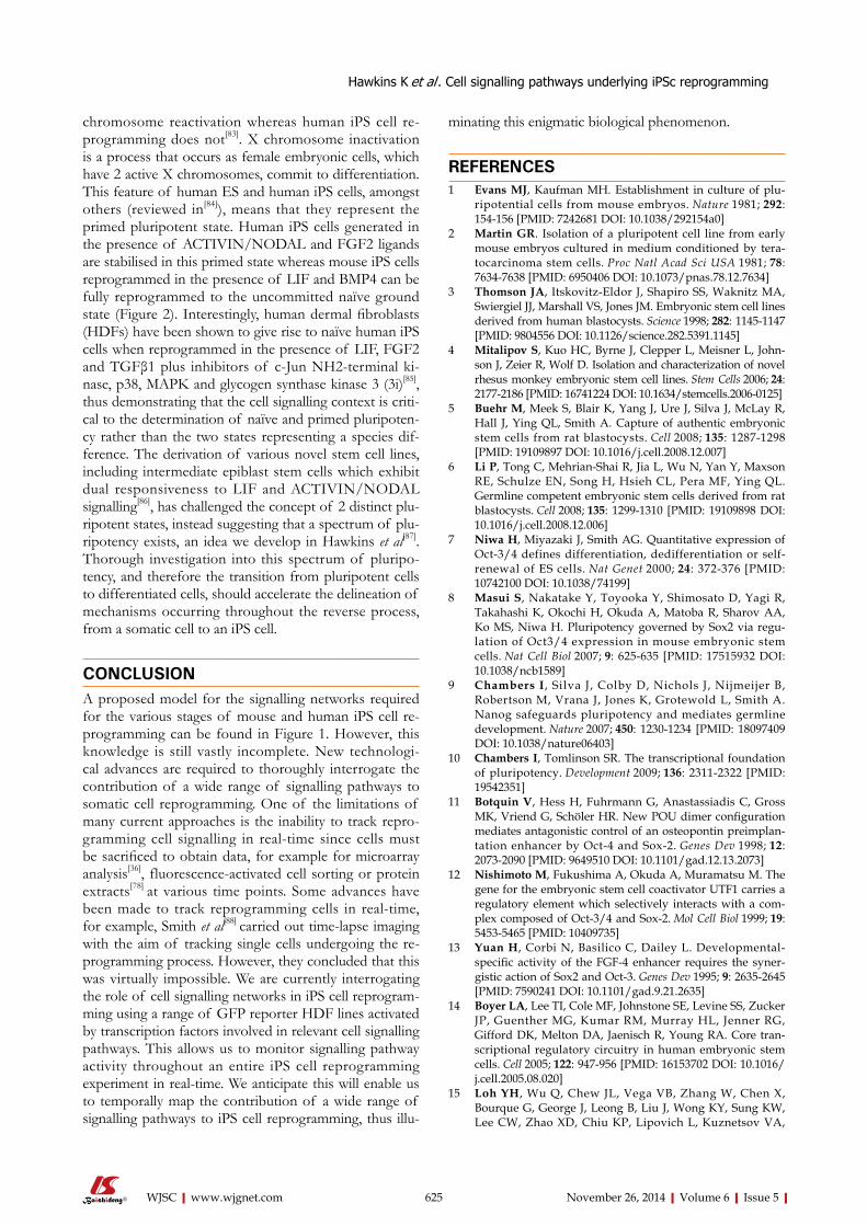
IPS REPROGRAMMING IS A STEPWISE PROCESS Much progress has been made in recent years to define the molecular mechanisms involved in iPS cell repro-gramming. This has led to the general acceptance of the model proposed by Samavarchi-Tehrani et al[36] that re-programming consists of 3 phases: initiation, maturation and stabilisation (Summarised in Figure 1). Throughout reprogramming various changes occur not only to the cell phenotype but also to gene and non-coding RNA ex-pression, epigenetic status and metabolism. In this review we will focus on cell signalling during the 3 stages of iPS cell reprogramming whilst other aspects are reviewed elsewhere by Papp et al[37] and Jia et al[38].
INITIATIONThe initiation phase of reprogramming occurs in virtu-ally all successfully transfected cells[39] and is characterised by somatic genes being switched off by methylation, an increase in cell proliferation, a metabolic switch from
oxidative phosphorylation to glycolysis, reactivation of telomerase activity and a mesenchymal-to-epithelial tran-sition (MET)[40]. MET is a feature of both mouse[41] and human[42] somatic cell reprogramming and involves the loss of mesenchymal characteristics such as motility and the acquisition of epithelial characteristics such as cell polarity and expression of the cell adhesion molecule E-CADHERIN, perhaps explaining why E-cadherin can replace Oct4 in the reprogramming process[43]. MET and the opposite transition, epithelial-to-mesenchymal transition (EMT), are key features of embryogenesis[44], tumour metastasis[45] and both mouse[46] and human[47] ES cell differentiation. Interestingly, the MET that marks the initiation of cellular reprogramming is reversible since removal of the reprogramming factors from mouse “pre-iPS” cells after induction of reprogramming has been shown to lead to reversion of the cells to a mesenchymal phenotype[36], thus demonstrating that continued trans-gene expression is necessary to allow cells to progress to the maturation stage.
Mechanistically, Sox2 suppresses expression of Snail, an EMT inducer[48], and Klf4 induces E-cadherin expres-sion, thus promoting MET[41]. In addition, Maekawa et al[49] have shown that the Glis family zinc finger 1 protein Glis1 can substitute cMyc in the reprogramming cocktail by inducing MET, thus initiating iPS cell reprogramming. MET can also be induced by chemicals, for example, various groups have demonstrated the ability of trans-forming growth factor (TGF)β inhibition to enhance the initiation stage of both mouse[50,51] and human[42] somatic cell reprogramming. This observation is supported by the finding that addition of recombinant TGFβ abro-gates iPS cell formation[42] and is likely due to the EMT-inducing action of TGFβ signalling, which then prevents the MET that is critical to successful iPS cell reprogram-
622 November 26, 2014|Volume 6|Issue 5|WJSC|www.wjgnet.com
Resistance toapoptosis/senescence
Metabolic shift
Resistance toapoptosis/senescence
Sox2 BMP
Klf4
METTGFβ
p53
LIN28 FGF2
Wnt
cMyc
Initiation
LIF/STAT3
Maturation
Demethylation of pluripotency gene promoters
X chromosomereactivation
Stabilisation
Transgeneindependence
OCT4SOX2
NANOG
Stabilisation
Transgeneindependence
OCT4SOX2
NANOG
Maturation
Demethylation of pluripotency gene promoters
LIN28Sox2
METTGFβ
Increasedproliferation
cMyc
p53
Initiation
MAPK
Metabolic shift
PI3K
Mouse
Human
Figure 1 The key stages in (A) mouse and (B) human induced pluripotent stem cell reprogramming and the signalling pathways that regulate them.
Hawkins K et al . Cell signalling pathways underlying iPSc reprogramming
A
Increased proliferation

Table 2 Small molecules that enhance induced pluripotent stem cell reprogramming
cycle arrest via p21 and thus p53 knockdown promotes proliferation[59].
Fibroblast growth factor (FGF) signalling has also been implicated at the initiation stage[60]. Araki et al[61] show that Fgf4 is upregulated on day 3 after induction of reprogramming in MEFs and Jiao et al[60] show that FGF2 can improve the reprogramming efficiency in the early phases of mouse somatic cell reprogramming, whereas it has adverse effects in the later stages. Mechanistically, this group have shown that FGF2 promotes the early stages of reprogramming through accelerating cell proliferation, facilitating MET and eliminating extracellular collagens. In addition to an increased proliferation rate, the minority of cells that undergo successful reprogramming also ex-hibit resistance to apoptosis and senescence, by transgene expression[56]. Recent studies have shown that miR-302 expression allows cells to overcome reprogramming-induced senescence[62] and that silencing of the INK4/ARF locus is also likely to be involved, since INK4/ARF blockade improves reprogramming efficiency[63,64]. The INK4/ARF locus encodes tumour suppressor genes that activate the retinoblastoma and p53 pathways. Its inac-tivation therefore blocks apoptosis and senescence and facilitates reprogramming.
The initiation phase is also characterised by a meta-bolic switch from oxidative phosphorylation to glycolysis[65] that occurs around 7 d after induction of reprogram-ming[66] and involves phosphatidylinositol-3-kinase (PI3K)/AKT signalling[53,67]. For example, Chen et al[67] have demonstrated that the PI3K/AKT pathway was activated during reprogramming in parallel with the up-regulation of glycolytic gene expression, showing spe-cifically that AKT activated 2 key glycolytic regulators, AS1060 and PFKB2. Zhu et al[53] have also shown that PS48, an activator of the PI3K/AKT pathway, is able to enhance reprogramming by upregulating glycolytic genes. By switching their metabolism from oxidative phos-
ming. TGFβ signalling promotes EMT via a wide variety of mechanisms, including mediating the disassembly of junctional complexes, reorganising the cell cytoskeleton, and EMT gene activation[52]. Various TGFβ inhibitors have been used to promote reprogramming, including A-83-01[41,53], E616452[25,50] (also known as RepSox) and SB431542[42] (Table 2). In addition to promoting MET, TGFβ inhibitors promote Nanog expression[50], thus providing 2 potential mechanisms for their ability to en-hance reprogramming. Mitogen-activated protein kinase (MAPK) signalling, activated by TGFβ, further induces the expression of mesodermal genes[52]. Inhibitors of MAPK signalling such as PD0325901 have therefore been used in combination with TGFβ inhibitors to pro-mote MET[42].
Bone morphogenetic protein (BMP) signalling also plays an important role in the initiation stage of mouse iPS cell reprogramming by promoting MET via upregula-tion of epithelial genes such as E-cadherin, Occludin and Epithelial cell adhesion molecule[36]. Chen et al[54] have shown that BMPs can replace Klf4 in the reprogramming cock-tail, allowing mouse embryonic fibroblasts (MEFs) to be reprogrammed using Oct4 alone. However, constitutive BMP activation prevents human somatic cell reprogram-ming. This was discovered through the observation that a naturally occurring Alk2 mutation, which causes fibro-dysplasia ossificans progressiva in humans, prevents iPS cell reprogramming and that this blockade can be rescued by inhibition of the ALK2 receptor[55].
Increased proliferation has been observed in cells undergoing reprogramming as early as 3 d after induc-tion of reprogramming[56] and is likely to be initiated by cMyc transgene expression[57]. Lin28 expression and p53 knockdown also increase the efficiency of iPS cell repro-gramming by stimulating cell proliferation[39]. Specifically, LIN28 has been shown to regulate cell cycle genes such as Cyclin A, Cyclin B and Cdk4[58] whilst p53 induces cell
623 November 26, 2014|Volume 6|Issue 5|WJSC|www.wjgnet.com
Small molecule Function Ref.
BIX-01294 Histone methyltransferase inhibitor Shi et al[51]
Bayk8644 Calcium channel agonist Shi et al[51]
RG108 DNA methyltransferase inhibitor Shi et al[51]
5-Aza-2’-Deoxycytidine DNA methyltransferase inhibitor Huangfu et al[89]
Dexamethasone Steroid glucocorticoid Huangfu et al[89]
Valproic acid HDAC inhibitor Huangfu et al[89]
Trichostatin A HDAC inhibitor Huangfu et al[89]
SAHA HDAC inhibitor Huangfu et al[89]
PD0325901 + CHIR99021
MAPK inhibition and GSK3 inhibition Shi et al[51], Silva et al[77]
SB 431542+ PD0325901 TGFβ inhibitor Lin et al[42]
And MAPK inhibitorA-83-01 TGFβ inhibitor Li et al[41], Zhu et al[53]
E616452 TGFβ inhibitor Ichida et al[90]
AMI-5 Protein arginine methyltransferase inhibitor Yuan et al[13]
Kenpaullone Unknown “novel function” Lyssiotis et al[91]
Adapted from Feng et al[73]. SAHA: Suberoylanilide hydroxamic acid; AMI: Arginine N-Methyltransferase Inhibitor.
Hawkins K et al . Cell signalling pathways underlying iPSc reprogramming

phorylation to anaerobic glycolysis, pre-iPS cells assume an ES cell-like phenotype[68]. ES cells are likely to have developed this form of metabolism as an adaptation to the hypoxic in vivo environment of the early embryo[69]. Interestingly, various groups have shown that iPS cell reprogramming is enhanced by hypoxia[70,71], likely due to the acceleration of this metabolic shift.
MATURATIONTanabe et al[72] have recently identified the maturation stage of iPS cell reprogramming as being a major bottle-neck in the process, which is likely to account for the low efficiency of the process generally. They demonstrate that LIN28, but not NANOG, shp53 or CYCLIN D1, promotes maturation of iPS cells. During maturation, epigenetic changes occur allowing expression of the first pluripotency-associated genes[40]. These genes include Fbxo15, Sall4, Oct4, Nanog and Esrrb. Interestingly, Esrrb has been shown to be sufficient to reprogram MEFs in collaboration with Sox2 and Oct4[73].
LIF/STAT3 signalling is required for the maturation phase of mouse iPS cell reprogramming[74]. Interestingly, pre-iPS cell colony formation has been observed in the absence of LIF, however, beyond day 6 of reprogram-ming these colonies detach. This is likely due to the requirement that cells undergoing the reprogramming process have for LIF signalling to maintain cMyc expres-sion[75]. In addition, Tang et al[74] demonstrate that LIF/STAT3 activation induces earlier formation of an in-creased number of pre-iPS cell colonies. Mechanistically, this group demonstrate that LIF/STAT3 signalling is re-quired for demethylation of pluripotency-associated gene promoters. Specifically, STAT3 signalling was shown to directly block the action of the DNA methyltransferase DNMT1 and Histone deacetylases 2, 3 and 8.
Wnt signalling also enhances the maturation phase of mouse somatic cell reprogramming whereby exogenous stimulation of the pathway using Wnt3a between days 6 and 9 after induction of reprogramming enhances the formation of Nanog positive colonies[76]. Various groups
have suggested that expression of Nanog is necessary for cells to advance from the maturation phase to the sta-bilisation stage[39,77] and thus, Samavarchi et al[36] suggest that Nanog expression alone is responsible for mediating the transition from pre-iPS cells to stably reprogrammed cells. This group demonstrate that removal of the re-programming factors from mouse iPS cells at day 9 after induction of reprogramming did not induce phenotypic reversion. Other groups, however, have reported dif-ferent time points for the stabilisation stage, including day 11[78,79] and day 16[80], suggesting that this can vary depending on discrete protocols and culture variations. It is clear that there remains substantial information to be learned regarding this critical intermediary step but NANOG appears to play a pivotal role in iPS cell matu-ration.
STABILISATIONOnly around 1% of cells that initiate reprogramming make it to the stabilisation stage[72]. This can be explained by the observation made by Golipour et al[81] that not all cells are “stabilisation competent”. This group identify a gene expression signature that distinguishes stabilisation competent and stabilisation incompetent cells and show that stabilisation competent cells require transgene re-pression to enter this stage. Since the stabilisation stage is characterised by transgene independence, only cells that have activated endogenous pluripotency gene expression are able to maintain pluripotency at this late stage. En-dogenous pluripotency gene expression is facilitated by demethylation of pluripotency gene promoters, thus ex-plaining why various DNA and histone methyltransferase inhibitors have been shown to accelerate iPS cell repro-gramming, amongst other small molecules (Table 2). This may also explain the ability of the H3K27 demethylase UTX to substitute for some of the original reprogram-ming factors[82].
The end-point of iPS cell reprogramming is a mat-ter of some controversy. For example, the stabilisation stage of mouse iPS cell reprogramming involves X
624 November 26, 2014|Volume 6|Issue 5|WJSC|www.wjgnet.com
LIF BMP4
gp13
0LI
FRJAK
STAT3
Klf4
Sox2 Oct4 Nanog Id genes
PI3K
AktGrb2
MAPKTbx3
Smads1/5/8
Smads1/5/8 Smad4
TGF β
RI
TGF β
RII
TGF β
RI
TGF β
RII
FGFR
FGFR
Sox2 Oct4 Nanog
MEK1/2
Smad4 Smad4
Smad2
Smad2
Smad3
Smad3PI3K
AKT
ERK1/2
Gene expression
Figure 2 The core signalling networks that maintain pluripotency in (A) naive and (B) primed pluripotent cells.
Hawkins K et al . Cell signalling pathways underlying iPSc reprogramming
A BNaive Primed
FGF2Activin/Nodal
chromosome reactivation whereas human iPS cell re-programming does not[83]. X chromosome inactivation is a process that occurs as female embryonic cells, which have 2 active X chromosomes, commit to differentiation. This feature of human ES and human iPS cells, amongst others (reviewed in[84]), means that they represent the primed pluripotent state. Human iPS cells generated in the presence of ACTIVIN/NODAL and FGF2 ligands are stabilised in this primed state whereas mouse iPS cells reprogrammed in the presence of LIF and BMP4 can be fully reprogrammed to the uncommitted naïve ground state (Figure 2). Interestingly, human dermal fibroblasts (HDFs) have been shown to give rise to naïve human iPS cells when reprogrammed in the presence of LIF, FGF2 and TGFβ1 plus inhibitors of c-Jun NH2-terminal ki-nase, p38, MAPK and glycogen synthase kinase 3 (3i)[85], thus demonstrating that the cell signalling context is criti-cal to the determination of naïve and primed pluripoten-cy rather than the two states representing a species dif-ference. The derivation of various novel stem cell lines, including intermediate epiblast stem cells which exhibit dual responsiveness to LIF and ACTIVIN/NODAL signalling[86], has challenged the concept of 2 distinct plu-ripotent states, instead suggesting that a spectrum of plu-ripotency exists, an idea we develop in Hawkins et al[87]. Thorough investigation into this spectrum of pluripo-tency, and therefore the transition from pluripotent cells to differentiated cells, should accelerate the delineation of mechanisms occurring throughout the reverse process, from a somatic cell to an iPS cell.
CONCLUSION A proposed model for the signalling networks required for the various stages of mouse and human iPS cell re-programming can be found in Figure 1. However, this knowledge is still vastly incomplete. New technologi-cal advances are required to thoroughly interrogate the contribution of a wide range of signalling pathways to somatic cell reprogramming. One of the limitations of many current approaches is the inability to track repro-gramming cell signalling in real-time since cells must be sacrificed to obtain data, for example for microarray analysis[36], fluorescence-activated cell sorting or protein extracts[78] at various time points. Some advances have been made to track reprogramming cells in real-time, for example, Smith et al[88] carried out time-lapse imaging with the aim of tracking single cells undergoing the re-programming process. However, they concluded that this was virtually impossible. We are currently interrogating the role of cell signalling networks in iPS cell reprogram-ming using a range of GFP reporter HDF lines activated by transcription factors involved in relevant cell signalling pathways. This allows us to monitor signalling pathway activity throughout an entire iPS cell reprogramming experiment in real-time. We anticipate this will enable us to temporally map the contribution of a wide range of signalling pathways to iPS cell reprogramming, thus illu-
minating this enigmatic biological phenomenon.
REFERENCES1 Evans MJ, Kaufman MH. Establishment in culture of plu-
ripotential cells from mouse embryos. Nature 1981; 292: 154-156 [PMID: 7242681 DOI: 10.1038/292154a0]
2 Martin GR. Isolation of a pluripotent cell line from early mouse embryos cultured in medium conditioned by tera-tocarcinoma stem cells. Proc Natl Acad Sci USA 1981; 78: 7634-7638 [PMID: 6950406 DOI: 10.1073/pnas.78.12.7634]
3 Thomson JA, Itskovitz-Eldor J, Shapiro SS, Waknitz MA, Swiergiel JJ, Marshall VS, Jones JM. Embryonic stem cell lines derived from human blastocysts. Science 1998; 282: 1145-1147 [PMID: 9804556 DOI: 10.1126/science.282.5391.1145]
4 Mitalipov S, Kuo HC, Byrne J, Clepper L, Meisner L, John-son J, Zeier R, Wolf D. Isolation and characterization of novel rhesus monkey embryonic stem cell lines. Stem Cells 2006; 24: 2177-2186 [PMID: 16741224 DOI: 10.1634/stemcells.2006-0125]
5 Buehr M, Meek S, Blair K, Yang J, Ure J, Silva J, McLay R, Hall J, Ying QL, Smith A. Capture of authentic embryonic stem cells from rat blastocysts. Cell 2008; 135: 1287-1298 [PMID: 19109897 DOI: 10.1016/j.cell.2008.12.007]
6 Li P, Tong C, Mehrian-Shai R, Jia L, Wu N, Yan Y, Maxson RE, Schulze EN, Song H, Hsieh CL, Pera MF, Ying QL. Germline competent embryonic stem cells derived from rat blastocysts. Cell 2008; 135: 1299-1310 [PMID: 19109898 DOI: 10.1016/j.cell.2008.12.006]
7 Niwa H, Miyazaki J, Smith AG. Quantitative expression of Oct-3/4 defines differentiation, dedifferentiation or self-renewal of ES cells. Nat Genet 2000; 24: 372-376 [PMID: 10742100 DOI: 10.1038/74199]
8 Masui S, Nakatake Y, Toyooka Y, Shimosato D, Yagi R, Takahashi K, Okochi H, Okuda A, Matoba R, Sharov AA, Ko MS, Niwa H. Pluripotency governed by Sox2 via regu-lation of Oct3/4 expression in mouse embryonic stem cells. Nat Cell Biol 2007; 9: 625-635 [PMID: 17515932 DOI: 10.1038/ncb1589]
9 Chambers I, Silva J, Colby D, Nichols J, Nijmeijer B, Robertson M, Vrana J, Jones K, Grotewold L, Smith A. Nanog safeguards pluripotency and mediates germline development. Nature 2007; 450: 1230-1234 [PMID: 18097409 DOI: 10.1038/nature06403]
10 Chambers I, Tomlinson SR. The transcriptional foundation of pluripotency. Development 2009; 136: 2311-2322 [PMID: 19542351]
11 Botquin V, Hess H, Fuhrmann G, Anastassiadis C, Gross MK, Vriend G, Schöler HR. New POU dimer configuration mediates antagonistic control of an osteopontin preimplan-tation enhancer by Oct-4 and Sox-2. Genes Dev 1998; 12: 2073-2090 [PMID: 9649510 DOI: 10.1101/gad.12.13.2073]
12 Nishimoto M, Fukushima A, Okuda A, Muramatsu M. The gene for the embryonic stem cell coactivator UTF1 carries a regulatory element which selectively interacts with a com-plex composed of Oct-3/4 and Sox-2. Mol Cell Biol 1999; 19: 5453-5465 [PMID: 10409735]
13 Yuan H, Corbi N, Basilico C, Dailey L. Developmental-specific activity of the FGF-4 enhancer requires the syner-gistic action of Sox2 and Oct-3. Genes Dev 1995; 9: 2635-2645 [PMID: 7590241 DOI: 10.1101/gad.9.21.2635]
14 Boyer LA, Lee TI, Cole MF, Johnstone SE, Levine SS, Zucker JP, Guenther MG, Kumar RM, Murray HL, Jenner RG, Gifford DK, Melton DA, Jaenisch R, Young RA. Core tran-scriptional regulatory circuitry in human embryonic stem cells. Cell 2005; 122: 947-956 [PMID: 16153702 DOI: 10.1016/j.cell.2005.08.020]
15 Loh YH, Wu Q, Chew JL, Vega VB, Zhang W, Chen X, Bourque G, George J, Leong B, Liu J, Wong KY, Sung KW, Lee CW, Zhao XD, Chiu KP, Lipovich L, Kuznetsov VA,
625 November 26, 2014|Volume 6|Issue 5|WJSC|www.wjgnet.com
Hawkins K et al . Cell signalling pathways underlying iPSc reprogramming

Robson P, Stanton LW, Wei CL, Ruan Y, Lim B, Ng HH. The Oct4 and Nanog transcription network regulates pluri-potency in mouse embryonic stem cells. Nat Genet 2006; 38: 431-440 [PMID: 16518401 DOI: 10.1038/ng1760]
16 Kopp JL, Ormsbee BD, Desler M, Rizzino A. Small increases in the level of Sox2 trigger the differentiation of mouse embryonic stem cells. Stem Cells 2008; 26: 903-911 [PMID: 18238855 DOI: 10.1634/stemcells.2007-0951]
17 Pan G, Li J, Zhou Y, Zheng H, Pei D. A negative feedback loop of transcription factors that controls stem cell pluripo-tency and self-renewal. FASEB J 2006; 20: 1730-1732 [PMID: 16790525]
18 Takahashi K, Yamanaka S. Induction of pluripotent stem cells from mouse embryonic and adult fibroblast cultures by defined factors. Cell 2006; 126: 663-676 [PMID: 16904174]
19 Takahashi K, Tanabe K, Ohnuki M, Narita M, Ichisaka T, Tomoda K, Yamanaka S. Induction of pluripotent stem cells from adult human fibroblasts by defined factors. Cell 2007; 131: 861-872 [PMID: 18035408]
20 Yu J, Vodyanik MA, Smuga-Otto K, Antosiewicz-Bourget J, Frane JL, Tian S, Nie J, Jonsdottir GA, Ruotti V, Stewart R, Slukvin II, Thomson JA. Induced pluripotent stem cell lines derived from human somatic cells. Science 2007; 318: 1917-1920 [PMID: 18029452]
21 Yu J, Hu K, Smuga-Otto K, Tian S, Stewart R, Slukvin II, Thomson JA. Human induced pluripotent stem cells free of vector and transgene sequences. Science 2009; 324: 797-801 [PMID: 19325077 DOI: 10.1126/science.1172482]
22 Fusaki N, Ban H, Nishiyama A, Saeki K, Hasegawa M. Ef-ficient induction of transgene-free human pluripotent stem cells using a vector based on Sendai virus, an RNA virus that does not integrate into the host genome. Proc Jpn Acad Ser B Phys Biol Sci 2009; 85: 348-362 [PMID: 19838014 DOI: 10.2183/pjab.85.348]
23 Wang W, Yang J, Liu H, Lu D, Chen X, Zenonos Z, Cam-pos LS, Rad R, Guo G, Zhang S, Bradley A, Liu P. Rapid and efficient reprogramming of somatic cells to induced pluripotent stem cells by retinoic acid receptor gamma and liver receptor homolog 1. Proc Natl Acad Sci USA 2011; 108: 18283-18288 [PMID: 21990348]
24 Zhou H, Wu S, Joo JY, Zhu S, Han DW, Lin T, Trauger S, Bien G, Yao S, Zhu Y, Siuzdak G, Schöler HR, Duan L, Ding S. Generation of induced pluripotent stem cells using re-combinant proteins. Cell Stem Cell 2009; 4: 381-384 [PMID: 19398399 DOI: 10.1016/j.stem.2009.04.005]
25 Hou P, Li Y, Zhang X, Liu C, Guan J, Li H, Zhao T, Ye J, Yang W, Liu K, Ge J, Xu J, Zhang Q, Zhao Y, Deng H. Pluripotent stem cells induced from mouse somatic cells by small-molecule compounds. Science 2013; 341: 651-654 [PMID: 23868920 DOI: 10.1126/science.1239278]
26 Kim JB, Sebastiano V, Wu G, Araúzo-Bravo MJ, Sasse P, Gentile L, Ko K, Ruau D, Ehrich M, van den Boom D, Meyer J, Hübner K, Bernemann C, Ortmeier C, Zenke M, Fleischmann BK, Zaehres H, Schöler HR. Oct4-induced plu-ripotency in adult neural stem cells. Cell 2009; 136: 411-419 [PMID: 19203577 DOI: 10.1016/j.cell.2009.01.023]
27 Eminli S, Utikal J, Arnold K, Jaenisch R, Hochedlinger K. Reprogramming of neural progenitor cells into induced pluripotent stem cells in the absence of exogenous Sox2 expression. Stem Cells 2008; 26: 2467-2474 [PMID: 18635867 DOI: 10.1634/stemcells.2008-0317]
28 Aasen T, Raya A, Barrero MJ, Garreta E, Consiglio A, Gonzalez F, Vassena R, Bilić J, Pekarik V, Tiscornia G, Edel M, Boué S, Izpisúa Belmonte JC. Efficient and rapid generation of induced pluripotent stem cells from human keratinocytes. Nat Biotechnol 2008; 26: 1276-1284 [PMID: 18931654]
29 Hanna J, Markoulaki S, Schorderet P, Carey BW, Beard C, Wernig M, Creyghton MP, Steine EJ, Cassady JP, Foreman R, Lengner CJ, Dausman JA, Jaenisch R. Direct reprogram-
ming of terminally differentiated mature B lymphocytes to pluripotency. Cell 2008; 133: 250-264 [PMID: 18423197]
30 Qin D, Gan Y, Shao K, Wang H, Li W, Wang T, He W, Xu J, Zhang Y, Kou Z, Zeng L, Sheng G, Esteban MA, Gao S, Pei D. Mouse meningiocytes express Sox2 and yield high efficiency of chimeras after nuclear reprogramming with exogenous factors. J Biol Chem 2008; 283: 33730-33735 [PMID: 18826945 DOI: 10.1074/jbc.M806788200]
31 Okita K, Yamakawa T, Matsumura Y, Sato Y, Amano N, Watanabe A, Goshima N, Yamanaka S. An efficient non-viral method to generate integration-free human-induced pluripotent stem cells from cord blood and peripheral blood cells. Stem Cells 2013; 31: 458-466 [PMID: 23193063 DOI: 10.1002/stem.1293]
32 Stadtfeld M, Brennand K, Hochedlinger K. Reprogram-ming of pancreatic beta cells into induced pluripotent stem cells. Curr Biol 2008; 18: 890-894 [PMID: 18501604 DOI: 10.1016/j.cub.2008.05.010]
33 Cyranoski D. Stem cells cruise to clinic. Nature 2013; 494: 413 [PMID: 23446394 DOI: 10.1038/494413a]
34 Park IH, Arora N, Huo H, Maherali N, Ahfeldt T, Shima-mura A, Lensch MW, Cowan C, Hochedlinger K, Daley GQ. Disease-specific induced pluripotent stem cells. Cell 2008; 134: 877-886 [PMID: 18691744 DOI: 10.1016/j.cell.2008.07.041]
35 Matsa E, Rajamohan D, Dick E, Young L, Mellor I, Stani-forth A, Denning C. Drug evaluation in cardiomyocytes de-rived from human induced pluripotent stem cells carrying a long QT syndrome type 2 mutation. Eur Heart J 2011; 32: 952-962 [PMID: 21367833 DOI: 10.1093/eurheartj/ehr073]
36 Samavarchi-Tehrani P, Golipour A, David L, Sung HK, Beyer TA, Datti A, Woltjen K, Nagy A, Wrana JL. Functional genomics reveals a BMP-driven mesenchymal-to-epithelial transition in the initiation of somatic cell reprogramming. Cell Stem Cell 2010; 7: 64-77 [PMID: 20621051 DOI: 10.1016/j.stem.2010.04.015]
37 Papp B, Plath K. Epigenetics of reprogramming to induced pluripotency. Cell 2013; 152: 1324-1343 [PMID: 23498940 DOI: 10.1016/j.cell.2013.02.043]
38 Jia W, Chen W, Kang J. The functions of microRNAs and long non-coding RNAs in embryonic and induced pluripo-tent stem cells. Genomics Proteomics Bioinformatics 2013; 11: 275-283 [PMID: 24096129 DOI: 10.1016/j.gpb.2013.09.004]
39 Hanna J, Saha K, Pando B, van Zon J, Lengner CJ, Creyghton MP, van Oudenaarden A, Jaenisch R. Direct cell reprogram-ming is a stochastic process amenable to acceleration. Na-ture 2009; 462: 595-601 [PMID: 19898493 DOI: 10.1038/na-ture08592]
40 David L, Polo JM. Phases of reprogramming. Stem Cell Res 2014; 12: 754-761 [PMID: 24735951 DOI: 10.1016/j.scr.2014.03.007]
41 Li R, Liang J, Ni S, Zhou T, Qing X, Li H, He W, Chen J, Li F, Zhuang Q, Qin B, Xu J, Li W, Yang J, Gan Y, Qin D, Feng S, Song H, Yang D, Zhang B, Zeng L, Lai L, Esteban MA, Pei D. A mesenchymal-to-epithelial transition initiates and is required for the nuclear reprogramming of mouse fibroblasts. Cell Stem Cell 2010; 7: 51-63 [PMID: 20621050 DOI: 10.1016/j.stem.2010.04.014]
42 Lin T, Ambasudhan R, Yuan X, Li W, Hilcove S, Abujarour R, Lin X, Hahm HS, Hao E, Hayek A, Ding S. A chemi-cal platform for improved induction of human iPSCs. Nat Methods 2009; 6: 805-808 [PMID: 19838168 DOI: 10.1038/nmeth.1393]
43 Redmer T, Diecke S, Grigoryan T, Quiroga-Negreira A, Birchmeier W, Besser D. E-cadherin is crucial for embryonic stem cell pluripotency and can replace OCT4 during somat-ic cell reprogramming. EMBO Rep 2011; 12: 720-726 [PMID: 21617704 DOI: 10.1038/embor.2011.88]
44 Yang J, Weinberg RA. Epithelial-mesenchymal transition: at the crossroads of development and tumor metastasis. Dev Cell 2008; 14: 818-829 [PMID: 18539112 DOI: 10.1016/j.devcel.2008.05.009]
626 November 26, 2014|Volume 6|Issue 5|WJSC|www.wjgnet.com
Hawkins K et al . Cell signalling pathways underlying iPSc reprogramming

45 Yao D, Dai C, Peng S. Mechanism of the mesenchymal-epithelial transition and its relationship with metastatic tumor formation. Mol Cancer Res 2011; 9: 1608-1620 [PMID: 21840933 DOI: 10.1158/1541-7786.MCR-10-0568]
46 Spencer HL, Eastham AM, Merry CL, Southgate TD, Perez-Campo F, Soncin F, Ritson S, Kemler R, Stern PL, Ward CM. E-cadherin inhibits cell surface localization of the pro-migratory 5T4 oncofetal antigen in mouse embryonic stem cells. Mol Biol Cell 2007; 18: 2838-2851 [PMID: 17507657 DOI: 10.1091/mbc.E06-09-0875]
47 Eastham AM, Spencer H, Soncin F, Ritson S, Merry CL, Stern PL, Ward CM. Epithelial-mesenchymal transition events during human embryonic stem cell differentiation. Can-cer Res 2007; 67 : 11254-11262 [PMID: 18056451 DOI: 10.1158/0008-5472.CAN-07-2253]
48 Liu X, Sun H, Qi J, Wang L, He S, Liu J, Feng C, Chen C, Li W, Guo Y, Qin D, Pan G, Chen J, Pei D, Zheng H. Sequen-tial introduction of reprogramming factors reveals a time-sensitive requirement for individual factors and a sequential EMT-MET mechanism for optimal reprogramming. Nat Cell Biol 2013; 15: 829-838 [PMID: 23708003 DOI: 10.1038/ncb2765]
49 Maekawa M, Yamaguchi K, Nakamura T, Shibukawa R, Kodanaka I, Ichisaka T, Kawamura Y, Mochizuki H, Gos-hima N, Yamanaka S. Direct reprogramming of somatic cells is promoted by maternal transcription factor Glis1. Na-ture 2011; 474: 225-229 [PMID: 21654807 DOI: 10.1038/na-ture10106]
50 Maherali N, Hochedlinger K. Tgfbeta signal inhibition co-operates in the induction of iPSCs and replaces Sox2 and cMyc. Curr Biol 2009; 19: 1718-1723 [PMID: 19765992 DOI: 10.1016/j.cub.2009.08.025]
51 Shi Y, Do JT, Desponts C, Hahm HS, Schöler HR, Ding S. A combined chemical and genetic approach for the genera-tion of induced pluripotent stem cells. Cell Stem Cell 2008; 2: 525-528 [PMID: 18522845 DOI: 10.1016/j.stem.2008.05.011]
52 Thiery JP, Sleeman JP. Complex networks orchestrate epi-thelial-mesenchymal transitions. Nat Rev Mol Cell Biol 2006; 7: 131-142 [PMID: 16493418 DOI: 10.1038/nrm1835]
53 Zhu S, Li W, Zhou H, Wei W, Ambasudhan R, Lin T, Kim J, Zhang K, Ding S. Reprogramming of human primary somatic cells by OCT4 and chemical compounds. Cell Stem Cell 2010; 7: 651-655 [PMID: 21112560 DOI: 10.1016/j.stem.2010.11.015]
54 Chen J, Liu J, Yang J, Chen Y, Chen J, Ni S, Song H, Zeng L, Ding K, Pei D. BMPs functionally replace Klf4 and sup-port efficient reprogramming of mouse fibroblasts by Oct4 alone. Cell Res 2011; 21: 205-212 [PMID: 21135873 DOI: 10.1038/cr.2010.172]
55 Hamasaki M, Hashizume Y, Yamada Y, Katayama T, Hohjoh H, Fusaki N, Nakashima Y, Furuya H, Haga N, Takami Y, Era T. Pathogenic mutation of ALK2 inhibits induced pluripotent stem cell reprogramming and main-tenance: mechanisms of reprogramming and strategy for drug identification. Stem Cells 2012; 30: 2437-2449 [PMID: 22949078 DOI: 10.1002/stem.1221]
56 Papp B, Plath K. Reprogramming to pluripotency: step-wise resetting of the epigenetic landscape. Cell Res 2011; 21: 486-501 [PMID: 21321600 DOI: 10.1038/cr.2011.28]
57 Apostolou E, Hochedlinger K. Chromatin dynamics during cellular reprogramming. Nature 2013; 502: 462-471 [PMID: 24153299 DOI: 10.1038/nature12749]
58 Xu B, Zhang K, Huang Y. Lin28 modulates cell growth and associates with a subset of cell cycle regulator mRNAs in mouse embryonic stem cells. RNA 2009; 15: 357-361 [PMID: 19147696 DOI: 10.1261/rna.1368009]
59 el-Deiry WS, Tokino T, Velculescu VE, Levy DB, Parsons R, Trent JM, Lin D, Mercer WE, Kinzler KW, Vogelstein B. WAF1, a potential mediator of p53 tumor suppression. Cell 1993; 75: 817-825 [PMID: 8242752 DOI: 10.1016/0092-8674(9
3)90500-P]60 Jiao J, Dang Y, Yang Y, Gao R, Zhang Y, Kou Z, Sun XF,
Gao S. Promoting reprogramming by FGF2 reveals that the extracellular matrix is a barrier for reprogramming fibro-blasts to pluripotency. Stem Cells 2013; 31: 729-740 [PMID: 23307593 DOI: 10.1002/stem.1318]
61 Araki R, Jincho Y, Hoki Y, Nakamura M, Tamura C, Ando S, Kasama Y, Abe M. Conversion of ancestral fibroblasts to induced pluripotent stem cells. Stem Cells 2010; 28: 213-220 [PMID: 20020427]
62 Banito A, Rashid ST, Acosta JC, Li S, Pereira CF, Geti I, Pinho S, Silva JC, Azuara V, Walsh M, Vallier L, Gil J. Se-nescence impairs successful reprogramming to pluripotent stem cells. Genes Dev 2009; 23: 2134-2139 [PMID: 19696146 DOI: 10.1101/gad.1811609]
63 Li H, Collado M, Villasante A, Strati K, Ortega S, Cañamero M, Blasco MA, Serrano M. The Ink4/Arf locus is a barrier for iPS cell reprogramming. Nature 2009; 460: 1136-1139 [PMID: 19668188 DOI: 10.1038/nature08290]
64 Utikal J, Polo JM, Stadtfeld M, Maherali N, Kulalert W, Walsh RM, Khalil A, Rheinwald JG, Hochedlinger K. Im-mortalization eliminates a roadblock during cellular repro-gramming into iPS cells. Nature 2009; 460: 1145-1148 [PMID: 19668190 DOI: 10.1038/nature08285]
65 Panopoulos AD, Yanes O, Ruiz S, Kida YS, Diep D, Taut-enhahn R, Herrerías A, Batchelder EM, Plongthongkum N, Lutz M, Berggren WT, Zhang K, Evans RM, Siuzdak G, Izpisua Belmonte JC. The metabolome of induced pluripo-tent stem cells reveals metabolic changes occurring in so-matic cell reprogramming. Cell Res 2012; 22: 168-177 [PMID: 22064701 DOI: 10.1038/cr.2011.177]
66 Park SJ, Yeo HC, Kang NY, Kim H, Lin J, Ha HH, Ven-drell M, Lee JS, Chandran Y, Lee DY, Yun SW, Chang YT. Mechanistic elements and critical factors of cellular repro-gramming revealed by stepwise global gene expression analyses. Stem Cell Res 2014; 12: 730-741 [PMID: 24727632 DOI: 10.1016/j.scr.2014.03.002]
67 Chen M, Zhang H, Wu J, Xu L, Xu D, Sun J, He Y, Zhou X, Wang Z, Wu L, Xu S, Wang J, Jiang S, Zhou X, Hoffman AR, Hu X, Hu J, Li T. Promotion of the induction of cell pluripo-tency through metabolic remodeling by thyroid hormone triiodothyronine-activated PI3K/AKT signal pathway. Bio-materials 2012; 33: 5514-5523 [PMID: 22575839 DOI: 10.1016/j.biomaterials.2012.04.001]
68 Kondoh H, Lleonart ME, Nakashima Y, Yokode M, Tanaka M, Bernard D, Gil J, Beach D. A high glycolytic flux sup-ports the proliferative potential of murine embryonic stem cells. Antioxid Redox Signal 2007; 9: 293-299 [PMID: 17184172 DOI: 10.1089/ars.2006.1467]
69 Ottosen LD, Hindkaer J, Husth M, Petersen DE, Kirk J, Ingerslev HJ. Observations on intrauterine oxygen ten-sion measured by fibre-optic microsensors. Reprod Biomed Online 2006; 13: 380-385 [PMID: 16984770 DOI: 10.1016/S1472-6483(10)61443-5]
70 Shimada H, Hashimoto Y, Nakada A, Shigeno K, Naka-mura T. Accelerated generation of human induced pluripo-tent stem cells with retroviral transduction and chemical inhibitors under physiological hypoxia. Biochem Biophys Res Commun 2012; 417: 659-664 [PMID: 22172948 DOI: 10.1016/j.bbrc.2011.11.111]
71 Yoshida Y, Takahashi K, Okita K, Ichisaka T, Yamanaka S. Hypoxia enhances the generation of induced pluripotent stem cells. Cell Stem Cell 2009; 5: 237-241 [PMID: 19716359 DOI: 10.1016/j.stem.2009.08.001]
72 Tanabe K, Nakamura M, Narita M, Takahashi K, Yamanaka S. Maturation, not initiation, is the major roadblock dur-ing reprogramming toward pluripotency from human fibroblasts. Proc Natl Acad Sci USA 2013; 110: 12172-12179 [PMID: 23812749]
73 Feng B, Jiang J, Kraus P, Ng JH, Heng JC, Chan YS, Yaw LP,
627 November 26, 2014|Volume 6|Issue 5|WJSC|www.wjgnet.com
Hawkins K et al . Cell signalling pathways underlying iPSc reprogramming

Zhang W, Loh YH, Han J, Vega VB, Cacheux-Rataboul V, Lim B, Lufkin T, Ng HH. Reprogramming of fibroblasts into induced pluripotent stem cells with orphan nuclear receptor Esrrb. Nat Cell Biol 2009; 11: 197-203 [PMID: 19136965 DOI: 10.1038/ncb1827]
74 Tang Y, Tian XC. JAK-STAT3 and somatic cell reprogramming. JAK-STAT 2013; 2: e24935 [PMID: 24470976 DOI: 10.4161/jkst.24935]
75 Yang J, van Oosten AL, Theunissen TW, Guo G, Silva JC, Smith A. Stat3 activation is limiting for reprogramming to ground state pluripotency. Cell Stem Cell 2010; 7: 319-328 [PMID: 20804969 DOI: 10.1016/j.stem.2010.06.022]
76 Ho R, Papp B, Hoffman JA, Merrill BJ, Plath K. Stage-specific regulation of reprogramming to induced pluripotent stem cells by Wnt signaling and T cell factor proteins. Cell Rep 2013; 3: 2113-2126 [PMID: 23791530 DOI: 10.1016/j.celrep.2013.05.015]
77 Silva J, Nichols J, Theunissen TW, Guo G, van Oosten AL, Bar-randon O, Wray J, Yamanaka S, Chambers I, Smith A. Nanog is the gateway to the pluripotent ground state. Cell 2009; 138: 722-737 [PMID: 19703398 DOI: 10.1016/j.cell.2009.07.039]
78 Hansson J, Rafiee MR, Reiland S, Polo JM, Gehring J, Okawa S, Huber W, Hochedlinger K, Krijgsveld J. Highly coordinated proteome dynamics during reprogramming of somatic cells to pluripotency. Cell Rep 2012; 2: 1579-1592 [PMID: 23260666 DOI: 10.1016/j.celrep.2012.10.014]
79 Polo JM, Anderssen E, Walsh RM, Schwarz BA, Nefzger CM, Lim SM, Borkent M, Apostolou E, Alaei S, Cloutier J, Bar-Nur O, Cheloufi S, Stadtfeld M, Figueroa ME, Rob-inton D, Natesan S, Melnick A, Zhu J, Ramaswamy S, Hochedlinger K. A molecular roadmap of reprogramming somatic cells into iPS cells. Cell 2012; 151: 1617-1632 [PMID: 23260147 DOI: 10.1016/j.cell.2012.11.039]
80 Brambrink T, Foreman R, Welstead GG, Lengner CJ, Wer-nig M, Suh H, Jaenisch R. Sequential expression of pluri-potency markers during direct reprogramming of mouse somatic cells. Cell Stem Cell 2008; 2: 151-159 [PMID: 18371436 DOI: 10.1016/j.stem.2008.01.004]
81 Golipour A, David L, Liu Y, Jayakumaran G, Hirsch CL, Trcka D, Wrana JL. A late transition in somatic cell repro-gramming requires regulators distinct from the pluripoten-cy network. Cell Stem Cell 2012; 11: 769-782 [PMID: 23217423 DOI: 10.1016/j.stem.2012.11.008]
82 Mansour AA, Gafni O, Weinberger L, Zviran A, Ayyash M,
Rais Y, Krupalnik V, Zerbib M, Amann-Zalcenstein D, Maza I, Geula S, Viukov S, Holtzman L, Pribluda A, Canaani E, Horn-Saban S, Amit I, Novershtern N, Hanna JH. The H3K27 demethylase Utx regulates somatic and germ cell epigenetic reprogramming. Nature 2012; 488: 409-413 [PMID: 22801502 DOI: 10.1038/nature11272]
83 Plath K, Lowry WE. Progress in understanding reprogram-ming to the induced pluripotent state. Nat Rev Genet 2011; 12: 253-265 [PMID: 21415849 DOI: 10.1038/nrg2955]
84 Nichols J, Smith A. Naive and primed pluripotent states. Cell Stem Cell 2009; 4: 487-492 [PMID: 19497275 DOI: 10.1016/j.stem.2009.05.015]
85 Gafni O, Weinberger L, Mansour AA, Manor YS, Chomsky E, Ben-Yosef D, Kalma Y, Viukov S, Maza I, Zviran A, Rais Y, Shipony Z, Mukamel Z, Krupalnik V, Zerbib M, Geula S, Caspi I, Schneir D, Shwartz T, Gilad S, Amann-Zalcenstein D, Benjamin S, Amit I, Tanay A, Massarwa R, Novershtern N, Hanna JH. Derivation of novel human ground state na-ive pluripotent stem cells. Nature 2013; 504: 282-286 [PMID: 24172903 DOI: 10.1038/nature12745]
86 Chang KH, Li M. Clonal isolation of an intermediate plu-ripotent stem cell state. Stem Cells 2013; 31: 918-927 [PMID: 23341219 DOI: 10.1002/stem.1330]
87 Hawkins K, Keramari M, Soncin F, Segal JM, Mohamet L, Miazga N, Ritson S, Bobola N, Merry CLR, Ward CM. Novel cell lines isolated from mES cells exhibiting de novo methylation of the E-cadherin promoter. CMB 2014; In press
88 Smith AG . Embryo-derived stem cells: of mice and men. Annu Rev Cell Dev Biol 2001; 17: 435-462 [PMID: 11687496 DOI: 10.1146/annurev.cellbio.17.1.435]
89 Huangfu D, Induction of pluripotent stem cells by defined factors is greatly improved by small-molecule compounds. Nat Biotechnol 2008; 26: 79-797 [PMID: 18568017 DOI: 10.1038/nbt1418]
90 Ichida JK, A small-molecule inhibitor of Tgf-β signaling replaces Sox2 in reprogramming by inducing Nanog. Cell Stem Cell 2009; 5: 491-503 [PMID: 19818703 DOI: 10.1016/ j.stem.2009.09.012]
91 Lyssiotis CA, Reprogramming of murine fibroblasts to induced pluripotent stem cells with chemical complementa-tion of Klf4. Proc Natl Acad Sci 2009; 106: 8912-8917 [PMID: 19447925 DOI: 10.1073/pnas.0903860106]
P- Reviewer: Imamura M, Niyibizi C, Niu W, Song J S- Editor: Song XX L- Editor: A E- Editor: Lu YJ
628 November 26, 2014|Volume 6|Issue 5|WJSC|www.wjgnet.com
Hawkins K et al . Cell signalling pathways underlying iPSc reprogramming

© 2014 Baishideng Publishing Group Inc. All rights reserved.
Published by Baishideng Publishing Group Inc8226 Regency Drive, Pleasanton, CA 94588, USA
Telephone: +1-925-223-8242Fax: +1-925-223-8243
E-mail: [email protected] Desk: http://www.wjgnet.com/esps/helpdesk.aspx
http://www.wjgnet.com