Cell migration in Immunology - Ruhr-Universität … · Cell migration in Immunology The right...
39
Cell migration in Immunology The right cell(s) to the right time at the right place Dr. Stefanie Gnipp
Transcript of Cell migration in Immunology - Ruhr-Universität … · Cell migration in Immunology The right...

Cell migration in Immunology
The right cell(s) to the right time at the right place
Dr. Stefanie Gnipp

2
1. General introduction
2. How do the leukocytes leave the bloodflow and enter the tissue?
vascular trespassing/extravasation molecular process
3. How do the leukocytes move within the tissue?
4. Cell motility and cell shape remodeling
5. How do the cells know that and where they have to migrate?
Soluble signals: Chemokines
6. Diseases associated with defective cellular locomotion
3
The movement, or migration, of cells (also known as cell motility) is an important part of many biological processes. Cell migration is observed in immune cells, during embryonic development, wound repair and in the formation of new blood vessels (a process known as angiogenesis). Further: placental development (trophoblast invasion) cancer; metastasis
4
Cell trafficking in immunology
The trafficking of leukocytes into and within lymphoid and peripheral tissues is central to immune cell development, immunosurveillance and effector function. Interstitial leukocyte trafficking is the result of amoeboid polarization and migration, guided by soluble or tissue-bound chemoattractant signals for positioning and local arrest.
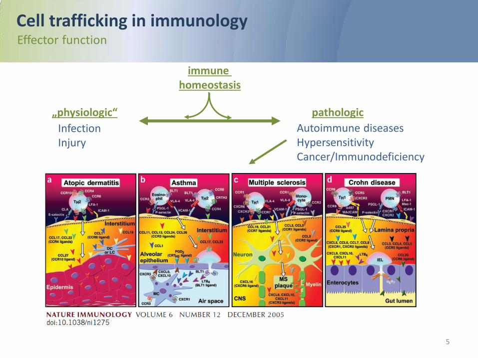
pathologic
Autoimmune diseases Hypersensitivity Cancer/Immunodeficiency
„physiologic“
Infection Injury
5
Cell trafficking in immunology Effector function
immune homeostasis

6
Cell trafficking in immunology Cell types
Both innate and adaptive immune functions depend upon interstitial leukocyte migration. After leaving the bone marrow by way of the blood, monocytes and granulocytes reach lymphoid or peripheral tissues, move toward their targets and execute effector functions. Dendritic cells (DCs) collect and process antigenic material and, in response to maturation signals, migrate from the periphery to lymphoid tissues to present antigens and trigger T cell activation. T lymphocytes emigrate from the thymus, become activated by a cascade of cell-cell interactions in secondary lymphoid organs and circulate to peripheral tissues for effector function. Similarly, B lymphocytes move within secondary lymphatic tissues to capture antigen, receive T cell help and recirculate and become resident in the bone marrow and other lymphoid organs as antibody- secreting plasma cells.

7
How do the leukocytes leave the bloodflow and enter the tissue?
How do they know where they have to leave the circulation?

8
Cell trafficking in immunology Scenario: Inflammation
1) Infectious agents: release of PAMPs (pathogen associated molecular patterns) 2) Tissue damage: release of DAMPs (damage associated molecular patterns)
resident cells of the innate immune system (mast cells; macrophages; DCs..) release of pro-inflammatory mediators/Cytokines
endothelial cells (microvasculature)
activation
Molecular changes: (E-and P-Selectin expression on surface) enables recognition by circulating leukocytes and recruitment to inflamed tissue

9
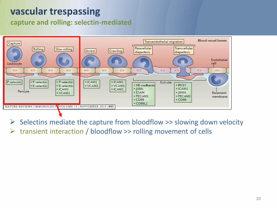
vascular trespassing/extravasation of leukocytes Overview
The recruitment of leukocytes from the circulation into tissues is a multistep cascade: 1. Slowing down velocity in the blood flow (selectin-dependent)= capture 2. movement on the vascular wall=rolling 3. Stimulation by sense signals, presented by the endothelium=activation 4. cell adhesion to the apical endothelial plasma membrane= adhesion 5. crawling through the endothelium into the underlying tissue= transmigration/ diapedesis 6. Migration within tissue
10
vascular trespassing capture and rolling: selectin-mediated
Selectins mediate the capture from bloodflow >> slowing down velocity transient interaction / bloodflow >> rolling movement of cells

11
vascular trespassing/extravasation Molecular principles
Selectins: Expression on endothelial cells upon
activation P-Selectin= „platelet“ selectin E-Selectin= „endothelial“ selectin
Integrins: trans-membrane receptors that mediate
dynamic interactions between the extracellular matrix and the actin cytoskeleton during cell motility
αβ heterodimers with a large extracellular domain that binds the extracellular matrix (ECM) and links to the actin cytoskeleton through a short cytoplasmic tail
Binding specificity is determined by the extracellular domain of integrins that recognize diverse matrix ligands including fibronectin, collagen and laminin
They recognize cell surface receptors including the Ig superfamily members ICAM-1 (by LFA1: αLβ2, αMβ2) or VCAM-1 (by VLA4:α4β1)
Adhesion upon activation (Chemokine..)!

12
vascular trespassing/extravasation alternative ways: paracelullar vs. transcellular
Ley et al.

13
How do the leukocytes move within the tissue?
14
Cell trafficking in immunology interstitial leukocyte migration
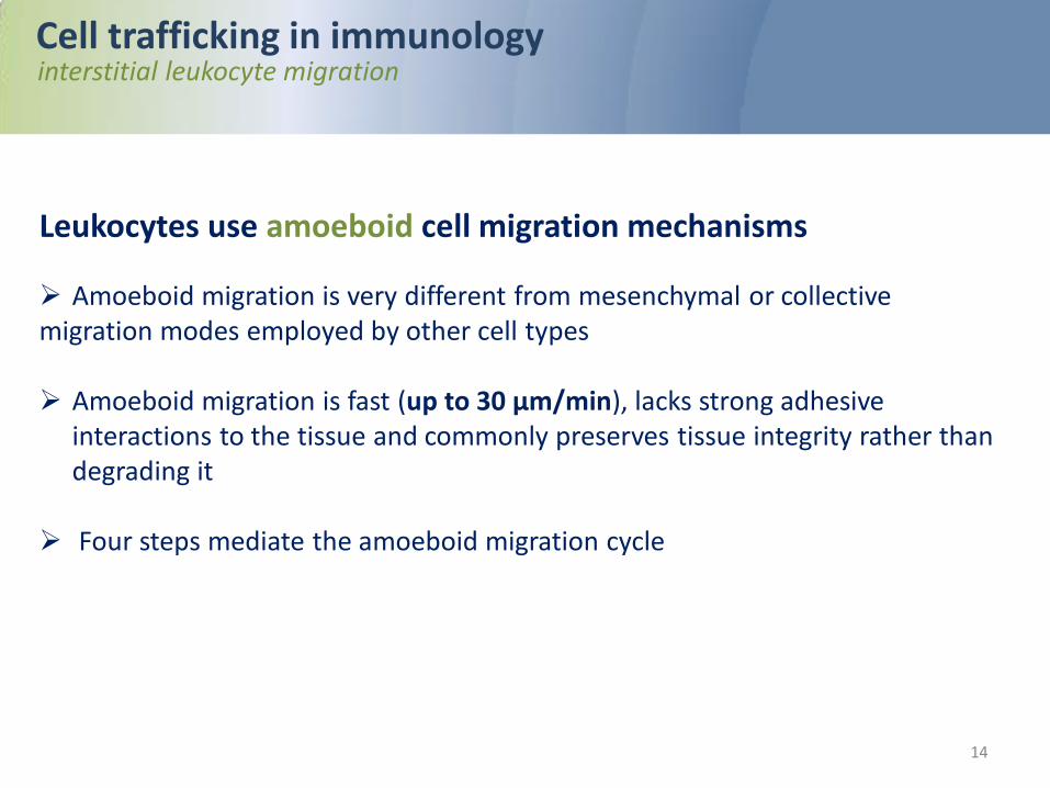
Leukocytes use amoeboid cell migration mechanisms
Amoeboid migration is very different from mesenchymal or collective migration modes employed by other cell types Amoeboid migration is fast (up to 30 μm/min), lacks strong adhesive
interactions to the tissue and commonly preserves tissue integrity rather than degrading it
Four steps mediate the amoeboid migration cycle

15
Cell trafficking in immunology interstitial leukocyte migration
4-step mechanisms of amoeboid migration 1. the leading edge protrudes one or several pseudopods by actin flow, 2. protruding membrane and surface receptors interact with the substrate, 3. actomyosin-mediated contraction of the cell body occurs in mid-region, and 4. so the rear of the cell moves forward. These steps occur in a cyclic manner, generating forward movement:

16
Cell trafficking in immunology interstitial leukocyte migration
GPCR-signaling: results in local activation of Rac Rac induces actin polymerization through WAVE (Scar) and Arp2/3. WAVE, a member of the WASP family of actin-binding proteins, mediates actin filament formation. Arp2/3 causes sideward branching of actin filaments. Together, these activities generate interconnected, branched networks.
Leading edge: high density of GPCR-receptors:
• fMLP (N-formyl-Met-Leu-Phe) receptor and • C5a receptor • chemokine receptors including CCR7, CXCR4, CXCR5,CCR3 • The leukotriene B4 receptor BLT1 • sphingosine-1-phosphate receptors • lysophosphatidic acid (LPA) receptors

17
Cell trafficking in immunology interstitial leukocyte migration
signaling key player: Rho and ROCK Upstream of myosin II (unclear mechanisms) PI(3)K-γ and possibly DOCK-2 suppress lateral protrusions The phosphatase PTEN also contributes to lateral stability by preventing ectopic protrusion formation. PTEN is excluded from the leading edge but active in lateral and rear cell parts, where it dephosphorylates kinases, including PI(3)K and Akt, as well as phosphatidylinositol-(3,4,5)-trisphosphate, and thereby counteracts protrusion formation
Mid-body: generates actomyosin-based stiffness and
contractility limits lateral protrusions and thereby maintains a
stable,bipolar cortex The cytoskeletal motor protein myosin II located in
the central and rear regions of leukocytes promotes actin filament contraction and limits lateral protrusions
Myosin II cross-links actin filaments in parallel, forming the contractile shell required to hold the extending cell together and propelling the cell nucleus, the most rigid part of the cell, forward

18
Cell trafficking in immunology interstitial leukocyte migration
Uropod=trailing edge: contains the highly glycosylated surface receptors CD43 and CD44, adhesion receptors
including intercellular adhesion molecule (ICAM)-1, ICAM-3, β1 integrins and ERM adaptor proteins The uropod mediates cell–matrix and cell–cell interactions during migration (putative
anchoring function) Contains rearward-polarized microtubules, the Golgi, and abundant actin-binding ERM (ezrin, radixin, moesin) proteins In association with microtubules, mitochondria localize to the rear of the cell, which, presumably owing to local ATP delivery to the region of ATP-dependent actomyosin contraction, is required for proper polarization, uropod retraction and migration

19
Cell motility relies on the remodeling of the cell shape:
How are the external motility signals integrated
to coordinate cell shape remodeling?

20
Cytoskeletal rearrangements General aspects
The initial response of a cell to a migration-promoting agent is to polarize and extend protrusions in the direction of migration. These protrusions can be large, broad lamellipodia or spike-like filopodia, are usually driven by actin polymerization, and are stabilized by adhering to the extracellular matrix (ECM) or adjacent cells via transmembrane receptors linked to the actin cytoskeleton. These adhesions serve as traction sites for migration as the cell moves forward over them, and they are disassembled at the cell rear, allowing it to detach.

21
Cytoskeletal rearrangements Actin
Actin cytoskeleton organization at the two poles of a migrating T cell. Schematic representations of the ultrastructure of the actin cytoskeleton networks at the leading and trailing edges of the migrating T cell. At the leading edge, the T cell that migrates on a 2D surface emits a protrusion that alternates between a lamellipodium and a pseudopodium. It contains a very dynamical and highly branched actin meshwork. At the trailing edge, the T cell uropod is made of a network of parallel actin bundles that can slide along each other to generate contractile forces.
Frontiers in Immunology | Dupré et al. I November 2015 | Volume 6 | Article 586

22
Cytoskeletal rearrangements Basic Regulation of Actin Assembly
Frontiers in Immunology | Dupré et al. I November 2015 | Volume 6 | Article 586
ATP-bound actin is added to the fast growing barbed end of filaments via the combined action of profilin, which prevents self-nucleation of actin monomers and actin-nucleating proteins (formin FMLN1 or WASP-family proteins) both of which are under the control of RhoGTPases.
Depolymerization is promoted by cofilin, which stimulates dissociation
of ADP-bound actin at the pointed end of filaments. The rate of cofilin-mediated depolymerization can be controlled by Rho via Rock.

23
Cytoskeletal rearrangements Basic Regulation of Actin Assembly
Frontiers in Immunology | Dupré et al. I November 2015 | Volume 6 | Article 586
In addition to be elongated by formins, actin filaments can build networks in multiple ways. Actin bundles or cables with parallel or anti-parallel orientation of actin filaments are assembled by cross-linking proteins such as fimbrin. Actin filaments can also be cross-linked in a non-parallel fashion via filamin to create a gelled network.
Branched networks are promoted by the Arp2/3 complex that initiates nucleation of branched filaments on the side of existing ones.
This activity is driven by WASP-family proteins and stabilized by HS-1 An additional important regulation of actin cytoskeleton networks is
mediated by capping proteins such as gelsolin, which bind the plus end of actin filaments to prevent monomer exchange.

24
Cytoskeletal rearrangements Basic Regulation of Actin Assembly
Frontiers in Immunology | Dupré et al. I November 2015 | Volume 6 | Article 586
Actin filaments not only generate forces while they elongate. They also generate the cell contractile forces via the intercalation of the molecular motor myosin between parallel actin filaments, which results in filament sliding.
Such process is regulated by the control of the myosin light chain phosphatase and kinase activities, as well as by the degree of actin cross-linking via α-actinin.

25
Basics of Actin Assembly
Actin Filament Elongation, Branching and Stabilization Actin Filament Crosslinking and Myosin-Driven Contraction
Frontiers in Immunology | Dupré et al. November 2015 | Volume 6 | Article 586

26
Cytoskeletal rearrangements Actin
Molecular actors contributing to actin cytoskeleton remodeling in T lymphocytes
Frontiers in Immunology | Dupré et al. I November 2015 | Volume 6 | Article 586
27
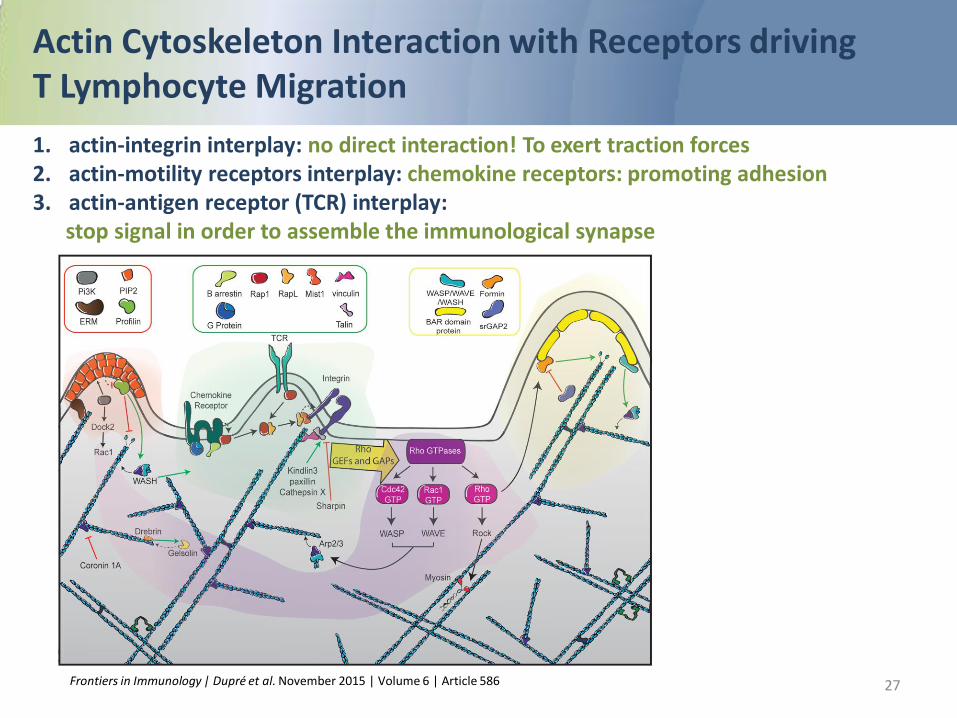
Actin Cytoskeleton Interaction with Receptors driving T Lymphocyte Migration 1. actin-integrin interplay: no direct interaction! To exert traction forces 2. actin-motility receptors interplay: chemokine receptors: promoting adhesion 3. actin-antigen receptor (TCR) interplay: stop signal in order to assemble the immunological synapse
Frontiers in Immunology | Dupré et al. November 2015 | Volume 6 | Article 586

How do the cells know that and where
they have to migrate?
Signals that induce and coordinate movement:
Chemokines
28

29
Chemokines
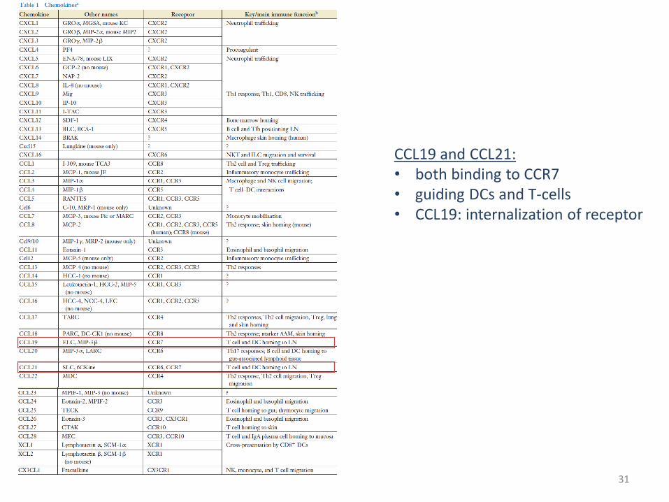
Definition: Chemokines are chemotactic cytokines that control the migratory patterns and positioning of immune cells. Chemokine function is critical for all immune cell movement ranging from the migration required for immune cell development and homeostasis, to that required for the generation of primary and amnestic cellular and humoral immune responses, to the pathologic recruitment of immune cells in disease. Chemokines are a group of small (∼8–14 kDa), mostly basic, structurally related molecules that regulate cell trafficking of various types of leukocytes through interactions with a subset of seven-transmembrane, G protein–coupled receptors. About 40 chemokines have now been identified in humans. They mainly act on neutrophils, monocytes, lymphocytes, and eosinophils and play a pivotal role in host defense mechanisms.
Chemokine classes CC CXC C CX3C

30
31
CCL19 and CCL21: • both binding to CCR7 • guiding DCs and T-cells • CCL19: internalization of receptor

32
J Immunol. May 1, 2006, 176 (9) 5153-5159
CCL19>> internalization of the receptor
CCL19/CCL21: chemokine signaling via CCR7 Homing of T-cells and guidance of DCs to lymph nodes

33
Cytokine & Growth Factor Reviews Volume 24, Issue 3, June 2013, Pages 269-283 https://doi.org/10.1016/j.cytogfr.2013.03.001

34
Which diseases originate/are associated from/with defective migration ?
impact of proper cellular locomotion

35
Luster et al.; VOLUME 6 NUMBER 12 DECEMBER 2005 NATURE IMMUNOLOGY

36 Frontiers in Immunology | www.frontiersin.org November 2015 | Volume 6 | Article 586

37
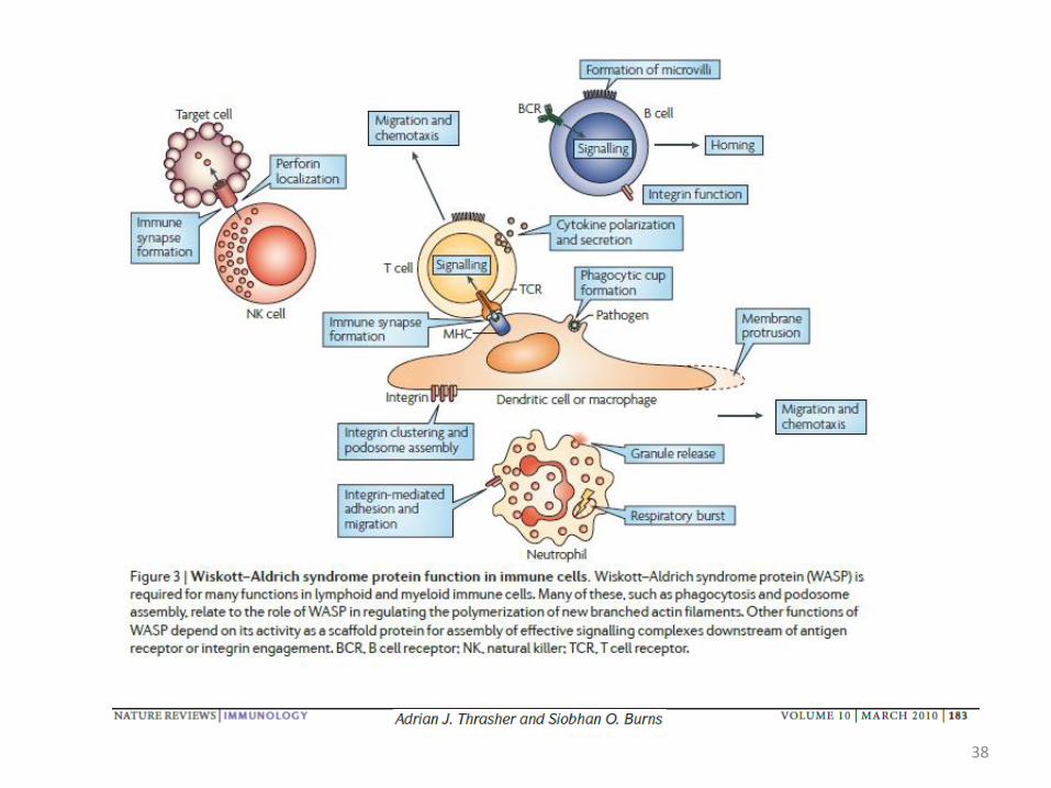
Example : The Wiskott–Aldrich syndrome
Loss of WASP (Wiskott–Aldrich syndrome protein) activity leads to Wiskott–Aldrich syndrome, an X-linked disease that is associated with defects in a broad range of cellular processes, resulting in complex immunodeficiency, autoimmunity and microthrombocytopenia.
Disease Markers 29 (2010) 157–175
38

39
Summary Trafficking of leukocytes ..is central to immune cell development, immunosurveillance and effector function Innate and adaptive immune functions depend upon interstitial leukocyte migration Inflammation: Activation of endothelial cells by pro-inflammatory cytokines from resident immune cells To enter tissues, leukocytes pass endothelial barrier (multistep cascade) in a selectin and integrin-dependent manner Within the tissue, leukocytes migrate in an amoeboid fashion (4 steps) which differs from mesenchymal migration Polarization of cells > cellular shape remodeling: leading edge; mid-body; uropod Cytoskeletal rearrangement: signaling that regulates actin assembly/disassembly Soluble factors that initiate migration/maturation: Chemokines CCL19/CCL21 as an example for guiding DCs and T-cells to and within lymphnodes
The Wiskott–Aldrich syndrome: defective WASP results in complex immunodeficiency


![Clinical & Cellular Skelton et al., Immunology migration into the graft, restriction of their Vβ-TCR repertoire [3], ... purified via positive T cell isolation kit using magnetic](https://static.fdocuments.us/doc/165x107/5ab499c17f8b9a7c5b8bf547/clinical-cellular-skelton-et-al-immunology-migration-into-the-graft-restriction.jpg)