Cell guidance by magnetic nanowires
7
Research Article Cell guidance by magnetic nanowires Fredrik Johansson ⁎ , Malin Jonsson, Kersti Alm, Martin Kanje Department of Cell and Organism Biology, Lund University, Helgonavägen 3b, SE-22362 Lund, Sweden ARTICLE INFORMATION ABSTRACT Article Chronology: Received 15 October 2008 Revised version received 12 November 2009 Accepted 16 December 2009 Available online 28 December 2009 The phenomenon of contact guidance on thin fibers has been known since the beginning of the 20th century when Harrison studied cells growing on fibers from spider's web. Since then many studies have been performed on structured surfaces and fibers. Here we present a new way to induce guidance of cells or cell processes using magnetic nanowires. We have manufactured magnetic Ni-nanowires (200 nm in diameter and 40 μm long) with a template-based electro- deposition method. Drops of a nanowire/ethanol suspension were placed on glass cover slips. The nanowires were aligned in an external magnetic field and adhered to the cover slips after evaporation of the ethanol. When the wires had adhered, the magnetic field was removed. L929 fibroblasts and dissociated dorsal root ganglia (DRG) neurons from mice were cultured on the nanowire-coated cover slips for 24 h and 72 h respectively. The fibroblasts were affected by the aligned nanowires and displayed contact guidance. Regenerated axons also displayed contact guidance on the wires. There were no overt signs of toxicity caused by Ni-wires. Aligned magnetic nanowires can be useful for lab-on-a-chip devices and medical nerve grafts. © 2010 Elsevier Inc. All rights reserved. Keywords: Nanowire Axon Fibroblast Magnetic Guidance Introduction Structurally well-defined magnetic nano-particles (MNP) and nanowires have become increasingly important tool in biotech- nology applications such as drug delivery, imaging and cell manipulation [1-6]. Cell manipulation by external magnetic fields, i.e. cell separation, has been made possible using phagocytosed MNPs in cells. Hultgren et al. demonstrated that for cell separation, magnetic nanowires have an advantage over spherical particles since elongated structures have a stronger magnetic moment and hence may exert a larger force on the cell. The advantages of wires as compared to particles are due to the high aspect ratio (elongated shape), resulting in higher magnetic anisotropy. Nanowires can be produced by many techniques. We have used electro-deposition of Ni, in nano-porous alumina membranes since it is a very successful method to obtain magnetic nanowires using standard laboratory equipment [7]. Such magnetic Ni-nanowires have typically a width of 200 nm, which corresponds to the pore size of the template, while lengths are determined by the membrane thickness or deposition time. The size of such wires is very suitable for cell interaction applications such as cell separation [2-4], where the wires should be internalized in eukaryotic cells that are typically 10 to 20 μm in diameter. Spatial cell organization can also be achieved among cells with internalized nanowires, either spontaneously in an end-to-end fashion, or by using external micro-magnet patterns [8,9]. Furthermore, it is possible to create chemically different functional sequences along the wires by altering the metal e.g. Au and Ni along the wires in a striated manner [6,10,11]. Then, link molecules that bind to specific metals, such as thiols for functionalization of gold, enables chemical patterning with any molecule of interest. Simple (nonfunctiona- lized) wires can, as in this study, also be used for easy production of nanotopographies on which cells can be cultured. The wires and hence the topography can be manipulated via an external magnetic EXPERIMENTAL CELL RESEARCH 316 (2010) 688 – 694 ⁎ Corresponding author. Fax: +46 462224539. E-mail address: [email protected] (F. Johansson). 0014-4827/$ – see front matter © 2010 Elsevier Inc. All rights reserved. doi:10.1016/j.yexcr.2009.12.016 available at www.sciencedirect.com www.elsevier.com/locate/yexcr
-
Upload
fredrik-johansson -
Category
Documents
-
view
212 -
download
0
Transcript of Cell guidance by magnetic nanowires

E X P E R I M E N T A L C E L L R E S E A R C H 3 1 6 ( 2 0 1 0 ) 6 8 8 – 6 9 4
ava i l ab l e a t www.sc i enced i r ec t . com
www.e l sev i e r . com/ loca te /yexc r
Research Article
Cell guidance by magnetic nanowires
Fredrik Johansson⁎, Malin Jonsson, Kersti Alm, Martin Kanje
Department of Cell and Organism Biology, Lund University, Helgonavägen 3b, SE-22362 Lund, Sweden
A R T I C L E I N F O R M A T I O N
⁎ Corresponding author. Fax: +46 462224539.E-mail address: [email protected]
0014-4827/$ – see front matter © 2010 Elseviedoi:10.1016/j.yexcr.2009.12.016
A B S T R A C T
Article Chronology:
Received 15 October 2008Revised version received12 November 2009Accepted 16 December 2009
Available online 28 December 2009
The phenomenon of contact guidance on thin fibers has been known since the beginning of the20th century when Harrison studied cells growing on fibers from spider's web. Since then manystudies have been performed on structured surfaces and fibers. Here we present a new way toinduce guidance of cells or cell processes using magnetic nanowires. We have manufacturedmagnetic Ni-nanowires (200 nm in diameter and 40 μm long) with a template-based electro-
deposition method. Drops of a nanowire/ethanol suspension were placed on glass cover slips. Thenanowires were aligned in an external magnetic field and adhered to the cover slips afterevaporation of the ethanol. When the wires had adhered, the magnetic field was removed. L929fibroblasts and dissociated dorsal root ganglia (DRG) neurons from mice were cultured on thenanowire-coated cover slips for 24 h and 72 h respectively. The fibroblasts were affected by thealigned nanowires and displayed contact guidance. Regenerated axons also displayed contactguidance on the wires. There were no overt signs of toxicity caused by Ni-wires. Aligned magneticnanowires can be useful for lab-on-a-chip devices and medical nerve grafts.
© 2010 Elsevier Inc. All rights reserved.
Keywords:
NanowireAxonFibroblastMagneticGuidance
Introduction
Structurally well-defined magnetic nano-particles (MNP) andnanowires have become increasingly important tool in biotech-nology applications such as drug delivery, imaging and cellmanipulation [1-6]. Cell manipulation by external magnetic fields,i.e. cell separation, has been made possible using phagocytosedMNPs in cells. Hultgren et al. demonstrated that for cell separation,magnetic nanowires have an advantage over spherical particlessince elongated structures have a stronger magnetic moment andhence may exert a larger force on the cell. The advantages of wiresas compared to particles are due to the high aspect ratio(elongated shape), resulting in higher magnetic anisotropy.
Nanowires can be produced bymany techniques.We have usedelectro-deposition of Ni, in nano-porous aluminamembranes sinceit is a very successful method to obtain magnetic nanowires usingstandard laboratory equipment [7]. Such magnetic Ni-nanowires
u.se (F. Johansson).
r Inc. All rights reserved.
have typically a width of 200 nm, which corresponds to the poresize of the template, while lengths are determined by themembrane thickness or deposition time. The size of such wires isvery suitable for cell interaction applications such as cell separation[2-4], where the wires should be internalized in eukaryotic cellsthat are typically 10 to 20 μm in diameter. Spatial cell organizationcan also be achieved among cells with internalized nanowires,either spontaneously in an end-to-end fashion, or by using externalmicro-magnet patterns [8,9]. Furthermore, it is possible to createchemically different functional sequences along the wires byaltering the metal e.g. Au and Ni along the wires in a striatedmanner [6,10,11]. Then, linkmolecules that bind to specific metals,such as thiols for functionalization of gold, enables chemicalpatterning with any molecule of interest. Simple (nonfunctiona-lized)wires can, as in this study, also be used for easy production ofnanotopographies on which cells can be cultured. The wires andhence the topography can bemanipulated via an externalmagnetic

689E X P E R I M E N T A L C E L L R E S E A R C H 3 1 6 ( 2 0 1 0 ) 6 8 8 – 6 9 4
field. The complexity of such nanotopography depends primarilyon the shape of the magnetic field applied.
For many applications such as tissue engineering and neuronalnetworks, guidance of cells or cellular processes is important. To thisend,mainly two approaches are used, namely chemical patterning ofsurfaces [12] or topographic patterning. Organized topography, i.e.grooves [13,14], steps [13,15], fibers [13,16,17], or even rows ofstanding nanowires [18], are among the structures which inducecontact guidance for migrating cells and/or outgrowing nerve cellprocesses, axons. The techniques required to obtain such topographicpatterns on cell substrates are fairly cumbersome and involvesprocesses such as e-beam-lithography, UV-lithography and epitaxy.Most such studies have been performed on micrometer-sizedstructures, such as grooves and ridges, due to technical limitations,but others and we have shown that the phenomenon of contactguidance is valid also for submicron grooves and ridges [14,19].
On rounded surfaces such as fibers, the substrate curvatureinfluences the growth direction of nerve cell processes. An elegantstudy, although in the micrometer range, by Smeal et al. demon-strated that thin fibers (Ø=35 μm) induce a more pronouncedguidance than fibers with larger diameter (Ø=68–500 μm) [17].From this results that indicate better guidance on thinner, smallradius fibers, one may expect that fibers such as a Ni nanowire(Ø=200 nm)would induce a clear contact guidance effect if alignedon a surface. This is indeed the case and has been demonstrated onelectrospun polymer nanofibers, although the fiber thickness ofthose experiments is usually spread from nano-tomicrometers [20].Patterns of spatially well defined, magnetically aligned nanowires
Fig. 1 – (A) SEM image showing a top view of the pores in an aluminarevealing the pores of a 60-μm thick alumina membrane. (C) ThisPhotograph of the setup used.
mayalso serveasguidelines for cells andcell processes. Furthermore,this would eliminate the need for special equipment/techniques,such as facilities for lithographic processing or electro-spinning ofthe substrate, to achieve cell patterning and could be valuable for theconstruction of neuronal networks in vitro.
Here, we have studied the guidance of outgrowing nerve cellprocesses from dorsal root ganglia (DRG), and guidance of L929mouse fibroblasts on aligned Ni nanowires. The nanowires wereproduced by template-based electro-deposition in commerciallyavailable alumina membranes.
Materials and methods
Nanowire synthesis
The synthesis of the Ni nanowires has been described in detailelsewhere [21]. In short, commercially available 60 μm thickalumina membranes (Anodisc, Whatman Inc) with Ø=200 nmpores, (Figs. 1A and B) were used as templates to grow the Ninanowires. One side of the Anodisc filter has a thin layer (∼1 μm)of smaller pores (Ø=20 nm) that merge into the larger pores. Thisside of the membrane was covered with Ga/In eutectic (AlfaAesar®, Germany) to form a conductive layer that was used ascathode. The filter was placed on a copper electrode with theconducting layer in contact with the electrode. A reservoir, sealedwith gaskets and secured by clippers was placed atop of the filter.The reservoir was filled with Ni plating solution (1 M NiCl2, pH 1)
membrane. (B) SEM image (tilted 45°) of a crackedmembrane,simplified sketch shows the electro-deposition setup. (D)
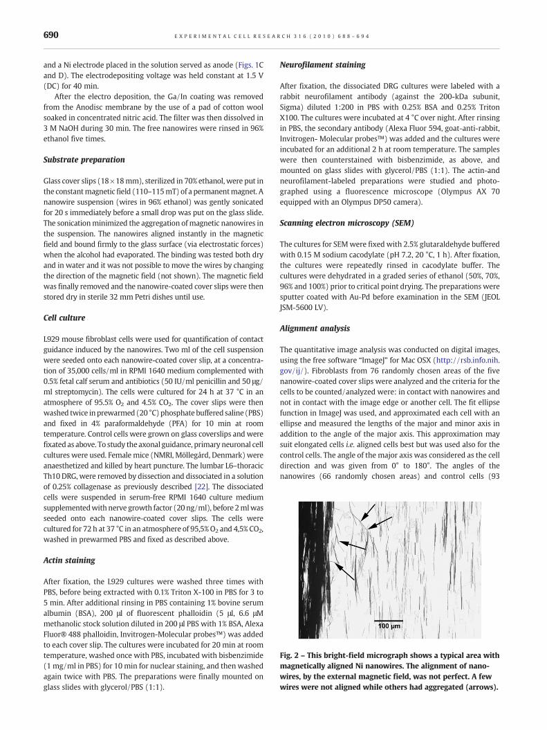
Fig. 2 – This bright-field micrograph shows a typical area withmagnetically aligned Ni nanowires. The alignment of nano-wires, by the external magnetic field, was not perfect. A fewwires were not aligned while others had aggregated (arrows).
690 E X P E R I M E N T A L C E L L R E S E A R C H 3 1 6 ( 2 0 1 0 ) 6 8 8 – 6 9 4
and a Ni electrode placed in the solution served as anode (Figs. 1Cand D). The electrodepositing voltage was held constant at 1.5 V(DC) for 40 min.
After the electro deposition, the Ga/In coating was removedfrom the Anodisc membrane by the use of a pad of cotton woolsoaked in concentrated nitric acid. The filter was then dissolved in3 M NaOH during 30 min. The free nanowires were rinsed in 96%ethanol five times.
Substrate preparation
Glass cover slips (18×18mm), sterilized in 70% ethanol, were put inthe constantmagnetic field (110–115mT) of a permanentmagnet. Ananowire suspension (wires in 96% ethanol) was gently sonicatedfor 20 s immediately before a small drop was put on the glass slide.The sonicationminimized the aggregation ofmagnetic nanowires inthe suspension. The nanowires aligned instantly in the magneticfield and bound firmly to the glass surface (via electrostatic forces)when the alcohol had evaporated. The binding was tested both dryand in water and it was not possible to move the wires by changingthe direction of the magnetic field (not shown). The magnetic fieldwas finally removed and the nanowire-coated cover slips were thenstored dry in sterile 32 mm Petri dishes until use.
Cell culture
L929 mouse fibroblast cells were used for quantification of contactguidance induced by the nanowires. Two ml of the cell suspensionwere seeded onto each nanowire-coated cover slip, at a concentra-tion of 35,000 cells/ml in RPMI 1640 medium complemented with0.5% fetal calf serum and antibiotics (50 IU/ml penicillin and 50 μg/ml streptomycin). The cells were cultured for 24 h at 37 °C in anatmosphere of 95.5% O2 and 4.5% CO2. The cover slips were thenwashed twice in prewarmed (20 °C) phosphate buffered saline (PBS)and fixed in 4% paraformaldehyde (PFA) for 10 min at roomtemperature. Control cells were grown on glass coverslips and werefixated as above. To study the axonal guidance, primary neuronal cellcultures were used. Female mice (NMRI, Möllegård, Denmark)wereanaesthetized and killed by heart puncture. The lumbar L6–thoracicTh10 DRG, were removed by dissection and dissociated in a solutionof 0.25% collagenase as previously described [22]. The dissociatedcells were suspended in serum-free RPMI 1640 culture mediumsupplementedwith nerve growth factor (20ng/ml), before 2mlwasseeded onto each nanowire-coated cover slips. The cells werecultured for 72 h at 37 °C in an atmosphere of 95,5%O2 and 4,5% CO2,washed in prewarmed PBS and fixed as described above.
Actin staining
After fixation, the L929 cultures were washed three times withPBS, before being extracted with 0.1% Triton X-100 in PBS for 3 to5 min. After additional rinsing in PBS containing 1% bovine serumalbumin (BSA), 200 μl of fluorescent phalloidin (5 μl, 6.6 μMmethanolic stock solution diluted in 200 μl PBS with 1% BSA, AlexaFluor® 488 phalloidin, Invitrogen-Molecular probes™) was addedto each cover slip. The cultures were incubated for 20 min at roomtemperature, washed once with PBS, incubated with bisbenzimide(1 mg/ml in PBS) for 10 min for nuclear staining, and then washedagain twice with PBS. The preparations were finally mounted onglass slides with glycerol/PBS (1:1).
Neurofilament staining
After fixation, the dissociated DRG cultures were labeled with arabbit neurofilament antibody (against the 200-kDa subunit,Sigma) diluted 1:200 in PBS with 0.25% BSA and 0.25% TritonX100. The cultures were incubated at 4 °C over night. After rinsingin PBS, the secondary antibody (Alexa Fluor 594, goat-anti-rabbit,Invitrogen- Molecular probes™) was added and the cultures wereincubated for an additional 2 h at room temperature. The sampleswere then counterstained with bisbenzimide, as above, andmounted on glass slides with glycerol/PBS (1:1). The actin-andneurofilament-labeled preparations were studied and photo-graphed using a fluorescence microscope (Olympus AX 70equipped with an Olympus DP50 camera).
Scanning electron microscopy (SEM)
The cultures for SEMwere fixed with 2.5% glutaraldehyde bufferedwith 0.15 M sodium cacodylate (pH 7.2, 20 °C, 1 h). After fixation,the cultures were repeatedly rinsed in cacodylate buffer. Thecultures were dehydrated in a graded series of ethanol (50%, 70%,96% and 100%) prior to critical point drying. The preparations weresputter coated with Au-Pd before examination in the SEM (JEOLJSM-5600 LV).
Alignment analysis
The quantitative image analysis was conducted on digital images,using the free software “ImageJ” for Mac OSX (http://rsb.info.nih.gov/ij/). Fibroblasts from 76 randomly chosen areas of the fivenanowire-coated cover slips were analyzed and the criteria for thecells to be counted/analyzed were: in contact with nanowires andnot in contact with the image edge or another cell. The fit ellipsefunction in ImageJ was used, and approximated each cell with anellipse and measured the lengths of the major and minor axis inaddition to the angle of the major axis. This approximation maysuit elongated cells i.e. aligned cells best but was used also for thecontrol cells. The angle of the major axis was considered as the celldirection and was given from 0° to 180°. The angles of thenanowires (66 randomly chosen areas) and control cells (93

691E X P E R I M E N T A L C E L L R E S E A R C H 3 1 6 ( 2 0 1 0 ) 6 8 8 – 6 9 4
randomly chosen areas) were measured in the same way. Cellswith amajor/minor axis ratio <1.5 were considered round, and nodirection could be measured. Such round cells were omitted in thealignment analysis, although counted.
Statistics
The absolute angle deviation of the cells from the averagenanowire direction, was tested relative the control cells. Theexperiment was repeated five times and the alignment analysiswas done from: 76 images resulting in 1048 cell counts onnanowires, 93 images resulting in 1844 control cell counts and 66images resulting in 3982 nanowire counts. A two-tailed t-test wasused for the comparison and p<0.05 was considered significant.
Fig. 3 – The images show L929 fibroblasts cultured on both the nanoThe L929 fibroblasts display contact guidance on the aligned Ni nancells show no alignment. (C) The SEM image shows fibroblasts botdifferent morphologies.
Results
Nanowires
The Ni nanowires differed somewhat in length. From SEMimages, the length of the wires was estimated to 40±10 μm(mean±SD). The diameter was 200 nm±20 nm (mean±SD),although some irregularities due to defect pores were found (notshown). The overall alignment of the nanowires was found to bevery good (Fig. 2) although not perfect. In this study, a highconcentration of wires was desirable to ensure cell-wireinteraction from many cells. Therefore some aggregation due tomagnetic interaction among neighbor wires was inevitable. The
wire-coated cover slips and smooth plastic control surfaces. (A)owires, and become elongated along the wires. (B) The controlh on nanowires and on the smooth glass surface. Note the

692 E X P E R I M E N T A L C E L L R E S E A R C H 3 1 6 ( 2 0 1 0 ) 6 8 8 – 6 9 4
nanowires are fixed to the surface by electrostatic and van derWaahls forces [23], and were not moved by the cells after24 hours of culture. This indicates a strong adhesion of the wiresto the naked glass surface.
L929 fibroblasts alignment
L929 fibroblasts cultured on nanowire-coated cover slips weremostly found on the nanowires and were clearly aligned andelongated along the nanowires (Fig. 3A). Fibroblasts that werenot in contact with the nanowires, and thereby excluded fromthe analysis, generally had a flattened shape with extensions indifferent directions, similar to that observed in the controlcultures (Figs. 3B and C). The alignment analysis showed asignificant (p<0.001) difference between cells on nanowires andcells on smooth control surfaces. This difference was also clearfrom the polar diagrams where the direction of the fibroblasts(90.5°±1.3°) coincides nicely with the direction of the alignednanowires (90.2°±0.4°), while the direction of the control cellswas more isotropic (Fig. 4) (angles reported as mean±SEM).Furthermore, there were more round cells, i.e. cells with amajor/minor axis ratio <1.5, on the control surfaces than on thenano-coated surfaces, 32.9% and 22.6% respectively.
DRG axon alignment
Regenerating axons from DRG neurons also displayed contactguidance, although the axons often changed nanowire in awandering fashion. Each single axon may change wire or evenignore to grow along a wire, in a seemingly random outgrowthprocess (Fig. 5). Even though the contact guidance cue from asingle nanowire may be questioned, the overall guiding effect isobvious from the images. This supports our previous study onaxonal guidance induced by grooves and ridges, where awandering outgrowth of axons still displayed a high degree ofoverall guidance [14].
Fig. 4 – The polar diagrams show the cell or nanowire distribution ioriented nanowires display a uniform direction at 90°. (B) Cells on sorientation of the aligned nanowires was well gathered around 90alignment. The angles of nanowires and aligned L929 fibroblasts cosymmetrical due to the angle measurement, that only give values b
Discussion
Previous studies have demonstrated that magnetic Ni nanowirescan be used to manipulate cells. After nanowire internalization byphagocytosis, the cells can be moved by external magnetic fields[2-4,8,9]. Here, for the first time, we demonstrate contact guidanceof whole cells (fibroblasts) and cell extensions (DRG axons)induced by aligned magnetic Ni nanowires. Guidance of cells byexternally controlled structures could find applications in manydifferent fields, e.g. “lab on a chip” devices, medical nerve grafts orfor the construction of tissue or organ like structures in vitro,including neuronal networks. Furthermore, the possibility tocombine different metals along the wire, e.g. Au-Ni-Au, and alsochemically functionalize the different parts of the wire, makesmagnetic nanowires highly interesting for further nanobiotech-nology applications [4,6,11].
In this study, the contact guidance due to thewireswas obviousin the L929 fibroblast cultures. The axons in the DRG neuronalculture showed a wandering outgrowth from one wire to another,suggesting that more powerful guidance cues (more wires) areneeded to induce good guidance.
To develop cell-guiding patterns, the alignment of the wiresmay be improved and alignment parameters have beenextensively studied [23,24]. The alignment may be affected bythe liquid flow during evaporation, where the drag force exceedsthe magnetic torque of the external field. Furthermore, an excessof nanowires results in aggregation due to magnetic interactionsamong the wires, resulting in alignment disturbances. Theintroduction of micro magnet arrays [6,9] may facilitate theproduction of complicated guiding patterns in real time withoutany predetermined lithographic patterning. This would enable amore dynamic way of testing guidance phenomenon in vitro andwould be valuable for the production of neuronal networks andcell patterns.
Ni at a high concentration is toxic to cells, and there could bea release of Ni in the culture medium, something that may be
n different directions. (A) The major axes of L929 fibroblasts onmooth control surfaces have random angle distribution. (C) The°, i.e. the direction of the external magnetic field duringincide, indicating contact guidance. Note that the diagrams areetween 0° and 180°.

Fig. 5 – The images show regenerating axons from dissociated DRG neurons interacting with the aligned nanowires. (A) Theguidance of axons was somewhat less than that found for the L929 fibroblasts, and most clearly detected when an excess ofnanowires forming large aggregates, was available. (Fluorescence image: neurons are red, nanowires faint green). Note therandomly growing axons outside the nanowire-coated area. (B) This SEM image shows axons growing along the nanowires for awhile, and their deflection in another direction. At areas with low concentrations of nanowires, the axons often changewire to growalong, as if the guiding cue of one wire is too weak to establish sufficient contact guidance. (C) This overview of axons on alignednanowires at low concentration shows that even if the axons occasionally changewire to grow along, the total arrangement, displaytypical contact guidance. (Fluorescence image: neurons are red, nanowires faint green and nuclei blue).
693E X P E R I M E N T A L C E L L R E S E A R C H 3 1 6 ( 2 0 1 0 ) 6 8 8 – 6 9 4
cumbersome for the viability of the cell culture. However, in cellseparation studies, using similar wires as here, Hultgren et al.found no signs of toxicity for NIH-3T3 mouse fibroblast culturedfor 3 days [2,4]. In MC3T3-E1 osteoblasts and marrow stromalcells, Prina-Mello et al. reported changes in organelle responsesbut the viability of the cells was not significantly compromisedby the internalization of the Ni nanowires [8]. Furthermore,Schedle et al. found a dose dependent inhibition of 3H-thymidineincorporation into L-929 fibroblasts when they were exposed todifferent metal cations. For L-929 fibroblasts, Ni2+was found toaffect proliferation less than Au3+, Cu2+and Co2+(an alternativeferromagnetic metal), but more than Zn2+, Pd2+or Cr2+[25]. It isalso speculated that insoluble Ni, internalized by phagocytosismay be mutagenic [26]. The issue of toxicity must be addressed,but for long time studies, covering the wires with inertbiocompatible materials, e.g. polymers, may control the releaseof Ni.
We have demonstrated that Ni nanowires that are attachedto a surface induce contact guidance for both fibroblasts andaxons. The molecular mechanism by which this occurs remainsto be elucidated. However, contact guidance involves rear-
rangement of focal adhesion points and cytoskeleton elements[27,28]. It has been reported that cells internalize free Ninanowires via an integrin-mediated phagocytosis [4,29]. Sinceintegrins are involved in both axonal outgrowth and celladhesion to surfaces, an interesting possibility is that thealignment of the fibroblasts and axons are due to theirattempts to phagocytose the firmly attached nanowires.Additional experiments with nanowires that release moreeasily from the surface after cell-alignment has occurred mayshow whether the same integrins are involved during theinternalization of the wires. This could be an importantguidance mechanism that to our knowledge has not beenpreviously suggested.
Conclusion
The present study showed that aligned magnetic Ni nanowiresinduce contact guidance both for single migrating cells (L-929fibroblasts) and outgrowing axons from dorsal root ganglionneurons. The construction of patterned substrates for the control

694 E X P E R I M E N T A L C E L L R E S E A R C H 3 1 6 ( 2 0 1 0 ) 6 8 8 – 6 9 4
of cellular growth opens a new avenue of medical and biotechno-logical applications for magnetic nanowires.
Acknowledgments
This work was supported by the Swedish Research Council. Thanksare due to Inger Antonson for her technical assistance. Thanks arealso due to Waldemar Hällström and PhD Per Gustavsson for theirvaluable input to the study.
R E F E R E N C E S
[1] D. Choi, A. Fung, H. Moon, D. Ho, Y. Chen, E. Kan, Y. Rheem, B. Yoo,N. Myung, Transport of living cells with magnetically assemblednanowires, Biomed. Microdevices 9 (2007) 143–148.
[2] A. Hultgren, M. Tanase, C.S. Chen, G.J. Meyer, D.H. Reich, Cellmanipulation using magnetic nanowires, J. Appl. Phys. 93 (2003)7554–7556.
[3] A. Hultgren, M. Tanase, C.S. Chen, D.H. Reich, High-yield cellseparations using magnetic nanowires, IEEE Trans. Magn. 40(2004) 2988–2990.
[4] A. Hultgren, M. Tanase, E.J. Felton, K. Bhadriraju, A.K. Salem, C.S.Chen, D.H. Reich, Optimization of yield in magnetic cellseparations using nickel nanowires of different lengths,Biotechnol. Prog. 21 (2005) 509–515.
[5] T.K. Jain, J. Richey, M. Strand, D.L. Leslie-Pelecky, C.A. Flask, V.Labhasetwar, Magnetic nanoparticles with dual functionalproperties: drug delivery and magnetic resonance imaging,Biomaterials 29 (2008) 4012–4021.
[6] D.H. Reich, M. Tanase, A. Hultgren, L.A. Bauer, C.S. Chen, G.J.Meyer, Biological applications of multifunctional magneticnanowires (invited), J. Appl. Phys. 93 (2003) 7275–7280.
[7] C.R. Martin, Nanomaterials: a membrane-based syntheticapproach, Science 266 (1994) 1961–1966.
[8] A. Prina-Mello, Z. Diao, J.M. Coey, Internalization offerromagnetic nanowires by different living cells,J. Nanobiotechnol. 4 (2006) 9.
[9] M. Tanase, E.J. Felton, D.S. Gray, A. Hultgren, C.S. Chen, D.H.Reich, Assembly of multicellular constructs and microarraysof cells using magnetic nanowires, Lab Chip 5 (2005)598–605.
[10] M. Hernandez-Velez, Nanowires and 1D arrays fabrication: anoverview, Thin Solid Films 495 (2006) 51–63.
[11] S. Kim, K.L. Shuford, H.M. Bok, S.K. Kim, S. Park, Intraparticlesurface plasmon coupling in quasi-one-dimensionalnanostructures, Nano. Lett. 8 (2008) 800–804.
[12] P. Gustavsson, F. Johansson, M. Kanje, L. Wallman, C.E. Linsmeier,Neurite guidance on protein micropatterns generated by apiezoelectric microdispenser, Biomaterials 28 (2007)1141–1151.
[13] R.G. Flemming, C.J. Murphy, G.A. Abrams, S.L. Goodman, P.F.Nealey, Effects of synthetic micro-and nano-structured surfaceson cell behavior, Biomaterials 20 (1999) 573–588.
[14] F. Johansson, P. Carlberg, N. Danielsen, L. Montelius, M. Kanje,Axonal outgrowth on nano-imprinted patterns, Biomaterials 27(2006) 1251–1258.
[15] P. Clark, P. Connolly, A.S. Curtis, J.A. Dow, C.D. Wilkinson,Topographical control of cell behaviour. I. Simple step cues,Development 99 (1987) 439–448.
[16] A.S. Badami, M.R. Kreke, M.S. Thompson, J.S. Riffle, A.S. Goldstein,Effect of fiber diameter on spreading, proliferation, anddifferentiation of osteoblastic cells on electrospun poly(lacticacid) substrates, Biomaterials 27 (2006) 596–606.
[17] R.M. Smeal, R. Rabbitt, R. Biran, P.A. Tresco, Substrate curvatureinfluences the direction of nerve outgrowth, Ann. Biomed. Eng. 33(2005) 376–382.
[18] C. Prinz, W. Hallstrom, T. Martensson, L. Samuelson, L. Montelius,M. Kanje, Axonal guidance on patterned free-standing nanowiresurfaces, Nanotechnology 19 (2008).
[19] A.I. Teixeira, G.A. McKie, J.D. Foley, P.J. Bertics, P.F. Nealey, C.J.Murphy, The effect of environmental factors on the response ofhuman corneal epithelial cells to nanoscale substrate topography,Biomaterials 27 (2006) 3945–3954.
[20] Z.Ma,M. Kotaki, R. Inai, S. Ramakrishna, Potential of nanofibermatrixas tissue-engineering scaffolds, Tissue Eng. 11 (2005) 101–109.
[21] A.K. Bentley, M. Farhoud, A.B. Ellis, G.C. Lisensky, A.M.L. Nickel,W.C. Crone, Template synthesis and magnetic manipulation ofnickel nanowires, J. Chem. Educ. 82 (2005) 765–768.
[22] C. Lindwall, L. Dahlin, G. Lundborg, M. Kanje, Inhibition of c-Junphosphorylation reduces axonal outgrowth of adult rat nodoseganglia and dorsal root ganglia sensory neurons, Mol. Cell.Neurosci. 27 (2004) 267–279.
[23] C.M. Hangarter, Y. Rheem, B. Yoo, E.H. Yang, N.V. Myung,Hierarchical magnetic assembly of nanowires, Nanotechnology18 (2007).
[24] C.M. Hangarter, N.V. Myung, Magnetic alignment of nanowires,Chem. Mater. 17 (2005) 1320–1324.
[25] A. Schedle, P. Samorapoompichit, X.H. Rausch-Fan, A. Franz, W.Fureder, W.R. Sperr, W. Sperr, A. Ellinger, R. Slavicek, G.Boltz-Nitulescu, P. Valent, Response of L-929 fibroblasts, humangingival fibroblasts, and human tissue mast cells to various metalcations, J. Dent. Res. 74 (1995) 1513–1520.
[26] K.S. Kasprzak, F.W. Sunderman Jr., K. Salnikow, Nickelcarcinogenesis, Mutat. Res. 533 (2003) 67–97.
[27] C. Oakley, D.M. Brunette, The sequence of alignment ofmicrotubules, focal contacts and actin filaments in fibroblastsspreading on smooth and grooved titanium substrata, J. Cell Sci.106 (Pt 1) (1993) 343–354.
[28] B. Wojciak-Stothard, A.S. Curtis, W. Monaghan, M. McGrath, I.Sommer, C.D. Wilkinson, Role of the cytoskeleton in the reactionof fibroblasts tomultiple grooved substrata, Cell Motil. Cytoskelet.31 (1995) 147–158.
[29] D.E. Owens III, N.A. Peppas, Opsonization, biodistribution, andpharmacokinetics of polymeric nanoparticles, Int. J. Pharm. 307(2006) 93–102.