Cell Differentiation during Sexual Development of the ...
12
1999, 19(1):450. Mol. Cell. Biol. Ulrich Kück Minou Nowrousian, Sandra Masloff, Stefanie Pöggeler and Activity macrospora Requires ATP Citrate Lyase Development of the Fungus Sordaria Cell Differentiation during Sexual http://mcb.asm.org/content/19/1/450 Updated information and services can be found at: These include: REFERENCES http://mcb.asm.org/content/19/1/450#ref-list-1 at: This article cites 60 articles, 14 of which can be accessed free CONTENT ALERTS more» articles cite this article), Receive: RSS Feeds, eTOCs, free email alerts (when new http://journals.asm.org/site/misc/reprints.xhtml Information about commercial reprint orders: http://journals.asm.org/site/subscriptions/ To subscribe to to another ASM Journal go to: on June 6, 2013 by guest http://mcb.asm.org/ Downloaded from
Transcript of Cell Differentiation during Sexual Development of the ...
1999, 19(1):450. Mol. Cell. Biol.
Ulrich KückMinou Nowrousian, Sandra Masloff, Stefanie Pöggeler and Activitymacrospora Requires ATP Citrate LyaseDevelopment of the Fungus Sordaria Cell Differentiation during Sexual
http://mcb.asm.org/content/19/1/450Updated information and services can be found at:
These include:
REFERENCEShttp://mcb.asm.org/content/19/1/450#ref-list-1at:
This article cites 60 articles, 14 of which can be accessed free
CONTENT ALERTS more»articles cite this article),
Receive: RSS Feeds, eTOCs, free email alerts (when new
http://journals.asm.org/site/misc/reprints.xhtmlInformation about commercial reprint orders: http://journals.asm.org/site/subscriptions/To subscribe to to another ASM Journal go to:
on June 6, 2013 by guesthttp://m
cb.asm.org/
Dow
nloaded from
MOLECULAR AND CELLULAR BIOLOGY,0270-7306/99/$04.0010
Jan. 1999, p. 450–460 Vol. 19, No. 1
Copyright © 1999, American Society for Microbiology. All Rights Reserved.
Cell Differentiation during Sexual Development of the FungusSordaria macrospora Requires ATP Citrate Lyase ActivityMINOU NOWROUSIAN, SANDRA MASLOFF, STEFANIE POGGELER, AND ULRICH KUCK*
Lehrstuhl fur Allgemeine Botanik, Ruhr-Universitat Bochum, D-44780 Bochum, Germany
Received 13 July 1998/Returned for modification 19 August 1998/Accepted 9 October 1998
During sexual development, mycelial cells from most filamentous fungi differentiate into typical fruitingbodies. Here, we describe the isolation and characterization of the Sordaria macrospora developmental mutantper5, which exhibits a sterile phenotype with defects in fruiting body maturation. Cytological investigationsrevealed that the mutant strain forms only ascus precursors without any mature spores. Using an indexedcosmid library, we were able to complement the mutant to fertility by DNA-mediated transformation. A singlecosmid clone, carrying a 3.5-kb region able to complement the mutant phenotype, has been identified. Se-quencing of the 3.5-kb region revealed an open reading frame of 2.1 kb interrupted by a 66-bp intron. The pre-dicted polypeptide (674 amino acids) shows significant homology to eukaryotic ATP citrate lyases (ACLs), with62 to 65% amino acid identity, and the gene was named acl1. The molecular mass of the S. macrospora ACL1polypeptide is 73 kDa, as was verified by Western blot analysis with a hemagglutinin (HA) epitope-tagged ACL1polypeptide. Immunological in situ detection of the HA-tagged polypeptide demonstrated that ACL is locatedwithin the cytosol. Sequencing of the mutant acl1 gene revealed a 1-nucleotide transition within the coding re-gion, resulting in an amino acid substitution within the predicted polypeptide. Further evidence that ACL1 isessential for fruiting body maturation comes from experiments in which truncated and mutated versions of theacl1 gene were used for transformation. None of these copies was able to reconstitute the fertile phenotype intransformed per5 recipient strains. ACLs are usually involved in the formation of cytosolic acetyl coenzyme A(acetyl-CoA), which is used for the biosynthesis of fatty acids and sterols. Protein extracts from the mutantstrain showed a drastic reduction in enzymatic activity compared to values obtained from the wild-type strain.Investigation of the time course of ACL expression suggests that ACL is specifically induced at the beginningof the sexual cycle and produces acetyl-CoA, which most probably is a prerequisite for fruiting body formation dur-ing later stages of sexual development. We discuss the contribution of ACL activity to the life cycle of S. macrospora.
The fruiting body maturation of filamentous ascomycetes isan attractive model system to study multicellular developmentin eukaryotes. It involves the formation of the outer structuresof the fruiting body but also development of mature ascosporeswithin the fruiting body itself (for reviews, see references 31and 47). Ascus development starts with the formation of fe-male gametangia called ascogonia. The ascogenous cells areenveloped by sterile hyphae to form fruiting body precursors.Subsequent tissue differentiation gives rise to an outer pig-mented peridial tissue, and following caryogamy, inner ascusinitials embedded in sterile paraphyses are formed. Maturefruiting bodies from most ascomycetes harbor 200 to 400 asci,which after meiosis and postmeiotic divisions contain eightascospores each. In many cases, ascospores are dischargedthrough an apical pore (ostiole) at the neck of the fruitingbody. Thus, fruiting body development requires the differen-tiation of the mycelia into several specialized tissues, and reg-ulation of these morphological and physiological changes willrequire a number of different genes. However, so far only alimited set of data about the genetic control of fruiting bodydevelopment is available.
Recently the mating type genes of several species have beencharacterized at the molecular level (for a review, see refer-ence 7). They regulate different stages of sexual developmentand encode putative transcription factors that control the ex-pression of developmental genes. Besides these, other genes
involved in morphogenesis have been cloned, most of themfrom the closely related pyrenomycetous fungi Neurosporacrassa and Podospora anserina. For example, the asd-1 genefrom N. crassa encodes a putative rhamnogalacturonase whichis essential for ascospore wall formation (32). Another exam-ple concerns the P. anserina car1 gene, which encodes a per-oxisomal membrane protein that is essential for peroxisomalassembly (3). car1 mutants show an impaired caryogamy lead-ing to a sterile phenotype. From these data the link betweenintact peroxisomes and fruiting body maturation becomes ev-ident.
It has been demonstrated for a number of ascomycetes thatseveral genes control not only sexual development but alsoasexual sporulation and vegetative growth. In N. crassa, mac-roconidia can serve as asexual spores or as male gametes.Among other factors, their formation is dependent on thenutritional state of the fungus and is controlled by a glucosetransporter protein, the product of the rco-3 gene (26). InP. anserina, development of female gametangia and senescenceare affected by the grisea gene (35). A failure in the expres-sion of grisea leads to a prolonged life span and to defectsin gametangium formation. Similarly, genes such as het-c inP. anserina and the mating type gene mt A-1 in N. crassa areresponsible for both vegetative incompatibility and sexual re-production (53, 54).
To isolate additional developmental genes from filamentousascomycetes, we have used UV mutagenesis to generate Sor-daria macrospora mutants with defects in fruiting body forma-tion. This homothallic pyrenomycetous fungus is closely re-lated to P. anserina and N. crassa, but in contrast to theseheterothallic species, single strains of S. macrospora produce
* Corresponding author. Mailing address: Lehrstuhl fur AllgemeineBotanik, Fakultat fur Biologie, Ruhr-Universitat Bochum, D-44780Bochum, Germany. Phone: 49-234-7006212. Fax: 49-234-7094184. E-mail: [email protected].
450
on June 6, 2013 by guesthttp://m
cb.asm.org/
Dow
nloaded from
fruiting bodies (perithecia) without the presence of a matingpartner. S. macrospora has already served as a model organismfor the investigation of meiotic pairing and recombination (69),and several mutants with defects in perithecium developmenthave long been reported (15). The development of moleculartools makes S. macrospora a suitable organism for studyingfruiting body maturation. Transformation to hygromycin B re-sistance is feasible (67), and an indexed cosmid library, allow-ing gene isolation, has been established (42). S. macrosporamating type genes are among the genes which have alreadybeen cloned and characterized, providing some insight intofruiting body development of homothallic ascomycetes (43).
In this paper we report on the molecular investigation of theS. macrospora sterile mutant per5. We succeeded in restoringfertilitybygenomiccomplementationbyusinganindexedS.mac-rospora cosmid library. The complementing factor was found tobe ATP citrate lyase (ACL), and to our knowledge, this is thefirst molecular analysis of a fungal ACL gene. ACL gene ex-pression in S. macrospora is developmentally regulated, and wediscuss the correlation between ACL expression and fruitingbody development.
MATERIALS AND METHODS
Strains and growth conditions. S. macrospora S 1957 and 3346 from ourlaboratory collection have a wild-type phenotype. The mutant per5 (strain S10938) was isolated from wild-type strain 3346 after UV mutagenesis (27a).Strains were propagated on BMM fructification medium (14), and spore germi-nation was achieved on BMM with 0.5% sodium acetate. For transformation andDNA isolation, S. macrospora was cultivated in CM medium [1% glucose, 0.2%tryptone, 0.2% yeast extract, 0.15% KH2PO4, 0.05% KCl, 0.05% MgSO4, 0.37%NH4Cl, and 10 mg each of ZnSO4, Fe(II)Cl2, and MnCl2 per liter].
Transformation of S. macrospora. Formation of protoplasts was done by pre-viously described procedures (42) with the following modifications. InoculatedFernbach flasks were incubated for 2 days at 27°C. Protoplasts were kept inprotoplast buffer (13 mM Na2HPO4, 45 mM KH2PO4, 600 mM KCl, pH 6.0)throughout the whole procedure. Transformation of S. macrospora was per-formed as described by Walz and Kuck (67) with the following modifications.Four hours after transformation, plates were overlaid with hygromycin B-topagar to a final concentration of 110 U of hygromycin B/ml. Transformantsappeared within 2 to 3 days after transformation. In order to transfer the trans-formants to fructification medium, plates were covered with filter paper andincubated for 12 h. The filter papers were then transferred to BMM plates withhygromycin B (110 U/ml) and incubated for another 12 h. Transformants weretransferred from the hygromycin plates to BMM plates without hygromycin B byrepeating the filter paper inoculation.
Preparation of RNA and genomic DNA and hybridization analysis. Prepara-tion of DNA was done as described by Poggeler et al. (42). Total RNA wasisolated from S. macrospora, using the method of Hoge et al. (19). Southern andNorthern blotting were performed as described by Sambrook et al. (51). DNAgels were soaked in 0.1 M HCl prior to denaturation.
Construction of plasmids. Cloning of S. macrospora DNA fragments, usingvectors pBluescriptII/KS(1) (Stratagene), pANsCos1 (34), and pBCHygro (59),was done by standard techniques (51). Cosmids and plasmids used in this inves-tigation are listed in Table 1. Construction of plasmid p85.1 was carried out asfollows. A fragment of 422 nucleotides (nt) was amplified from wild-type DNAby PCR with oligonucleotide 1037 (59 TTCGACAAGGGCCTAAGCC 39) andthe mutated oligonucleotide 1038 (59 TTCATCCCAAGGATGACGG 39) asprimers. The fragment was cloned into plasmid p59.3 which had previously beendigested with EcoRV and HindIII and was subsequently treated with Klenowpolymerase to fill in the HindIII overlap. The correct orientation and sequenceof the cloned fragment were checked by sequence analysis. Plasmid p94.1 wasconstructed by first hybridizing oligonucleotides 1049 (59 ATACCCCTACGACGTCCCCGATTACGCCTTGCA 39) and 1050 (59 AGGCGTAATCGGGGACGTCGTAGGGGTATTGCA 39), which encode the hemagglutinin (HA)epitope (58), and this double-stranded molecule was then ligated into the PstIsite of the acl1 open reading frame (ORF) found in plasmid p58.1 (Table 1). Theresulting plasmid was digested with ApaI, and the 3.5-kb DNA fragment con-taining the complete acl1 ORF was cloned into the vector pBCHygro. Theconstruction of a cosmid library was described previously (42).
DNA sequencing and sequence comparison. The dideoxynucleotide chain ter-mination procedure (52) was carried out with the T7 polymerase sequencing kit(Pharmacia, Freiburg, Germany). Sequencing products were separated by elec-trophoresis on 4% polyacrylamide gels as described by Lang and Burger (25).Wild-type cDNA and parts of the per5 mutant acl1 allele were sequenced byMWG-Biotech Customer Service (Ebersberg, Germany). Comparisons of nucle-otide and amino acid sequences were performed with FASTA (36) and withprograms from the HUSAR/Genius server, Heidelberg, Germany.
Preparation of crude extracts from S. macrospora. Fernbach flasks containing150 ml of BMM medium were inoculated with five or six 0.5-cm3 agar plugs takenfrom an S. macrospora BMM plate culture and incubated for 1 to 6 days at 27°C.The mycelium was filtered, washed with distilled water, and homogenized with 1to 2 ml of extraction buffer (0.02 M Tris-HCl, 20% [wt/vol] glycerol, 2 mMMgCl2, 1 mM EDTA, 5 mM b-mercaptoethanol, and 1 mM dithiothreitol [pH8.0 for the glucose-6-phosphate dehydrogenase test and pH 8.4 for the ACLtest]). After centrifugation (15,000 3 g, 10 min, 4°C) the protein content of thesupernatant was determined as described by Bradford (5).
Enzyme activities. (i) Malate dehydrogenase-coupled ACL test. The ACL (EC4.1.3.8) activity was measured as described by Srere (62) with the followingmodifications. Four hundred microliters of 0.5 M Tris-HCl (pH 8.4), 100 ml of2 mM acetyl coenzyme A (acetyl-CoA), 20 ml of 10 mM NADH, 50 ml of 0.2 MMgCl2, 0.7 ml of b-mercaptoethanol, 100 ml of 0.2 M sodium citrate, 10 ml of 1 MNaN3, 1 U of malate dehydrogenase (Boehringer, Mannheim, Germany), andS. macrospora crude extract containing 0.2 mg of protein were mixed in areaction tube. Distilled water was added to a volume of 900 ml. The decrease ofabsorption at 340 nm was measured at 25°C for 5 min at 30-s intervals. Thereaction was started by the addition of 100 ml of 0.2 M ATP, and measurementswere taken for another 5 min at 30-s intervals. The net decrease of absorptionwas calculated from the difference between the values obtained before and afterthe addition of ATP. The average NADH oxidation before the addition of ATPwas about 1 to 2 nmol per min per mg of protein. For each crude extract, at leastthree independent measurements were carried out. The average of the absorp-tion decrease is directly proportional to the ACL activity.
(ii) CAT-coupled ACL test. The chloramphenicol acetyltransferase (CAT)-coupled ACL test was done as described by Pentyala and Benjamin (37) with thefollowing modifications. A 255-ml volume of reaction buffer (59 mM Tris-HCl[pH 8.4], 12 mM dithiothreitol, 24 mM MgCl2, 0.39 mM CoA, 3.5 mM sodium
TABLE 1. Cosmids and plasmids used for transformation or hybridization experiments
Plasmid Vector Insert Reference
B3 pANsCos1 S. macrospora genomic DNA 42p27.7 pANsCos1 Deletion derivative of cosmid clone B3 containing the complete acl1 ORF This workp41.1 pBCHygro 3-kb SalI restriction fragment of cosmid clone B3 containing the N-terminal part of
the acl1 ORFThis work
p49.4 pANsCos1 Deletion derivative of cosmid clone B3 containing none of the acl1 ORF This workp52.9 pBCHygro 1.8-kb EcoRV restriction fragment of cosmid clone B3 containing C-terminal parts of
the acl1 ORFThis work
p58.1 pBluescriptII/KS(1) 4.8-kb EcoRV-HindIII fragment of cosmid clone B3 containing the complete acl1ORF; p58.1 was used for construction of plasmids p59.3, p61.2, and p94.1
This work
p59.3 pBCHygro 3.5-kb ApaI restriction fragment of cosmid clone B3 containing the complete acl1ORF cloned into the ApaI and SmaI sites of pBCHygro
This work
p61.2 pBCHygro 3.5-kb ApaI restriction fragment of cosmid clone B3 containing the complete acl1ORF cloned into the ApaI site of pBCHygro
This work
p85.1 p59.3 Nucleotide exchange from T to A at the EcoRV restriction site within the acl1 ORF This workp94.1 pBCHygro 3.5-kb ApaI restriction fragment of cosmid clone B3 containing the complete acl1
ORF with the HA epitope introduced into the PstI site of the ORFThis work
VOL. 19, 1999 FUNGAL CELL DIFFERENTIATION REQUIRES ACL ACTIVITY 451
on June 6, 2013 by guesthttp://m
cb.asm.org/
Dow
nloaded from
citrate, 21 mM [1,5-14C]citric acid [0.6 mCi per assay], 0.24 mM NADH, 1.4 U ofmalate dehydrogenase per ml, 70 U of CAT per ml, 1.4 mM chloramphenicol)was mixed with 15 ml of crude protein extract (2 mg/ml). The reaction was startedby the addition of 30 ml of 25 mM ATP or of 30 ml of distilled water in controlsamples. Incubation was done for 5 min at 25°C, and then the reaction wasstopped by heating to 65°C for 3 min. Twenty microliters of 0.1 M Tris-HCl (pH8.7) and 900 ml of ice-cold ethyl acetate were added and mixed. After centrifu-gation (3 min, 12,000 3 g), the upper phase (ethyl acetate) was removed and thelower phase again was extracted with 900 ml of ethyl acetate. The resulting upperphase was combined with the upper phase from the first extraction step, addedto 10 ml of scintillation fluid (LSC Cocktail Hydroluma; Baker), and assayed forradioactivity. For each crude extract, at least three independent measurementswere carried out. ACL activity was calculated from the differences betweenvalues obtained with and without addition of ATP. The average value withoutaddition of ATP was 0.1 nmol per min per mg of protein.
(iii) Glucose-6-phosphate dehydrogenase test. The glucose-6-phosphate dehy-drogenase test was done as described by Scott (55) and Shepherd (57) with thefollowing modifications. An 840-ml volume of reaction buffer (0.1 M Tris-HCl, 10mM MgCl2, pH 8.0), 50 ml of 50 mM NADP, and 50 ml of 50 mM glucose-6-phosphate were added to a reaction tube. The reaction was started by the addi-tion of S. macrospora crude extract containing 0.2 mg of protein. The increase ofabsorption at 340 nm was measured at 25°C for 5 min at 30-s intervals. For eachcrude extract, at least three independent measurements were done. The initialslopes of absorption increase are a measurement of the glucose-6-phosphatedehydrogenase activity.
SDS-PAGE and Western blot analysis. Proteins were separated by sodiumdodecyl sulfate-polyacrylamide gel electrophoresis (SDS-PAGE) as described byLaemmli (24). The immunological detection of the recombinant ACL1 polypep-tide was done with a polyclonal anti-HA antibody (Santa Cruz Biotechnolo-gy), a peroxidase-coupled antirabbit antibody, and a chemiluminescent sub-strate (Boehringer) according to the manufacturers’ protocols.
Oligonucleotides. Oligonucleotides were synthesized by the b-cyano-ethyl-phosphoamidite method (61) with an Applied Biosystems (Weiterstadt, Ger-many) 318A DNA synthesizer. High-pressure liquid chromatography purificationwas described previously (23).
PCR and RT-PCR amplification. PCR and reverse transcription-PCR (RT-PCR) were performed as described by Kempken and Kuck (20) with somemodifications. A DNA template (10 to 100 ng) was amplified by using 40 ng ofeach primer and 1.25 U of Goldstar polymerase (Eurogentec, Cologne, Ger-many) in a total volume of 50 ml. The amplification reaction consisted of 40cycles of 1 min at 92°C, 1 min at 50 to 55°C (depending on the primers used), and1 to 1.5 min at 72°C (depending on the length of the amplification product). ForRT-PCR, 5 mg of RNA was treated with 20 U of DNase (Boehringer) in anappropriate buffer for 1 h at 37°C. After phenol treatment and precipitation ofthe RNA, the pellet was redissolved in 20 ml of distilled water. The primer (20ng of oligonucleotide 917 [59 CATGATTGTAACCGCTCCG 39] or 946 [59ATGGCAACACCCTCATAAACACC 39]) was added to 10 ml of the RNAsolution, and the sample was denatured for 10 min at 85°C. Reverse transcriptionwas done with 80 U of avian myeloblastosis virus (AMV) reverse transcriptase(Boehringer) in the presence of 20 U of RNasin (Boehringer) at 45°C for 1 h ina final volume of 20 ml. After reverse transcription, 30 ml of distilled water wasadded, and 5 ml was used for PCR as described above. In order to detect anyDNA contamination, reverse transcription was also done without AMV reversetranscriptase. Aliquots of these samples gave no PCR product, showing thecomplete degradation of DNA by the previous DNase treatment.
Primer extension. Primer extension was carried out as described by Krug andBerger (22) and Kennell and Pring (21) with some modifications. Twenty nano-grams of oligonucleotide 1035 (59 GTTATGTGAATTGGTGACTCTCCC 39)was 59 labeled with [g-32P]dATP and precipitated with 50 mg of S. macrosporaRNA. The pellet was resuspended in distilled water and denatured at 85°C for 5min, and reverse transcription was undertaken at 45°C for 1 h with 25 U of AMVreverse transcriptase (Boehringer) in the presence of 25 U of RNasin (Boehr-inger). After phenol treatment and precipitation, the pellet was dissolved in 5 mlof distilled water, and 3.6 ml of stop solution from the T7 polymerase sequencingkit (Pharmacia) was added. One to four microliters of each sample was separatedon a polyacrylamide gel (see “DNA sequencing and sequence comparison” above);a sequencing reaction mixture containing just the oligonucleotide primer wasused for reference.
Fluorescence microscopy. For observations of nuclei, asci were fixed in carnoyfixative (49) and stained with DAPI (49,69-diamidino-2-phenylindole) (0.5 mg/ml). Immunological detection of ACL1::HA within S. macrospora hyphae wasperformed as described by Oakley et al. (33) with the following modifications.Strains were grown on cover slides for 48 h. For cell wall digestion, specimenswere incubated for 60 min at 27°C in a solution containing 10 mg of Novozyme234 (Novo Industrie AIS, Bagsvaerd, Denmark) per ml, 50% egg white, 25 mMPIPES [piperazine-N,N9-bis(2-ethanesulfonic acid)] (pH 6.7), 12.5 mM EGTA,and 2.5 mM MgSO4. Incubation in antibody solutions was performed in 50 mMTris (pH 7.5)–150 mM NaCl. Polyclonal anti-HA antibody was used as theprimary antibody; as a secondary antibody fluorescein isothiocyanate-labeledantirabbit antibody (Santa Cruz Biotechnology) was used. Mounting of speci-mens was performed in 50% glycerol–25 mM PIPES (pH 6.7)–12.5 mM EGTA–2.5 mM MgSO4. Observations were performed with a Zeiss Axiophot microscope
with the appropriate Zeiss filter combinations for DAPI or fluorescein isothio-cyanate. Photographs were taken with T-Max 400 (Kodak) or Provia 1600 (Fuji).
RESULTS
The developmental mutant per5 shows a defect in fruitingbody maturation. The sterile mutant per5, isolated from thewild-type strain after UV mutagenesis, displays normal vege-tative growth. The growth rates of the mutant and wild-typestrains are identical, and there seems to be no general impair-ment in essential vegetative functions (data not shown). Wheninoculated on fructification medium, the mutant strain shows afivefold reduction in the number of perithecia compared to thewild-type strain. The fruiting body neck is shorter in mutantper5 than in the wild type (Fig. 1a and b). Most importantly, incomparison with those of the wild-type strain, the fruitingbodies of the mutant strain harbor only immature asci contain-ing no ascospores (Fig. 1c to e). However, there seems to be noimpairment in karyogamy or meiotic and postmeiotic divisions.As shown in Fig. 1e, we observed up to eight nuclei in imma-ture asci after DAPI staining. In order to investigate whether asingle gene is responsible for the mutant phenotype, per5 wascrossed against the wild-type strain. A total of 119 tetrads wereanalyzed and showed a Mendelian segregation (4:4) of themutant phenotype. These data indicate the involvement of asingle gene locus in the mutant phenotype and lead to a cal-culated distance between the per5 locus and the centromere of26 centimorgans.
Complementation of the sterile mutant per5 by using anindexed cosmid library. Mutant per5 was complemented tofertility by transformation with an indexed cosmid library rep-resenting the S. macrospora genome. The cosmid library con-sists of 96 cosmid pools, each containing 48 individual cosmidclones (42). Seventy cosmid pools were used in transformationexperiments, and a total of 5,100 transformants were screenedfor restoration of fertility. Fertile transformants, identified bytheir ability to eject mature ascospores, occurred after trans-formation with five of the cosmid pools. In order to prove ge-nomic complementation, transformations were repeated withthese five putative complementing pools. In addition, spores ofthe fertile transformants were genetically analyzed for linkagebetween fertility and hygromycin B resistance. These investi-gations showed that one of the five cosmid pools contained acomplementing cosmid clone. Tetrad analysis demonstratedthat fertile transformants obtained by transformation with thefour other pools were due to suppressor mutations (data notshown).
From the complementing cosmid pool, a single complement-ing cosmid clone was isolated and designated B3. Southernhybridization analysis of cosmid B3 and wild-type S. macro-spora DNA confirmed that the cosmid clone B3 contains anative 41-kb fragment of genomic DNA showing no DNA re-arrangements (data not shown). In order to identify the com-plementing region of clone B3, restriction fragments werecloned into vector pBCHygro (59) and used in transformationexperiments. In addition, DNA fragments from cosmid B3were eluted from agarose gels and cotransformed with plasmidpBCHygro as described by Timberlake et al. (64). As a result,we identified a complementing 3.5-kb ApaI DNA fragmentwhich was cloned into the recombinant plasmid p59.3 as shownin Fig. 2.
Sequence analysis of the complementing DNA fragment.Sequencing of a total of 4.8 kb containing the 3.5-kb comple-menting fragment and adjacent regions revealed an ORF of 2.1kb with a single intron of 66 nt (Fig. 3). The ORF encodes apredicted 674-amino acid-protein having significant homology
452 NOWROUSIAN ET AL. MOL. CELL. BIOL.
on June 6, 2013 by guesthttp://m
cb.asm.org/
Dow
nloaded from

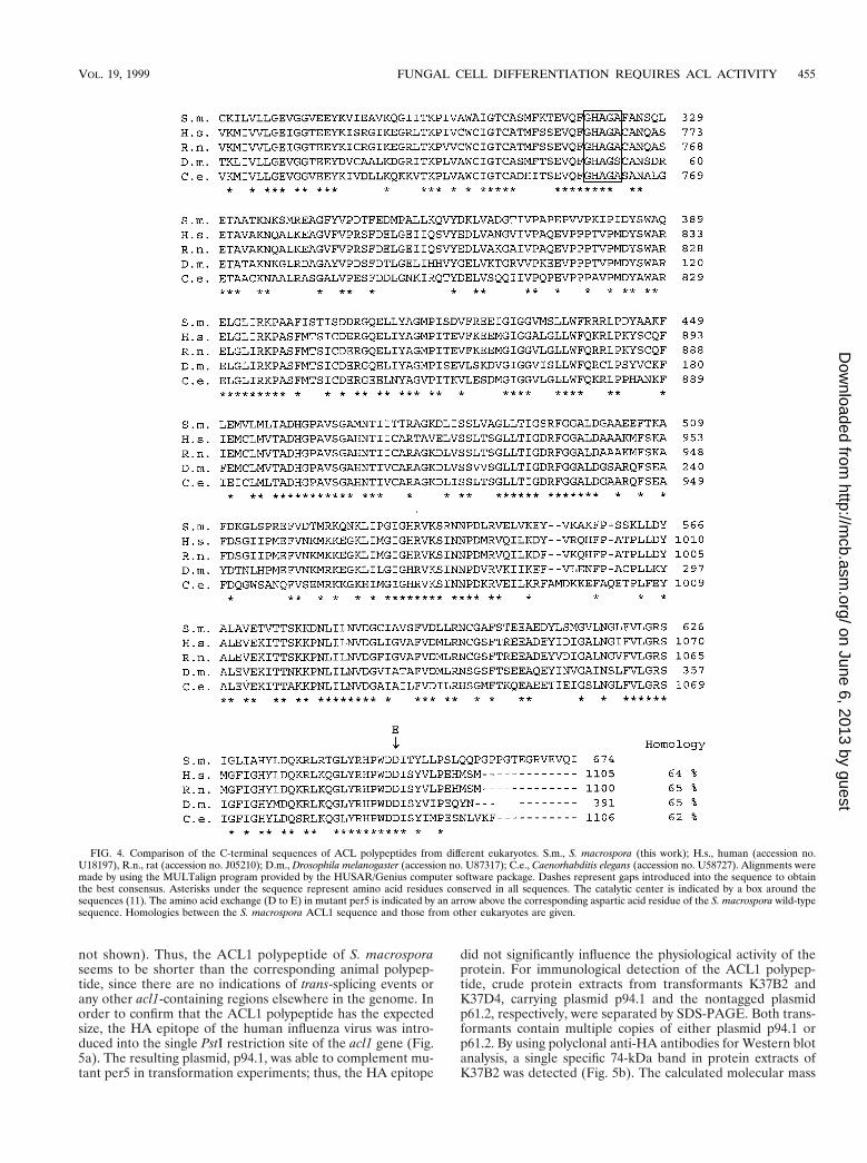
with higher eukaryotic ACLs. As shown in Fig. 4, amino acidsequence homologies to animal ACLs vary between 62 and65% over a length of about 600 amino acids. Translation ismost probably initiated at the first ATG (position 1357 [Fig. 3])of the ORF, and flanking sequences show the highest level ofsimilarity with translation initiation sites from other S. macro-spora genes (41). The putative polypeptide from S. macrosporahas a calculated molecular mass of 73 kDa and thus has onlyabout two-thirds of the molecular mass of animal ACL poly-
peptides (11, 12). The S. macrospora ACL1 polypeptide hashomology with the C-terminal part of the corresponding ani-mal polypeptides, which carries the enzyme’s proposed cata-lytic center, including the histidine residue that is autophos-phorylated during the catalyzed reaction (Fig. 4). Southernhybridization analysis indicates that the acl1 gene is a single-copy gene (data not shown). The length of the ORF (2.1 kb) isconsistent with data from Northern hybridizations showing a2.7-kb transcript when the acl1 ORF is used as a probe (data
FIG. 1. Phenotypes of the S. macrospora wild-type strain (wt) and mutant per5. Strains were grown for 7 days at 27°C. (a and b) Scanning electron micrographs ofperithecia from the wild-type strain (a) and mutant per5 (b). The magnifications in panels a and b are the same. (c and d) Differential interference contrast lightmicrographs of a wild-type ascus (c) and a mutant ascus (d). (e) Fluorescence micrograph of the mutant ascus in panel d stained with DAPI. Eight nuclei can bedistinguished within the ascus. Two pairs of nuclei, not yet completely separated after postmeiotic mitosis, are marked by arrows. The magnifications in panels c, d,and e are the same.
FIG. 2. Partial map of cosmid clone B3 together with derivatives used in transformation experiments. Cosmid clone B3 complements mutant per5 and contains thegene for ACL (acl1). The ORF of the acl1 gene and the direction of transcription are indicated by an arrow. Plasmids p59.3, p49.4, p52.9, p41.1, and p85.1 are derivativesof cosmid clone B3 (Table 1). The site of mutation within plasmid p85.1 is shown by an arrow. The values on the right give the total number of transformants (transf.)obtained with the corresponding plasmid and the number and corresponding percentage of fertile transformants. Abbreviations for restriction enzymes: A, ApaI; B,BamHI; E, EcoRV; H, HindIII; P, PstI; S, SalI; X, XhoI.
VOL. 19, 1999 FUNGAL CELL DIFFERENTIATION REQUIRES ACL ACTIVITY 453
on June 6, 2013 by guesthttp://m
cb.asm.org/
Dow
nloaded from

FIG. 3. Nucleotide and derived amino acid sequences for the S. macrospora acl1 gene and its flanking regions. Intron sequences are indicated in lowercase, andcharacteristic intron sequences are underlined. The transcription initiation site is marked by an arrow; a putative CAAT box is marked by a line above the sequence.The catalytic center is indicated by a box around the sequence (11). The single nucleotide exchange present in mutant per5 at position 3372 (T to A) and the resultingamino acid exchange (aspartic to glutamic acid) are shown in boldface above and below the corresponding wild-type sequences, respectively. The nucleotide anddeduced polypeptide sequence are numbered on the left, starting with nucleotide 626 according to the numbering of the complete sequence (4,847 bp) deposited inthe EMBL sequence database under accession no. AJ224922.
454 NOWROUSIAN ET AL. MOL. CELL. BIOL.
on June 6, 2013 by guesthttp://m
cb.asm.org/
Dow
nloaded from
not shown). Thus, the ACL1 polypeptide of S. macrosporaseems to be shorter than the corresponding animal polypep-tide, since there are no indications of trans-splicing events orany other acl1-containing regions elsewhere in the genome. Inorder to confirm that the ACL1 polypeptide has the expectedsize, the HA epitope of the human influenza virus was intro-duced into the single PstI restriction site of the acl1 gene (Fig.5a). The resulting plasmid, p94.1, was able to complement mu-tant per5 in transformation experiments; thus, the HA epitope
did not significantly influence the physiological activity of theprotein. For immunological detection of the ACL1 polypep-tide, crude protein extracts from transformants K37B2 andK37D4, carrying plasmid p94.1 and the nontagged plasmidp61.2, respectively, were separated by SDS-PAGE. Both trans-formants contain multiple copies of either plasmid p94.1 orp61.2. By using polyclonal anti-HA antibodies for Western blotanalysis, a single specific 74-kDa band in protein extracts ofK37B2 was detected (Fig. 5b). The calculated molecular mass
FIG. 4. Comparison of the C-terminal sequences of ACL polypeptides from different eukaryotes. S.m., S. macrospora (this work); H.s., human (accession no.U18197), R.n., rat (accession no. J05210); D.m., Drosophila melanogaster (accession no. U87317); C.e., Caenorhabditis elegans (accession no. U58727). Alignments weremade by using the MULTalign program provided by the HUSAR/Genius computer software package. Dashes represent gaps introduced into the sequence to obtainthe best consensus. Asterisks under the sequence represent amino acid residues conserved in all sequences. The catalytic center is indicated by a box around thesequences (11). The amino acid exchange (D to E) in mutant per5 is indicated by an arrow above the corresponding aspartic acid residue of the S. macrospora wild-typesequence. Homologies between the S. macrospora ACL1 sequence and those from other eukaryotes are given.
VOL. 19, 1999 FUNGAL CELL DIFFERENTIATION REQUIRES ACL ACTIVITY 455
on June 6, 2013 by guesthttp://m
cb.asm.org/
Dow
nloaded from
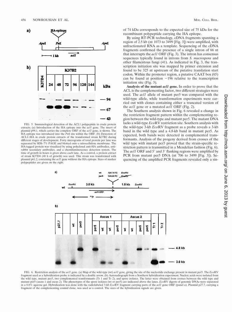
of 74 kDa corresponds to the expected size of 75 kDa for therecombinant polypeptide carrying the HA epitope.
By using RT-PCR technology, cDNA fragments spanning aregion of 2.5 kb (nt 1073 to 3499 [Fig. 3]) were amplified, withunfractionated RNA as a template. Sequencing of the cDNAfragments confirmed the presence of a single intron of 66 ntthat interrupts the acl1 ORF (Fig. 3). The intron has consensussequences typically found in introns from S. macrospora andother filamentous fungi (41). As indicated in Fig. 3, the tran-scription initiation site was mapped by primer extension andfound to be 325 nt upstream of the putative translation startcodon. Within the promoter region, a putative CAAT box (65)can be found at position 2196 relative to the transcriptioninitiation site (Fig. 3).
Analysis of the mutant acl1 gene. In order to prove that theACL is the complementing factor, two different strategies wereused. The acl1 allele of mutant per5 was compared with thewild-type allele, while transformation experiments were car-ried out with clones containing either a truncated version ofthe acl1 gene or a mutated acl1 ORF (Fig. 2).
The Southern analysis shown in Fig. 6 revealed a change inthe restriction fragment pattern within the complementing re-gion between the wild-type and mutant per5. The mutant DNAlacks a wild-type EcoRV restriction site. Southern analysis withthe wild-type 3-kb EcoRV fragment as a probe reveals a 3-kbband in the wild type and a 4.8-kb band in mutant per5. Asexpected, both bands were detected in complemented trans-formants. Analysis of the progeny derived from crosses of thewild type with mutant per5 proved that the strain-specific re-striction pattern is transmitted in a Mendelian fashion (Fig. 6).The acl1 ORF and 59 and 39 flanking regions were amplified byPCR from mutant per5 DNA (nt 706 to 3499 [Fig. 3]). Se-quencing of the amplified PCR fragments revealed only a sin-
FIG. 5. Immunological detection of the ACL1 polypeptide in crude proteinextracts. (a) Introduction of the HA epitope into the acl1 gene. The insert ofplasmid p94.1, which carries the complete ORF of the acl1 gene, is shown. TheHA epitope was introduced into the PstI site within the ORF. (b) Detection ofACL1::HA in crude protein extracts of the transformed strain K37B2 duringdifferent stages of development. Forty micrograms of total protein per lane wasseparated by SDS–7% PAGE and blotted onto a nitrocellulose membrane. TheHA-tagged protein was visualized by using polyclonal anti-HA antibodies, anti-rabbit secondary antibodies, and a chemiluminescence detection system. Thetime of growth in hours is given above each lane. As a control, a protein extractof strain K37D4 (48 h of growth) was used. This strain was transformed withplasmid p61.2, containing the acl1 gene without the HA epitope. Sizes of markerpolypeptides are given on the right.
FIG. 6. Restriction analysis of the acl1 gene. (a) Map of the wild-type (wt) acl1 gene, giving the site of the nucleotide exchange present in mutant per5. The EcoRVfragment used as a hybridization probe is indicated by a double arrow. (b) Autoradiograph from a Southern hybridization experiment. Nucleic acids were isolated fromthe wild type, mutant per5, two complemented transformants (Tr 1 and Tr 2), and spore isolates. The latter were obtained from crosses between the wild type andmutant per5 (ascus 1 and ascus 2). The phenotypes of the spore isolates (wt or per5) are indicated above the lanes. EcoRV digests of genomic DNAs were separatedin a 0.8% agarose gel. Hybridization was done with the radiolabeled 3-kb EcoRV fragment carrying parts of the acl1 gene ORF (panel a). Plasmid p27.7, carrying afragment of the complementing cosmid clone, was used as a control. The sizes of the hybridization signals are given.
456 NOWROUSIAN ET AL. MOL. CELL. BIOL.
on June 6, 2013 by guesthttp://m
cb.asm.org/
Dow
nloaded from
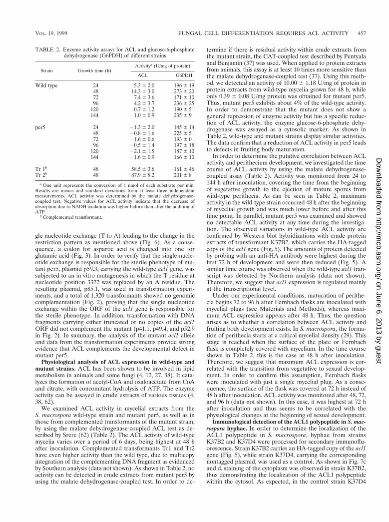
gle nucleotide exchange (T to A) leading to the change in therestriction pattern as mentioned above (Fig. 6). As a conse-quence, a codon for aspartic acid is changed into one forglutamic acid (Fig. 3). In order to verify that the single nucle-otide exchange is responsible for the sterile phenotype of mu-tant per5, plasmid p59.3, carrying the wild-type acl1 gene, wassubjected to an in vitro mutagenesis in which the T residue atnucleotide position 3372 was replaced by an A residue. Theresulting plasmid, p85.1, was used in transformation experi-ments, and a total of 1,320 transformants showed no genomiccomplementation (Fig. 2), proving that the single nucleotideexchange within the ORF of the acl1 gene is responsible forthe sterile phenotype. In addition, transformation with DNAfragments carrying either truncated or no copies of the acl1ORF did not complement the mutant (p41.1, p49.4, and p52.9in Fig. 2). In summary, the analysis of the mutant acl1 alleleand data from the transformation experiments provide strongevidence that ACL complements the developmental defect inmutant per5.
Physiological analysis of ACL expression in wild-type andmutant strains. ACL has been shown to be involved in lipidmetabolism in animals and some fungi (4, 12, 27, 38). It cata-lyzes the formation of acetyl-CoA and oxaloacetate from CoAand citrate, with concomitant hydrolysis of ATP. The enzymeactivity can be assayed in crude extracts of various tissues (4,38, 62).
We examined ACL activity in mycelial extracts from theS. macrospora wild-type strain and mutant per5, as well as inthose from complemented transformants of the mutant strain,by using the malate dehydrogenase-coupled ACL test as de-scribed by Srere (62) (Table 2). The ACL activity of wild-typemycelia varies over a period of 6 days, being highest at 48 hafter inoculation. Complemented transformants Tr1 and Tr2have even higher activity than the wild type, due to multicopyintegration of the complementing DNA fragment as evidencedby Southern analysis (data not shown). As shown in Table 2, noactivity can be detected in crude extracts from mutant per5 byusing the malate dehydrogenase-coupled test. In order to de-
termine if there is residual activity within crude extracts fromthe mutant strain, the CAT-coupled test described by Pentyalaand Benjamin (37) was used. When applied to protein extractsfrom animals, this assay is at least 10 times more sensitive thanthe malate dehydrogenase-coupled test (37). Using this meth-od, we detected an activity of 10.00 6 1.18 U/mg of protein inprotein extracts from wild-type mycelia grown for 48 h, whileonly 0.39 6 0.08 U/mg protein was obtained for mutant per5.Thus, mutant per5 exhibits about 4% of the wild-type activity.In order to demonstrate that the mutant does not show ageneral repression of enzyme activity but has a specific reduc-tion of ACL activity, the enzyme glucose-6-phosphate dehy-drogenase was assayed as a cytosolic marker. As shown inTable 2, wild-type and mutant strains display similar activities.The data confirm that a reduction of ACL activity in per5 leadsto defects in fruiting body maturation.
In order to determine the putative correlation between ACLactivity and perithecium development, we investigated the timecourse of ACL activity by using the malate dehydrogenase-coupled assay (Table 2). Activity was monitored from 24 to144 h after inoculation, covering the time from the beginningof vegetative growth to the ejection of mature spores fromwild-type perithecia. As can be seen in Table 2, maximumactivity in the wild-type strain occurred 48 h after the beginningof mycelial growth and was much lower before and after thistime point. In parallel, mutant per5 was examined and showedno detectable ACL activity at any time during the investiga-tion. The observed variations in wild-type ACL activity areconfirmed by Western blot hybridizations with crude proteinextracts of transformant K37B2, which carries the HA-taggedcopy of the acl1 gene (Fig. 5). The amounts of protein detectedby probing with an anti-HA antibody were highest during thefirst 72 h of development and were then reduced (Fig. 5). Asimilar time course was observed when the wild-type acl1 tran-script was detected by Northern analysis (data not shown).Therefore, we suggest that acl1 expression is regulated mainlyat the transcriptional level.
Under our experimental conditions, maturation of perithe-cia begins 72 to 96 h after Fernbach flasks are inoculated withmycelial plugs (see Materials and Methods), whereas maxi-mum ACL expression appears after 48 h. Thus, the questionarises as to whether a correlation between ACL activity andfruiting body development exists. In S. macrospora, the forma-tion of perithecia starts at a critical mycelial density (29). Thisstage is reached when the surface of the plate or Fernbachflask is completely covered with mycelium. In the time courseshown in Table 2, this is the case at 48 h after inoculation.Therefore, we suggest that maximum ACL expression is cor-related with the transition from vegetative to sexual develop-ment. In order to confirm this assumption, Fernbach flaskswere inoculated with just a single mycelial plug. As a conse-quence, the surface of the flask was covered at 72 h instead of48 h after inoculation. ACL activity was monitored after 48, 72,and 96 h (data not shown). In this case, it was highest at 72 hafter inoculation and thus seems to be correlated with thephysiological changes at the beginning of sexual development.
Immunological detection of the ACL1 polypeptide in S. mac-rospora hyphae. In order to determine the localization of theACL1 polypeptide in S. macrospora, hyphae from strainsK37B2 and K37D4 were processed for secondary immunoflu-orescence. Strain K37B2 carries an HA-tagged copy of the acl1gene (Fig. 5), while strain K37D4, carrying the correspondingnontagged plasmid, was used as a control. As shown in Fig. 7cand d, staining of the cytoplasm was observed in strain K37B2,thus demonstrating the localization of the ACL1 polypeptidewithin the cytosol. As expected, in the control strain K37D4
TABLE 2. Enzyme activity assays for ACL and glucose-6-phosphatedehydrogenase (G6PDH) of different strains
Strain Growth time (h)Activitya (U/mg of protein)
ACL G6PDH
Wild type 24 5.3 6 2.0 196 6 1948 14.3 6 3.0 273 6 2072 7.4 6 3.6 171 6 1096 4.2 6 3.7 236 6 25
120 0.7 6 1.2 190 6 5144 1.0 6 0.9 235 6 9
per5 24 21.3 6 2.0 145 6 1448 20.8 6 1.6 225 6 572 21.6 6 0.6 193 6 096 20.5 6 1.4 197 6 18
120 22.1 6 1.5 187 6 10144 21.6 6 0.9 166 6 10
Tr 1b 48 58.5 6 3.6 161 6 46Tr 2b 48 87.9 6 8.2 201 6 8
a One unit represents the conversion of 1 nmol of each substrate per min.Results are means and standard deviations from at least three independentmeasurements. ACL activity was determined by the malate dehydrogenase-coupled test. Negative values for ACL activity indicate that the decrease ofabsorption due to NADH oxidation was higher before than after the addition ofATP.
b Complemented transformant.
VOL. 19, 1999 FUNGAL CELL DIFFERENTIATION REQUIRES ACL ACTIVITY 457
on June 6, 2013 by guesthttp://m
cb.asm.org/
Dow
nloaded from

only autofluorescence of the septa was observed (Fig. 7a andb). By this method, we cannot exclude the possibility that ACLalso resides within other subcellular compartments. In general,however, our data are in accordance with subcellular fraction-ation experiments that detected ACL within the cytosol insome yeasts and Aspergillus niger (4, 38).
DISCUSSIONThe ACL gene is highly conserved. In this paper we describe
the isolation and characterization of per5, a sterile mutant ofS. macrospora which was complemented to fertility by the geneencoding ACL. ACL produces acetyl-CoA, which in eukaryotesis used mainly in fatty acid and sterol biosynthesis. ACL islocalized in the cytosol in animals and fungi (4, 38), whereas inplants it resides in the chloroplasts, which are the sites of fattyacid biosynthesis within photoautotrophic organisms (46). InS. macrospora, ACL also resides within the cytoplasm, as wasdemonstrated by immunofluorescence analysis (Fig. 7).
So far, the acl genes of several vertebrates have been se-quenced, among them the genes from humans and rats (11, 12,30). To our knowledge, we report the first molecular analysis ofan acl gene from a lower eukaryote. The S. macrospora ACL1polypeptide corresponds to the C-terminal part of the animalACL1 polypeptides. These polypeptides are 62 to 65% identi-cal over a length of 600 amino acids, including the proposedcatalytic center (Fig. 4). Epitope tagging demonstrated that the73-kDa S. macrospora ACL1 polypeptide is smaller than its120-kDa animal counterparts (11). In animals and in the yeastRhodotorula gracilis, the ACL protein is a homotetramer offour identical subunits (56, 60), whereas in the filamentousfungi Aspergillus nidulans and Penicillium spiculisporum, itseems to consist of two 55- and 70-kDa subunits forming ahexamer of about 380 kDa (1, 27). Thus, the 73-kDa ACL1polypeptide encoded by the S. macrospora acl1 gene couldbe part of a multimeric protein, additional subunits of whichmight be encoded by other genes.
ACL is functional in S. macrospora, and its activity is detect-able in crude protein extracts (Table 2). As can be concludedfrom the site of mutation in the mutant acl1 allele and bytransformation with in vitro-mutagenized plasmid DNA, thehighly conserved aspartic acid in the C-terminal part of the
polypeptide is important for ACL activity (Fig. 4). Besides thehistidine residue in the catalytic center, which is autophos-phorylated during the catalyzed reaction, the human and ratacl genes contain three additional phosphorylation sites (39,44). Phosphorylation of these sites is dependent on develop-ment or physiological state (2, 45), and enzymatic activity isinfluenced by phosphorylation (37). There are no sequenceshomologous to these three sites in the Sordaria polypeptide,indicating that regulation of ACL expression is not conservedamong these organisms.
Actually, ACL seems to perform quite different functions inanimals and fungi. In S. macrospora, it is important for sexualdevelopment, whereas its full activity is not required for veg-etative growth, since mutant per5 displays wild-type vegetativegrowth. In animals, the highest levels of ACL expression arefound in the liver. However, ACL inhibitors have no toxiceffect (63), suggesting that ACL may be a putative target forhypolipidemic intervention in humans (17). In Saccharomycescerevisiae and some other yeasts, no ACL has been detected(4), indicating that corresponding enzymatic activities are per-formed by other enzymes such as acetyl-CoA synthetases.
So far, no equivalent genes have been cloned from pro-karyotes. However, ACL activity has been detected in somearchaeal and bacterial species (66). In eukaryotes ACL is in-volved in lipid and sterol biosynthesis, whereas in prokaryotesit appears to be part of the reverse tricarboxylic acid cycle (66).This pathway is an alternative route for carbon fixation used insome archaea and bacteria (for a review, see reference 48). Acomparison of prokaryotic and eukaryotic acl gene sequencesshould prove interesting, particularly with respect to any con-served amino acids essential for catalytic functions. In general,ACL seems to be an evolutionarily ancient enzyme which hasachieved quite different physiological functions within diverseorganisms.
ACL is essential for fruiting body development. Analysis ofthe S. macrospora mutant per5 has demonstrated that althougha drastic reduction of ACL activity does not impair vegetativegrowth, ACL is an essential requirement for fruiting bodymaturation. ACL produces acetyl-CoA and oxaloacetate, andso far no further function has been attributed to the protein.The acetyl-CoA produced by ACL is used mainly in fatty acid
FIG. 7. Immunological detection of the ACL1 polypeptide in S. macrospora hyphae. Hyphae from strain K37D4 (a and b) (transformed with plasmid p61.2, withoutthe HA epitope) and strain K37B2 (c and d) (transformed with plasmid p94.1, carrying the HA epitope) were grown on cover slides and processed for immunoflu-orescence. (a and c) Differential interference contrast light micrographs; (b and d) fluorescence of ACL1::HA in the same hyphae. Magnifications are all the same.
458 NOWROUSIAN ET AL. MOL. CELL. BIOL.
on June 6, 2013 by guesthttp://m
cb.asm.org/
Dow
nloaded from

and sterol biogenesis. Fatty acids and sterols play importantroles in many cellular processes, such as the generation ofbiomembranes, hormones, and secondary messengers. Besidesthese general functions, many developmental processes in dif-ferent organisms are dependent on fatty acid metabolism. Inplants, the formation of pollen grains and seeds is closelycorrelated with lipid production. Several genes of the fatty acidbiosynthesis pathway of Brassica napus are tightly regulated ina spatiotemporal manner, e.g., those for acyl carrier proteinsand stearoyl-acyl carrier protein desaturases (8, 40). In ani-mals, enzymes for lipid biosynthesis and fatty acid beta-oxida-tion are both regulated during morphogenesis, as can be seenin developing rats (9, 13). In fungi, fatty acid biosynthesishas been well studied at the cellular level, but only in a fewcases have more specialized functions been attributed tolipid metabolism (for a review, see reference 6). For example,in N. crassa fatty acids were shown to be involved in thecircadian rhythm, and they are also needed for mitosis inSchizosaccharomyces pombe (28, 50). Lipids as well as sterolderivatives serve as growth factors or pheromones in somefungal species (for a review, see reference 10).
Investigation of mutant per5 indicates that fatty acids andsterols are essential for fruiting body development in S. mac-rospora. As the mutant displays a normal vegetative growthrate, it can be concluded that sufficient acetyl-CoA and lipidsare produced for this process. This can be achieved either bythe residual ACL activity present in mutant per5 or by enzymesother than ACL, such as acetyl-CoA synthetase. Although thiswork demonstrates a specific role for ACL, producing acetyl-CoA for fruiting body development, it is worth noting that thewild-type strain displayed ACL activity at every time pointduring development (Table 2), not just during peritheciumformation. Nevertheless, the crucial role of ACL seems to be infruiting body maturation, and it can be assumed that the ste-rility of mutant per5 is due to the fact that a certain amount ofacetyl-CoA and its derivatives is a prerequisite for peritheciummaturation. This is consistent with the finding that a partialrestoration of the wild-type phenotype can be achieved bysupplementation of growth media with fatty acids, such asoleate (unpublished results). Obviously, other enzymes pro-ducing acetyl-CoA cannot compensate for the reduced ACLactivity during sexual development, indicating that ACL is aspecific and probably the only relevant enzyme producingacetyl-CoA for fruiting body development. Our findings sup-port the view that some housekeeping functions might be cir-cumvented to a certain degree but are essential under specialphysiological conditions such as sexual reproduction.
The expression of acl1 is developmentally regulated, beinghighest during the transition from vegetative to sexual devel-opment. One of the reasons for this expression pattern mightinvolve the demand for energy. Different biosynthetic routesfor generating cytosolic acetyl-CoA influence the metaboliccosts for biosynthesis of macromolecules (18). Thus, the im-portance of ACL for S. macrospora fruiting body developmentmight be due to the fact that acetyl-CoA production has tomeet certain energetic demands which cannot be fulfilled byother metabolic pathways. Another reason for the observedexpression pattern might be that metabolites for the formationof fungal fruiting bodies are at least partially supplied by thevegetative mycelium. Therefore, the mycelium has to gain acertain competence before fruiting body formation is induced(for a review, see reference 68). As was recently shown forN. crassa, asci within perithecia contain far more oleate thanperithecial wall tissues (16). It may be speculated that the lipidcomposition is the same in the closely related species N. crassaand S. macrospora. As oleate is a metabolic derivative of
acetyl-CoA, this may explain why mutant per5 is able to formperithecial walls but no mature asci (Fig. 1).
In S. macrospora, ACL activity is highest at 48 h after inoc-ulation, when mycelial density reaches a critical value andsexual development is induced (Table 2). We propose theexistence of a yet-unidentified signal that regulates acl1 geneexpression, which delivers acetyl-CoA that is required duringperithecium formation. In general, our findings support theview that not only is the basic metabolism of cells regulatedaccording to the developmental requirements, but differentproteins are involved in producing the same metabolic inter-mediates at different developmental stages.
ACKNOWLEDGMENTS
We thank S. Schlewinski for performing the S. macrospora crosses,H. J. Rathke for the artwork, and T. Stutzel for help with the scanningelectron microscopy.
This work was supported by a grant from the Graduiertenforderungdes Landes Nordrhein-Westfalen (NRW) (Germany) and by theDeutsche Forschungsgemeinschaft, Bonn-Bad Godesberg.
REFERENCES
1. Adams, I. P., S. Dack, F. M. Dickinson, M. Midgley, and C. Ratledge. 1997.ATP: citrate lyase from Aspergillus nidulans. Biochem. Soc. Trans. 25:670.
2. Benjamin, W. B., S. N. Pentyala, J. R. Woodgett, Y. Hod, and D. Marshak.1994. ATP citrate-lyase and glycogen synthase kinase-3 beta in 3T3-L1 cellsduring differentiation into adipocytes. Biochem. J. 300:477–482.
3. Berteaux-Lecellier, V., M. Picard, C. Thompson-Coffe, D. Zickler, A. Pan-vier-Adoutte, and J. M. Simonet. 1995. A nonmammalian homolog of thePAF1 gene (Zellweger syndrome) discovered as a gene involved in caryo-gamy in the fungus Podospora anserina. Cell 81:1043–1051.
4. Boulton, C. A., and C. Ratledge. 1981. Correlation of lipid accumulation inyeasts with possession of ATP:citrate lyase. J. Gen. Microbiol. 127:169–176.
5. Bradford, M. M. 1976. A rapid and sensitive method for the quantification ofmicrogram quantities of protein utilizing the principle of protein-dye bind-ing. Anal. Biochem. 72:248–254.
6. Chopra, A., and G. K. Khuller. 1984. Lipid metabolism in fungi. Crit. Rev.Microbiol. 11:209–271.
7. Coppin, E., R. Debuchy, S. Arnaise, and M. Picard. 1997. Mating types andsexual development in filamentous ascomycetes. Microbiol. Mol. Biol. Rev.61:411–428.
8. De Silva, J., S. J. Robinson, and R. Safford. 1992. The isolation and func-tional characterisation of a B. napus acyl carrier protein 59 flanking regioninvolved in the regulation of seed storage lipid synthesis. Plant Mol. Biol. 18:1163–1172.
9. Djouadi, F., B. Riveau, C. Merlet-Benichou, and J. Bastin. 1997. Tissue-specific regulation of medium-chain acyl-CoA dehydrogenase gene by thy-roid hormones in the developing rat. Biochem. J. 324:289–294.
10. Dyer, P. S., D. S. Ingram, and K. Johnstone. 1992. The control of sexualmorphogenesis in the ascomycotina. Biol. Rev. 67:421–458.
11. Elshourbagy, N. A., J. C. Near, P. J. Kmetz, G. M. Sathe, C. Southan, J. E.Strickler, M. Gross, J. F. Young, T. N. C. Wells, and P. H. E. Groot. 1990.Rat ATP citrate-lyase. J. Biol. Chem. 265:1430–1435.
12. Elshourbagy, N. A., J. C. Near, P. J. Kmetz, T. N. C. Wells, P. H. E. Groot,B. A. Saxty, S. A. Hughes, M. Franklin, and I. S. Gloger. 1992. Cloning andexpression of a human ATP-citrate lyase cDNA. Eur. J. Biochem. 204:491–499.
13. Eritani, N., H. Fukuda, and Y. Matsumura. 1993. Lipogenic enzyme geneexpression in rat liver during development after birth. J. Biochem. 113:519–525.
14. Esser, K. 1982. Cryptogams—cyanobacteria, algae, fungi, lichens. CambridgeUniversity Press, London, United Kingdom.
15. Esser, K., and J. Straub. 1958. Genetische Untersuchungen an Sordariamacrospora Auersw., Kompensation und Induktion bei genbedingten En-twicklungsdefekten. Z. Vererbungsl. 89:729–746.
16. Goodrich-Tanrikulu, M., K. Howe, A. Stafford, and M. A. Nelson. 1998.Changes in fatty acid composition of Neurospora crassa accompany sexualdevelopment and ascospore germination. Microbiology 144:1713–1720.
17. Gribble, A. D., R. E. Dolle, A. Shaw, D. McNair, R. Novelli, C. E. Novelli,B. P. Slingsby, V. P. Shah, D. Tew, B. A. Saxty, M. Allen, P. H. Goot, N.Pearce, and J. Yates. 1996. ATP-citrate lyase as a target for hypolipidemicintervention. Design and synthesis of 2-substituted butanedioic acids asnovel, potent inhibitors of the enzyme. J. Med. Chem. 39:3569–3584.
18. Henriksen, C. M., L. H. Christensen, J. Nielsen, and J. Villadsen. 1996.Growth energetics and metabolic fluxes in continuous cultures of Penicilliumchrysogenum. J. Biotechnol. 45:149–164.
19. Hoge, J. H. C., J. Springer, B. Zantige, and J. G. H. Wessels. 1982. Absence
VOL. 19, 1999 FUNGAL CELL DIFFERENTIATION REQUIRES ACL ACTIVITY 459
on June 6, 2013 by guesthttp://m
cb.asm.org/
Dow
nloaded from

of differences in polysomal RNA from vegetative monokaryotic and dikary-otic cells of the fungus Schizophyllum commune. Exp. Mycol. 6:225–232.
20. Kempken, F., and U. Kuck. 1996. restless, an active Ac-like transposon fromthe fungus Tolypocladium inflatum: structure, expression, and alternativeRNA splicing. Mol. Cell. Biol. 16:6563–6572.
21. Kennell, J. C., and D. R. Pring. 1989. Initiation and processing of atp6,T-urf13 and ORF221 transcripts from mitochondria of T cytoplasm maize.Mol. Gen. Genet. 216:16–24.
22. Krug, M. S., and S. L. Berger. 1987. First strand cDNA synthesis primed witholigo (dT). Methods Enzymol. 152:316–323.
23. Kuck, U., Y. Choquet, M. Schneider, M. Dron, and P. Bennoun. 1987.Structural and transcription analysis of two homologous genes for the P700chlorophyll a-apoproteins in Chlamydomonas reinhardtii: evidence for in vivotrans-splicing. EMBO J. 6:2185–2195.
24. Laemmli, U. K. 1970. Cleavage of structural proteins during the assembly ofthe head of bacteriophage T4. Nature 227:680–685.
25. Lang, B. F., and G. Burger. 1990. A rapid, high resolution DNA sequencinggel system. Anal. Biochem. 188:176–180.
26. Madi, L., S. A. McBride, L. A. Bailey, and D. J. Ebbole. 1997. rco-3, a geneinvolved in glucose transport and conidiation in Neurospora crassa. Genetics146:499–508.
27. Måhlen, A. 1973. Purification and some properties of ATP citrate lyase fromPenicillium spiculisporum. Eur. J. Biochem. 36:342–346.
27a.Masloff, S. Unpublished data.28. Mattern, D. L. 1985. Unsaturated fatty acid isomers: effects on the circadian
rhythm of a fatty-acid-deficient Neurospora crassa mutant. Arch. Biochem.Biophys. 237:402–407.
29. Molowitz, R., M. Bahn, and B. Hock. 1976. The control of fruiting bodyformation in the ascomycete Sordaria macrospora Auersw. by arginine andbiotin: a two-factor analysis. Planta 128:143–148.
30. Moon, Y. A., K. S. Kim, S. W. Park, and Y. S. Kim. 1996. Cloning andidentification of exon-intron organization of the rat ATP-citrate lyase gene.Biochim. Biophys. Acta 1307:280–284.
31. Nelson, M. A. 1996. Mating systems in ascomycetes: a romp in the sac.Trends Genet. 12:69–74.
32. Nelson, M. A., S. T. Merino, and R. L. Metzenberg. 1997. A putative rham-nogalacturonase required for sexual development of Neurospora crassa. Ge-netics 146:531–540.
33. Oakley, B. R., C. E. Oakley, Y. Yoon, and M. K. Jung. 1990. g-Tubulin is acomponent of the spindle pole body that is essential for microtubule functionin Aspergillus nidulans. Cell 61:1289–1301.
34. Osiewacz, H. D. 1994. A versatile shuttle cosmid vector for the efficientconstruction of genomic libraries and for the cloning of fungal genes. Curr.Genet. 26:87–90.
35. Osiewacz, H. D., and U. Nuber. 1996. GRISEA, a putative copper-activatedtranscription factor from Podospora anserina involved in differentiation andsenescence. Mol. Gen. Genet. 252:115–124.
36. Pearson, W. R. 1990. Rapid and sensitive sequence comparison with FASTPand FASTA. Methods Enzymol. 183:63–98.
37. Pentyala, S. N., and W. B. Benjamin. 1995. Effect of oxaloacetate andphosphorylation on ATP-citrate lyase activity. Biochemistry 34:10961–10969.
38. Pfitzner, A., C. P. Kubicek, and M. Rohr. 1987. Presence and regulation ofATP:citrate lyase from the citric acid producing fungus Aspergillus niger.Arch. Microbiol. 147:88–91.
39. Pierce, M. W., J. L. Palmer, H. T. Keutmann, and J. Avruch. 1981. ATP-citrate lyase. Structure of a tryptic peptide containing the phosphorylationsite directed by glycagon and the cAMP-dependent protein kinase. J. Biol.Chem. 256:8867–8870.
40. Piffanelli, P., J. H. E. Ross, and D. J. Murphy. 1997. Intra- and extracellularlipid composition and associated gene expression patterns during pollendevelopment in Brassica napus. Plant J. 11:549–562.
41. Poggeler, S. 1997. Sequence characteristics within nuclear genes from Sor-daria macrospora. Fungal Genet. Newsl. 44:41–44.
42. Poggeler, S., M. Nowrousian, S. Jacobsen, and U. Kuck. 1997. An efficientprocedure to isolate fungal genes from an indexed cosmid library. J. Micro-biol. Methods 29:49–61.
43. Poggeler, S., S. Risch, U. Kuck, and H. D. Osiewacz. 1997. Mating-type genesfrom the homothallic fungus Sordaria macrospora are functionally expressedin a heterothallic ascomycete. Genetics 147:567–580.
44. Ramakrishna, S., G. D’Angelo, and W. B. Benjamin. 1990. Sequence of siteson ATP-citrate lyase and phosphatase inhibitor 2 phosphorylated by multi-functional protein kinase (a glycogen synthase kinase 3 like kinase). Bio-chemistry 29:7617–7624.
45. Ramakrishna, S., K. S. Murthy, and W. B. Benjamin. 1989. Effect of insulinon ATP-citrate lyase phosphorylation: regulations of peptide A and peptideB phosphorylations. Biochemistry 28:856–860.
46. Ratledge, C., M. D. V. Bowater, and P. N. Taylor. 1997. Correlation ofATP/citrate lyase activity with lipid accumulation in developing seeds ofBrassica napus L. Lipids 32:7–12.
47. Read, N. D., and A. Beckett. 1996. Ascus and ascospore morphogenesis.Mycol. Res. 100:1281–1314.
48. Romano, A. H., and T. Conway. 1996. Evolution of carbohydrate metabolicpathways. Res. Microbiol. 147:448–455.
49. Romeis, B. 1968. Mikroskopische Technik. R Oldenbourg, Munich, Ger-many.
50. Saitoh, S., K. Takahashi, K. Nabeshima, Y. Yamashita, Y. Nakaseko, Y.Hirata, and M. Yanagida. 1996. Aberrant mitosis in fission yeast mutantsdefective in fatty acid synthetase and acetyl CoA carboxylase. J. Cell Biol.134:949–961.
51. Sambrook, J., E. F. Fritsch, and T. Maniatis. 1989. Molecular cloning: alaboratory manual, 2nd ed. Cold Spring Harbor Laboratory Press, ColdSpring Harbor, N.Y.
52. Sanger, F., S. Nicklen, and A. R. Coulson. 1977. DNA sequencing withchain-terminating inhibitors. Proc. Natl. Acad. Sci. USA 74:5436–5467.
53. Saupe, S., C. Descamps, B. Turcq, and J. Begueret. 1994. Inactivation of thePodospora anserina vegetative incompatibility locus het-c, whose productresembles a glycolipid transfer protein, drastically impairs ascospore produc-tion. Proc. Natl. Acad. Sci. USA 91:5927–5931.
54. Saupe, S., L. Stenberg, K. T. Shiu, A. J. F. Griffiths, and N. L. Glass. 1996.The molecular nature of mutations in the mt A-1 gene of the Neurosporacrassa A idiomorph and their relation to mating-type function. Mol. Gen.Genet. 250:115–122.
55. Scott, W. A. 1975. Glucose-6-phosphate dehydrogenase from Neurosporacrassa. Methods Enzymol. 41:177–182.
56. Shashi, K., A. K. Bachhawat, and R. Joseph. 1990. ATP:citrate lyase ofRhodotorula gracilis: purification and properties. Biochim. Biophys. Acta1033:23–30.
57. Shepherd, M. G. 1975. Glucose-6-phosphate dehydrogenase from Penicil-lium duponti. Methods Enzymol. 41:201–205.
58. Shiio, Y., M. Itoh, and J. I. Inoue. 1995. Epitope tagging. Methods Enzymol.254:497–502.
59. Silar, P. 1995. Two new easy to use vectors for transformations. FungalGenet. Newsl. 42:73.
60. Singh, M., E. G. Richards, A. Mukherjee, and P. A. Srere. 1976. Structure ofATP citrate lyase from rat liver. Physicochemical studies and proteolyticmodification. J. Biol. Chem. 251:5242–5250.
61. Sinha, N. D., J. Biernat, J. McManus, and H. Koster. 1984. Polymer supportoligonucleotide synthesis. XVIII. Use of b-cyanoethyl-N,N-diacylamino-/N-morpholino-phosphoramidite of deoxynucleosides for the synthesis of DNAfragments simplifying deprotection and isolation of the final product. NucleicAcids Res. 12:4539–4557.
62. Srere, P. A. 1962. Citrate-cleavage enzyme. Methods Enzymol. 5:641–644.63. Sullivan, A. C., J. Triscari, J. G. Hamilton, O. N. Miller, and V. R. Wheatley.
1973. Effect of (2)-hydroxy-citrate upon the accumulation of lipid in the rat.I. Lipogenesis. Lipids 9:121–128.
64. Timberlake, W. E., M. T. Boylan, M. B. Cooley, P. M. Mirabito, E. B.O’Hara, and C. Willett. 1985. Rapid identification of mutation-complement-ing restriction fragments from Aspergillus nidulans cosmids. Exp. Mycol. 9:351–355.
65. Unkles, S. E. 1992. Gene organization in industrial filamentous fungi, p. 28–53. In J. R. Kinghorn and G. Turner (ed.), Applied molecular genetics offilamentous fungi. Blackie Academic & Professional, Glasgow, United King-dom.
66. Wahlund, T. M., and F. R. Tabita. 1997. The reductive tricarboxylic acidcycle of carbon dioxide assimilation: initial studies and purification of ATP-citrate lyase from the green sulfur bacterium Chlorobium tepidum. J. Bacte-riol. 179:4859–4867.
67. Walz, M., and U. Kuck. 1995. Transformation of Sordaria macrospora tohygromycin B resistance: characterization of transformants by electro-phoretic karyotyping and tetrad analysis. Curr. Genet. 29:88–95.
68. Wessels, J. G. H. 1993. Fruiting in the higher fungi. Adv. Microb. Physiol. 34:147–202.
69. Zickler, D. 1977. Development of the synaptonemal complex and the “re-combination nodules” during meiotic prophase in the seven bivalents of thefungus Sordaria macrospora Auersw. Chromosoma 61:289–316.
460 NOWROUSIAN ET AL. MOL. CELL. BIOL.
on June 6, 2013 by guesthttp://m
cb.asm.org/
Dow
nloaded from