Cell Biology Outline - Cells • Prokaryotic Cells ... 178 Fall 2008/B178F08... · 4 19 Eukaryotic...
7
1 Cell Biology 2 Outline - Cells • Cell Theory • Cell Size • Microscopes • Prokaryotic Cells • Eukaryotic Cells • Cell Membranes 3 • 1665 - Robert Hooke – Describes small rooms in tree bark • 1838-39 Matt Schleiden & Ted Schann – All organisms are composed of one or more cells. – Cells are the smallest living units of all living organisms. • Late 1800’s Rudolf Virchow’s contribution – Cells arise only by division of a previously existing cell. – Life on earth represents a continuous line of descent Historical Observations of Cells 4 Cell Theory 1. All organisms are composed of cells. 2. Cells are the smallest living things. 3. Cells arise only from pre-existing cells. All cells today represent a continuous line of descent from the first living cells. Cell Theory 5 Surface area to volume ratio 3:1 0.3:1 Small cells have an optimum surface area to volume ratio Why Are Cells Small? 6 Cell size Electron microscopes resolve structures 0.2nm apart. Light microscopes resolve structures 200nm apart.
Transcript of Cell Biology Outline - Cells • Prokaryotic Cells ... 178 Fall 2008/B178F08... · 4 19 Eukaryotic...

1
Cell Biology
2
Outline - Cells
• Cell Theory• Cell Size• Microscopes• Prokaryotic Cells• Eukaryotic Cells• Cell Membranes
3
• 1665 - Robert Hooke – Describes small rooms in tree bark
• 1838-39 Matt Schleiden & Ted Schann– All organisms are composed of one or more cells.– Cells are the smallest living units of all living organisms.
• Late 1800’s Rudolf Virchow’s contribution– Cells arise only by division of a previously existing cell.– Life on earth represents a continuous line of descent
Historical Observations of Cells
4
Cell Theory1. All organisms are composed of cells.2. Cells are the smallest living things.3. Cells arise only from pre-existing cells.
All cells today represent a continuous line of descent from the first living cells.
Cell Theory
5Surface area to volume ratio 3:1 0.3:1
Small cells have an optimum surface area to volume ratio
Why Are Cells Small?
6
Cell size
Electron microscopesresolve structures 0.2nm apart.
Light microscopesresolve structures 200nm apart.
2
7
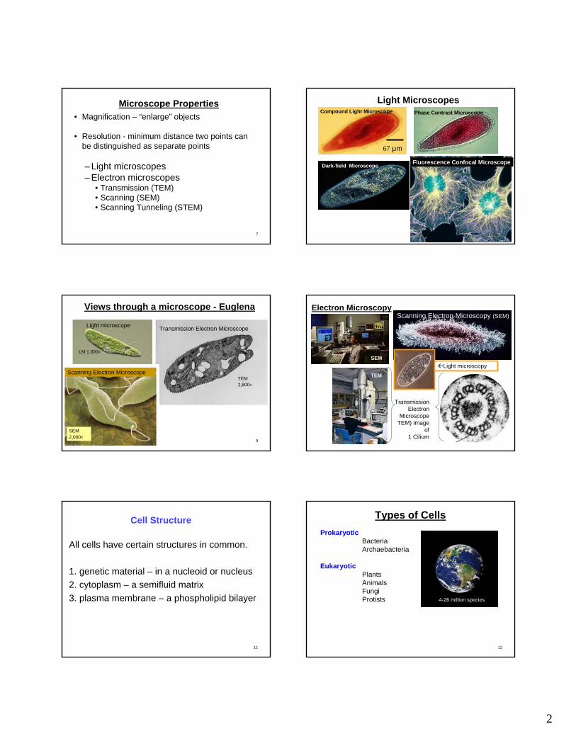
• Magnification – “enlarge” objects
• Resolution - minimum distance two points can be distinguished as separate points
– Light microscopes– Electron microscopes
• Transmission (TEM) • Scanning (SEM)• Scanning Tunneling (STEM)
Microscope Properties
8
Light Microscopes
67 µm
Compound Light Microscope Phase Contrast Microscope
Dark-field Microscope Fluorescence Confocal Microscope
9
LM 1,000×
Light microscope
TEM 2,800×
Transmission Electron Microscope
SEM 2,000×
Scanning Electron Microscope
Views through a microscope - Euglena
10
Transmission Electron
Microscope TEM) Image
of 1 Cilium
Scanning Electron Microscopy (SEM)
67 µm
Electron Microscopy
SEM
TEM
Light microscopy
11
All cells have certain structures in common.
1. genetic material – in a nucleoid or nucleus2. cytoplasm – a semifluid matrix3. plasma membrane – a phospholipid bilayer
Cell Structure
12
Types of CellsProkaryotic
BacteriaArchaebacteria
EukaryoticPlantsAnimalsFungiProtists 4-26 million species

3
Copyright © 2005 Pearson Education, Inc. Publishing as Benjamin Cummings
Prokaryotic Cell Structure
14
Gram Positive
Gram Negative
Bacterial Cell Walls
15
Rods
Spirals
Spheres
Cell walls give bacteria different shapes
16
Prokaryotic Cells - SummarySimplest organisms – all bacteriaPlasma Membrane – barrier & regulates
Inside the Plasma MembraneCytoplasmRibosomesDNA
Outside the Plasma MembraneCell wall – Peptidoglycan layer(s)
gram-positive or gram-negativeCapsule – Polysaccharide: Adhesion & HydrationPili - cell adhesion or DNA transfer
Common characteristics
1. Nucleus
2. Cytoplasm
3. Membranes Form compartmentsSeparate metabolic activityIncrease surface area
Plasma Membrane
Nucleus
Cytoplasm
Eukaryotic Cells
Copyright © 2005 Pearson Education, Inc. Publishing as Benjamin Cummings
Nucleus
Nucleus1. Repository of genetic information2. Synthesis of RNA
for ribosome constructionfor protein synthesis

4
19
Eukaryotic Cell Structure – Nucleus
Nuclear pores
20
• Ribosomes– RNA-protein complexes
composed of two subunits– Site of protein synthesis– Assembled in nucleoli
Ribosomes
Fig. 4.11
Ribosomes and Endoplasmic Reticulum
Rough endoplasmic
reticulum
Rough endoplasmic
reticulum
Smooth endoplasmic
reticulum
22
Endoplasmic ReticulumForms compartmentsLarge surface area for metabolism
Rough ERProtein synthesis by ribosomesTransport
Smooth ER Lipid synthesisDetoxification
23
Golgi viewed with fluorescence microscope
Golgi Apparatus
Incoming Transport Vesicles
Outgoing Vesicles
Golgi function Collect, package, and distribute molecules
24
Endomembrane System
5
25
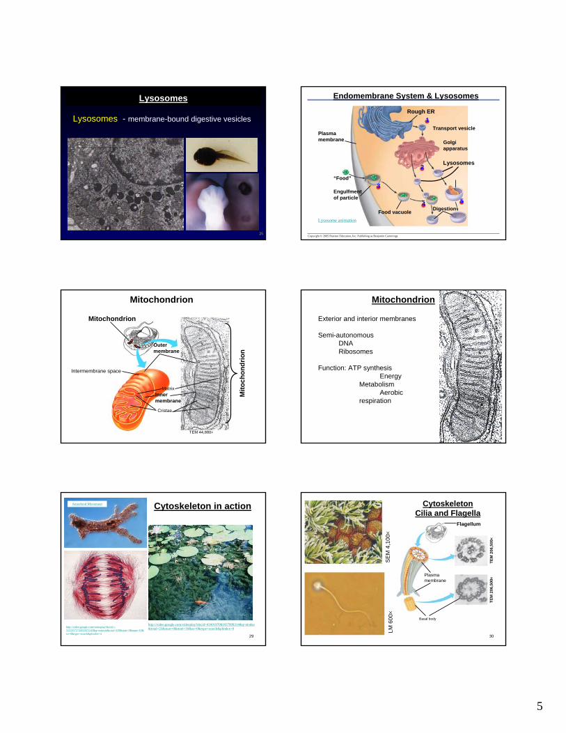
Lysosomes - membrane-bound digestive vesicles
Lysosomes
Copyright © 2005 Pearson Education, Inc. Publishing as Benjamin Cummings
Golgiapparatus
Transport vesiclePlasmamembrane
“Food”
Food vacuole
Lysosomes
Digestion
1
2
54
3Engulfmentof particle
Rough ER
Lysosome animation
Endomembrane System & Lysosomes
Mitochondrion
Outermembrane
Intermembrane space
MatrixInnermembrane
Cristae
TEM 44,880×
Mito
chon
drio
n
Mitochondrion Mitochondrion
Exterior and interior membranes
Semi-autonomousDNARibosomes
Function: ATP synthesis Energy
MetabolismAerobic
respiration
29
Amoeboid Movement
http://video.google.com/videoplay?docid=4349197081937999314&q=elodea&total=23&start=0&num=10&so=0&type=search&plindex=0http://video.google.com/videoplay?docid=-
5522357274832025243&q=mitosis&total=329&start=0&num=10&so=0&type=search&plindex=4
Cytoskeleton in action
30
SE
M 4
,100×
LM 6
00×
Cytoskeleton Cilia and Flagella
Flagellum
Basal body
TEM
206
,500×
TEM
206
,500×
Plasmamembrane

6
Copyright © 2005 Pearson Education, Inc. Publishing as Benjamin Cummings
Protein Fibers of the cytoskeleton
Actin subunit
Microfilaments7 nm
Fibrous subunits
10 nm
Intermediate filament
Microtubule
25 nm
Tubulin subunit
Network of protein fibers supporting cell shape, movement and anchoring organelles
Cell shape Cell movement
Movement organelles, chromosomes, flagellaCell shape
Actin and Microfilaments
Actin Microfilaments
Microtubules Intermediate Filaments
Intermediate Filaments Rat epithelial cell
Red blood cellsKeratins: Hair & Nails
• Network of protein fibers – Shape– Movement– anchoring organelles
Microfilaments - made of actin protein• Cell shape & movement (cell extensions, contractions)
Microtubules - made of tubulin protein• Organelle & chromosome movement
Intermediate filaments – made of fibrous protein• Structural stability
Cytoskeleton - Summary
Centriole
Lysosome
Mitochondrion
Ribosomes
Roughendoplasmicreticulum
CytoplasmNucleus
Nucleolus
Nuclearenvelope
Smoothendoplasmicreticulum
Golgiapparatus Plasma
membrane
Microvilli
Peroxisome
CytoskeletonActin filament
Intermediatefilament
Microtubule
Eukaryotic Cell Structure

7
Central vacuole
Chloroplast
Cell wall
Plasma membrane
Plant Cell Plant Cell Walls
Plasmodesmata
Plant Cells - VacuolePlant Cells
Chloroplasts
Chloroplast structureTwo external membranesInternal membranesDNARibosomes
Function: Photosynthesis
• Central vacuole– Large compartments in mature plant cells,– Storage facility for water & other materials– Produce turgor (pressure) for cell rigidity
• Cell wall– Cellulose & other polysaccharides– Support of Cells, Tissues, Organs, Whole Plant
• Chloroplasts– Membranes: 2 in envelope & many internal– Semiautonomous: DNA & ribosomes– Photosynthesis
Plant Cell Unique Characteristics
End Cells